Article

Viral Metagenomics Reveals a Putative Novel HPV Type in Anogenital Wart Tissues

1
Department of Dermatology, Affiliated Hospital of Jiangsu University, Zhenjiang 212001, China
2
Department of Dermatology, The Affiliated Huai’an Hospital of Xuzhou Medical University and The Second People’s Hospital of Huai’an, Huaian 223002, China
3
Department of Clinical Laboratory, Women’s Hospital of Nanjing Medical University, Nanjing 210004, China
*
Author to whom correspondence should be addressed.
Pathogens 2022, 11(12), 1452; https://doi.org/10.3390/pathogens11121452
Submission received: 28 October 2022 / Revised: 27 November 2022 / Accepted: 28 November 2022 / Published: 1 December 2022

Abstract

:
Viral metagenomics is widely employed to identify novel viruses in biological samples. Recently, although numerous novel human papillomavirus (HPV) types have been identified in clinical samples including anogenital warts (AGWs), many novel HPV sequences remain to be discovered. In this study, a putative novel HPV type designated as HPV-JDFY01 was discovered from library GW05 with 63 sequence reads by the viral metagenomic technique. Its complete genomic sequence was determined by PCR to bridge the gaps between contigs combining Sanger sequencing. The complete genome of HPV-JDFY01 is a 7186 bp encoding 7 open reading frames (ORFs) (E6, E7, E1, E2, E4, L2 and L1) and contains a 487 bp long control region (LCR) between L1 and E6. Sequence and phylogeny analysis indicated that HPV-JDFY01 shared the highest sequence identity of 74.2% with HPV-mSK_244 (MH777383) and well clustered into the genus Gammapapillomavirus. It has the classical genomic organization of Gammapapillomaviruses. Epidemiological investigation showed that one out of the 413 AGW tissue samples was positive for HPV-JDFY01. Further research with large size and different type of samples should be performed to elucidate the epidemiologic status of HPV-JDFY01.

1. Introduction

HPVs are present as small, nonenveloped and circular double-stranded DNA viruses with an icosahedral capsid, and they consist of an average genome length of 8000 bp [1,2]. The whole genome can be broadly grouped into early regions, late regions and a long control region according to their different functions [3,4,5]. The outer capsid of HPV is comprised predominantly of seventy-two major capsid protein L1, which plays an essential role in the attachment during the viral entry and the assembling of virions [6,7]. L1 protein has also been applied for the classification or defining of a novel HPV type if the variability proportion is over 10% [3].
The International Human Papillomaviruses Reference Center has confirmed 223 HPV types with complete genome as of 5/April/2022 <https://www.hpvcenter.se/>. These HPVs are only distributed over five evolutionary genera, including Alphapapillomavirus, Betapapillomavirus, Gammapapillomavirus, Mupapillomavirus and Nupapillomavirus.
Gammapapillomavirus accounts for over 50% of total HPVs and owns 83.3% of the novel types from 2014, which demands significant attention [8]. Little is known about its genetic characteristics, but it has been reported that all of the Gamma-6 lack the E6 protein to promote carcinogenesis [9]. In addition, patients with warts, hypogammaglobulinemia, infections or myelokathexis (WHIM) syndrome seem to be inclined to be infected by Gammapapillomavirus [10].
In the past four years, only four novel HPV types had been identified, including HPV226 (Gamma-6), HPV227 (Beta-2), HPV228 (Gamma-27) and HPV229 (Gamma-7) (https://www.hpvcenter.se/), three of which belonged to Gammapapillomavirus. Latsuzbaia et al. [11] discovered that the strain HPV226 contained one TATA box and three palindromic E2-binding sites in LCR, one zinc-binding domain in E7 and one ATP-binding site in E1. Other special structural features, such as polyadenylation sites and LxCxE motif were identified in HPV227 [12]. Both HPV226 and HPV227 were detected by next-generation sequencing (NGS) after rolling circular amplification [11,12].
Viral Metagenomics is based on NGS and can detect all the viral nucleic acid directly in biological samples so as to effectively find possible new viruses. In our study, using viral metagenomic method, a putative novel HPV (HPV-JDFY01) was discovered from AGWs tissue samples and the complete genome was acquired by combining PCR and Sanger sequencing. Sequence and phylogeny analysis were further performed to fully characterized this putative novel type of HPV.

2. Materials and Methods

2.1. Sample Collection and Preparation

A total of 110 tissue samples of wart from AGW patients, including 34 females and 76 males, were collected from September 2016 to August 2017 from the Department of Dermatology and Venerology at the Affiliated Hospital of Jiangsu University. The mean age of AGW patients was 42.5 years and the mean disease duration was 3.26 months. Each sample was transferred into 1.5 mL test tube with 1 mL dulbecco’s phosphate-buffered saline (DPBS) and then added into several 5 mm sterile stainless-steel beads to fully grind tissue for 5 min. After freeze and thaw 3 times on dry ice for 3 min each time, the tissue supernatants were obtained after centrifuging for 10 min at 12,000× g under the conditions of 4 °C.

2.2. Viral Metagenomic and Bioinformatics Analysis

The 110 supernatants of wart tissues were randomly pooled into 11 sample pools, each containing 10 samples. These sample pools were filtered through a 0.45 μm filter (Merck Millipore Ltd., Tullagreen, Carrigtwohill, Darmstadt, Germany, Co. Cork, IRL) to remove cellular particles in the sample [13,14]. The filtrates were digested by DNase and RNase at 37 °C for 60 min to remove non-viral encapsidated nucleic acids [15,16]. Then, a QIAamp Viral RNA Mini Kit (QIAGEN GmbH, QIAGEN Strasse 1, 40724 Hilden, Germany) was utilized to extract the remaining nucleic acids in the samples [17]. Reverse transcription (RT) and Klenow reactions were conducted to synthesize double-stranded DNA, which was prepared for constructing the 11 cDNA libraries by Nextera XT DNA Sample Preparation Kit (Illumina, Inc., San Diego, CA 92122 USA) [18,19]. Libraries were subjected to NGS on the MiSeq Illumina sequencing platform. The generated NGS data was processed by in-house analysis pipeline. Briefly, the bioinformatics steps were performed on a cluster of 32 nodes running Linux, and taking the Phred Score Q30 as the threshold trimmed the low sequencing quality tails [20]. Adaptors and primer sequences were trimmed by VecScreen in the default parameters [21]. These cleaned reads from sequencing were de novo assembled into contigs within each barcode group by Ensemble assembler [22]. The assembled contigs, along with unassembled reads, were matched against viral proteome database by BLASTx with an E-value cutoff of <10−5, where the viral proteome databased included NCBI virus reference proteome together with viral proteins sequences from NCBI nr fasta file.

2.3. Acquiring of Complete Genome and PCR Reactions

Finding six contigs ranging from 123 bp to 645 bp in length in GW05 pool, which showed low sequence similarity to known HPVs, 2 sets of specific PCR primers derived from a contig belonging to L1 gene of HPV were, respectively designed to confirm the existence of the putative novel HPV sequence in samples from GW05 pool. PCR primer sequences are displayed in Table 1. The screening conditions of PCR were as follows: 95 °C for 5 min, 31 cycles of 95 °C for 30 s, 48 °C (for the first round) or 52 °C (for the second round) for 30 s and 72 °C for 40 s and a final extension at 72 °C for 5 min. The circular complete genome of HPV-JDFY01 was acquired by PCR primers bridging the gaps between contigs and the PCR products were cloned and subjected to Sanger sequencing. In order to assess the prevalence of HPV-JDFY01 in AGW samples, an epidemiological investigation was performed by PCR screening method described above for a total of 413 AGW specimens collected from August 2017 to August 2020.

2.4. Phylogenetic Analysis

Phylogenetic analysis was performed based on amino acid sequences of predicted ORFs to analyze the genetic relationships between HPV-JDFY01 and the other related HPV types which included the representative types of Alphapapillomavirus, Betapapillomavirus and Gammapapillomavirus. Multiple alignments were conducted using ClustalW in MEGA7 with the default settings [23]. The aligned file was converted to Nexus (.nex) formmat by Geneious 11.1.2 and subsequently modified by Notapade++ [24]. The mean standard deviation of split frequencies was controlled as low as possible when generating phylogenetic trees by Mrbayes 3.2.7 with Bayesian inference (BI) [25].

3. Results

The NGS results indicated that the 11 libraries generated a total of 6,509,970 reads, 726,777 (11.16%) of which showed significant sequence similarity to HPV. The library GW02 generated the largest number of total reads (1,300,146) while the library GW09 contained the most abundant HPV-related sequence reads (227,374). Among the HPV sequence reads in these libraries, Alpha-10 (26,708) ranking first was followed by Alpha-8 (20,929), Alpha-14 (1622) and Alpha-9 (1066). HPV11 occupied the dominant part in Alpha-10 (95.41%) and HPV of the GW05 library (33.87%), respectively, in accordance with that of the literature reported previously [26,27]. HPV6, another common subtype of AGW, accounted for 0.42% in GW05 library. Except for unclassified papillomaviridae, the other HPV types identified in the 110 wart samples were presented in Table S1. Given that six contigs ranging from 123 bp to 645 bp in length in the GW05 pool showed low sequence similarity to known HPVs, 10 individual samples in this pool were screened by the nested PCR. The result indicated only one sample was positive. PCR combining Sanger sequencing was then used to bridge the sequence gaps and obtain the whole genome of the putative novel HPV eventually.

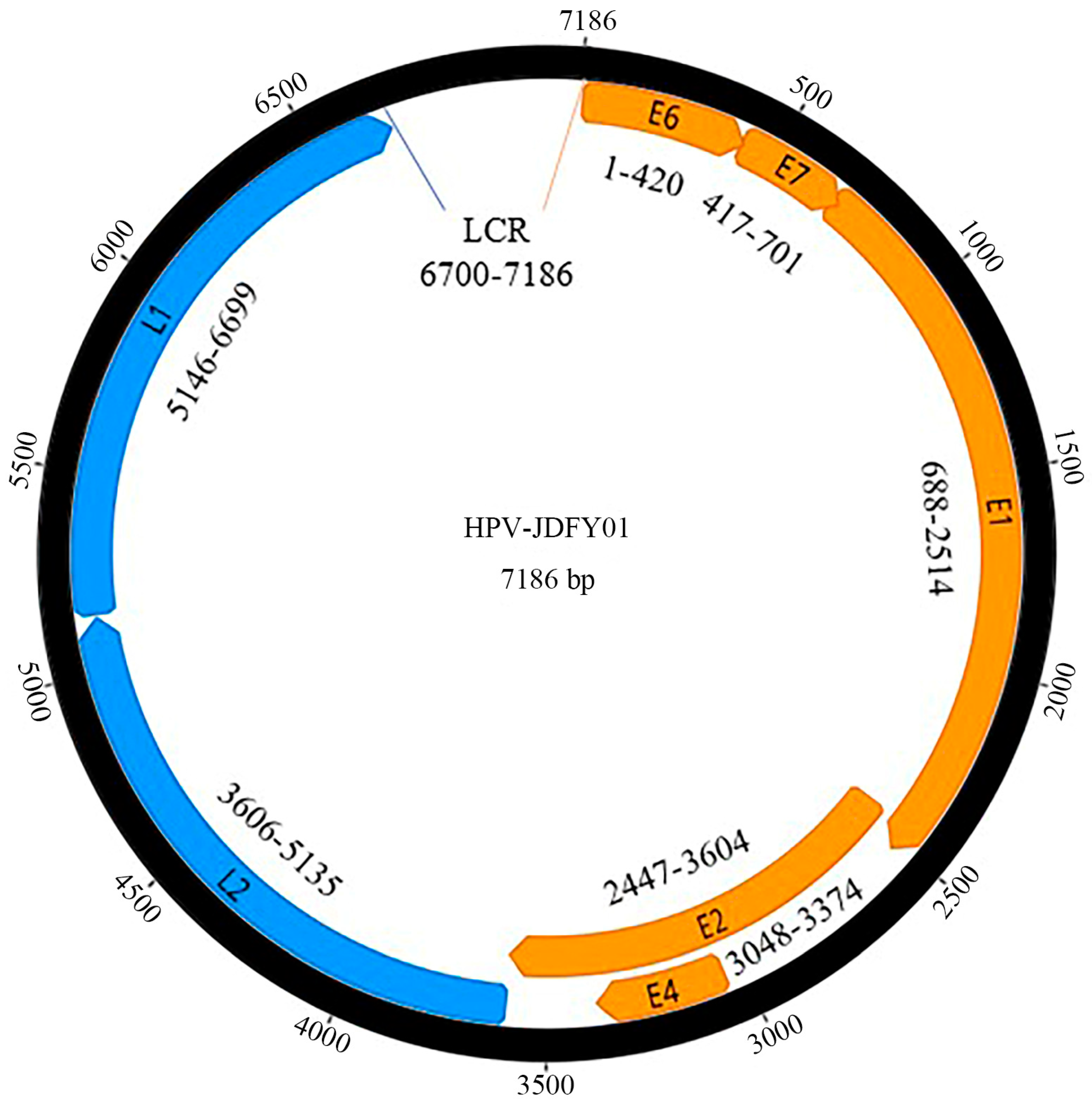
3.1. Complete Genomic Structure of HPV-JDFY01

A putative novel HPV type (HPV-JDFY01) was discovered from AGW, of which the complete circular genome is 7186 bp in length with a GC content of 36.6%. The details of the seven predicted ORFs are described as follows: Early proteins: E6 (nucleotide (nt) 1–420, 420 bp), E7 (nt 417–701, 285 bp), E1 (nt 688–2514, 1827 bp), E2 (nt 2447–3604, 1158 bp) and E4 (nt 3048–3374, 327 bp); Late proteins: L2 (nt 3606–5135, 1530 bp) and L1 (nt 5146–6699, 1554 bp); and a 487 bp long control region (LCR) is located at 6700 to 7186 of the genome between L1 and E6 (Figure 1). The putative E6 protein of HPV-JDFY01 contains two conserved zinc-finger domains, CxxC(x)29CxxC being located at nucleotide positions of 79–189 and 298–408, which are separated by 36 amino acids [28,29]. Three putative DZ binding domains (PTAL, nucleotide positions 16–27; LSLV, nucleotide positions 139–150 and ESLV, nucleotide positions 256–267) were also identified [30]. The putative E7, a downstream protein of E6, contains one zinc-finger domain (nucleotide positions 558–668), but no binding domain (LxCxE) for the conserved retinoblastoma tumor suppressor protein (pRB) [31]. As the largest protein of HPV-JDFY01, the putative E1 has one conserved ATP-binding site of the ATP-dependent DNA helicase with a consensus sequence (GPPDTGKS, nucleotide positions 1966–1989) and one typical bipartite nuclear localization signal (NLS) (KRK, nucleotide positions 919–927; KRRL, nucleotide positions 1009–1020). A leucine-rich nuclear export signal (NES) (LSPRLEAVKI, nucleotide positions 961–990) situates between the two clusters of NLS [32]. The putative E4 protein, with the highest proline content (12.8%), lies fully inside the E2. One highly conserved furin cleavage motif (RAKR, nucleotide positions 3620–3631) has been found at the N-terminal part of the L2 protein. In addition, a putative polyadenilation site (AATAAA, nucleotide positions 3695–3700) and a L2 transmembrane domain-like aa sequence (IIYLGGLGIGTGKGTG, nucleotide positions 3749–3796) are also observed [33]. NLS-like signals, which are used to direct HPV virions to import into the nucleus, are identified both at the C-terminal part of L2 protein (nucleotide positions 5099–5119) and in L1 of HPV-JDFY01 (nucleotide positions 6622–6696). The 487-bp LCR, also known as upstream regulatory region (URR), contains one putative polyadenilation site (AATAAA, nucleotide positions 6754–6759) and three palindromic E2-binding sites (ACC-AGTTCT-GGT, nucleotide positions 6914–6925; ACC-GATAAC-GGT, nucleotide positions 6955–6966 and ACC-AAGTGT-GGT, nucleotide positions 7135–7146) [34]. A TATA box (TATAAA, nucleotide positions 7150–7155) is located at the 31 nt upstream of E6’s first start codon. Most of the above-mentioned genetic characteristics could be examined in all other Gamma-8 species members. In view of the highest sequence identity of HPV-JDFY01 was only 74.2% with HPV-mSK_244 (MH777383), it needs to be confirmed as one putative novel HPV type by the current demarcation criteria of International Committee on the Taxonomy of Viruses (ICTV) [1,3].

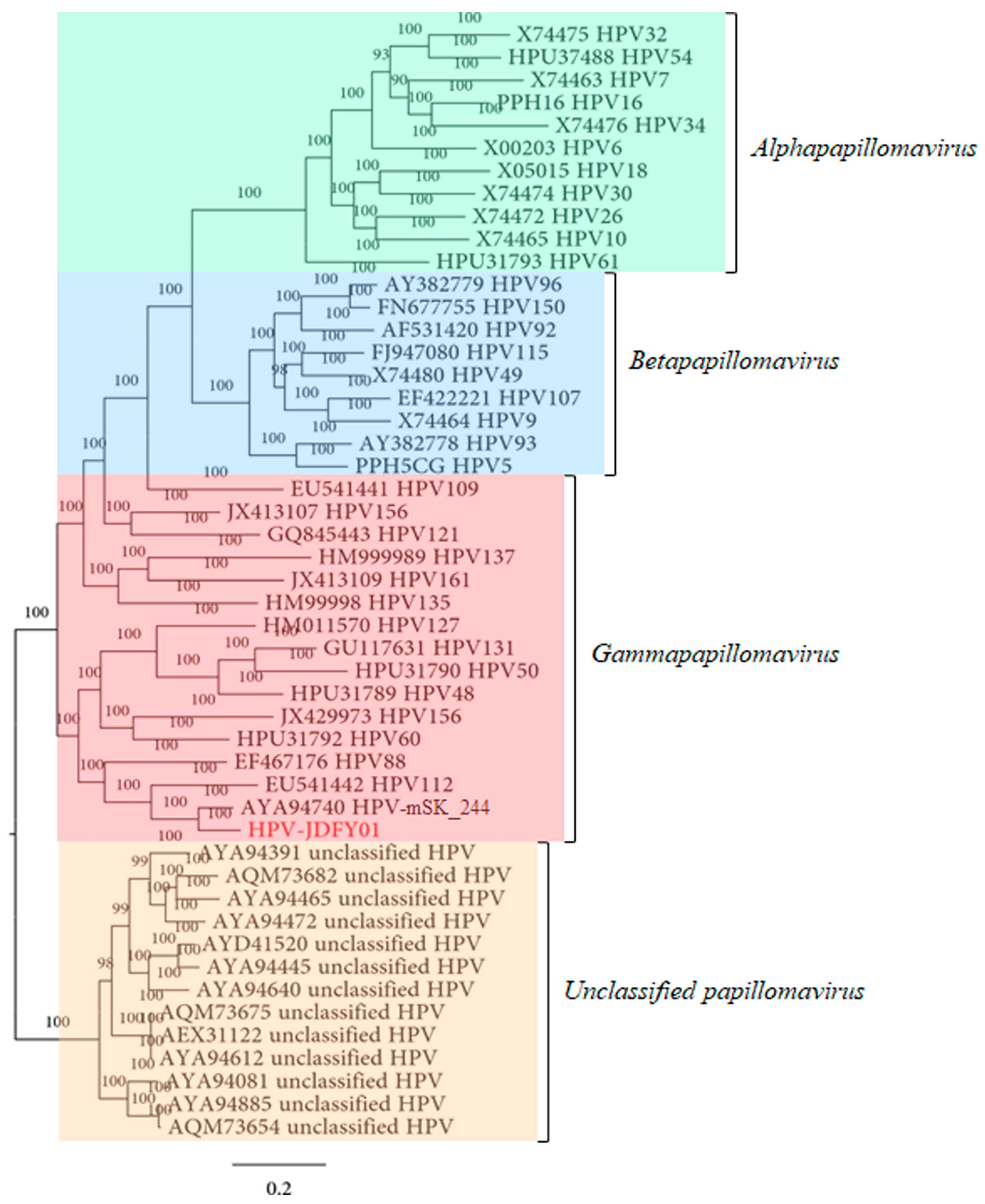
3.2. Phylogenetic Analysis of HPV-JDFY01

The amino acid sequence of HPV-JDFY01 L1 was aligned with 11 typical reference sequences from Alphapapillomavirus, 9 from Betapapillomavirus, 15 from Gammapapillomavirus and 13 from unclassified papillomavirus so as to generate the phylogenetic tree that clearly reflected the evolutionary relationships of these representative HPV types. From the results of the phylogenetic analysis, the HPV-JDFY01, together with HPV-mSK_244 (Figure 2, Figures S1–S5) was clustered into the same clade of Gammapapillomavirus, a genus with the highest number of novel HPVs in recent years [8]. Both HPV-JDFY01 and HPV-mSK_244 were directly adjacent to the HPV112 which belongs to the Gamma-8 species. The sequence identity and phylogenetic analysis supported that HPV-JDFY01 was closely related to Gamma-8 types.

3.3. Epidemiological Analysis of HPV-JDFY01

Epidemiological investigation showed that only one out of the 413 AGW tissue samples was positive for HPV-JDFY01. The typically clinical and pathological manifestations were shown in Figure 3A,B.

3.4. GenBank Accession Number

The complete genomic sequence of HPV-JDFY01 was submitted to the GenBank database under accession number MW311489.

4. Discussion

Viral metagenomics, based on NGS, can detect nucleic acid of all viral pathogens from samples and have the potential to become crucial for clinical diagnosis in the near future [35,36,37,38]. We report a genome of HPV-JDFY01, a putative novel type in Gammapapillomavirus, identified by viral metagenomics from AGWs collected in Jiangsu Province, China.
Previous literatures considered that HPVs of the same species share ≥ 71% L1 similarity [1,3]. Phylogenetic analysis indicated that HPV-JDFY01 shared the highest sequence identity of 74.2% with HPV-mSK_244 based on the nucleotide sequence of L1. Thus, HPV-JDFY01 could be assumed as a putative novel HPV type and may have similar biological functions with HPV-mSK_244. HPV-mSK_244 was identified from 207 skin and nares swabs from DOCK8-deficient patients, who have recurrent cutaneous and systemic infections and can suffer from the predisposition and susceptibility to cancer [7]. However, HPV-JDFY01 was found in immunocompetent AGW patients. Additionally, in the phylogenetic analyses of L1 protein, HPV-JDFY01 and HPV-mSK_244 were clustered together with HPV112, which belongs to the Gamma-8 species. It is worth noting that HPV112, a member of the Gamma-8 species, was also cloned from a 25-year-old male with condyloma acuminata [39]. The other two types of Gamma-8, HPV164 and HPV168, were isolated from exfoliated skin cell samples of healthy individuals [40,41]. In summary, the above evidences indicate that HPV types from the Gamma-8 species may engage in much milder relationships with human beings. Additionally, the relative abundances of DNA viruses, mostly Papillomaviridae and Polyomaviridae, were higher on the skin than in the oral cavity and feces of DOCK8-deficient patients [7]. HPV-JDFY01 was isolated from tissue samples of wart while HPV112, HPV164 and HPV168 were all isolated from skin [39,40]. Therefore, it can be stated that the members of Gamma-8 tend to infect skin and may be distinguished into cutaneotropic HPV types.
Even though further studies are needed to assess the relationship between HPV-JDFY01 and Gamma-8, the genetic characteristics of HPV-JDFY01 were further analyzed by comparing with the previous literatures, which were related to other HPV members of Gamma-8. We found that most of specific characteristics of Gamma-8 could be identified in HPV-JDFY01, including zinc-finger domains, PDZ binding domains, ATP-binding sites, nuclear localization signals, nuclear export signals, NLS-like signals, furin cleavage motifs, polyadenilation sites, palindromic E2-binding sites and TATA boxes. In previous literature, it has been reported that the E7 protein of HPV112 contained a pRB protein binding domain, although it was not totally conserved [39]. However, pRB protein binding motif (LxCxE) have not been detected in E7 protein of HPV-JDFY01. This means lacking the LxCxE motif may have the effect of attenuating the association between E7 and pRB [42] and indicate transforming properties of HPV-JDFY01. A highly conserved NLS motif can promote nuclear localization and bind to the nuclear matrix [43], which was also not identified in E2 of HPV-JDFY01. Moreover, three internal PDZ-binding motifs (PTAL, LSLV and ESLV) were identified in the putative E6 protein. Nevertheless, these motifs may not possess biological functions [44]. Further structural analysis will be needed to better understand the relationship between HPV-JDFY01 and the other members from Gamma-8.
On the basis of the highly conserved, immunogenic and self-assembly natures in HPV L1 protein, virus-like particles (VLPs) of HPV have been designed and developed for the purpose of prophylactic HPV vaccines [45]. Further prediction analyses of advanced structure for novel HPV L1 protein are thus warranted.

5. Conclusions

In this study, a putative novel Gammapapillomavirus named HPV-JDFY01 was detected by a viral metagenomics method from AGWs, whose complete genome was determined and genomic structure and phylogeny were fully characterized. The functional, biophysical and carcinogenic properties of HPV-JDFY01 need to be elucidated in the future research.

Supplementary Materials

The following supporting information can be downloaded at: https://www.mdpi.com/article/10.3390/pathogens11121452/s1, Figure S1: Phylogenetic analysis based on the amino acid sequence of HPV-JDFY01 L2 protein.; Figure S2: Phylogenetic analysis based on the amino acid sequence of HPV-JDFY01 E6 protein.; Figure S3: Phylogenetic analysis based on the amino acid sequence of HPV-JDFY01 E7 protein.; Figure S4: Phylogenetic analysis based on the amino acid sequence of HPV-JDFY01 E1 protein.; Figure S5: Phylogenetic analysis based on the amino acid sequence of HPV-JDFY01 E2 protein.; Table S1: The other HPV types identified in the 110 wart samples.

Author Contributions

Conceptualization, H.X., Y.L. (Yumei Li), Y.L. (Yu Ling) and H.H.; methodology, Y.L., H.H. and H.W.; software, Y.L. (Yu Ling), H.H. and X.W.; validation, N.Z.; investigation, H.H. and N.Z.; resources, H.X.; data curation, Y.L. (Yu Ling); writing—original draft preparation, H.H.; writing—review and editing, H.X.; supervision, H.X. and Y.L. (Yumei Li); project administration, H.X. and Y.L. (Yumei Li); funding acquisition, H.X. and Y.L. (Yumei Li). All authors have read and agreed to the published version of the manuscript.

Funding

This research was funded by “the National Key Research and Development Programs of China, grant number 2022YFC2603801”, “the Maternal and Child Health Project of Jiangsu Province (F201717)”, “the Doctor Project of Affiliated Hospital of Jiangsu University (jdfyrc2019003)” and “the Gynecology Project of Zhenjiang Traditional Chinese Medicine (SS202204-KFC01)”.

Institutional Review Board Statement

The study was conducted in accordance with the Declaration of Helsinki, and approved by Medical Ethical Committee at the Affiliated Hospital of Jiangsu University (Reference code: ujsah2016219).

Informed Consent Statement

Informed consent was obtained from all subjects involved in the study.

Data Availability Statement

The data presented in this study are available on request from the corresponding author.

Acknowledgments

We thank all members of Department of Microbiology, School of Medicine, Jiangsu University, Zhenjiang, Jiangsu, China.

Conflicts of Interest

The authors declare no conflict of interest.

References

  1. de Villiers, E.M.; Fauquet, C.; Broker, T.R.; Bernard, H.U.; zur Hausen, H. Classification of papillomaviruses. Virology 2004, 324, 17–27. [Google Scholar] [CrossRef] [PubMed] [Green Version]
  2. Seedorf, K.; Krammer, G.; Durst, M.; Suhai, S.; Rowekamp, W.G. Human papillomavirus type 16 DNA sequence. Virology 1985, 145, 181–185. [Google Scholar] [CrossRef] [PubMed]
  3. Bernard, H.-U.; Burk, R.D.; Chen, Z.; van Doorslaer, K.; Hausen, H.z.; de Villiers, E.-M. Classification of papillomaviruses (PVs) based on 189 PV types and proposal of taxonomic amendments. Virology 2010, 401, 70–79. [Google Scholar] [CrossRef] [PubMed] [Green Version]
  4. Baker, C.C.; Phelps, W.C.; Lindgren, V.; Braun, M.J.; Gonda, M.A.; Howley, P.M. Structural and transcriptional analysis of human papillomavirus type 16 sequences in cervical carcinoma cell lines. J. Virol. 1987, 61, 962–971. [Google Scholar] [CrossRef] [PubMed] [Green Version]
  5. Howley, P.M.; Munger, K.; Werness, B.A.; Phelps, W.C.; Schlegel, R. Molecular mechanisms of transformation by the human papillomaviruses. Princess Takamatsu Symp. 1989, 20, 199–206. [Google Scholar] [PubMed]
  6. Gheit, T. Mucosal and Cutaneous Human Papillomavirus Infections and Cancer Biology. Front. Oncol. 2019, 9, 355. [Google Scholar] [CrossRef] [Green Version]
  7. Tirosh, O.; Conlan, S.; Deming, C.; Lee-Lin, S.Q.; Huang, X.; Program, N.C.S.; Su, H.C.; Freeman, A.F.; Segre, J.A.; Kong, H.H. Expanded skin virome in DOCK8-deficient patients. Nat. Med. 2018, 24, 1815–1821. [Google Scholar] [CrossRef]
  8. Muhr, L.S.A.; Eklund, C.; Dillner, J. Towards quality and order in human papillomavirus research. Virology 2018, 519, 74–76. [Google Scholar] [CrossRef]
  9. Van Doorslaer, K.; McBride, A.A. Molecular archeological evidence in support of the repeated loss of a papillomavirus gene. Sci. Rep. 2016, 6, 33028. [Google Scholar] [CrossRef]
  10. Pastrana, D.V.; Peretti, A.; Welch, N.L.; Borgogna, C.; Olivero, C.; Badolato, R.; Notarangelo, L.D.; Gariglio, M.; FitzGerald, P.C.; McIntosh, C.E.; et al. Metagenomic Discovery of 83 New Human Papillomavirus Types in Patients with Immunodeficiency. mSphere 2018, 3, e00645-18. [Google Scholar] [CrossRef]
  11. Latsuzbaia, A.; Arbyn, M.; Dutta, S.; Fischer, M.; Gheit, T.; Tapp, J.; Tommasino, M.; Weyers, S.; Mossong, J. Complete Genome Sequence of a Novel Human Gammapapillomavirus Isolated from a Cervical Swab in Luxembourg. Genome Announc. 2018, 6, e00114-18. [Google Scholar] [CrossRef] [PubMed] [Green Version]
  12. Brancaccio, R.N.; Robitaille, A.; Dutta, S.; Rollison, D.E.; Tommasino, M.; Gheit, T. Isolation of a Novel Beta-2 Human Papillomavirus from Skin. Microbiol. Resour. Announc. 2019, 8, e01628-18. [Google Scholar] [CrossRef] [PubMed] [Green Version]
  13. Liu, Z.; Yang, S.; Wang, Y.; Shen, Q.; Yang, Y.; Deng, X.; Zhang, W.; Delwart, E. Identification of a novel human papillomavirus by metagenomic analysis of vaginal swab samples from pregnant women. Virol. J. 2016, 13, 122. [Google Scholar] [CrossRef] [PubMed] [Green Version]
  14. Ling, Y.; Wang, J.; Yin, J.; Xu, J.; Wu, Y.; Zhou, R.; Lu, J.; Yang, S.; Wang, X.; Shen, Q.; et al. Genomic organization of a Gamma-6 papillomavirus metagenomic discovered from vaginal swab samples of Chinese pregnant women. Virol. J. 2020, 17, 44. [Google Scholar] [CrossRef] [Green Version]
  15. Zhang, W.; Li, L.; Deng, X.; Blumel, J.; Nubling, C.M.; Hunfeld, A.; Baylis, S.A.; Delwart, E. Viral nucleic acids in human plasma pools. Transfusion 2016, 56, 2248–2255. [Google Scholar] [CrossRef]
  16. Zhang, W.; Yang, S.; Shan, T.; Hou, R.; Liu, Z.; Li, W.; Guo, L.; Wang, Y.; Chen, P.; Wang, X.; et al. Virome comparisons in wild-diseased and healthy captive giant pandas. Microbiome 2017, 5, 90. [Google Scholar] [CrossRef] [Green Version]
  17. Wang, H.; Li, S.; Mahmood, A.; Yang, S.; Wang, X.; Shen, Q.; Shan, T.; Deng, X.; Li, J.; Hua, X.; et al. Plasma virome of cattle from forest region revealed diverse small circular ssDNA viral genomes. Virol. J. 2018, 15, 11. [Google Scholar] [CrossRef] [Green Version]
  18. Sabatier, M.; Bal, A.; Destras, G.; Regue, H.; Queromes, G.; Cheynet, V.; Lina, B.; Bardel, C.; Brengel-Pesce, K.; Navratil, V.; et al. Comparison of Nucleic Acid Extraction Methods for a Viral Metagenomics Analysis of Respiratory Viruses. Microorganisms 2020, 8, 1539. [Google Scholar] [CrossRef]
  19. Xiao, Y.; Wang, H.; Feng, L.; Pan, J.; Chen, Z.; Wang, H.; Yang, S.; Shen, Q.; Wang, X.; Shan, T.; et al. Fecal, oral, blood and skin virome of laboratory rabbits. Arch. Virol. 2020, 165, 2847–2856. [Google Scholar] [CrossRef]
  20. Pichler, M.; Coskun, O.K.; Ortega-Arbulu, A.S.; Conci, N.; Worheide, G.; Vargas, S.; Orsi, W.D. A 16S rRNA gene sequencing and analysis protocol for the Illumina MiniSeq platform. Microbiologyopen 2018, 7, e00611. [Google Scholar] [CrossRef]
  21. McGinnis, S.; Madden, T.L. BLAST: At the core of a powerful and diverse set of sequence analysis tools. Nucleic Acids Res. 2004, 32, W20–W25. [Google Scholar] [CrossRef] [PubMed]
  22. Deng, X.; Naccache, S.; Ng, T.; Federman, S.; Li, L.; Chiu, C.; Delwart, E. An ensemble strategy that significantly improves de novo assembly of microbial genomes from metagenomic next-generation sequencing data. Nucleic Acids Res. 2015, 43, e46. [Google Scholar] [CrossRef] [PubMed] [Green Version]
  23. Bibi, Z.; Fatima, A.; Rani, R.; Maqbool, A.; Khan, S.; Naz, S.; Waseem, S. Genetic characterization of Plasmodium vivax isolates from Pakistan using circumsporozoite protein (pvcsp) and merozoite surface protein-1 (pvmsp-1) genes as genetic markers. Malar. J. 2021, 20, 112. [Google Scholar] [CrossRef] [PubMed]
  24. Liu, Q.; Wang, H.; Ling, Y.; Yang, S.X.; Wang, X.C.; Zhou, R.; Xiao, Y.Q.; Chen, X.; Yang, J.; Fu, W.G.; et al. Viral metagenomics revealed diverse CRESS-DNA virus genomes in faeces of forest musk deer. Virol. J. 2020, 17, 61. [Google Scholar] [CrossRef] [Green Version]
  25. Huelsenbeck, J.; Ronquist, F. MRBAYES: Bayesian inference of phylogenetic trees. Bioinformatics 2001, 17, 754–755. [Google Scholar] [CrossRef] [Green Version]
  26. Grennan, D. Genital Warts. JAMA 2019, 321, 520. [Google Scholar] [CrossRef]
  27. Domenech-Vinolas, M.; Leon-Maldonado, L.; Ramirez-Palacios, P.; Flores, Y.N.; Granados-Garcia, V.; Brown, B.; Corona, E.; Liu, S.; Lazcano-Ponce, E.; Salmeron, J. Incidence, psychosocial burden, and economic impact of genital warts in Mexico. Salud Publica Mex. 2018, 60, 624–632. [Google Scholar] [CrossRef]
  28. Ogawa, T.; Tomita, Y.; Okada, M.; Shirasawa, H. Complete genome and phylogenetic position of bovine papillomavirus type 7. J. Gen. Virol. 2007, 88, 1934–1938. [Google Scholar] [CrossRef]
  29. Bernard, H.U. Regulatory elements in the viral genome. Virology 2013, 445, 197–204. [Google Scholar] [CrossRef] [Green Version]
  30. Kohler, A.; Gottschling, M.; Manning, K.; Lehmann, M.D.; Schulz, E.; Kruger-Corcoran, D.; Stockfleth, E.; Nindl, I. Genomic characterization of ten novel cutaneous human papillomaviruses from keratotic lesions of immunosuppressed patients. J. Gen. Virol. 2011, 92, 1585–1594. [Google Scholar] [CrossRef]
  31. McLaughlin-Drubin, M.E.; Munger, K. The human papillomavirus E7 oncoprotein. Virology 2009, 384, 335–344. [Google Scholar] [CrossRef] [PubMed] [Green Version]
  32. Bergvall, M.; Melendy, T.; Archambault, J. The E1 proteins. Virology 2013, 445, 35–56. [Google Scholar] [CrossRef] [PubMed] [Green Version]
  33. Wang, J.W.; Roden, R.B. L2, the minor capsid protein of papillomavirus. Virology 2013, 445, 175–186. [Google Scholar] [CrossRef] [PubMed] [Green Version]
  34. Lace, M.J.; Isacson, C.; Anson, J.R.; Lorincz, A.T.; Wilczynski, S.P.; Haugen, T.H.; Turek, L.P. Upstream regulatory region alterations found in human papillomavirus type 16 (HPV-16) isolates from cervical carcinomas increase transcription, ori function, and HPV immortalization capacity in culture. J. Virol. 2009, 83, 7457–7466. [Google Scholar] [CrossRef] [Green Version]
  35. Gu, W.; Miller, S.; Chiu, C.Y. Clinical Metagenomic Next-Generation Sequencing for Pathogen Detection. Annu. Rev. Pathol. 2019, 14, 319–338. [Google Scholar] [CrossRef]
  36. Liao, Z.H.; Yang, S.; Li, Q.; Lu, H.Z. The role of metagenomic next-generation sequencing as a promising technology for diagnosing HIV-TB coinfection. Zoonoses 2022, 2, 32. [Google Scholar] [CrossRef]
  37. Slavov, S.N. Viral Metagenomics for Identification of Emerging Viruses in Transfusion Medicine. Viruses 2022, 14, 2448. [Google Scholar] [CrossRef]
  38. Miller, S.; Chiu, C. The Role of Metagenomics and Next-Generation Sequencing in Infectious Disease Diagnosis. Clin. Chem. 2021, 68, 115–124. [Google Scholar] [CrossRef]
  39. Ekstrom, J.; Forslund, O.; Dillner, J. Three novel papillomaviruses (HPV109, HPV112 and HPV114) and their presence in cutaneous and mucosal samples. Virology 2010, 397, 331–336. [Google Scholar] [CrossRef] [Green Version]
  40. Li, J.; Pan, Y.; Deng, Q.; Cai, H.; Ke, Y. Identification and characterization of eleven novel human gamma-papillomavirus isolates from healthy skin, found at low frequency in a normal population. PLoS ONE 2013, 8, e77116. [Google Scholar] [CrossRef]
  41. Li, J.; Cai, H.; Xu, Z.; Wang, Q.; Hang, D.; Shen, N.; Liu, M.; Zhang, C.; Abliz, A.; Ke, Y. Nine complete genome sequences of cutaneous human papillomavirus genotypes isolated from healthy skin of individuals living in rural He Nan province, China. J. Virol. 2012, 86, 11936. [Google Scholar] [CrossRef] [Green Version]
  42. Hatterschide, J.; Bohidar, A.E.; Grace, M.; Nulton, T.J.; Kim, H.W.; Windle, B.; Morgan, I.M.; Munger, K.; White, E.A. PTPN14 degradation by high-risk human papillomavirus E7 limits keratinocyte differentiation and contributes to HPV-mediated oncogenesis. Proc. Natl. Acad Sci. USA 2019, 116, 7033–7042. [Google Scholar] [CrossRef] [Green Version]
  43. Zou, N.; Lin, B.Y.; Duan, F.; Lee, K.Y.; Jin, G.; Guan, R.; Yao, G.; Lefkowitz, E.J.; Broker, T.R.; Chow, L.T. The hinge of the human papillomavirus type 11 E2 protein contains major determinants for nuclear localization and nuclear matrix association. J. Virol. 2000, 74, 3761–3770. [Google Scholar] [CrossRef] [Green Version]
  44. Nagasaka, K.; Kawana, K.; Osuga, Y.; Fujii, T. PDZ domains and viral infection: Versatile potentials of HPV-PDZ interactions in relation to malignancy. Biomed. Res. Int. 2013, 2013, 369712. [Google Scholar] [CrossRef] [Green Version]
  45. Huber, B.; Wang, J.W.; Roden, R.B.S.; Kirnbauer, R. RG1-VLP and Other L2-Based, Broad-Spectrum HPV Vaccine Candidates. J. Clin. Med. 2021, 10, 1044. [Google Scholar] [CrossRef]
Figure 1. Genomic organization of HPV-JDFY01 isolated from AGW. Early proteins: E6 (nt 1–420, 420 bp); E7 (nt 417–701, 285 bp); E1 (nt 688–2514, 1827 bp); E2 (nt 2447–3604, 1158 bp) and E4 (nt 3048–3374, 327 bp). Late proteins: L2 (nt 3606–5135, 1530 bp) and L1 (nt 5146–6699, 1554 bp), and a 487 bp long control region (LCR) located at 6700 to 7186 of the genome between L1 and E6. The sizes of overlapping fragments are 4 bp between E6 and E7, 14 bp between E7 and E1, and 68 bp between E1 and E2. Even the entire E4 lies fully inside the E2 ORF.
Pathogens 11 01452 g001
Figure 2. Phylogenetic analysis based on the amino acid sequence of HPV-JDFY01 L1 protein. HPV-JDFY01 is labeled in red.
Pathogens 11 01452 g002
Figure 3. Clinical and pathological manifestations of AGW with HPV-JDFY01 positive by epidemiological screening. (A) Genital warts present as multiple, flat papules on the inguinal skin. (B) Representative pathological findings with HE staining in 100-fold magnification indicate hyperkeratosis, parakeratosis, acanthosis, papillomatosis, and characteristic vacuolated parakeratotic and granular cells (koilocytes).
Pathogens 11 01452 g003
Table 1. Specific primer sequences used to screen and amplify the whole genome of HPV-JDFY01.
Primer IDApplicationPrimer Sequences (5′-3′)
Screening WFFirst roundAGGCCTCCATTACCTCTTCTG
Screening WR AGCACAGATGGTTCAGTTGT
Screening NFSecond roundAGAGTTTGATCTGCGCCATTTGAT
Screening NR AGATGATGGCGAGCTGG
Gap1 WFFirst roundTTGCGCGCAAAAATGAGAGG
Gap1 WR GAATTCCCCTGCTCCAGCTC
Gap1 NFSecond roundTTGCGCGCAAAAATGAGAGG
Gap1 NR GCACACCGTGGACAAAGAAG
Gap2 WFFirst roundAATCTGTCCCGTCTCCTGGA
Gap2 WR TGGAACCCCGAATGTGGATG
Gap2 NFSecond roundAATCTGTCCCGTCTCCTGGA
Gap2 NR TGCCAGCAGCTTTACAATGC
Gap3 WFFirst roundTGGTCCACCAGATACCGGAA
Gap3 WR TGGAAGTAGGAGTGGGCTGT
Gap3 NFSecond roundGCAAAACACAAACATCCACAGC
Gap3 NR TGGAAGTAGGAGTGGGCTGT
Gap4 WFFirst roundAGCTCAGCGCATGTAGTTTTG
Gap4 WR ACCCGAAACTGAGAACCTGAC
Gap4 NFSecond roundCTCAGCGCATGTAGTTTTGCA
Gap4 NR CCACTGGTTTTTGGGGAGGT
Gap5 WFFirst roundTGGCAAACGAGAACAAGGTT
Gap5 WR TCCTCCTCCTCTGCTTCAGT
Gap5 NFSecond roundTGCCAGACATTACTTTGCCAG
Gap5 NR TGCAAGCTTTCATTCCGACA
WF: upstream primer of first round, WR: downstream primer of first round, NF: upstream primer of second round, NR: downstream primer of second round.
Publisher’s Note: MDPI stays neutral with regard to jurisdictional claims in published maps and institutional affiliations.

