Article

Genetic Characterization of Cryptosporidium cuniculus from Rabbits in Egypt

1
Department of Hygiene and Zoonoses, Faculty of Veterinary Medicine, Mansoura University, Mansoura 35516, Egypt
2
Division of Foodborne, Waterborne, and Environmental Diseases, National Center for Emerging and Zoonotic Infectious Diseases, Centers for Disease Control and Prevention, Atlanta, GA 30329, USA
3
Department of Poultry Diseases, Faculty of Veterinary Medicine, Mansoura University, Mansoura 35516, Egypt
4
Center for Emerging and Zoonotic Diseases, College of Veterinary Medicine, South China Agricultural University, Guangzhou 510642, China
*
Author to whom correspondence should be addressed.
Pathogens 2021, 10(6), 775; https://doi.org/10.3390/pathogens10060775
Submission received: 28 May 2021 / Revised: 16 June 2021 / Accepted: 17 June 2021 / Published: 20 June 2021

Abstract

:
Rabbits are increasingly farmed in Egypt for meat. They are, however, known reservoirs of infectious pathogens. Currently, no information is available on the genetic characteristics of Cryptosporidium spp. in rabbits in Egypt. To understand the prevalence and genetic identity of Cryptosporidium spp. in these animals, 235 fecal samples were collected from rabbits of different ages on nine farms in El-Dakahlia, El-Gharbia, and Damietta Provinces, Egypt during the period from July 2015 to April 2016. PCR-RFLP analysis of the small subunit rRNA gene was used to detect and genotype Cryptosporidium spp. The overall detection rate was 11.9% (28/235). All 28 samples were identified as Cryptosporidium cuniculus. The 16 samples successfully subtyped by the sequence analysis of the partial 60 kDa glycoprotein gene belonged to two subtypes, VbA19 (n = 1) and VbA33 (n = 15). As C. cuniculus is increasingly recognized as a cause of human cryptosporidiosis, Cryptosporidium spp. in rabbits from Egypt have zoonotic potential.

1. Introduction

Cryptosporidiosis is a common cause of diarrhea in humans and animals [1,2]. It is one of the most important diseases in both developing countries and industrialized nations due to its importance in diarrhea-associated death in young children and foodborne and waterborne outbreaks of illness [1,3,4,5,6]. The etiologic agents of cryptosporidiosis, Cryptosporidium spp., have over 40 established species and many genotypes of unknown species status [7]. Among them, approximately 20 species and genotypes have been found in humans [8]. Most human cryptosporidiosis cases are caused by C. parvum and C. hominis. Other human-pathogenic Cryptosporidium spp. include C. meleagridis, C. ubiquitum, C. cuniculus, C. felis, C. canis, C. viatorum, and C. muris [7].
Rabbits are a supply of high-quality protein to humans. Reports of the Food and Agriculture Organization of the United Nations (FAO) showed that Egypt was the fourth largest producer of rabbit meat in the world, with approximately 7.6 million rabbits [9,10]. Results of recent studies indicate that rabbits can serve as reservoirs of many zoonotic pathogens [11,12,13]. They are commonly infected with several Cryptosporidium species, especially C. cuniculus [13,14,15,16,17,18,19,20]. In recent years, there have been increasing reports of C. cuniculus in humans [21,22,23,24]. In the United Kingdom and New Zealand, C. cuniculus is the third most common Cryptosporidium species in patients with diarrhea [24,25].
In recent years, molecular epidemiological studies have been conducted to understand the transmission of Cryptosporidium spp. in humans, livestock, and companion animals in Egypt [26,27,28,29,30,31,32,33,34]. Although rabbits are commonly farmed in Egypt, to the authors’ knowledge there have been no thorough studies on the distribution and genetic identity of Cryptosporidium spp. in rabbits in the country. Therefore, this study was conducted to examine the occurrence, genetic characteristics, and zoonotic potential of Cryptosporidium spp. in rabbits from three provinces (El-Dakahlia, El-Gharbia, and Damietta) in Egypt.

2. Results

2.1. Cryptosporidium Infection on Rabbit Farms

Cryptosporidium spp. were detected by PCR analysis of the SSU rRNA gene in 28 (11.9%) of the 235 fecal samples analyzed in the study. Eight of the nine farms examined were positive for Cryptosporidium spp. Among the eight positive farms, the infection rates ranged from 4% to 24% (Table 1). The farms El-Dakahlia 3 and El-Gharbia 2 had high infection rates of 21% and 24%, respectively, while no infection was detected on farm El-Gharbia 1. By age, Cryptosporidium spp. were detected in rabbits of all ages, with a higher infection rate found in rabbits of <3 months (20%; Fisher’s exact test = 11.237, p = 0.003 in the overall comparison) (Table 2). By animal breed, Cryptosporidium spp. were identified in all breeds, with slightly higher detection rates in Hi-Plus rabbits (15%) than in New Zealand (11%) and Rex (7%), although these rates were not statistically different (Fisher’s exact test = 2.283, p = 0.333 in the overall comparison). Farms in El-Dakahlia province recorded higher Cryptosporidium occurrence (17%) than El-Gharbia (11%) and Damietta (7%) provinces (Fisher’s exact test = 3.685, p = 0.157 in the overall comparison).

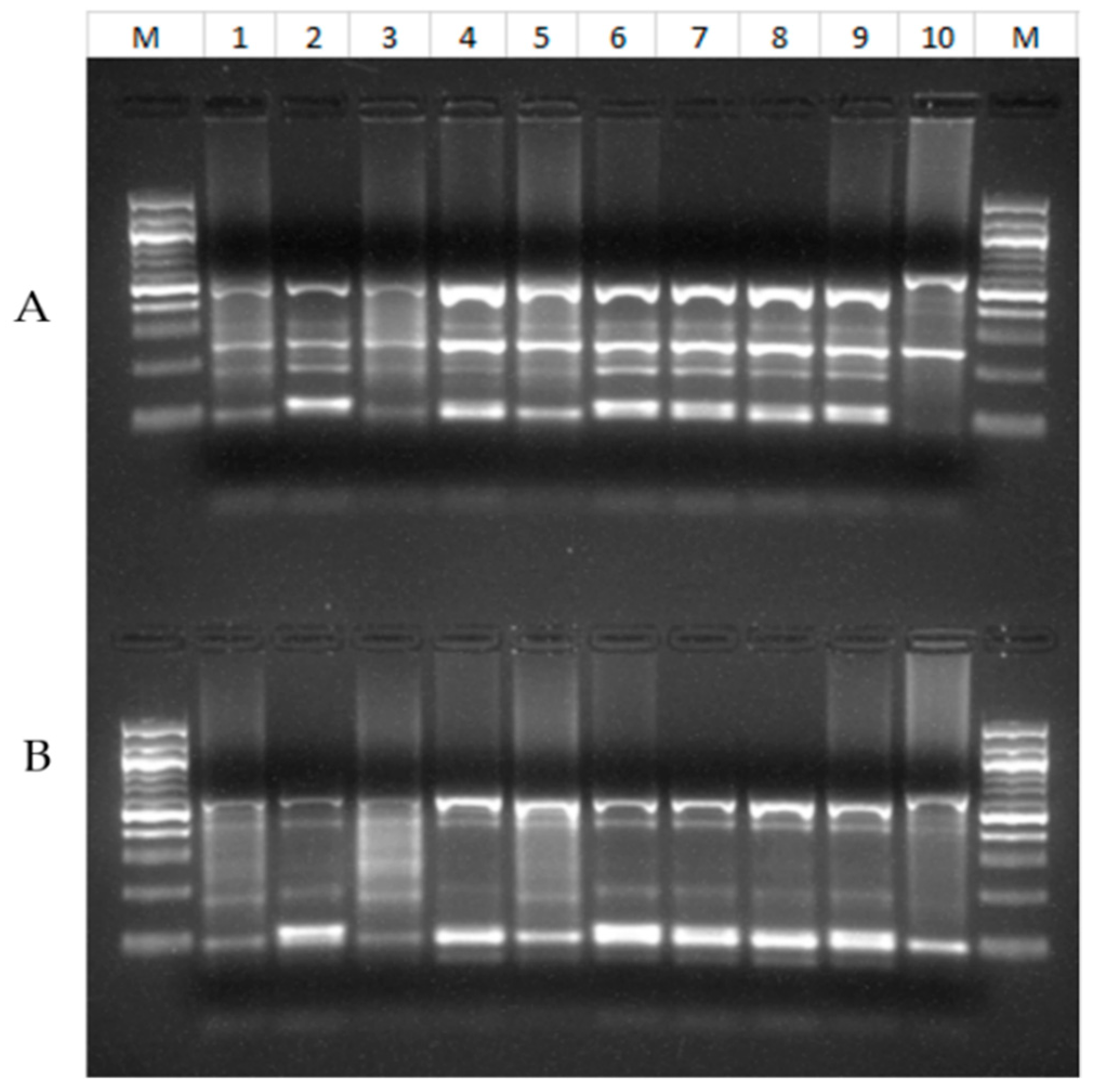
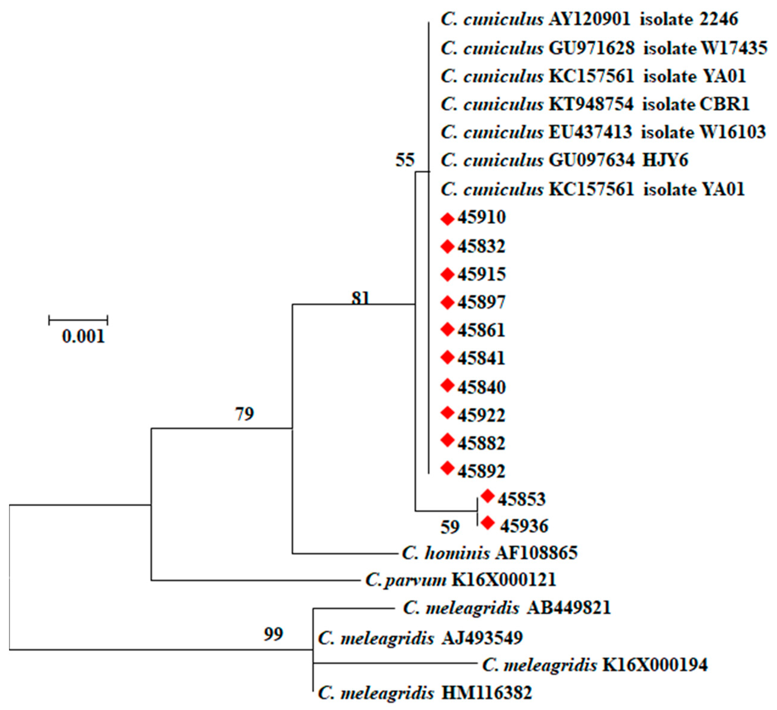
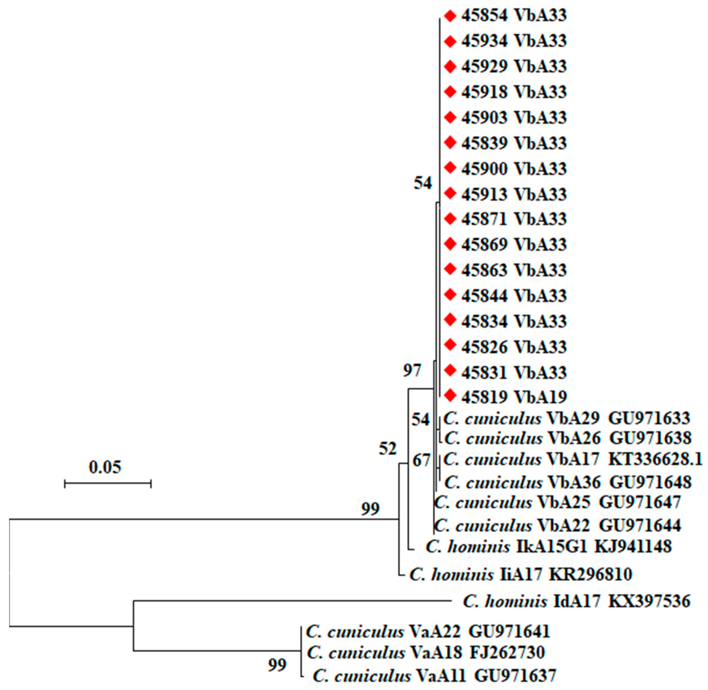
2.2. Cryptosporidium Genotypes and Subtypes

All 28 samples amplified by PCR analysis of the SSU rRNA gene had C. cuniculus by RFLP analysis (Figure 1). They produced two types of nucleotide sequences. Among them, ten sequences were identical to those in GenBank (AY120901, FJ262724, etc.), while two sequences had an A to T substitution near the 5′ end of the partial gene. In the phylogenetic analysis of the SSU rRNA sequences, C. cuniculus sequences obtained from the 12 samples clustered with reference sequences from GenBank (Figure 2). Of the 28 C. cuniculus samples, 16 were successfully subtyped by sequence analysis of the gp60 gene, with two subtypes being identified: VbA19 (n = 1) and VbA33 (n = 15). These sequences were identical to each other in the non-repeat regions but had one A to T substitution compared to sequences (KU852732, GU097641, GU097647, GU971639, etc.) in GenBank (Figure 3). The VbA19 subtype was found only in a 6-month-old rabbit from farm El-Gharbia 3, while the VbA33 subtype was found on other Cryptosporidium-positive farms. Among the samples from four cages of animals with diarrhea, one sample from a cage of 4-month-old rabbits on El-Dakahlia 1 was positive for C. cuniculus VbA33 (Table 3).

3. Discussion

The results of the present study suggest a common occurrence of Cryptosporidium spp. in rabbits in the study areas. In this study, the overall occurrence of Cryptosporidium spp. in rabbits was 11.9% (28/235). This is similar to the infection rates of 11.2% (24/215) in a study of two rabbit farms in Heilongjiang Province, China [35], and 13.2% (14/106) in rabbits residing in Sydney drinking water catchments [36]. It is, however, higher than infection rates found in rabbits from Australia (6.8% or 12/176 and 8.4% or 22/263) [37,38], and Nigeria (3.7% or 4/107) [19], but lower than the infection rate recorded in pet rabbits in Japan (21.9% or 21/96) [16]. Several reports from China showed low infection rates of 1.03% (3/290), 2.4% (9/378), 3.4% (37/1081), and 3.4% (11/321) [13,39,40,41]. The differences in the infection rates of Cryptosporidium spp. among studies may be attributed to differences in sample size, rabbit breeds, management systems, geographic regions, and sample collection seasons. In one study, the infection rate of Cryptosporidium spp. in dead juvenile rabbits suffering from diarrhea was significantly higher than healthy ones (30.3% vs 3.3%) [16]. Among the nine farms examined in the present study, the occurrence of Cryptosporidium spp. on El-Gharbia 2 (24%) was higher than other farms (0–21%), possibly because of the poor hygiene and management practices on the farm.
Like in other animals, the infection rate of Cryptosporidium spp. is significantly higher in rabbits of youngest age. In this study, rabbits of <3 months had a significantly higher Cryptosporidium infection rate than older rabbits. Our findings are in agreement with observations in earlier studies, where the highest prevalence of Cryptosporidium spp. was recorded in young rabbits [13,39]. Similar age-associated occurrence of Cryptosporidium spp. has been reported in humans, cattle, and bamboo rats [33,34,42]. In the present study, a higher detection rate of Cryptosporidium spp. was recorded in Hi-Plus rabbits than Rex and New Zealand ones, possibly because of the high number of samples and sampling of many young animals. In contrast to our results, Rex and New Zealand rabbits were more susceptible than other breeds to Cryptosporidium infection in some earlier studies in China [13,35].
Generally, few clinical signs have been associated with cryptosporidiosis in rabbits, especially adult ones, and the infection is often not recognized due to the asymptomatic oocyst shedding [39,41]. This is in line with our results, where most rabbits were apparently healthy. Although two reports observed fatality in outbreaks of diarrhea in rabbits due to cryptosporidiosis [16,43], Cryptosporidium was detected in only one of the four samples from animals with diarrhea.
All isolates of Cryptosporidium spp. detected in our study were genotyped as C. cuniculus, which is one of the causes of human cryptosporidiosis and has zoonotic significance [44]. In some countries, such as the UK, Australia and New Zealand, many sporadic cases of human cryptosporidiosis have been attributed to infections with C. cuniculus [22,24,25,45]. It was recognized as the third most important Cryptosporidium species causing cryptosporidiosis in humans in the UK during 2007 to 2008 and New Zealand during 2009 to 2019 [24,25]. It was also associated with a waterborne outbreak of cryptosporidiosis due to contamination of treated drinking water by wild rabbits [46,47]. Humans may be infected with C. cuniculus via contaminated water or direct contact with rabbits [22].
In this study, based on gp60 sequence analysis, the C. cuniculus isolates belong to two subtypes (VbA19 and VbA33) in the Vb subtype family. Previously, the VbA19 subtype was isolated from rabbits in the Czech Republic [46], while the VbA33 subtype was detected in humans in the UK [48]. The two subtypes identified in the present study, however, differed from them by one nucleotide in the non-repeat region. Currently, Va and Vb are the only two subtype families within C. cuniculus. Between them, subtypes in the Va subtype family are more commonly seen in humans while those in the Vb subtype family are more commonly seen in rabbits [35]. The occurrence of similar subtypes of C. cuniculus in humans and rabbits supports the zoonotic potential of C. cuniculus [41].
In conclusion, to the best of our knowledge, this is the first study on the genetic identity of Cryptosporidium spp. in rabbits in Egypt. The results of this study suggest a common occurrence of C. cuniculus in farm rabbits in several areas of the country. The detection of C. cuniculus in this study supports the potential role of rabbits as a source of human infections. Further studies from other localities in Egypt are needed to improve our understanding of the clinical and public health significance of Cryptosporidium spp., in rabbits in Egypt.

4. Materials and Methods

4.1. Ethics Statement

Permission was obtained from the owners of the farms before collection of fecal specimens. All fieldwork associated with this study was conducted in compliance with the Guide for the Care and Use of Laboratory Animals in Egypt. The study protocol was approved by the Ethics Committee of the Faculty of Veterinary Medicine, Mansoura University, Egypt.

4.2. Specimen Collections

A total of 235 fresh fecal specimens were collected between July 2015 and April 2016 from nine rabbit farms randomly selected in El-Dakahlia, El-Gharbia, and Damietta provinces in Egypt. All farms sampled in this study were medium-sized farms housing 700–900 rabbits. Fecal specimens were randomly collected from at least 20% of rabbit cages on each farm. Each specimen consisted of 3–5 fresh fecal pellets gathered from each cage. Each collection from each cage (containing 4–7 rabbits) was regarded as one specimen. The fecal pellets were placed into a sterile disposable plastic bag labeled with the age and breed of the animals and sampling date and location. Animals in four cages showed clinical signs of enteric diseases (emaciation, dehydration, and diarrhea) at the time of specimen collection. The rabbits were divided into three age groups: <3-month-old, 3–6-month-old, and >6-month-old. Specimens were stored in 70% ethanol at 4 °C until being transported to the Centers for Disease Control and Prevention, Atlanta, Georgia, USA for DNA extraction and molecular analysis.

4.3. DNA Extraction and PCR Amplification

The fecal specimens were washed twice with distilled water by centrifugation to remove ethanol before DNA extraction. Extraction of genomic DNA from specimens was performed using the FastDNA SPIN Kit for Soil (BIO 101, Carlsbad, CA, USA). The genomic DNA was eluted with 100 μL reagent-grade water and stored at −20 °C until PCR analysis.

4.4. Cryptosporidium Detection, Genotyping and Subtyping

Cryptosporidium spp. in the specimens were detected by nested PCR analysis of a ∼830-bp fragment of the small subunit rRNA (SSU rRNA) gene as previously described [49]. Cryptosporidium species were identified by restriction fragment length polymorphism (RFLP) analysis of the secondary PCR products of SSU rRNA gene using SspI (New England BioLabs, Ipswich, MA, USA) and VspI (Promega, Madison, WI, USA) restriction enzymes [50]. All Cryptosporidium-positive specimens were selected for further subtyping by PCR and sequence analysis of the 60-kDa glycoprotein (gp60) gene [51]. Each specimen was analyzed twice for each genetic target, using C. baileyi DNA as the positive control for the SSU rRNA-based PCR, C. parvum DNA as the positive control for gp60-based PCR, and reagent-grade water as the negative control for both PCR assays.

4.5. DNA Sequence and Phylogenetic Analysis

Montage PCR filters (Millipore, Bedford, MA, USA) were used to purify all secondary PCR products of both genes. The purified products were sequenced in both directions using the secondary PCR primers and Big Dye® Terminator v3.1 Cycle Sequencing Kit (Applied Biosystems, Foster City, CA, USA) on an ABI 3130 Genetic Analyzer (Applied Biosystems). ChromasPro (version 1.5) (www.technelysium.com.au/ChromasPro.html/, accessed on 22 June 2009) was used to edit and assemble the DNA sequences, while ClustalX 2.0.11 (http://www.clustal.org/, accessed on 1 June 2018) was used to align the obtained nucleotide sequences against each other and reference sequences from GenBank to determine the genetic relatedness of various C. cuniculus subtype families. A phylogenetic tree was constructed using the maximum likelihood algorithm implemented in MEGA version 7.0.26 (www.megasoftware.net/, accessed on 1 May 2017) based on substitution rates calculated with the general time reversible model. Bootstrap analysis was applied to evaluate the reliability of cluster formation in the phylogenetic tree with 1000 replicates.

4.6. Statistical Analysis

Differences in infection rates of Cryptosporidium spp. among rabbits of different age groups, localities, and breeds were estimated using the Fisher’s exact test. The SPSS software version 20.0 (IBM, Armonk, NY, USA) was used in the statistical analysis of the data. Differences were considered significant at p ≤ 0.05.

Author Contributions

Conceptualization, D.N., L.X.; methodology, D.N., N.A., D.M.R. and L.X.; software, D.N and L.X.; validation, D.N. and L.X.; formal analysis, D.N.; investigation, D.N.; resources, D.N. and L.X.; data curation, D.N.; writing—original draft preparation, D.N.; writing—review and editing, D.N., N.A., D.M.R. and L.X.; visualization, L.X.; supervision, D.M.R. and L.X.; project administration, D.M.R. and L.X.; funding acquisition, D.N. and L.X. All authors have read and agreed to the published version of the manuscript.

Funding

This study was supported partially by the National Natural Science Foundation of China (31820103014), 111 Project (D20008), Innovation Team Project of Guangdong University (2019KCXTD001), and the Centers for Disease Control and Prevention.

Institutional Review Board Statement

Permission was obtained from the owners of the farms before collections of fecal specimens. All fieldwork associated with this study was conducted in compliance with the Guide for the Care and Use of Laboratory Animals in Egypt. The study protocol was approved by the Ethics Committee of the Faculty of Veterinary Medicine, Mansoura University, Egypt.

Informed Consent Statement

Not applicable.

Data Availability Statement

All relevant data are within the paper.

Acknowledgments

The findings and conclusions in this report are those of the authors and do not necessarily represent the represent of the official position of the U.S. Centers for Disease Control and Prevention.

Conflicts of Interest

The authors declare that they have no conflict of interest.

References

  1. Kotloff, K.L.; Nataro, J.P.; Blackwelder, W.C.; Nasrin, D.; Farag, T.H.; Panchalingam, S.; Wu, Y.; Sow, S.O.; Sur, D.; Breiman, R.F.; et al. Burden and aetiology of diarrhoeal disease in infants and young children in developing countries (the Global Enteric Multicenter Study, GEMS): A prospective, case-control study. Lancet 2013, 382, 209–222. [Google Scholar] [CrossRef]
  2. Meganck, V.; Hoflack, G.; Opsomer, G. Advances in prevention and therapy of neonatal dairy calf diarrhoea: A systematical review with emphasis on colostrum management and fluid therapy. Acta Vet. Scand. 2014, 56, 75. [Google Scholar] [CrossRef] [Green Version]
  3. Platts-Mills, J.A.; Babji, S.; Bodhidatta, L.; Gratz, J.; Haque, R.; Havt, A.; McCormick, B.J.; McGrath, M.; Olortegui, M.P.; Samie, A.; et al. Pathogen-specific burdens of community diarrhoea in developing countries: A multisite birth cohort study (MAL-ED). Lancet Glob. Health 2015, 3, e564–e575. [Google Scholar] [CrossRef] [Green Version]
  4. Efstratiou, A.; Ongerth, J.E.; Karanis, P. Waterborne transmission of protozoan parasites: Review of worldwide outbreaks—An update 2011–2016. Water Res. 2017, 114, 14–22. [Google Scholar] [CrossRef]
  5. Ryan, U.; Hijjawi, N.; Xiao, L. Foodborne cryptosporidiosis. Int. J. Parasitol. 2018, 48, 1–12. [Google Scholar] [CrossRef] [Green Version]
  6. GBD. Estimates of global, regional, and national morbidity, mortality, and aetiologies of diarrhoeal diseases: A systematic analysis for the Global Burden of Disease Study 2015. Lancet Infect. Dis. 2017, 17, 909–948. [Google Scholar] [CrossRef] [Green Version]
  7. Feng, Y.; Ryan, U.M.; Xiao, L. Genetic diversity and population structure of Cryptosporidium. Trends Parasitol. 2018, 34, 997–1011. [Google Scholar] [CrossRef]
  8. Xiao, L. Molecular epidemiology of cryptosporidiosis: An update. Exp. Parasitol. 2010, 124, 80–89. [Google Scholar] [CrossRef] [PubMed]
  9. FAO. Statistical Yearbook; World Food and Agriculture Organization: Rome, Italy, 2013; Available online: https://reliefweb.int/report/world/fao-statistical-yearbook-2013-world-food-and-agriculture (accessed on 19 June 2013).
  10. FAO. FAO Database. 2019. Available online: http://www.faostat.fao.org (accessed on 5 March 2019).
  11. Ni, X.; Qin, S.; Lou, Z.; Ning, H.; Sun, X. Seroprevalence and risk factors of Chlamydia infection in domestic rabbits (Oryctolagus cuniculus) in China. BioMed Res. Int. 2015, 2015, 460473. [Google Scholar] [CrossRef] [PubMed] [Green Version]
  12. Xie, X.; Bil, J.; Shantz, E.; Hammermueller, J.; Nagy, E.; Turner, P.V. Prevalence of lapine rotavirus, astrovirus, and hepatitis E virus in Canadian domestic rabbit populations. Vet. Microbiol. 2017, 208, 146–149. [Google Scholar] [CrossRef] [PubMed]
  13. Zhang, X.; Qi, M.; Jing, B.; Yu, F.; Wu, Y.; Chang, Y.; Zhao, A.; Wei, Z.; Dong, H.; Zhang, L. Molecular characterization of Cryptosporidium spp., Giardia duodenalis, and Enterocytozoon bieneusi in rabbits in Xinjiang, China. J. Eukaryot. Microbiol. 2018, 65, 854–859. [Google Scholar] [CrossRef]
  14. Sturdee, A.P.; Chalmers, R.M.; Bull, S.A. Detection of Cryptosporidium oocysts in wild mammals of mainland Britain. Vet. Parasitol. 1999, 80, 273–280. [Google Scholar] [CrossRef]
  15. Ryan, U.; Xiao, L.; Read, C.; Zhou, L.; Lal, A.A.; Pavlasek, I. Identification of novel Cryptosporidium genotypes from the Czech Republic. Appl. Environ. Microbiol. 2003, 69, 4302–4307. [Google Scholar] [CrossRef] [Green Version]
  16. Shiibashi, T.; Imai, T.; Sato, Y.; Abe, N.; Yukawa, M.; Nogami, S. Cryptosporidium infection in juvenile pet rabbits. J. Vet. Med. Sci. 2006, 68, 281–282. [Google Scholar] [CrossRef] [Green Version]
  17. Robinson, G.; Chalmers, R.M. The European rabbit (Oryctolagus cuniculus), a source of zoonotic cryptosporidiosis. Zoonoses Public Health 2010, 57, e1–e13. [Google Scholar] [CrossRef] [PubMed]
  18. Mosier, D.A.; Cimon, K.Y.; Kuhls, T.L.; Oberst, R.D.; Simons, K.R. Experimental cryptosporidiosis in adult and neonatal rabbits. Vet. Parasitol. 1997, 69, 163–169. [Google Scholar] [CrossRef]
  19. Ayinmode, A.B.; Agbajelola, V.I. Molecular identification of Cryptosporidium parvum in rabbits (Oryctolagus cuniculus) in Nigeria. Ann. Parasitol. 2019, 65, 237–243. [Google Scholar] [CrossRef] [PubMed]
  20. Yang, Z.; Yang, F.; Wang, J.; Cao, J.; Zhao, W.; Gong, B.; Yan, J.; Zhang, W.; Liu, A.; Shen, Y. Multilocus sequence typing and population genetic structure of Cryptosporidium cuniculus in rabbits in Heilongjiang Province, China. Infect. Genet. Evol. 2018, 64, 249–253. [Google Scholar] [CrossRef]
  21. Molloy, S.F.; Kirwan, P.; Asaolu, S.O.; Holland, C.V.; Nichols, R.A.B.; Connelly, L.; Smith, H.V. Identification of a high diversity of Cryptosporidium species genotypes and subtypes in a pediatric population in Nigeria. Am. J. Trop. Med. Hyg. 2010, 82, 608–613. [Google Scholar] [CrossRef] [PubMed] [Green Version]
  22. Koehler, A.V.; Whipp, M.J.; Haydon, S.R.; Gasser, R.B. Cryptosporidium cuniculus--new records in human and kangaroo in Australia. Parasites Vectors 2014, 7, 492. [Google Scholar] [CrossRef]
  23. Martinez-Ruiz, R.; de Lucio, A.; Fuentes, I.; Carmena, D. Autochthonous Cryptosporidium cuniculus infection in Spain: First report in a symptomatic paediatric patient from Madrid. Enferm. Infecc. Microbiol. Clin. 2016, 34, 532–534. [Google Scholar] [CrossRef]
  24. Garcia, R.J.; Pita, A.B.; Velathanthiri, N.; French, N.P.; Hayman, D.T.S. Species and genotypes causing human cryptosporidiosis in New Zealand. Parasitol. Res. 2020, 119, 2317–2326. [Google Scholar] [CrossRef]
  25. Chalmers, R.M.; Elwin, K.; Hadfield, S.J.; Robinson, G. Sporadic human cryptosporidiosis caused by Cryptosporidium cuniculus, United Kingdom, 2007–2008. Emerg. Infect. Dis. 2011, 17, 536–538. [Google Scholar] [CrossRef]
  26. Amer, S.; Honma, H.; Ikarashi, M.; Tada, C.; Fukuda, Y.; Suyama, Y.; Nakai, Y. Cryptosporidium genotypes and subtypes in dairy calves in Egypt. Vet. Parasitol. 2010, 169, 382–386. [Google Scholar] [CrossRef]
  27. Amer, S.; Zidan, S.; Adamu, H.; Ye, J.; Roellig, D.; Xiao, L.; Feng, Y. Prevalence and characterization of Cryptosporidium spp. in dairy cattle in Nile River delta provinces, Egypt. Exp. Parasitol. 2013, 135, 518–523. [Google Scholar] [CrossRef]
  28. El-Badry, A.A.; Al-Antably, A.S.; Hassan, M.A.; Hanafy, N.A.; Abu-Sarea, E.Y. Molecular seasonal, age and gender distributions of Cryptosporidium in diarrhoeic Egyptians: Distinct endemicity. Eur. J. Clin. Microbiol. Infect. Dis. 2015, 34, 2447–2453. [Google Scholar] [CrossRef]
  29. Ibrahim, M.A.; Abdel-Ghany, A.E.; Abdel-Latef, G.K.; Abdel-Aziz, S.A.; Aboelhadid, S.M. Epidemiology and public health significance of Cryptosporidium isolated from cattle, buffaloes, and humans in Egypt. Parasitol. Res. 2016, 115, 2439–2448. [Google Scholar] [CrossRef] [PubMed]
  30. Helmy, Y.A.; Krucken, J.; Nockler, K.; von Samson-Himmelstjerna, G.; Zessin, K.H. Molecular epidemiology of Cryptosporidium in livestock animals and humans in the Ismailia province of Egypt. Vet. Parasitol. 2013, 193, 15–24. [Google Scholar] [CrossRef]
  31. Mahfouz, M.E.; Mira, N.; Amer, S. Prevalence and genotyping of Cryptosporidium spp. in farm animals in Egypt. J. Vet. Med. Sci. 2014, 76, 1569–1575. [Google Scholar] [CrossRef] [Green Version]
  32. Gharieb, R.M.A.; Merwad, A.M.A.; Saleh, A.A.; Abd El-Ghany, A.M. Molecular screening and genotyping of Cryptosporidium species in household dogs and in-contact children in Egypt: Risk factor analysis and zoonotic importance. Vector Borne Zoonotic Dis. 2018, 18, 424–432. [Google Scholar] [CrossRef]
  33. Naguib, D.; El-Gohary, A.H.; Roellig, D.; Mohamed, A.A.; Arafat, N.; Wang, Y.; Feng, Y.; Xiao, L. Molecular characterization of Cryptosporidium spp. and Giardia duodenalis in children in Egypt. Parasites Vectors 2018, 11, 403. [Google Scholar] [CrossRef] [PubMed] [Green Version]
  34. Naguib, D.; El-Gohary, A.H.; Mohamed, A.A.; Roellig, D.M.; Arafat, N.; Xiao, L. Age patterns of Cryptosporidium species and Giardia duodenalis in dairy calves in Egypt. Parasitol. Int. 2018, 67, 736–741. [Google Scholar] [CrossRef] [PubMed]
  35. Yang, Z.; Zhao, W.; Shen, Y.; Zhang, W.; Shi, Y.; Ren, G.; Yang, D.; Ling, H.; Yang, F.; Liu, A.; et al. Subtyping of Cryptosporidium cuniculus and genotyping of Enterocytozoon bieneusi in rabbits in two farms in Heilongjiang Province, China. Parasite 2016, 23, 52. [Google Scholar] [CrossRef]
  36. Zahedi, A.; Monis, P.; Aucote, S.; King, B.; Paparini, A.; Jian, F.; Yang, R.; Oskam, C.; Ball, A.; Robertson, I.; et al. Zoonotic Cryptosporidium species in animals inhabiting Sydney water catchments. PLoS ONE 2016, 11, e0168169. [Google Scholar] [CrossRef] [Green Version]
  37. Nolan, M.J.; Jex, A.R.; Koehler, A.V.; Haydon, S.R.; Stevens, M.A.; Gasser, R.B. Molecular-based investigation of Cryptosporidium and Giardia from animals in water catchments in southeastern Australia. Water Res. 2013, 47, 1726–1740. [Google Scholar] [CrossRef]
  38. Nolan, M.J.; Jex, A.R.; Haydon, S.R.; Stevens, M.A.; Gasser, R.B. Molecular detection of Cryptosporidium cuniculus in rabbits in Australia. Infect. Genet. Evol. 2010, 10, 1179–1187. [Google Scholar] [CrossRef]
  39. Shi, K.; Jian, F.; Lv, C.; Ning, C.; Zhang, L.; Ren, X.; Dearen, T.K.; Li, N.; Qi, M.; Xiao, L. Prevalence, genetic characteristics, and zoonotic potential of Cryptosporidium species causing infections in farm rabbits in China. J. Clin. Microbiol. 2010, 48, 3263–3266. [Google Scholar] [CrossRef] [Green Version]
  40. Liu, X.; Zhou, X.; Zhong, Z.; Chen, W.; Deng, J.; Niu, L.; Wang, Q.; Peng, G. New subtype of Cryptosporidium cuniculus isolated from rabbits by sequencing the gp60 gene. J. Parasitol. 2014, 100, 532–536. [Google Scholar] [CrossRef] [PubMed]
  41. Zhang, W.; Shen, Y.; Wang, R.; Liu, A.; Ling, H.; Li, Y.; Cao, J.; Zhang, X.; Shu, J.; Zhang, L. Cryptosporidium cuniculus and Giardia duodenalis in rabbits: Genetic diversity and possible zoonotic transmission. PLoS ONE 2012, 7, e31262. [Google Scholar] [CrossRef]
  42. Li, F.; Zhang, Z.; Hu, S.; Zhao, W.; Zhao, J.; Kvac, M.; Guo, Y.; Li, N.; Feng, Y.; Xiao, L. Common occurrence of divergent Cryptosporidium species and Cryptosporidium parvum subtypes in farmed bamboo rats (Rhizomys sinensis). Parasites Vectors 2020, 13, 149. [Google Scholar] [CrossRef]
  43. Kaupke, A.; Kwit, E.; Chalmers, R.M.; Michalski, M.M.; Rzezutka, A. An outbreak of massive mortality among farm rabbits associated with Cryptosporidium infection. Res. Vet. Sci. 2014, 97, 85–87. [Google Scholar] [CrossRef]
  44. Ryan, U.; Fayer, R.; Xiao, L. Cryptosporidium species in humans and animals: Current understanding and research needs. Parasitology 2014, 141, 1667–1685. [Google Scholar] [CrossRef] [Green Version]
  45. Elwin, K.; Hadfield, S.J.; Robinson, G.; Chalmers, R.M. The epidemiology of sporadic human infections with unusual cryptosporidia detected during routine typing in England and Wales, 2000–2008. Epidemiol. Infect. 2012, 140, 673–683. [Google Scholar] [CrossRef]
  46. Chalmers, R.M.; Robinson, G.; Elwin, K.; Hadfield, S.J.; Xiao, L.; Ryan, U.; Modha, D.; Mallaghan, C. Cryptosporidium sp. rabbit genotype, a newly identified human pathogen. Emerg. Infect. Dis. 2009, 15, 829–830. [Google Scholar] [CrossRef]
  47. Puleston, R.L.; Mallaghan, C.M.; Modha, D.E.; Hunter, P.R.; Nguyen-Van-Tam, J.S.; Regan, C.M.; Nichols, G.L.; Chalmers, R.M. The first recorded outbreak of cryptosporidiosis due to Cryptosporidium cuniculus (formerly rabbit genotype), following a water quality incident. J. Water Health 2014, 12, 41–50. [Google Scholar] [CrossRef] [Green Version]
  48. Robinson, G.; Wright, S.; Elwin, K.; Hadfield, S.J.; Katzer, F.; Bartley, P.M.; Hunter, P.R.; Nath, M.; Innes, E.A.; Chalmers, R.M. Re-description of Cryptosporidium cuniculus Inman and Takeuchi, 1979 (Apicomplexa: Cryptosporidiidae): Morphology, biology and phylogeny. Int. J. Parasitol. 2010, 40, 1539–1548. [Google Scholar] [CrossRef] [PubMed]
  49. Xiao, L.; Singh, A.; Limor, J.; Graczyk, T.K.; Gradus, S.; Lal, A. Molecular characterization of Cryptosporidium oocysts in samples of raw surface water and wastewater. Appl. Env. Microbiol. 2001, 67, 1097–1101. [Google Scholar] [CrossRef] [Green Version]
  50. Xiao, L.; Lal, A.A.; Jiang, J. Detection and differentiation of Cryptosporidium oocysts in water by PCR-RFLP. Methods Mol. Biol. 2004, 268, 163–176. [Google Scholar] [CrossRef] [PubMed]
  51. Alves, M.; Xiao, L.; Sulaiman, I.; Lal, A.A.; Matos, O.; Antunes, F. Subgenotype analysis of Cryptosporidium isolates from humans, cattle, and zoo ruminants in Portugal. J. Clin. Microbiol. 2003, 41, 2744–2747. [Google Scholar] [CrossRef] [PubMed] [Green Version]
Figure 1. RFLP analysis of PCR products of SSU rRNA gene from Cryptosporidium cuniculus from rabbits using SspI (A) and VspI (B) restriction enzymes. Lane M: 100 bp molecular markers; Lane 1–9: C. cuniculus; Lane 10: positive control (C. baileyi).
Pathogens 10 00775 g001
Figure 2. Phylogenetic relationships among Cryptosporidium spp. based on the nucleotide sequences of the SSU rRNA gene through a maximum likelihood analysis based on substitution rates calculated with the general time reversible model. Numbers at the internodes represent bootstrap values (>50%) from 1000 replicates. The Cryptosporidium cuniculus samples identified in this study are labeled with red rhombus.
Pathogens 10 00775 g002
Figure 3. Phylogenetic tree of Cryptosporidium spp. based on gp60 sequences through a maximum likelihood analysis based on substitution rates calculated with the general time reversible model. Numbers at the internodes represent bootstrap values (>70%) from 1000 replicates. The Cryptosporidium cuniculus samples identified in this study are labeled with red rhombus.
Pathogens 10 00775 g003
Table 1. Distribution of Cryptosporidium spp. in rabbits by farm and age group in El Dakahlia, El-Gharbia, and Damietta provinces, Egypt.
FarmAge (Month)No. of SamplesCryptosporidium spp. *
No. Positive (%)95% Confidence Interval
Lower LimitUpper Limit
El-Dakahlia 1<3102--
3–6111--
>6101--
Subtotal314 (13%)1.1024.69
El-Dakahlia 2<392--
3–672--
>670--
Subtotal234 (17%)1.9032.89
El-Dakahlia 3<3113--
3–6112--
>661--
Subtotal286 (21%)6.2036.59
El-Gharbia 1<380--
3–680--
>6100--
Subtotal260 (0%)0.000.00
El-Gharbia 2<374--
3–6122--
>660--
Subtotal256 (24%)7.2540.74
El-Gharbia 3<3122--
3–6111--
>670--
Subtotal303 (10%)−0.7320.73
Damietta 1<391--
3–6100--
>680--
Subtotal271 (4%)−3.4210.82
Damietta 2<341--
3–681--
>680--
Subtotal202 (10%)−3.1423.14
Damietta 3<391--
3–681--
>680--
Subtotal252 (8%)−2.6318.63
Total-23528 (11.9%)--
* Fisher’s exact test: t = 12.258, p = 0.106.
Table 2. Factors associated with Cryptosporidium infection in rabbits in Egypt.
FactorsSample SizeCryptosporidium spp.
No. Positive
(%)
95% Confidence IntervalFisher’s Exact Testp
Lower LimitUpper Limit
Age
(month)
<37916 (20)11.3829.1111.2370.003
3–68610 (12)4.8518.40
>6702 (3)−1.036.79
BreedRex574 (7)0.3013.602.2830.333
Hi-Plus9815 (15)8.1022.40
New Zealand809 (11)4.3018.20
LocalityEl-Dakahlia8214 (17)8.9025.203.6850.157
El-Gharbia819 (11)4.2017.90
Damietta725 (7)1.0012.70
Table 3. Distribution of 16 Cryptosporidium cuniculus subtypes at the gp60 locus in rabbits in Egypt.
Sample IDC. cuniculus SubtypeAge (Month)LocationBreed
45819VbA196El-Gharbia 3Hi-Plus
45826VbA331El-Dakahlia 1Rex
45831 *VbA334El-Dakahlia 1Rex
45834VbA334Damietta 3New Zealand
45839VbA333Damietta 2Hi-Plus
45844VbA334El-Gharbia 2Hi-Plus
45854VbA333El-Gharbia 2Hi-Plus
45863VbA332El-Gharbia 2Hi-Plus
45869VbA332El-Gharbia 2Hi-Plus
45871VbA331El-Gharbia 2Hi-Plus
45900VbA331Damietta 3New Zealand
45903VbA332El-Dakahlia 3New Zealand
45913VbA331El-Dakahlia 3New Zealand
45918VbA331El-Dakahlia 3New Zealand
45929VbA331El-Dakahlia 2Hi-Plus
45934VbA332El-Dakahlia 2Hi-Plus
* From rabbit with diarrhea.
Publisher’s Note: MDPI stays neutral with regard to jurisdictional claims in published maps and institutional affiliations.

