
Human Anthrax in Dolj County, Romania—A Series of Three Cases


 ,
,
Abstract
:1. Introduction
2. Results
2.1. Case 1
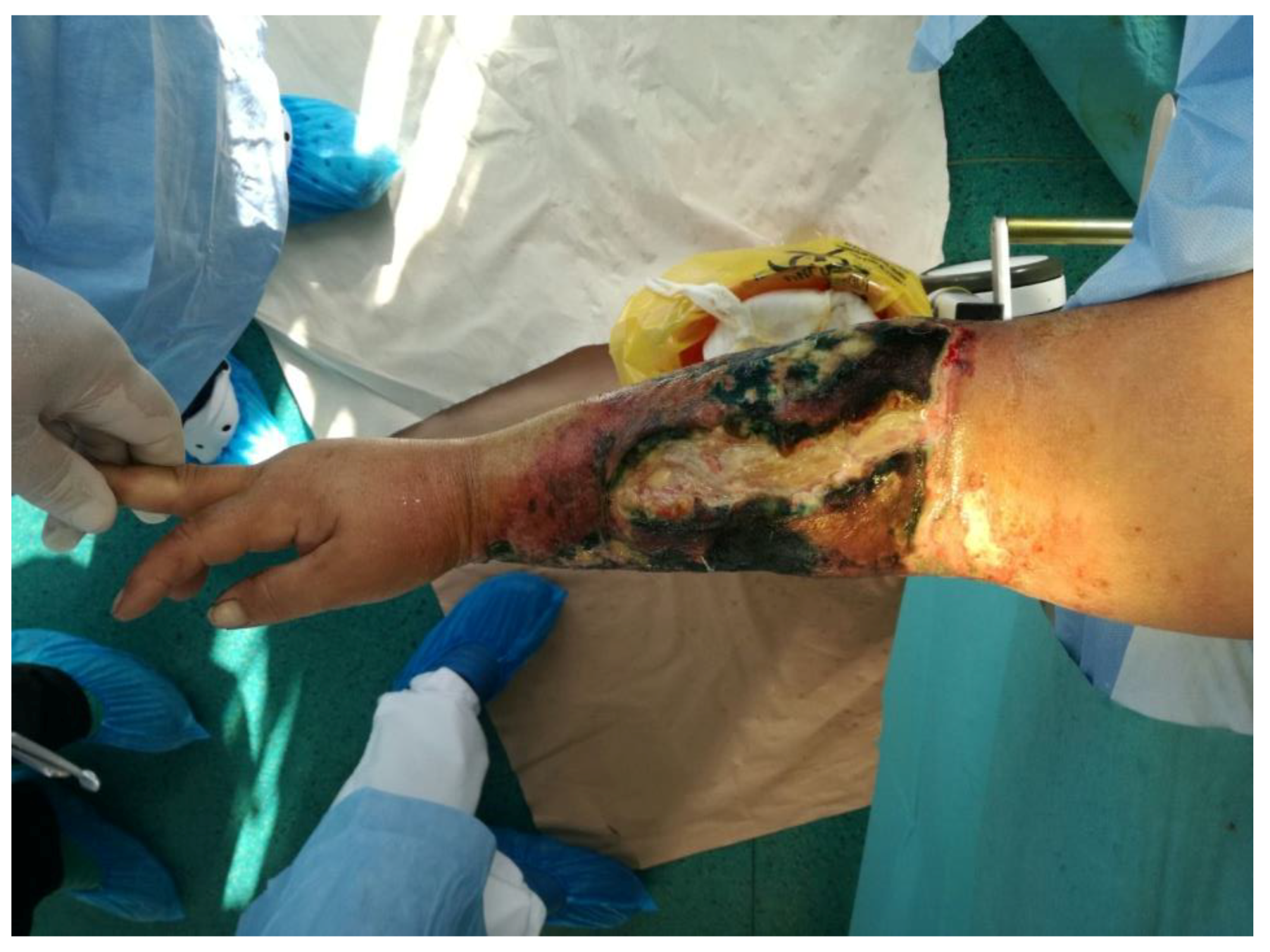
2.2. Case 2
2.3. Case 3
3. Discussion
4. Materials and Methods
5. Conclusions
Author Contributions
Funding
Institutional Review Board Statement
Informed Consent Statement
Data Availability Statement
Conflicts of Interest
References
- World Health Organization. Anthrax in Humans and Animals, 4th ed.; World Health Organization: Geneva, Switzerland, 2008; pp. 4–82. [Google Scholar]
- Goel, A.K. Anthrax: A disease of biowarfare and public health importance. World J. Clin. Cases 2015, 3, 20–33. [Google Scholar] [CrossRef] [PubMed]
- Antonation, K.S.; Grützmacher, K.; Dupke, S. Bacillus cereus biovar anthracis causing anthrax in sub-Saharan Africa—Chromosomal monophyly and broad geographic distribution. PLoS Negl. Trop. Dis. 2016, 10, e000492. [Google Scholar] [CrossRef] [PubMed]
- Cupșa, A. Boli Infecțioase Transmisibile; Editura Medicală Universitară Craiova: Craiova, Romania, 2007; pp. 15.7–15.13. [Google Scholar]
- Cennimo, D.J. Anthrax. 2021. Available online: https://emedicine.medscape.com/article/212127-overview (accessed on 5 April 2021).
- Giubelan, L.; Dumitrescu, F.; Dragonu, L.; Stoian, A.C. Boli Infecțioase; Editura Medicală Universitară Craiova: Craiova, Romania, 2020; pp. 348–354. [Google Scholar]
- Ozkurt, Z.; Parlak, M.; Tastan, R.; Dinler, U.; Saglam, Y.S.; Ozyurek, S.F. Anthrax in eastern Turkey, 1992–2004. Emerg. Infect. Dis. 2005, 11, 1939–1941. [Google Scholar] [CrossRef] [PubMed] [Green Version]
- Doganay, M. Anthrax. In Infectious Diseases, 3rd ed.; Cohen, J., Opal, S.M., Powderly, W.G., Eds.; Mosby-Elsevier: Beijing, China, 2010; pp. 1257–1261. [Google Scholar]
- European Centre for Disease Prevention and Control. Annual Epidemiological Report 2016—Anthrax. Available online: https://ecdc.europa.eu/en/publications-data/anthrax-annual-epidemiological-report-2016-2014-data (accessed on 5 October 2017).
- 2010 Case Definition. Available online: https://wwwn.cdc.gov/nndss/conditions/anthrax/case-definition/2010/ (accessed on 1 May 2021).
- Godyn, J.J.; Reyes, L.; Siderits, R.; Hazra, A. Cutaneous anthrax: Conservative or surgical treatment? Adv. Skin Wound Care 2005, 18, 146–150. [Google Scholar] [CrossRef] [PubMed]
- Siloşi, C.A.; Siloşi, I.; Pădureanu, V.; Bogdan, M.; Mogoantă, S.S.; Ciurea, M.E.; Cojocaru, M.; Boldeanu, L.; Avrămescu, C.S.; Boldeanu, M.V.; et al. Sepsis and identification of reliable biomarkers for postoperative period prognosis. Rom. J. Morphol. Embryol. 2018, 59, 77–91. [Google Scholar] [PubMed]
- Tuncali, D.; Akbuga, U.B.; Aslan, G. Cutaneous anthrax of the hand: Some clinical observations. Indian J. Plast. Surg. 2004, 37, 131–133. [Google Scholar] [CrossRef] [Green Version]
- Bal, A.; Gökdemir, O. Anthrax: A case report. J. Pak. Med. Assoc. 2014, 64, 1201–1202. [Google Scholar] [PubMed]
- Aslan, G.; Terzioglu, A. Surgical management of cutaneous anthrax. Ann. Plast. Surg. 1998, 41, 468–470. [Google Scholar] [PubMed]
- Turnbull, P.C.; Sirianni, N.M.; LeBron, C.I.; Samaan, M.N.; Sutton, F.N.; Reyes, A.E.; Peruski, L.F. MICs of selected antibiotics for Bacillus anthracis, Bacillus cereus, Bacillus thuringiensis and Bacillus mycoides from a range of clinical and environmental sources as determined by the Etest. J. Clin. Microbiol. 2004, 42, 3626–3634. [Google Scholar] [CrossRef] [PubMed] [Green Version]
- Martin, G.J.; Friedlander, A.M. Bacillus anthracis (Anthrax). In Mandell, Douglas, and Bennett’s Principles and Practice of Infectious Diseases, 8th ed.; Churchill Livingstone: London, UK, 2014; pp. 2391–2409. [Google Scholar]
- Kobuch, W.E.; Davis, J.; Fleischer, K.; Isaacson, M.; Turnbull, P.C.B. A clinical and epidemiological study of 621 patients with anthrax in western Zimbabwe. Salisb. Med. Bull. 1990, 68, 34–38. [Google Scholar]
- Friedlander, A.M. Anthrax: Clinical features, pathogenesis, and potential biological warfare threat. Curr. Clin. Top. Infect. Dis. 2000, 20, 335–349. [Google Scholar] [PubMed]
- Harrison, L.H.; Ezzell, J.W.; Abshire, T.G.; Kidd, S.; Kaufmann, A.F. Evaluation of serologic tests for diagnosis of anthrax after an outbreak of cutaneous anthrax in Paraguay. J. Infect. Dis. 1989, 160, 706–710. [Google Scholar] [CrossRef] [PubMed]
- Drăgoescu, A.N.; Pădureanu, V.; Stănculescu, A.D.; Chiuțu, L.C.; Florescu, D.N.; Gheonea, I.A.; Pădureanu, R.; Stepan, A.; Streba, C.T.; Drocaș, A.I.; et al. Presepsin as a potential prognostic marker for sepsis according to actual practice guidelines. J. Pers. Med. 2020, 11, 2. [Google Scholar] [CrossRef] [PubMed]


| Laboratory Test Report | Normal Range | Unit | At Admission | After 4 Days | After 7 Days | After 10 Days | At Discharge |
|---|---|---|---|---|---|---|---|
| White blood cells (WBC) | 4.0–9.0 | ×103/mm3 | 13.4 | 22.3 | 20.5 | 15.8 | 10.9 |
| Mature neutrophils | 34–67 | % | 77 | 79 | 76 | 55 | 65 |
| Immature neutrophils | 0–3.0 | % | 7 | 8 | 12 | 12 | 4 |
| Eosinophils | 0–5 | % | 0 | 0 | 1 | 3 | 7 |
| Basophils | 0–1 | % | 0 | 0 | 0 | 0 | 1 |
| Lymphocytes | 20–50 | % | 12 | 8 | 7 | 24 | 16 |
| Monocytes | 4–8 | % | 4 | 5 | 4 | 6 | 7 |
| Red blood cells (RBC) | 3.50–4.50 | ×106/mm3 | 4.91 | 4.52 | 4.04 | 3.97 | 3.84 |
| Hemoglobin (HGB) | 11.5–14.5 | g/dL | 14.5 | 13.4 | 12.1 | 12 | 11.4 |
| Hematocrit (HCT) | 35.0–43.0 | % | 46.4 | 42.9 | 38.5 | 34.6 | 34.6 |
| Mean corpuscular volume (MCV) | 75.0–95.0 | μm3 | 95 | 95 | 95 | 87 | 90 |
| Mean corpuscular hemoglobin (MCH) | 26.0–32.0 | pg | 29.5 | 29.6 | 30 | 30.3 | 29.7 |
| Mean corpuscular hemoglobin concentration (MCHC) | 31.0–37.9 | g/dL | 31.2 | 31.2 | 31.5 | 34.8 | 32.9 |
| Red cell distribution width (RDW) | 11.0–16.0 | % | 11.3 | 11.4 | 11.4 | 11.3 | 12.1 |
| Platelets (PLT) | 150–450 | ×103/mm3 | 187 | 167 | 213 | 291 | 276 |
| Mean platelet volume (MPV) | 9.0–13.0 | μm3 | 8.1 | 7.7 | 7.7 | 7.3 | 7.6 |
| Erythrocyte sedimentation rate (ESR) | 1–12/1 h | mm/hour | 16 | 80 | 20 | ||
| 4–25/2 h | 33 | 100 | 44 | ||||
| Fibrinogen | 150–400 | mg/dL | 451 | 251 | |||
| INR | 0.98 | 1.01 | 0.97 | 0.99 | 0.96 | ||
| GPT | 10–35 | U/L | 20.3 | 24.6 | 72.3 | 79.3 | 78.9 |
| GOT | 0–32 | U/L | 23.6 | 24 | 55 | 34.6 | 36.2 |
| Na+ | 136–145 | mmol/L | 130.1 | 127.1 | 129.8 | 127.6 | 129.5 |
| K+ | 3.30–5.10 | mmol/L | 3.53 | 3.79 | 3.89 | 4.07 | 4.73 |
| Serum total protein | 66–87 | g/L | 47.7 | 46.2 | 68 | ||
| Blood glucose level | 70–115 | mg/dL | 126.2 | 127 | 121.6 | 64.8 | 89.3 |
| Serum creatinine | 0.50–0.90 | mg/dL | 0.68 | 0.75 | 0.63 | 0.50 | 0.48 |
| Serum urea | 10–50 | mg/dL | 30.3 | 27 | 38.8 | 51.9 | 36.8 |
| Total bilirubin | 0–1 | mg/dL | 0.25 | 0.2 | 0.14 | 0.17 | |
| C-Reactive Protein | <10 | mg/L | 12 | <6 | |||
| Procalcitonin | <0.25 | ng/mL | <0.25 |
| Case | Clinical Aspect | Gram Smear/Culture | Serological Tests in Dynamics | Epidemiological Link |
|---|---|---|---|---|
| 1 | Cutaneous anthrax | Negative | Positive | Yes |
| 2 | Malignant edema | Positive | Positive | Yes |
| 3 | Cutaneous anthrax | Negative | Negative | Yes |
| Case | Culture | Gram Smear | Previous Antibiotic | Antibodies anti B. anthracis at Admission | Antibodies anti B. anthracis after 10–14 (Days) |
|---|---|---|---|---|---|
| 1 | Negative (a) | Negative (a) | Yes | Negative (b) | Positive (T = 1/10) (b) |
| 2 | Positive (a, b) | Positive (a, b) | No | Negative (b) | Positive (T = 1/10) (b) |
| 3 | Negative (a) | Negative (a) | Yes | Negative(b) | Negative (b) |
Publisher’s Note: MDPI stays neutral with regard to jurisdictional claims in published maps and institutional affiliations. |
© 2021 by the authors. Licensee MDPI, Basel, Switzerland. This article is an open access article distributed under the terms and conditions of the Creative Commons Attribution (CC BY) license (https://creativecommons.org/licenses/by/4.0/).
Share and Cite
Dumitrescu, F.; Georgescu, E.F.; Giubelan, L.; Pădureanu, V.; Stoian, A.C.; Dincă, V.; Georgescu, M.; Dragonu, L.; Marinescu, D. Human Anthrax in Dolj County, Romania—A Series of Three Cases. Pathogens 2021, 10, 644. https://doi.org/10.3390/pathogens10060644
Dumitrescu F, Georgescu EF, Giubelan L, Pădureanu V, Stoian AC, Dincă V, Georgescu M, Dragonu L, Marinescu D. Human Anthrax in Dolj County, Romania—A Series of Three Cases. Pathogens. 2021; 10(6):644. https://doi.org/10.3390/pathogens10060644
Chicago/Turabian StyleDumitrescu, Florentina, Eugen Florin Georgescu, Lucian Giubelan, Vlad Pădureanu, Andreea Cristina Stoian, Viorica Dincă, Milena Georgescu, Livia Dragonu, and Daniela Marinescu. 2021. "Human Anthrax in Dolj County, Romania—A Series of Three Cases" Pathogens 10, no. 6: 644. https://doi.org/10.3390/pathogens10060644