
Cutaneous Granulomatosis Revealing Whipple’s Disease: Value of Tropheryma whipplei Polymerase Chain Reaction Assay for the Diagnosis

 , , ,
, , , 
Abstract
:1. Introduction
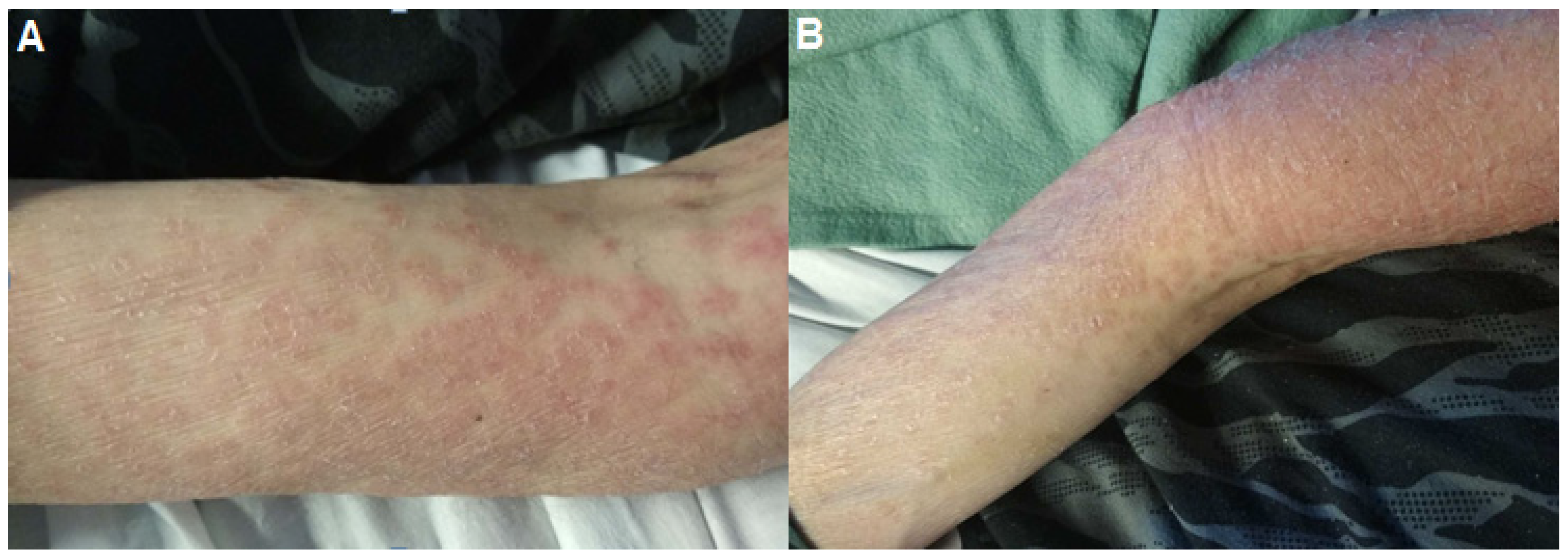
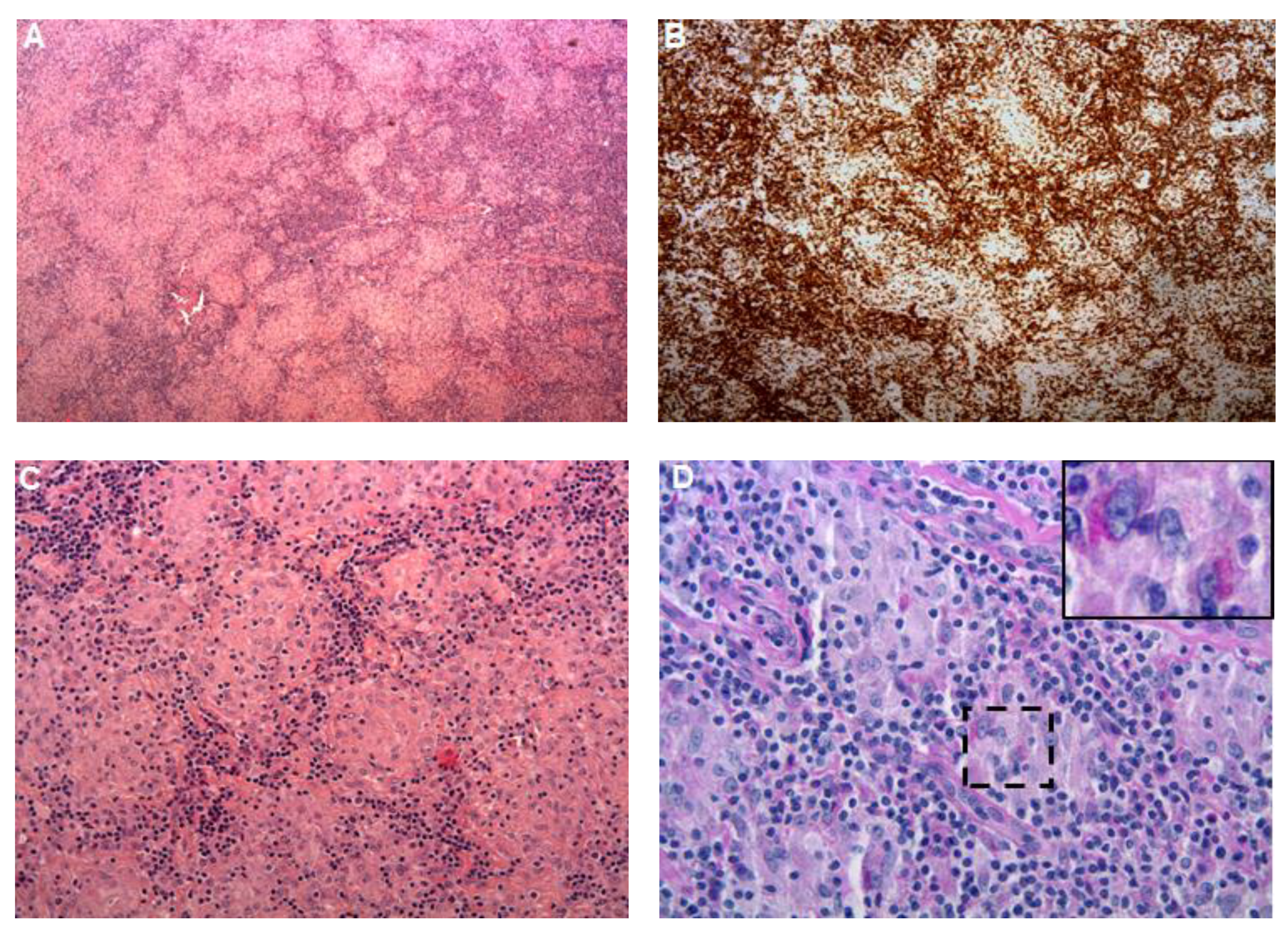
2. Observation
3. Discussion
4. Conclusions
Author Contributions
Funding
Institutional Review Board Statement
Informed Consent Statement
Data Availability Statement
Conflicts of Interest
References
- Antunes, C.; Singhal, M. Whipple Disease. In StatPearls; StatPearls Publishing: Treasure Island, FL, USA, 2021. Available online: http://www.ncbi.nlm.nih.gov/books/NBK441937/ (accessed on 9 July 2021).
- Marth, T.; Moos, V.; Müller, C.; Biagi, F.; Schneider, T. Tropheryma whipplei infection and Whipple’s disease. Lancet Infect. Dis. 2016, 16, 13–22. [Google Scholar] [CrossRef]
- Boumaza, A.F.; Arrindell, J.; Ben Azzouz, E.; Desnues, B. Phenotypic diversity of Tropheryma whipplei clinical isolates. Microb. Pathog. 2021, 158, 105074. [Google Scholar] [CrossRef] [PubMed]
- Dolmans, R.A.V.; Boel, C.H.E.; Lacle, M.M.; Kusters, J.G. Clinical Manifestations, Treatment, and Diagnosis of Tropheryma whipplei Infections. Clin. Microbiol. Rev. 2017, 30, 529–555. [Google Scholar] [CrossRef] [PubMed] [Green Version]
- Krusche, M.; Boro, D.; Bertolini, J.; Kötter, I. Rare erosive arthritis and dermatitis syndrome in Whipple’s disease. Z. Rheumatol. 2019, 78, 180–182. [Google Scholar] [CrossRef] [PubMed]
- Guérin, A.; Kerner, G.; Marr, N.; Markle, J.G.; Fenollar, F.; Wong, N.; Boughorbel, S.; Avery, D.T.; Ma, C.S.; Bougarn, S.; et al. IRF4 haploinsufficiency in a family with Whipple’s disease. eLife 2018, 7, e32340. [Google Scholar] [CrossRef] [PubMed]
- Borretta, L.; Walsh, N.M.; Bakowsky, V.; Arnason, T.; Croul, S.; Pasternak, S. A Case of Whipple Disease With Cutaneous Manifestations. Am. J. Dermatopathol. 2021, 43, e104–e106. [Google Scholar] [CrossRef] [PubMed]
- Canal, L.; de la Fuente, D.; Rodriguez-Moreno, J.; Penin, R.M.; Marcoval, J. Specific cutaneous involvement in Whipple disease. Am. J. Dermatopathol. 2014, 36, 344–346. [Google Scholar] [CrossRef] [PubMed]
- Friedmann, A.C.; Perera, G.K.; Jayaprakasam, A.; Forgacs, I.; Salisbury, J.R.; Creamer, D. Whipple’s disease presenting with symmetrical panniculitis. Br. J. Dermatol. 2004, 151, 907–911. [Google Scholar] [CrossRef]
- Frenk, E.; Merot, Y.; Perez, I.; Chamot, A.M.; Gerster, J.C. Whipple’s disease with sarcoidosis-like cutaneous manifestations. Ann. Dermatol. Venereol. 1991, 118, 115–118. [Google Scholar] [PubMed]
- Cho, C.; Linscheer, W.G.; Hirschkorn, M.A.; Ashutosh, K. Sarcoidlike granulomas as an early manifestation of Whipple’s disease. Gastroenterology 1984, 87, 941–947. [Google Scholar] [CrossRef]
- Sanchez, A.; Del Giudice, P.; Mantion, C.; Mazellier, S.; Boukari, F.; Roger, P.-M.; Courjon, J. Erythematous skin nodules during treatment of Whipple’s disease. Infect. Dis. Now 2021, 51, 397–399. [Google Scholar] [CrossRef] [PubMed]
- Schaller, J.; Carlson, J.A. Erythema nodosum-like lesions in treated Whipple’s disease: Signs of immune reconstitution inflammatory syndrome. J. Am. Acad. Dermatol. 2009, 60, 277–288. [Google Scholar] [CrossRef] [PubMed]
- Totschnig, D.; Seitz, T.; Zoufaly, A.; Hagenauer-Drektraan, S.; Wenisch, C. Whipple’s disease diagnosed in a patient with suspected sarcoidosis. Int. J. Infect. Dis. IJID Off. Publ. Int. Soc. Infect. Dis. 2021, 106, 41–42. [Google Scholar] [CrossRef]
- Walters, S.; Valliani, T.; Przemioslo, R.; Rooney, N. Whipple’s disease: An unexpected finding in a peripheral lymph node biopsy. Lancet Lond. Engl. 2014, 383, 2268. [Google Scholar] [CrossRef]
- Geissdörfer, W.; Moos, V.; Moter, A.; Loddenkemper, C.; Jansen, A.; Tandler, R.; Morguet, A.J.; Fenollar, F.; Raoult, D.; Bogdan, C.; et al. High frequency of Tropheryma whipplei in culture-negative endocarditis. J. Clin. Microbiol. 2012, 50, 216–222. [Google Scholar] [CrossRef] [PubMed] [Green Version]
- Edouard, S.; Fenollar, F.; Raoult, D. The rise of Tropheryma whipplei: A 12-year retrospective study of PCR diagnoses in our reference center. J. Clin. Microbiol. 2012, 50, 3917–3920. [Google Scholar] [CrossRef] [PubMed] [Green Version]
- Günther, U.; Moos, V.; Offenmüller, G.; Oelkers, G.; Heise, W.; Moter, A.; Loddenkemper, C.; Schneider, T. Gastrointestinal diagnosis of classical Whipple disease: Clinical, endoscopic, and histopathologic features in 191 patients. Medicine 2015, 94, e714. [Google Scholar] [CrossRef] [PubMed]
- Lepidi, H.; Fenollar, F.; Gerolami, R.; Mege, J.-L.; Bonzi, M.-F.; Chappuis, M.; Sahel, J.; Raoult, D. Whipple’s disease: Immunospecific and quantitative immunohistochemical study of intestinal biopsy specimens. Hum. Pathol. 2003, 34, 589–596. [Google Scholar] [CrossRef]
- Baisden, B.L.; Lepidi, H.; Raoult, D.; Argani, P.; Yardley, J.H.; Dumler, J.S. Diagnosis of Wihipple disease by immunohistochemical analysis: A sensitive and specific method for the detection of Tropheryma whipplei (the Whipple bacillus) in paraffin-embedded tissue. Am. J. Clin. Pathol. 2002, 118, 742–748. [Google Scholar] [CrossRef] [PubMed] [Green Version]
- Kutlu, O.; Şengiz, E.S.; Gökden, Y.; Kandemir, Ö.; Tükek, T. Whipple′s Disease: A Case Report. Med. Princ. Pract. 2020, 29, 90–93. [Google Scholar] [CrossRef] [PubMed]


Publisher’s Note: MDPI stays neutral with regard to jurisdictional claims in published maps and institutional affiliations. |
© 2021 by the authors. Licensee MDPI, Basel, Switzerland. This article is an open access article distributed under the terms and conditions of the Creative Commons Attribution (CC BY) license (https://creativecommons.org/licenses/by/4.0/).
Share and Cite
Zayet, S.; Isnard, P.; Bustamante, J.; Boutboul, D.; Abroug, S.; Belfeki, N. Cutaneous Granulomatosis Revealing Whipple’s Disease: Value of Tropheryma whipplei Polymerase Chain Reaction Assay for the Diagnosis. Pathogens 2021, 10, 1438. https://doi.org/10.3390/pathogens10111438
Zayet S, Isnard P, Bustamante J, Boutboul D, Abroug S, Belfeki N. Cutaneous Granulomatosis Revealing Whipple’s Disease: Value of Tropheryma whipplei Polymerase Chain Reaction Assay for the Diagnosis. Pathogens. 2021; 10(11):1438. https://doi.org/10.3390/pathogens10111438
Chicago/Turabian StyleZayet, Souheil, Pierre Isnard, Jacinta Bustamante, David Boutboul, Sarra Abroug, and Nabil Belfeki. 2021. "Cutaneous Granulomatosis Revealing Whipple’s Disease: Value of Tropheryma whipplei Polymerase Chain Reaction Assay for the Diagnosis" Pathogens 10, no. 11: 1438. https://doi.org/10.3390/pathogens10111438