1. Introduction
Mosquito-borne diseases continue to be a significant health concern, causing millions of deaths globally. Traditional broad-spectrum methods of vector control, such as insecticide spraying and bed nets, have proven to be effective but have limitations in terms of sustainability and the emergence of insecticide resistance. The discovery of RNA interference (RNAi)—a virus defense tool naturally found in eukaryotic organisms [
1]—offered an opportunity for the development of species-specific approaches for mosquito control.
RNAi can be used to manipulate gene expression via post-translational gene silencing by degrading or inhibiting the translation of gene-specific messenger RNA (mRNA) molecules [
2]. In mosquitoes, the RNAi pathway has been exploited for decades to study gene function, as well as for the development of RNAi-based vector and pathogen management approaches [
3]. Methods of dsRNA delivery in mosquitoes are diverse and vary according to their intended application.
The development of field-relevant RNAi-based mosquito management strategies often exploits the natural feeding behavior of mosquitoes for dsRNA delivery. Mosquito larvae feed on microorganisms (e.g., bacteria, yeasts) in their aquatic habitats, and these can be genetically engineered to produce dsRNA that silences the expression of critical fitness-related genes. For example, in
Aedes aegypti, two chitin synthase genes were successfully knocked down by adding bacterial lysate containing dsRNA into rearing water [
4]. In another species,
Ae. albopictus, a viral (densovirus)-mediated interference system was developed for larvae, downregulating V-ATPase synthesis and causing high mortality [
5]. Microbial methods are also being developed for adult control. In
Anopheles gambiae, dsRNA in bacterial lysate mixed with glucose solution was used as a sugar source for feeding adult mosquitoes [
4,
6]. Mysore and co-authors [
7] have also employed genetically engineered yeast that produced short hairpin RNAs (shRNAs) to suppress
Rbfox1 genes in larvae and adult
Aedes albopictus,
Anopheles gambiae, and
Culex quinquefasciatus via feeding, causing high larval and adult mortality.
Alternative nonmicrobial feeding-based delivery methods include feeding larval and adult stages with nanoparticles as dsRNA carriers. Nanoparticles can protect dsRNA from degradation by endogenic nucleases and improve its stability during digestion processes in the midgut. The potential of various nanoparticles has been explored, such as chitosan, carbon quantum dots, and silica nanoparticles [
8,
9,
10,
11], and Effectene (transfection reagent)-based liposomes [
8]. While nanoparticles offer great potential in field applications, challenges remain in terms of optimizing nanoparticle formulations for maximum dsRNA loading, stability, and appropriate release within the mosquito body. The efficiency and specificity of dsRNA uptake by feeding varies according to life stage, and feeding-based dsRNA delivery is not feasible for the management of mosquitoes in embryonic or pupal (non-feeding) life stages.
To study gene function, the delivery of dsRNA into mosquitoes may occur by feeding but also by microinjection, electroporation, or soaking. Microinjection can be performed at virtually every life stage, starting from an embryo, and is the most common technique used for gene knockdown in mosquitoes [
9,
10,
12]. Although effective, microinjection involves physically puncturing the mosquito cuticle with a fine needle and is a delicate procedure that requires considerable skill and precision. The procedure can cause localized tissue damage, which may affect both mortality rates and experimental outcomes [
10,
12,
13]. High mortality rates have been recorded for microinjections of
Culex pupae [
12,
13,
14], for example. Electroporation utilizes an electrical pulse to facilitate dsRNA delivery. It was first successfully employed in Odonata larvae and later used to facilitate the knockdown of
AsCPR128 in
An. sinensis [
15]. Both methods may be challenging to scale up for high-throughput testing of gene function or screening for potential vector control targets, although microinjections have been used for this purpose [
16].
Soaking is an alternative method of dsRNA delivery that involves immersing mosquito larvae or pupae in a solution containing dsRNA. The dsRNA can penetrate exposed cell membranes and reach the cytoplasm, thus initiating the RNAi pathway. Soaking assays are less technically challenging compared to microinjections because they do not require expensive equipment and can also target the non-feeding pupal stage. Double-stranded RNA delivery by soaking has been efficiently used in several studies. For example, soaking was first applied to first instar
Ae. aegypti by Singh and co-authors. They suppressed the expression of
chitin synthase-1 and
-2,
heat shock protein 83, and
β-tubulin genes by soaking larvae for 2 h in dsRNA [
17]. This same delivery method was successfully used to knock down the expression of fertility genes in
Ae. aegypti by soaking first through fourth instars [
18]. Another study demonstrated the knockdown of the gene encoding
heat shock protein 90 in
Cx. pipiens by dehydration of larvae in salt solution and subsequent rehydration in dsRNA [
19].
Most of the above-listed approaches target genes expressed in the larval and adult stages. However, functional studies for genes initially expressed in the pupal stage and critical for adult development are limited because this life stage does not feed and is fragile. Recently, the
CYP4G35 gene, which is expressed at high levels in the pupal stage, was efficiently suppressed in
Ae. aegypti pupae simply by soaking them in water with added dsRNA. This effect lasted into early adulthood [
20]. To our knowledge, no studies have yet used soaking for dsRNA delivery to study gene function in
Culex pupae.
Here, we propose a rehydration-driven soaking assay that can be utilized for the efficient delivery of dsRNA in life stages and organisms where microinjection and feeding protocols are challenging or impractical. Soaking is an attractive approach to dsRNA delivery, particularly when screening large numbers of pupae or genes because it reduces the handling time of individual insects over microinjections. As proof of concept for our dsRNA delivery method, we targeted
laccase 2, a gene encoding an enzyme that plays a crucial role in the melanin synthesis pathway, in
Cx. pipiens. Laccase 2 is involved in the final steps of melanin synthesis and utilizes molecular oxygen to oxidize phenolic substrates. This oxidative reaction generates reactive quinones, which can then undergo polymerization to produce melanin [
21]. The knockdown of
laccase 2 expression should disrupt melanin production and consequently impair crucial physiological functions in mosquitoes. Based on previous studies of
Tribolium castaneum [
22] and
An. sinensis [
23],
laccase 2 showed promise as a gene target for this silencing proof-of-concept study. Knockdown mutants with reduced
laccase 2 expression showed defects in melanization. Moreover, in
An. sinensis, the pale cuticle was significantly thinner in
dsAsLac2 mosquitoes, and the immune response to pathogens was weakened. We reasoned that if
laccase 2 has a similar functional role in
Cx. pipiens, the defects resulting from the knockdown of expression would allow for easy phenotypic scoring following dsRNA delivery via soaking. While
laccase 2 served as a straightforward gene target for our proof of concept, the manipulation of
laccase 2 expression can also aid in understanding its specific physiological role in
Cx. pipiens and potential application to mosquito control.
Cx. pipiens pupae are able to survive short periods of desiccation. We leveraged this to silence laccase 2 expression by the dehydration and subsequent soaking of newly molted pupae in concentrated dsRNA. The knockdown of laccase 2 expression resulted in visible alterations in mosquito phenotype and fitness. The pupae in our assay had much lower mortality rates compared to other assays, perhaps because dehydration, soaking, and rearing were performed in the same compartment, reducing handling. Our work establishes pupal dehydration and soaking as a feasible approach for studying genes that are expressed and functional during the pupal stage.
2. Materials and Methods
2.1. Mosquito Rearing
Mosquito colonies (
Cx. pipiens aboveground Northfield and belowground Cal1 populations) were maintained at 25 °C and 60–70% humidity under a photoperiod of 16 h light–8 h dark cycle. Larvae were fed with bovine liver powder every 48 h and kept at low density to maximize the number of simultaneously pupating individuals. Further details on collection and rearing procedures for these strains can be found in Noreuil and Fritz [
24].
2.2. Analysis of Laccase Protein Sequence and Structure
To identify the
laccase 2 gene among 5 laccase orthologs in
Cx. quinquefasciatus, all homologous
Culex laccase sequences were retrieved from the NCBI database (
http://www.ncbi.nlm.nih.gov/, accessed on 1 April 2022) and Vectorbase (
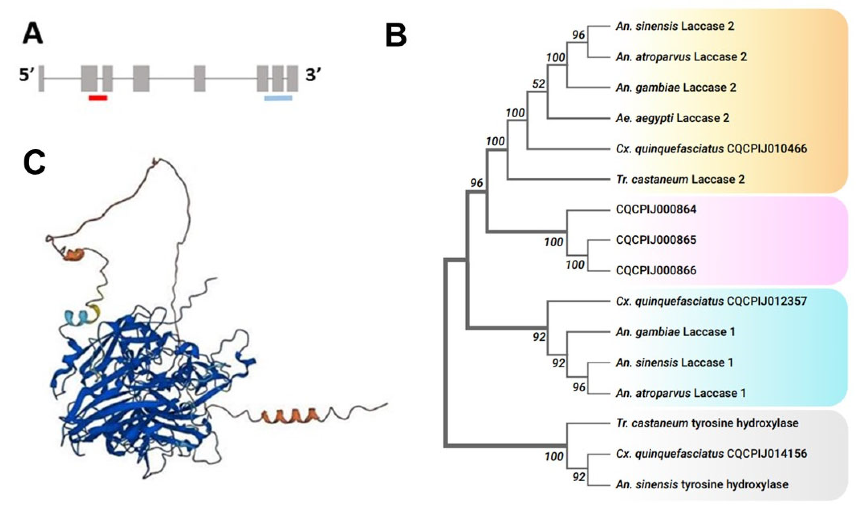
http://www.vectorbase.org/, accessed on 1 April 2022) using the BLASTP program. They included CPIJ012357-PA, CPIJ010466-PA, CPIJ000864-PA, CPIJ000865-PA, and CPIJ000866-PA. We also obtained orthologous Laccase 2 sequences from other insects with the following accession numbers:
Anopheles gambiae (Lac1: AGAP003738-PA; Lac2:AGAP006176-PB),
Anopheles atroparvus (Lac1: AATR020818-PA; Lac2: AATR020769-PA),
Anopheles sinensis (ASIC018958-PA),
Aedes aegypti (Lac1: AAE026602; Lac2: AAE016992-PA), and
Tribolium castaneum (Lac1: NP_001034514; Lac2: NP_001034487). Tyrosine hydroxylase amino acid sequences from
Cx. quinquefasciatus,
Tr. castaneum, and
An. sinensis served as an outgroup (CPIJ014156-PA, NC_007418.3, and ASIC011233-PA, respectively). ClustalW (Kalign [
25]) was used for multiple protein sequence alignments, and MEGA11 [
26] was employed to construct a phylogenetic tree using the maximum likelihood (ML) analysis. The bootstrap values were obtained based on 1000 bootstrap replications. To predict protein structure and confirm the similarity of
Cx. quinquefasciatus Laccase 2 to other Laccase 2 structures, we used the AlphaFold Protein Structure Database [
27].
2.3. Semi-Quantitative PCR and Temporal Expression Analysis of CpLac2
The timing of
laccase 2 expression initiation in pupal and adult
Cx. pipiens (
CpLac2) was previously unknown. To establish an optimal time for pupal soaking, three pupae were collected at 0, 1, 2, 5, 18, 24, 28, 32, and 47 h after pupation (2 biological replicates) for temporal expression analysis of
CpLac2. Total RNA was extracted from pooled mosquito samples with a QIAGEN RNeasy Mini kit (Germantown, MD, USA, cat. no. 74104) using the manufacturer’s instructions. Additionally, 1000 ng of the resulting RNA template was used in the cDNA synthesis reaction. cDNA was produced using a BIO-RAD iScript cDNA Synthesis Kit (Hercules, CA, USA, cat. no. 1708890) according to the manufacturer’s instructions. For the semi-quantitative polymerase chain reaction (PCR), cDNA was amplified using the standard protocol for HotStart ImmoMix Polymerase (BIOLINE USA Inc., Taunton, MA, USA, cat. no. BIO-25020). A negative control containing no template and
actin primers was included. PCR was performed with a 10 min initial denaturation at 97 °C, followed by cycles of 10 s at 95 °C, 30 s at 58 °C, 30 s at 72 °C, and a final extension for 5 min at 72 °C. Our semi-quantitative approach included multiple rounds of PCR replicated across samples and with varying numbers of cycles from 25 to 35. The timing of
laccase 2 expression initiation was determined by the visualization of amplicons on a 2% agarose gel using
actin primers as an internal control. Gel images showed the clearest differences in the initiation of expression after 35 PCR cycles, and subsequent analysis of the expression differences focused on samples amplified under those thermal cycler conditions. All primer sequences are listed in
Table 1.
The relative quantification of band intensities was performed using Image J (ImageJ bundled with 64-bit Java 8, v.1.8.0_345) gel analysis [
28,
29]. The
laccase 2 band intensities were standardized to
actin band intensities for each sample loaded onto the gel. Fold change in band intensities were calculated relative to the
actin band intensity at 24 h timepoint using the following formula:
where Y
max =
laccase 2 band intensity at 24 h and Y
n =
laccase 2 band intensity at a single time point. Z
max =
actin band intensity at 24 h and Z
n =
actin band intensity at the same time point selected for Y
n.
2.4. Knockdown of CpLac2 and Test of Pupal dsRNA Soaking
Generation of Linear DNA Template for dsRNA synthesis. To generate templates for in vitro dsRNA synthesis, we used HotStart ImmoMix Polymerase mix (BIOLINE USA Inc., Taunton, MA, USA, cat. no. BIO-25020) and primers containing T7 promoters from the 5′ end (
Table 1). The annealing temperature for the first 5 PCR cycles was 60 °C, followed by an additional 34 cycles performed at Tm 69 °C. Five μL of the product was examined on a 2% agarose gel before in vitro transcription (IVT) was performed to assess the product quality and concentration, as well as to verify that the products were unique (only one band visible) and of the expected size. PCR products were excised from the gel and purified using a Wizard SV Gel and PCR Clean-Up Kit (Promega, Madison, WI, USA, cat. no. A9281). Before using the PCR template in an IVT reaction, the product was cut from the gel, extracted using the Wizard SV Gel and PCR Clean-Up System (Promega, Fitchburg, WI, USA), and additional PCR was performed with the same primers to generate more template. Product concentrations were measured with a NanoDrop 1000 Spectrophotometer (Thermo Fisher Scientific, Waltham, MA, USA), and 10 μL of PCR product was sent for Sanger sequencing from the 5′ and 3′ ends. The resulting sequences were aligned with the target genomic sequence to validate the product specificity.
dsRNA Preparation. To design primers for dsRNA synthesis, we used Primer3 v4.1.0 software [
30,
31,
32]. The primers are listed in
Table 1. Three dsRNA constructs were employed in the assays: 437 bp
dsLaccase 2-5′ spanning exons 2 and 3 close to the 5′-end of the gene; 379 bp
dsLaccase 2-3′ spanning exons 6, 7, and 8 close to the 3′-end of the gene (
Figure 1A); and
dsGFP (used as a negative control), 699 bp amplified from 3788..4486 Turbo GFP locus of circular plasmid (Addgene, supplied by L. Pick). Double-stranded RNA was synthesized using an in vitro transcription MEGAscript T7 Transcription Kit (Thermo Fisher Scientific, Waltham, MA, USA, cat.no. AM1334). The reaction was incubated overnight at 37 °C. After the incubation, a brief DNA digestion with DNAse was performed to remove the remaining template. The whole reaction was then denatured in a heat block at 97 °C for 3 min and slowly cooled to room temperature to ensure the annealing of RNA strands. One μL of the product was then mixed with 3 μL nuclease-free water and examined on 2% agarose gel containing 1% bleach. The dsRNA product was then mixed with 3.5× volumes of ice-cold isopropanol and 0.1× volume of sodium acetate and precipitated overnight at −20 °C. The resulting dsRNA pellet was dissolved in 80–100 μL of nuclease-free water, and dsRNA was stored at −20 °C. The product concentration was measured using a NanoDrop 1000 Spectrophotometer (Thermo Fisher Scientific, Waltham, MA, USA).
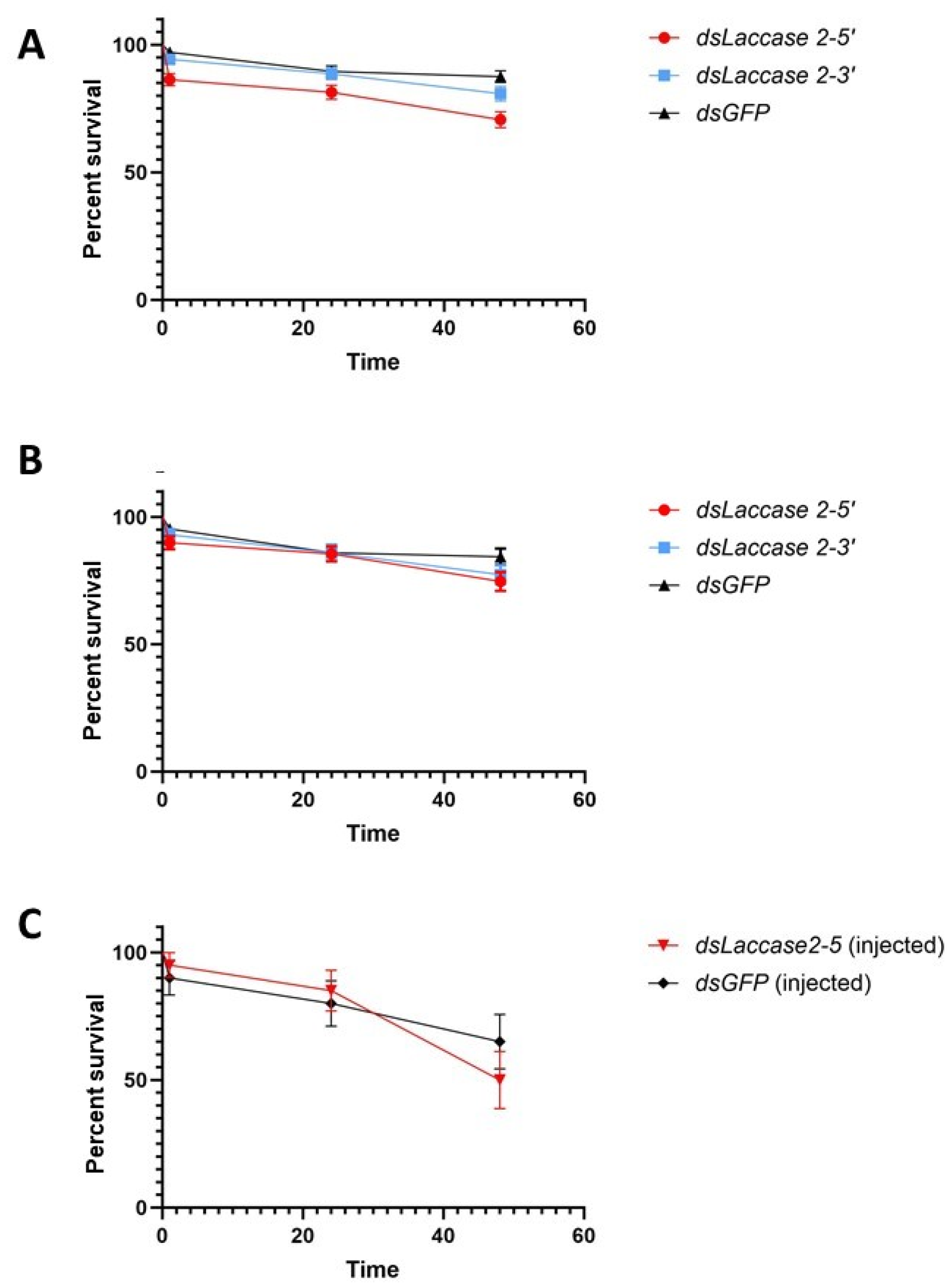
Dehydration and soaking in dsRNA. All pupae were removed from larval rearing pans 2 h before assay setup. In the subsequent 2 h window, freshly molted pupae that were pale in color were removed from the rearing pans, washed in cups containing deionized water, and placed in individual cells of 96-well cell culture plates using a trimmed plastic Pasteur pipette. Water that was inadvertently added was removed, and the well was dried using Kimwipes. Pupae were desiccated at room temperature for 30 min. After 30 min, 10 µL of the solution containing 1 μg of either dsLaccase 2-5′ (hereafter dsLac2-5), dsLaccase 2-3′ (hereafter dsLac2-3), dsGFP (negative control), or ddH2O mixed with Brilliant Blue dye (0.001 mg/mL) was added to each well. The mortality did not differ significantly between the dsGFP and ddH2O groups; therefore, the ddH2O group was excluded from further testing after our first replicate. After another 30 min, any dead individuals were removed, dye uptake was validated using a binocular scope (Olympus SZ61 stereoscope, Olympus America Inc., Waltham, MA, USA), and 150 μL of deionized water was added to each well containing the pupae. The mortality in experimental and control groups was assessed at 1 h, 24 h, and 47 h timepoints following desiccation. Cell culture plates containing pupae were held in mesh cages. After mosquitoes started to emerge, the cages were monitored every 30 min until the last mosquito emerged. The phenotypes of adults in the control and experimental groups were evaluated at 1 h post-emergence. To examine gene expression, three pupae from each timepoint were saved in 500 μL of RNAlater™ RNA-stabilizing solution (Thermo Fisher Scientific, Waltham, MA, USA AM7021) and held at 4 °C until RNA isolation. Total RNA was extracted from the pooled mosquito samples and cDNA was synthesized as described above for 3 pupae at 1, 24, and 47 h after the soaking assay for each of the replicates. The remaining pupae were allowed to emerge as adults and their phenotypes were scored. Per treatment, there were 20–36 pupae in each replicate, and the experiment was performed in 6 biological replicates.
dsRNA Microinjections. To further confirm that dsLac2 knockdown phenotypes were associated with dsRNA and not desiccation, we performed dsRNA microinjections. Twenty 2–3 h-old pupae were collected and rinsed in a Petri dish filled with deionized water. Individual pupae were transferred onto a 60-well cell culture microplate using a trimmed plastic Pasteur pipette. Microinjection needles were pulled using a Sutter P-97 micropipette puller (Sutter Instrument Company, Novato, CA, USA). After loading the dsRNA solution, the microinjection needle was inserted into the tip of a Pasteur pipette and fixed with parafilm. The injection needle was inserted by applying light pressure and sliding movements to the area near the 5–6th antennal segments under the antennal plate. Approximately 100 nL of 2 µg/µL dsLac2-5 (or dsGFP for the control) solution mixed with Brilliant Blue dye (0.001 mg/mL) was injected into the hemolymph according to gradations added to the needle. Pupae were gently transferred into a new cup containing 5 mL of ddH2O treated with 50 µL of 100× penicillin/streptomycin solution (10,000 UI/mL penicillin/10,000 µg/mL streptomycin, Millipore Sigma, Milwaukee, WI, USA) and placed into mesh mosquito cages. After adults started to emerge, treatment and control groups were monitored every 30 min until the last mosquito emerged. Adult mosquitoes were assessed at 1 h post emergence. Dead pupae were removed and counted at 1 h, 24 h, and 47 h after the assay, and samples for RNA extraction were taken from the dsLac2-5 and control groups at 24 and 47 h.
2.5. qRT-PCR Evaluation of Gene Expression
Laccase 2 expression in the knockdown and control sample pools was determined by qRT-PCR with
actin primers as an internal control. One µg of DNase-treated RNA was used for cDNA synthesis. cDNA was diluted 10× in nuclease-free water and used as a template in the qRT-PCR assay. In a 10 µL qRT-PCR, 0.5 µL of cDNA was used. Each sample was run in duplicate wells of a 96-well plate. No template and no RT controls were included in each reaction. qRT-PCR was performed on a Roche Lightcycler 480 System [
33] using SYBR Green master mix (Bio-Rad Laboratories, Inc. Hercules, CA, USA) and
laccase 2-specific primers (
Table 1). The reactions were performed for 1 min at 95 °C, followed by 39 cycles of 10 s at 95 °C, 30 s at 58 °C, and 30 s at 72 °C. The melting curve was analyzed at 60–95 °C to ensure a single peak, and thereby primer specificity. An initial standard curve analysis was performed for both
laccase 2 (target) and
actin (internal control) genes (primers are listed in
Table 1). Primer efficiencies were calculated as
and were 93.8% and 95.5% for
laccase 2 and
actin, respectively.
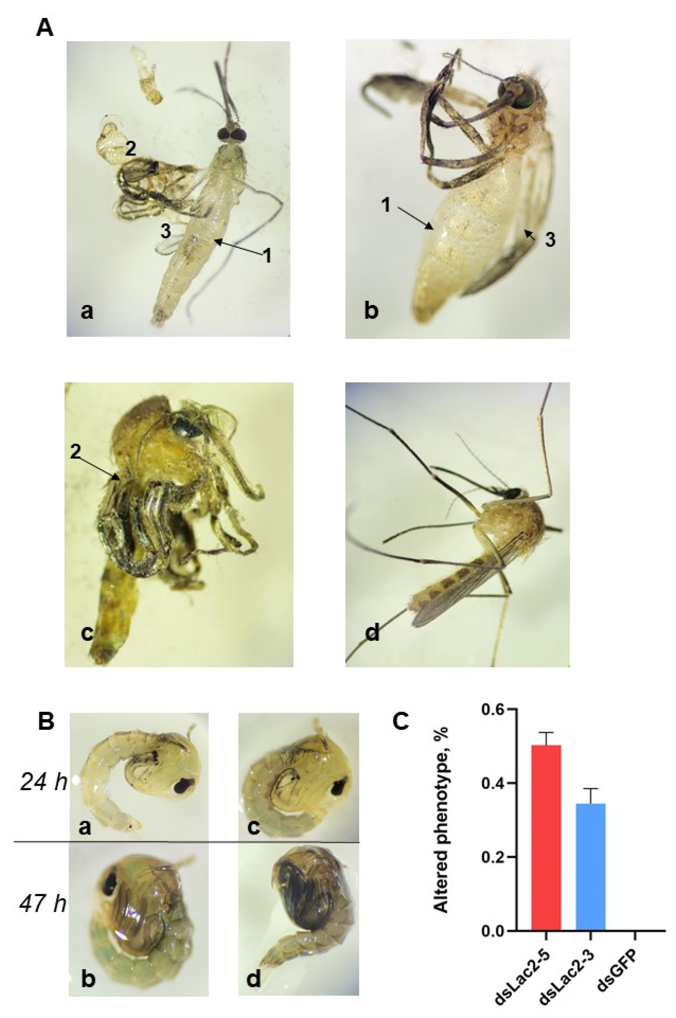
2.6. Phenotypic Evaluation
Mosquito phenotypes were evaluated via an Olympus SZ61 stereoscope at 30X magnification. Deformations to the cuticle, wings, legs, and body segments, as well as a lack of pigmentation, were recorded. Inability to successfully emerge from the pupal enclosure, as was observed by Du and co-authors [
23], was also documented. Mosquitoes were scored as having a knockdown phenotype if we observed one or more of the traits described above.
2.7. Data Analysis
A statistical analysis of mosquito survivorship following treatment with dsRNA was performed using a Mantel–Cox log-rank test for survival curves with GraphPad Prism (version 10.0.0 for Windows, GraphPad Software, Boston, MA, USA). The differential expression of
laccase 2 was quantified in pupae exposed to
dsLac2-5,
dsLac2-3, and
dsGFP at multiple timepoints following exposure with the ΔΔCt method according to [
34]. The phenotypes produced by
dsLac2-5 and
dsLac2-3 were similar, as were the reductions in gene expression following dsRNA treatment. Therefore, the samples exposed to both dsRNAs targeting
laccase 2 were analyzed together in comparison to dsGFP-exposed pupae collected at the same timepoint and run together on the same qRT-PCR plate. Welch’s unpaired t-tests of the ΔCt values were used to test for statistically significant gene expression differences with an a priori α-value of 0.05.
4. Discussion
The development of new dsRNA delivery methods is an active area of research [
13,
36], and here we document the strengths and weaknesses of dsRNA delivery by dehydration and soaking for
Cx. pipiens pupae. This approach has not been previously used to target this
Cx. pipiens life stage with RNAi. Our method represents the integration of an improvement over two previously described approaches: one that dehydrated and then soaked
Cx. pipiens larvae in dsRNA [
23], and another that soaked
Ae. aegypti pupae in dsRNA without dehydration [
24]. In our hands, soaking freshly molted
Cx. pipiens pupae in dsRNA without dehydration failed to elicit a phenotypic response, but dehydration and subsequent rehydration of pupae in concentrated dsRNA solution did. While we attempted multiple dehydration times in preliminary studies, dehydration times <15 min resulted in 5% or fewer individuals with knockdown phenotypes, and longer dehydration times increased mortality. A 30-minute dehydration followed by dsRNA soaking produced low mortality rates while still eliciting a strong phenotypic response.
Following dehydration and soaking, we observed the clear phenotypic effects of dsRNA exposure in up to 64% of exposed individuals. This effect size is sufficient for the screening of mosquito gene function, and indicates that the method of pupal dehydration and soaking offers an accessible and cost-effective approach to dsRNA delivery in labs without specialized equipment. Once optimized, dehydration had little effect on
Cx. pipiens pupal survival and most likely enhanced the uptake of dsRNA during the rehydration process. The survival of mosquitoes in our assays ranged from 56 to 75% in the
dsLac2-5′ group, 66 to 83% in the
dsLac2-3′ group, and 80 to 94% in the
dsGFP group, which is generally higher than in injected pupae [
37] (
Supplementary Figure S1). Furthermore, our approach reduced the manipulation and handling of pupae to improve survivorship. As a result, our pupal mortality rates (17–44% in the
dsLac2 groups and 16–20% in the
dsGFP group) were much lower than documented in previous studies [
20]. Our technique eliminated or minimized the risk of damage to pupae and reduced undesirable stress-related effects on mosquito fitness and development [
38]. Soaking offers a flexible and rapid screening approach for gene function and would likely allow for the simultaneous screening of multiple genes, enabling a better understanding of the impact of genetic interactions in complex traits.
Challenges for screening of gene function by dehydration and dsRNA soaking do exist, however. As one example, the saline buffer (2 M NaCl) used for dehydration in [
23] resulted in significant mortality (over 90%) in fourth instar larvae when used in our assays. We ceased to use it, and instead exposed pupae to desiccation on the benchtop. Prolonged dehydration of pupae (over 40 min) also resulted in increased mortality, and the optimal timeframe between dehydration and rehydration was 30 min. Dehydration success may depend upon species, pupal size, and even relative air humidity, presenting a challenge to the uniform dehydration of pupae and affecting the amount of successfully delivered dsRNA; therefore, this timeframe should be selected and adjusted based on pupae size, cuticle thickness, and tolerance to desiccation.
Timing of dsRNA application for soaking assays was important in our work, as well as in previous studies [
20]. We used freshly molted (0–2 h) pupae hours before any signs of cuticle tanning appeared. It had been previously demonstrated that soaking efficacy was reduced when applied to older pupae [
20]. We also did not observe any phenotypic changes when 12–20 h old pupae were soaked in the same concentrations of dsRNA used in our assays. This lack of effect might be related to the lower permeability of their cuticles, which could reduce the efficiency of dsRNA uptake.
When we compared phenotypical defects resulting from dehydration and dsRNA soaking to those from microinjection, we noted similarities in both the
dsLac2-5- and
dsLac2-3-treated groups, as well as the
dsLac2-5-microinjected group. The proportion of mosquitoes with altered phenotypes in our study was variable and significantly higher in the
dsLac2-5 group. This suggests that dsRNA efficacy depends on the design of the dsRNA construct, the specificity of protein synthesis and modifications, and the abovementioned physical properties of pupae. Employing various constructs, as suggested by others [
38,
39,
40,
41], or increasing the dsRNA concentration might positively impact dsRNA-mediated gene silencing efficacy in future experiments.
Interestingly, in our microinjection assay, the survival of pupae was similar or higher (up to 65%) compared to the
Culex spp. pupae microinjections in other studies (up to 40% in [
12,
13]). In these studies, freshly molted pupae were injected into the base dorsal segments close to the trumpet. In our work, we found that injecting pupae near the antennae by inserting the needle tip between cuticle plates gave higher survivorship, and we encourage the adoption of this pupal injection site in future
Culex work.
The results from our study also support the critical role for
laccase 2 in the formation of the
Cx. pipiens pupal cuticle, specifically around the period of intense cuticle darkening. Disruption of the melanin pathway in our assay caused consistent effects similar to those observed in
dsLac2 An. sinensis mutants (softer and lighter cuticle and delayed emergence of adults). We also observed that, in many mosquitoes from the
dsLac2-5- and
dsLac2-3-treated groups, the abdominal segments were shorter, whereas the legs and wings were severely malformed (curved). These individuals also showed delayed development and reduced fitness, and often struggled to leave the pupal enclosure, which resulted in the death of the individual. Observed malformations are likely to be the result of improper cuticle hardening, as was shown for
An. sinensis [
23] and
Tr. castaneum [
22]. These findings suggest that
laccase 2, via melanin synthesis, might play a substantial role in orchestrating the timely progression of pupal development and facilitating the successful transition from the pupal to the adult stage.
Emerging insecticide resistance remains one of the major challenges in mosquito control [
42,
43,
44,
45].
Laccase 2 and
tyrosine hydroxylase were included in a list of promising genetic targets for RNAi-based mosquito control to overcome insecticide-resistant populations [
5]. Inadequate or incomplete cuticle sclerotization leads to compromised protection from environmental threats, such as UV radiation exposure, viral, bacterial, and fungal pathogens, and decreased fitness. By targeting
laccase 2 and disrupting the melanin synthesis pathway, the survival and general fitness of mosquitoes are severely affected. Interfering with melanin synthesis can lead to compromised immune defenses and increased vulnerability to environmental stresses, ultimately reducing mosquito population sizes. More importantly,
laccase 2 can bind to multiple substrates and is a substantial component in the processes of detoxification in certain insecticides, as has been shown for insecticide-resistant strains of
Ae. aegypti [
46]. Targeting
laccase 2 may disrupt this detoxification mechanism, making mosquitoes more susceptible to insecticides that they were previously resistant to. Ultimately, silencing
laccase 2 expression can be combined with existing mosquito control strategies, such as insecticide-based interventions, to enhance their effectiveness. Overall, our results expand researcher options for target gene functional screening, which can facilitate the development of novel mosquito control applications.


