Article

A Novel Model of Pathogenesis of Metarhizium anisopliae Propagules through the Midguts of Aedes aegypti Larvae

by
Ricardo de Oliveira Barbosa Bitencourt
1,*,
Jacenir Reis dos Santos-Mallet
2,3,4,
Carl Lowenberger
5,
Adriana Ventura
6,
Patrícia Silva Gôlo
7,
Vânia Rita Elias Pinheiro Bittencourt
7 and
Isabele da Costa Angelo
8,*
1
Graduate Program in Veterinary Sciences, Veterinary Institute, Federal Rural University of Rio de Janeiro, Seropédica 23890-000, RJ, Brazil
2
Oswaldo Cruz Foundation, IOC-FIOCRUZ-RJ, Rio de Janeiro 21040-900, RJ, Brazil
3
Oswaldo Cruz Foundation, IOC-FIOCRUZ-PI, Teresina 64001-350, PI, Brazil
4
Laboratory of Surveillance and Biodiversity in Health, Iguaçu University-UNIG, Nova Iguaçu 28300-000, RJ, Brazil
5
Centre for Cell Biology, Development and Disease, Department of Biological Sciences, Simon Fraser University, Burnaby, BC V5A 1S6, Canada
6
Department of Animal Biology, Institute of Health and Biological Sciences, Federal Rural University of Rio de Janeiro, Seropédica 23890-000, RJ, Brazil
7
Department of Animal Parasitology, Veterinary Institute, Federal Rural University of Rio de Janeiro, Seropédica 23890-000, RJ, Brazil
8
Department of Epidemiology and Public Health, Veterinary Institute, Federal Rural University of Rio de Janeiro, Seropédica 23890-000, RJ, Brazil
*
Authors to whom correspondence should be addressed.
Insects 2023, 14(4), 328; https://doi.org/10.3390/insects14040328
Submission received: 16 February 2023 / Revised: 20 March 2023 / Accepted: 21 March 2023 / Published: 28 March 2023
(This article belongs to the Section Insect Behavior and Pathology)

Abstract

:

Simple Summary

The mosquito Aedes aegypti is the principal vector of multiple arboviruses including dengue (DENV), zika (ZIKV), and chikungunya (CHIKV) that cause major human disease and suffering. Control is based on the application of synthetic insecticides to reduce mosquito populations. However, the overuse of synthetic insecticides has led to mosquito resistance. Studies on the use of entomopathogenic fungi (EPF) such as Metarhizium anisopliae to reduce mosquito populations are gaining interest as they are ecologically safe and can kill different mosquito life stages. Despite their lethality towards mosquito larvae, their mechanisms of infection are unclear. In this study, we first evaluated the production of blastospores of three M. anisopliae isolates and assessed their effects against Ae. aegypti larvae. We assessed, and describe in detail, the mechanism of infection of one fungal isolate against Ae. aegypti larvae. Overall, our findings indicate that EPF are able to kill mosquito larvae by infecting the insect midgut, disrupting enterocytes, and causing brush border degradation.

Abstract

We assessed the effect of the entomopathogenic fungus Metarhizium anisopliae against Aedes aegypti. Conidia of M. anisopliae strains CG 489, CG 153, and IBCB 481 were grown in Adamek medium under different conditions to improve blastospore production. Mosquito larvae were exposed to blastospores or conidia of the three fungal strains at 1 × 10⁷ propagules mL−1. M. anisopliae IBCB 481 and CG 153 reduced larval survival by 100%, whereas CG 489 decreased survival by about 50%. Blastospores of M. anisopliae IBCB 481 had better results in lowering larval survival. M. anisopliae CG 489 and CG 153 reduced larval survival similarly. For histopathology (HP) and scanning electron microscopy (SEM), larvae were exposed to M. anisopliae CG 153 for 24 h or 48 h. SEM confirmed the presence of fungi in the digestive tract, while HP confirmed that propagules reached the hemocoel via the midgut, damaged the peritrophic matrix, caused rupture and atrophy of the intestinal mucosa, caused cytoplasmic disorganization of the enterocytes, and degraded the brush border. Furthermore, we report for the first time the potential of M. anisopliae IBCB 481 to kill Ae. aegypti larvae and methods to improve the production of blastospores.

1. Introduction

Metarhizium anisopliae s.l. (Metsch) Sorok (Ascomycota: Clavicipitaceae) is a complex of entomopathogenic fungi (EPF) widely used as a biological pesticide to mitigate arthropod pests in agricultural settings [1]. In addition, this fungal complex has been shown to control larvae of Aedes aegypti [2,3,4], the principal mosquito species that transmits several arboviruses that affect millions of people around the world [5]. EPF have been used in biological control applications due to their diversity, which allows the identification of appropriate isolates to target specific insect pests [6]. Also, EPF are non-toxic to vertebrates and have limited non-target effects [7]. Thus, several researchers have proposed that EPF represent a novel approach to control insects of public health importance, such as mosquitoes [8,9,10,11,12,13,14,15,16].
Unlike other entomopathogenic organisms such as the bacteria Bacillus thuringiensis and the virus Granulovirus sp. [17] that must be ingested by the target organism, EPF can infect arthropods by penetrating directly through the tegument [18,19]. The classic infection process of M. anisopliae starts with the adhesion of conidia to the host cuticle that involves protein adhesions such as adhesin-like Mad1 and Mad2 [20]. After adhesion, the conidia produce a germ tube and deliver lipases, chitinases (Chit1, Chit2), and proteases (Pr1, Pr2) to breach and penetrate the insect cuticle [21,22]. Inside the insect hemocoel, the fungus produces yeast-like cells denominated hyphal bodies (blastospores) that deploy an extensive cocktail of extracellular metabolites such as the destruxins A, B, and C to eliminate opportunistic organisms and downregulate the innate immune system of the insect host [1,23,24]. The hyphae colonize the insect, take up nutrients from the host, and once the nutrients are exhausted, the fungi emerge from the body to produce conidia [1,20].
Although EPF classically use the tegument as the main route of infection [20] (Butt et al., 2016) they also may infect mosquitoes through the digestive or respiratory tracts [4,8,10,14,16]. In aquatic larvae, the exact mode of infection of M. anisopliae is still unclear. Some researchers reported that EPF conidia cannot adhere to the larval cuticle or germinate in the midgut, thus killing Ae. aegypti larvae by occlusion of natural orifices, the mouth, and the respiratory siphon [16,25]. Greenfield et al. [26] reported the absence of hydrocarbons in Ae. aegypti larvae required for the conidia to germinate. Interestingly, Alkhaibari et al. [8] reported that M. anisopliae ARSEF 4556 blastospores attach to the Ae. aegypti larval exoskeleton and enterocytes to invade the larval hemocoel for colonization. Noskov et al. [14] reported conidial development in the gut of Aedes larvae infected with Metarhizium robertsii (strain MB-1). de Oliveira Barbosa Bitencourt et al. [3] observed M. anisopliae ARSEF 2211 and B. bassiana CG 206 in the midguts of Ae. aegypti larvae. Thus, the route of infection seems not to be the same for all fungal strains. In fact, there is much to elucidate on how these routes vary depending on the fungal isolate, the propagules, and the insect target.
Blastospores and conidia have distinct biological features [20]. Conidia are hydrophobic propagules produced in a solid substrate within 2 weeks and have a long shelf life [27,28]. On the other hand, blastospores are pleomorphic and hydrophilic spores that are considered analogs of hyphal bodies [20]. They can be produced in vitro [29] with shorter production cycles (~3 days) and possess morphological and physiological features to escape from insect innate immune responses [29,30]. Usually, producing a high mass of conidia is easier than producing blastospores, and the major challenge to the broad-spectrum use of blastospores as biological control agents includes production optimization and effective formulation technologies to improve their shelf life [31]. Some authors have reported the efficacy of blastospores and conidia against Ae. aegypti larvae [10,11,15,16]. Depending on the isolate, blastospores seem more effective against mosquito larvae than conidia [8,9,32]. However, for other isolates, blastospores, and conidia had similar virulence against Ae. aegypti larvae [11]. Interestingly, conidia of Beauveria bassiana CG 206 were slightly more virulent than blastospores against mosquito larvae. In contrast, blastospores and conidia of M. anisopliae CG 489 had similar results in reducing larval survival [3].
Despite the efficiency of EPF in invading the digestive tract of larvae, the gut is composed of a layer of epithelial cells, the enterocytes, that secrete the peritrophic matrix, digestive enzymes, and defense molecules to absorb nutrients as well as to protect themselves from pathogens [33].
Although there are reports of the presence of EPF in the digestive tract of Ae. aegypti larvae, it doesn’t have much information about to mechanism of action related to their ability to damage the larval gut, enter the hemocoel, and kill the larvae. We reported previously on the damage caused by EPF on the midgut of mosquito larvae [4]. Accordingly, the aims of this study were to: (1) assess the influence of shaker rotation for the production of blastospores; (2) assess and compare the virulence of blastospores and conidia of three M. anisopliae strains against Ae. aegypti larvae; (3) elucidate the damage to the larvae midgut caused by M. anisopliae CG 153; and (4) determine if the midgut could be an infection route for specific strains of EPF.

2. Materials and Methods

2.1. Culture and Preparation of M. anisopliae Conidia

Metarhizium anisopliae sensu lato (s.l.) CG 153 and CG 489 were obtained from the National Center for Genetic Resources-CENARGEN, EMBRAPA, Brazil. Metarhizium anisopliae s.l. IBCB 481 was obtained from the Biological Institute-IBCB, SP, Brazil. The fungal strains were cultured on potato dextrose agar medium and incubated under controlled conditions [25 ± 1 °C; relative humidity (RH) ≥ 80%]. After 15 days of incubation, the conidia were harvested and suspended in sterile 0.03% Tween 80 in dechlorinated tap water (v/v). Conidia were quantified using a Neubauer hemocytometer (KASVI®, São Paulo, SP, Brazil), and their concentration was adjusted to 1 × 10⁸ propagules mL−1 [34].

2.2. Influence of Shaker Rotations and the Time of Incubation on Blastospore Production

Three milliliters of conidial suspension at 1 × 10⁸ conidia mL−1 were inoculated into 125 mL Erlenmeyer flasks (N = 3) containing 42 mL of modified Adamek’s medium. The medium was prepared using 31.58 mL milhocine, 42.1 g of yeast extract (KASVI®, São José do Pinhais, PR, Brazil), 42.1 g of glucose D (+) anhydrous (Isofar®, Duque de Caxias, RJ, Brazil), and 21.03 mL of Tween 80 at 0.1% in 1000 mL of distilled water of final volume. The milhocine was prepared with 5 g of cornstarch (Maizena/Duryea©, Mogi Guaçu, SP, Brazil) dissolved in 250 mL of distilled water. The medium was prepared according to Duarte [35]. The pH was adjusted to 5.9, combining sodium hydroxide (NaOH) and hydrochloric acid (HCl) using a pH meter (mPA-201, MS Tecnopon®, Piracicaba, SP, Brazil]. The Erlenmeyer flasks were covered by aluminum foil and plastic wrap and then incubated at 27 °C at 150 rpm or 220 rpm (TE-424®, Tecnal, Brazil). After 48 h and 72 h, the medium was filtered using a sterile lint followed by centrifugation at 5000 rpm for five minutes (Rotina 380R, Hettich©, Tuttlingen, Baden-Württemberg, Germany). The supernatant was discarded, and 10 mL of 0.03% Tween 80 solution (v/v) was added. The blastospores were homogenized using a vortex (KASVI©, São José do Pinhais, PR, Brazil). The centrifugation, supernatant removal, water addition, and homogenization were conducted as described [3]. Blastospores were quantified using a Neubauer chamber [34]. The experiments were performed three different times using three different batches of propagules.

2.3. Insects

The Laboratory of Physiology and Arthropods Control (Fundação Oswaldo Cruz-FIOCRUZ, Rio de Janeiro, Brazil) provided the eggs of Ae. aegypti (Rockefeller strain). The eggs were maintained in tap water (2 L) under controlled conditions [27 ± 1 °C; (RH) ≥ 80%] on a 12 h light/dark cycle. After hatching, the larvae were fed daily with pulverized fish food (0.05 g/L).

2.4. Survival Analyses

For survival analyses, blastospores and conidial suspensions were prepared and adjusted to 1 × 10⁷ propagules mL−1. Then, 10 Ae. aegypti second instar larvae (L2) (N = 30) were placed in plastic cups (50 mL; 4.5 cm width × 4 cm height) containing 10 mL of pure blastospores (BLA) or conidial (CON) suspensions. The control group was exposed to sterile dechlorinated tap water plus 0.03% Tween 80. The cups were covered with mesh and sealed with a rubber band and maintained under controlled conditions [27 ± 1 °C; RH ≥ 80%] on a 12:12 h light/dark cycle. The larvae were fed daily with a pinch of sterile fish food in each cup. The survival rate was monitored daily for seven days, and dead larvae were removed daily [3]. The experiments were performed three times using three batches of propagules and larvae. The tests were performed using 90 larvae per replicate (each group), totaling 630 mosquitoes for all experiments.
The fungal viability was also tested. The fungal suspensions were diluted to 1 × 10⁵ propagules mL−1 by serial dilution (Alves, 1998); 10 μL of conidial or blastospore suspensions were inoculated onto Petri dishes containing PDA supplemented with 0.05% chloramphenicol. PDA plates were incubated at 25 ± 1 °C and a relative humidity (RH) of ≥80%]. After 16 h, three hundred conidia or blastospores were counted randomly using an optical microscope at 400× magnification [34]. Conidia were considered to have germinated if they developed the germ tube and the blastospores if they formed hyphae [36].

2.5. Scanning Electron Microscopy and Histopathological Analysis

The histopathology (HP) and the scanning electron microscopy (SEM) were conducted using larvae (N = 3) exposed to M. anisopliae CG 153. This strain demonstrated high efficacy in killing mosquito larvae and good results in producing blastospores. Larvae were exposed to fungal propagules as described above and examined at 24 h or 48 h post-exposure by HP and SEM. To conduct HP, the larvae were fixed in Bouin’s solution [37] for 24 h at 4 °C and processed for histological examination (dehydrated in ethanol series; diaphanisation in Xylol followed by xylol-alcohol solution according to Bitencourt et al.) [3]. The blocks of paraffin were cut to 3 μm thickness, and cross-sectioned larvae were stained with hematoxylin and eosin (HE) and examined under a light microscope (Primo Star, ZEISS®, São Paulo, Brazil) with an integrated camera AxioCam ICc 1 (ZEISS®) at a magnification of 400×.
For SEM, the larvae were fixed for 24 h in 2.5% (v/v) glutaraldehyde. The samples (N = 3) then were treated with 1% osmium tetroxide for 60 min and dehydrated through an increasing concentration series of ethanol washes as described previously [3]. The samples were photographed using a JEOL JSM-6390LV (JEOL BRASIL©, São Paulo, Brazil) scanning electron microscope at 15 kV.

2.6. Statistical Analyses

Kaplan-Meier analysis was used to calculate the cumulative survival curve. Pairwise comparisons were used, and the median survival time (S50) was calculated using the Log-rank test. The data on the blastospore production were submitted to the Shapiro-Wilk normality test, analyzed by two-way ANOVA followed by Tukey test for multiple comparisons with a significance level of 95% (p ≤ 0.05). All data were analyzed by GraphPad Prism v8.00, Inc. (GraphPad Software, Boston, MA, USA).

3. Results

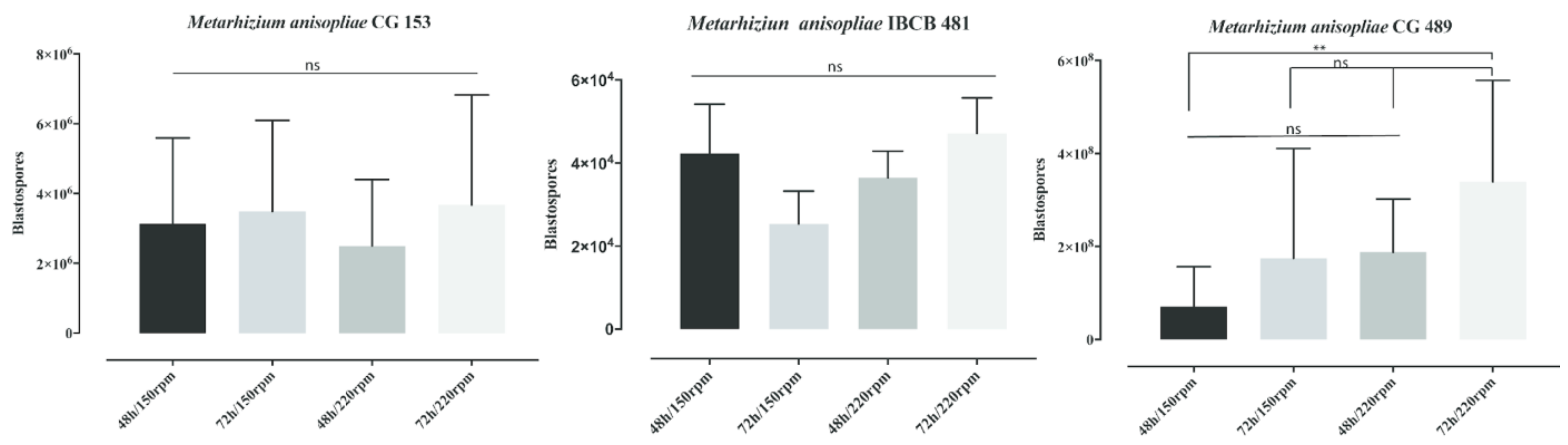
3.1. Influence of Shaker Rotations and the Time of Incubation on Blastospore Production

Neither the rotation speed nor the incubation time affected blastospore production in M. anisopliae strains IBCB 481 and CG 153 (Figure 1). Metarhizium anisopliae IBCB 481 incubated at 72 h/150 rpm did not improve its blastospore production significantly compared with 48 h/150 rpm (p = 0.5074), 48 h/220 rpm (p = 0.7947), or 72 h/220 rpm (p = 0.2968). The blastospore production of M. anisopliae IBCB 481 incubated at 72 h/220 rpm did not differ significantly compared with 48 h/150 rpm (p = 0.9786) or 48 h/220 rpm (p = 0.8137). In addition, the same strain incubated at 48 h/150 rpm did not differ significantly (p = 0.9612) compared with 48 h/220 rpm.
The blastopore production of M. anisopliae CG 153 after 72 h/150 rpm was similar compared with 48 h/150 rpm (p = 0.9917), 48 h/220 rpm (p = 0.8566), and 72 h/220 rpm (p = 0.9989). The production of blastospores was not statistically different at 72 h/220 rpm compared with 48 h/150 rpm (p = 0.9726) and 48 h/220 rpm (p = 0.7843). Metarhizium anisopliae CG 153 incubated at 48 h/150 rpm did not differ significantly (p = 0.9563) from 48 h/220 rpm.
After 72 h of incubation, M. anisopliae CG 489, at 220 rpm, produced more blastospores (p = 0.0087) compared with 48 h/150 rpm (Figure 1), but similar numbers to 72 h/150 rpm (p = 0.1636) and 48 h/220 rpm (p = 0.2201). Here, the blastospore production was also not statistically different compared with 72 h/150 rpm vs. 48 h/150 rpm (p = 0.9726); 72 h/150 rpm vs. 48 h/220 rpm (p = 0.9988); and 48 h/150 rpm vs. 48 h/220 rpm (p = 4315). Metarhizium anisopliae CG 489 produced the most blastospores, followed by M. anisopliae CG 153 and M. anisopliae IBCB 481 (Figure 1).

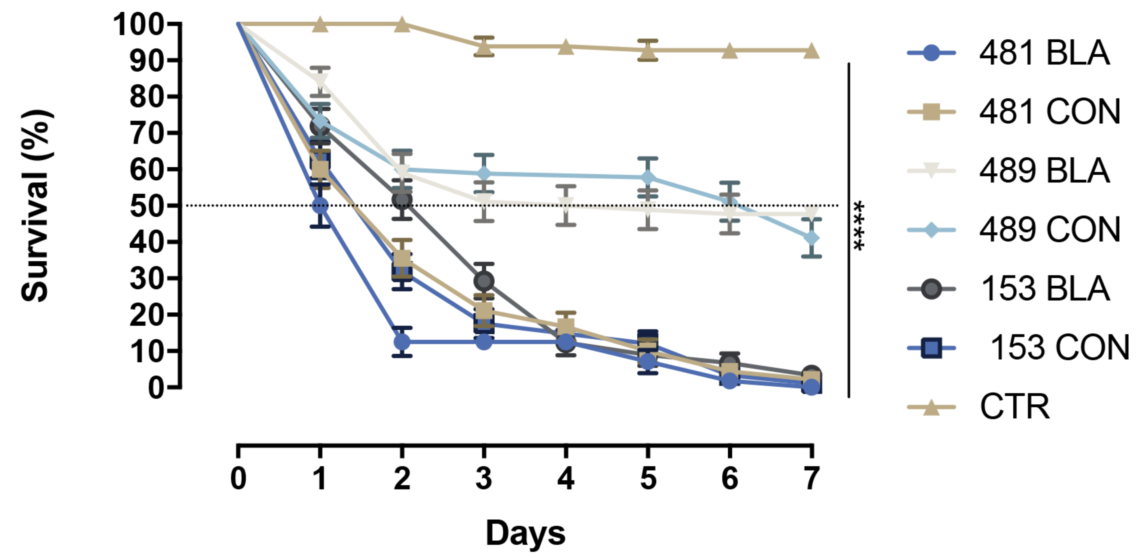
3.2. Survival Analyzes

Only 10% of Ae. aegypti larvae survived after three days of exposure to M. anisopliae IBCB 481 blastospores. For larvae exposed to M. anisopliae IBCB 481 conidia, the survival rate was 21% after 3 days. At the same time, 17% and 29% of larvae survived after exposure to conidia or blastospores of M. anisopliae CG 153, respectively. Also, after three days, 50% and 59% of larvae survived after exposure to blastospores and conidia of M. anisopliae CG 489, respectively (Figure 2). By day 7, the survival rate was as follows: 0% for larvae exposed to blastospores or conidia of M. anisopliae CG 153 or M. anisopliae IBCB 481, and 40% and 47% respectively, for larvae exposed to conidia and blastospores of M. anisopliae CG 489 (Figure 2). Therefore, blastospores and conidia of M. anisopliae IBCB 481 had the lowest S50 of larvae, followed by M. anisopliae CG 153 and M. anisopliae CG 489. The S50 ranged from 1 to 7 days (Table 1).
Regardless of the fungal strain, blastospores and conidia reduced the larval survival compared with the controls (Table 1, Figure 2). However, both propagules of the M. anisopliae CG 489 strain were significantly less virulent against Ae. aegypti larvae than M. anisopliae IBCB 481 (χ2 = 108.5; df = 3; p < 0.0001) and M. anisopliae CG 153 (χ2 = 87.13; df = 3; p < 0.0001). Comparing propagules of the same strain, blastospores and conidia of M. anisopliae CG 489 or propagules of M. anisopliae CG 153 had similar results in reducing larval survival. Blastospores of M. anisopliae IBCB 481 were less virulent than conidia of the same strain (χ2 = 5.461; p = 0.0195). Blastospores of M. anisopliae IBCB 481 were significantly more effective as larvicides compared with either the blastospores or conidia of M. anisopliae CG153 (χ2 = 14.89; p < 0.0001 and χ2 = 5.9; p = 0.0151, respectively). Conidia of M. anisopliae IBCB 481 were not found to differ significantly from either propagule of M. anisopliae CG 153 (χ2 = 2.324; df = 3; p = 0.3129).
Considering the blastospore production and the results of the bioassays, M. anisopliae CG 153 was chosen for subsequent experiments.

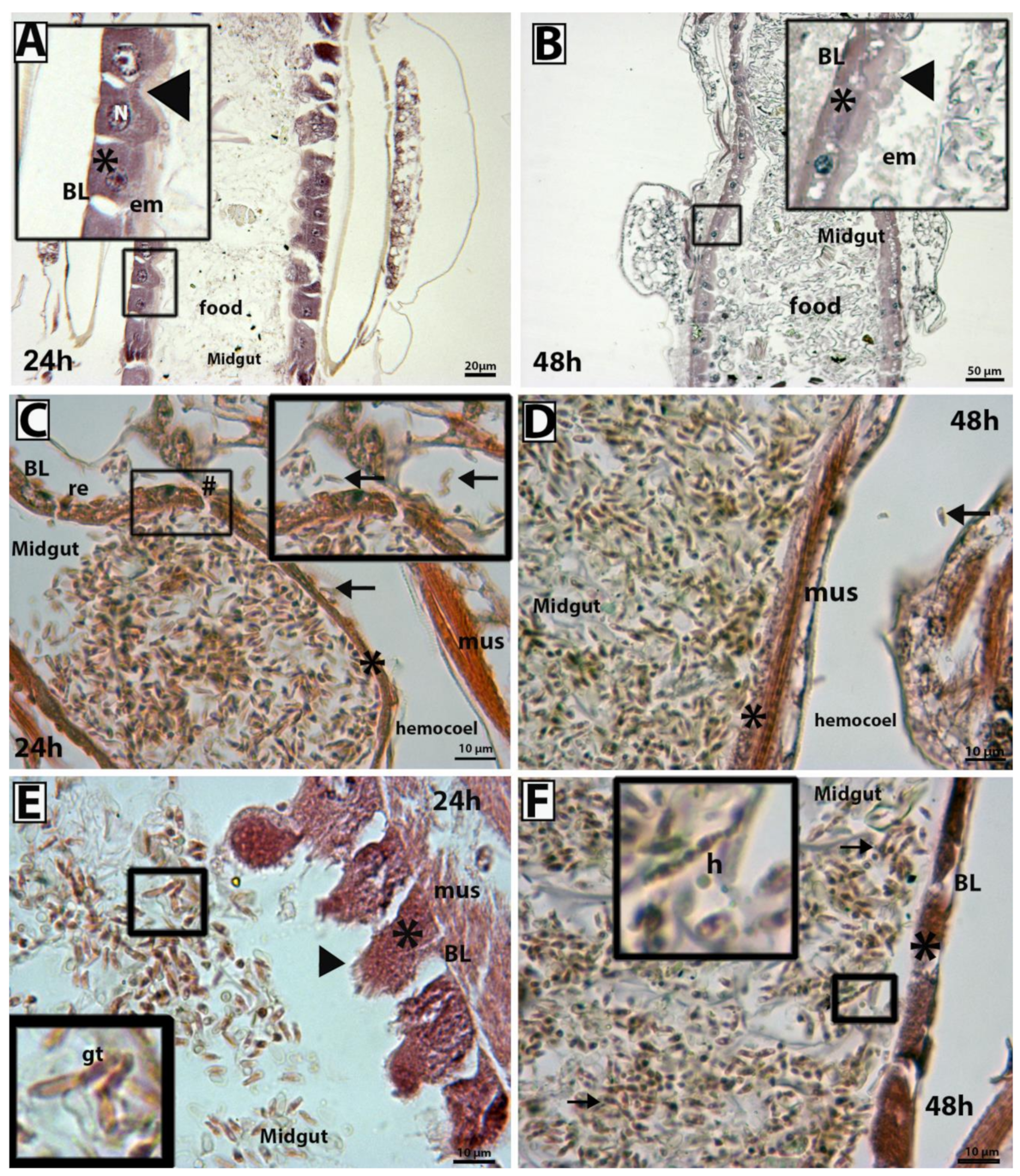
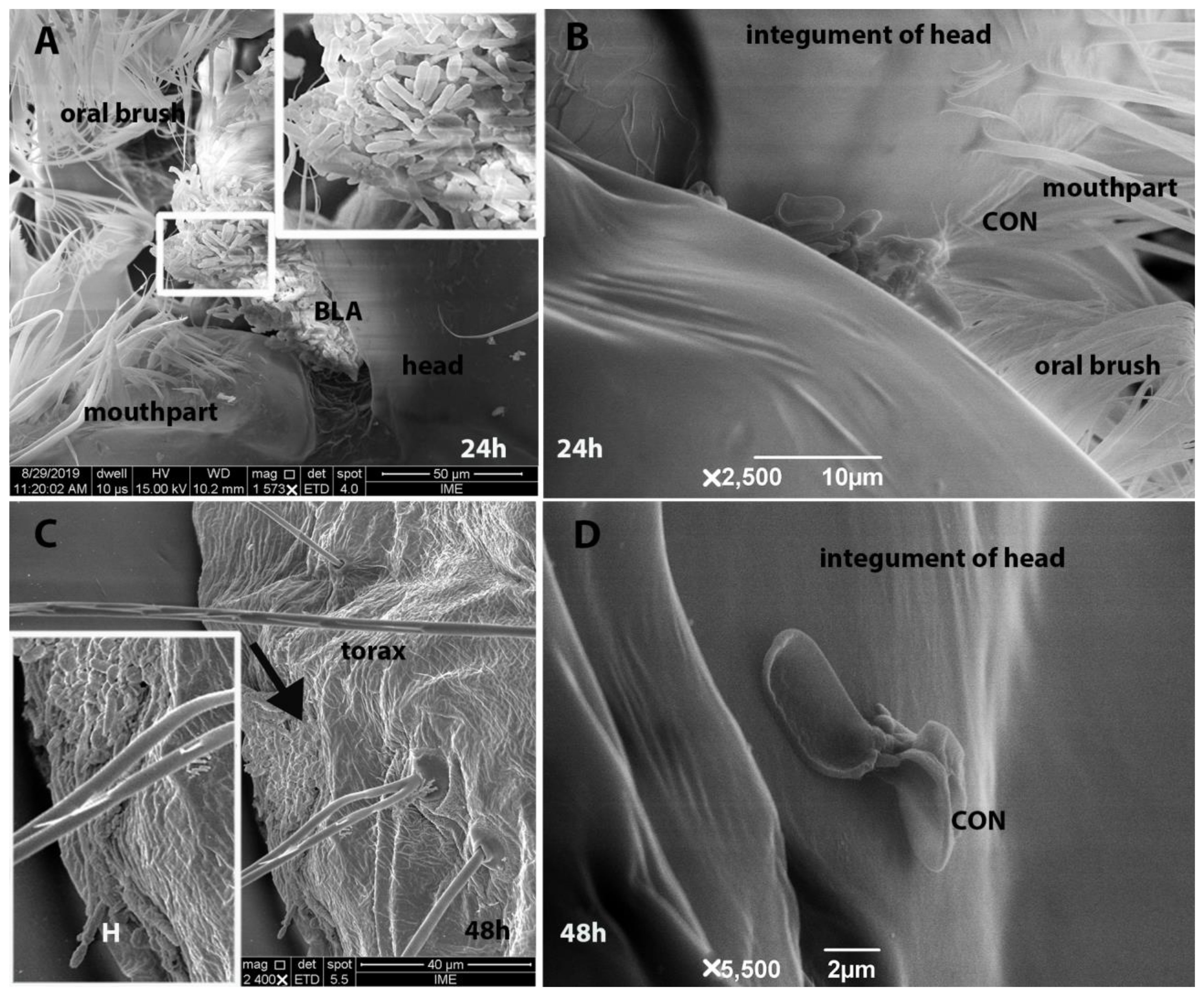
3.3. Scanning Electron Microscopy and Histopathological Analysis

At both 24 h and 48 h, M. anisopliae CG 153 was found to have infected 100% of the larvae via ingestion (Figure 3B–F and Figure 4A–D). Propagules on the one larval cuticle, however, were observed to produce hyphae (Figure 4C,D). After 24 h and 48 h, enterocytes of all samples from the control group exhibited normal morphology, spherical nuclei located in the parabasal region, and slightly acidophilic cytoplasm. The brush border and peritrophic membranes of 100% of the larvae from the control groups were both intact (Figure 3A,B) with columnar cells. Both fungal propagules infected the insect midgut (Figure 3C–F). Larvae exposed to fungi exhibited atrophy of the intestinal mucosa (appearing as squamous enterocytes) (Figure 3C,F). Although we did not observe fungal penetration through the cuticle, there was a rupture of the intestinal mucosa and invasion of the hemocoel by blastospores in one of the larvae (33%) after 24 h of exposure (Figure 3C) in mosquitoes exposed to conidia of M. anisopliae CG 153. Also, we observed germ tube development by blastospores (66%-2 larvae) (Figure 3D). Forty-eight hours post-exposure to conidial suspensions, 100% of the larval gut contained several hyphal bodies, and some hyphal bodies were present in the hemocoel (Figure 3D). After 24 h, 66% of larvae exposed to blastospores of M. anisopliae CG 153 exhibited disrupted enterocytes without nuclei and with a disorganized and acidophilic cytoplasm. Also, the brush border showed significant damage with an irregular surface or a lack of brush on the enterocytes (100% of the larvae-3 larvae) (Figure 3E). By 48 h, the three larvae exposed to blastospores showed flattened enterocytes (Figure 3D). The peritrophic matrix and extracellular material normally produced by enterocytes were not observed at either 24 h or 48 h in all larvae exposed to fungal propagules.

4. Discussion

Biocontrol strategies involving M. anisopliae represent a new weapon against larvae of the principal dengue vector, Ae. aegypti [8,9,13] (Alkhaibari et al., 2016, 2017; De Paula et al., 2021). We demonstrate that M. anisopliae CG 153 caused significant damage to the midgut integrity, affecting the larval enterocytes, which may explain the virulence of this fungus on larvae through a non-classical route. Furthermore, our experiments demonstrated the substantial effectiveness of three strains of M. anisopliae (CG 153, CG 489 and IBCB 481) against Ae. aegypti larvae.
We also modified the production parameters and improved the production of blastospores in one fungal strain (M. anisopliae CG 489). Blastospores are yeast-like cells produced in vitro conditions in a short incubation time [31]. Here, blastospore production increased after extending the incubation time [38]. However, only M. anisopliae CG 489 improved blastospore production at 200 rpm. This result suggests that although the three strains belong to the genus Metarhizium, the strains have intra-specific variations with specific features and requirements for energy resources that distinguish them from each other, which might directly affect their blastospore production [39,40]. Therefore, additional studies should be conducted to understand these intrinsic fungal characteristics and requirements to improve blastospore production.
Blastospores of M. anisopliae CG 489 demonstrated slightly better efficacy than conidia in reducing the survival of Ae. aegypti larvae, corroborating the results of Alkhaibari et al. [8] (2016). However, blastospores and conidia of M. anisopliae CG 153 and M. anisopliae IBCB 481 had similar effectiveness, corroborating the reports of de Oliveira Barbosa Bitencourt et al. [3,11] (2018, 2021a). Blastospores are in vitro propagules analogous to hyphal bodies and rapidly colonize aquatic targets such as mosquito larvae [8,41]. Although conidia are known to colonize terrestrial invertebrates [1], our findings demonstrate that they also can infect mosquito larvae in the aquatic environment and reduce larval survival. However, the efficacy of blastospores or conidia also depends on the genus and strain of the fungus, environmental conditions, and the target species [20].
Both propagules of M. anisopliae CG 153 use the mosquito’s digestive tract as the main front of interaction, as described previously [3,42]. However, this fungus also colonizes the hemocoel [8,14]. In addition, fungal propagules damaged the enterocytes and brush borders, which also might lead to the death of the larvae and invasion of the hemocoel. Interestingly, Silva et al. [43] reported epithelial tissue degeneration in the larval gut of Aedes albopictus exposed to Bacillus thuringiensis, and Jiraungkoorskul [44] and Al-Mehmadi and Al-Khalaf [45] reported similar results using plant extracts against Culex quinquefasciatus.
The histological analyses revealed a high concentration of hyphae and propagules inside the midgut, which caused damage to the intestinal mucosa, peritrophic membrane, necrosis of enterocytes, and intestinal disruption. The Ae. aegypti midgut is composed of absorptive enterocytes and non-absorptive hormone-producing cells [46]. Enterocytes play an important role in insect physiology, contributing to digestion, homeostasis, and protection against invading microorganisms, such as the formation of a peritrophic matrix, production of reactive oxygen species, as well as cell proliferation and differentiation [47,48,49]. Additionally, the digestive tract is a niche for microbiota. Taracena et al. [48] reported the damage to enterocytes and, at the same time, the proliferative ability of the intestinal cells of Ae. aegypti adults after being infected with the Dengue virus. In addition, Buchon et al. [50,51] reported that the presence of the microbiota and pathogenic bacteria increases the rate of epithelial turnover. However, substantial destruction of the enterocytes could result in a fatal loss of gut barrier integrity [40]. Taracena et al. [48] also observed an increase in mitosis of the peritrophic matrix after mosquito adults were exposed to the entomopathogenic bacteria Pseudomonas entomophila. Enterocytes produce a type 1 peritrophic matrix for adults and type 2 in larvae [52,53]. Interestingly, we did not observe a peritrophic matrix in larvae exposed to M. anisopliae CG 153. Our data suggest that the midgut serves as a gateway for fungi to colonize the hemocoel. Once the mucosa is damaged, the digestive process of the larvae becomes affected by interaction with this fungus, causing death.
Besides damaging the enterocytes, EPF might produce several secondary metabolites, such as the toxins destruxins and beauvericin [1,4,54]. Destruxins have insecticidal activity and play a crucial role in blocking calcium channels causing flaccid paralysis and inhibiting host immune responses [23,55]. Furthermore, Dumas et al. [56] reported the cytotoxic effect of destruxin E on brush border and epithelial cells of the Galleria mellonella midgut. Although we did not conduct experiments on the production of toxins, we suggest that M. anisopliae CG 153 might deliver secondary metabolites into the larval midgut, delaying enterocyte proliferation and differentiation. Additionally, the toxins would contribute to the degeneration of the brush borders, reduced secretion of the peritrophic matrix, and affect the gut microbiota, which also affects the enterocytes. Together these effects of the EPF on midgut physiology could explain the pathogenesis we observed.

5. Conclusions

Control of mosquitoes is essential to reduce the transmission of vector-borne diseases. Therefore, new biocontrol approaches to control Ae. aegypti adults and larvae need to be developed, and EPF have demonstrated their potential as effective insecticides. Here, low speed and shorter incubations only reduced the blastospores yield of M. anisopliae CG 489, providing new information on how blastospores might be produced on a larger scale. Blastospores and conidia of M. anisopliae CG 489, M. anisopliae CG 153, and M. anisopliae IBCB 481 showed strong potential as larvicidal agents. Additionally, we demonstrated the digestive tract as the major route of infection in the aquatic environment. Furthermore, we have shown details of enterocyte damage and brush border degradations in mosquito larvae exposed to EPF, which might explain the virulence of M. anisopliae CG 153. Although this isolate cannot successfully infect the mosquito’s integument, it can enter through the digestive tract, and move to the hemocoel and kill the larvae, reinforcing its potential use against Ae. aegypti larvae.

Author Contributions

Conceptualization, R.d.O.B.B. and I.d.C.A.; methodology, R.d.O.B.B. and J.R.d.S.-M.; validation, R.d.O.B.B., C.L., A.V. and I.d.C.A.; investigation, R.d.O.B.B.; resources, J.R.d.S.-M., C.L., V.R.E.P.B.; data curation, R.d.O.B.B., C.L., I.d.C.A., P.S.G. and A.V.; writing—original draft preparation, R.d.O.B.B.; writing—review and editing, R.d.O.B.B.; C.L., A.V. and I.d.C.A.; supervision, C.L. and I.d.C.A.; funding acquisition, C.L., P.S.G., V.R.E.P.B. and I.d.C.A. All authors have read and agreed to the published version of the manuscript.

Funding

Financial support was provided by the Natural Sciences and Engineering Research Council of Canada Discovery Grants (RGPIN261940 and 261940 to C.L).

Data Availability Statement

All the data generated from the current work are available upon reasonable request to the corresponding authors.

Acknowledgments

This study was financed in part by the Coordenação de Aperfeiçoamento de Pessoal de Nível Superior (CAPES) from Brazil, finance code 001, the Fundação Carlos Chagas Filho de Amparo a Pesquisa do Estado do Rio de Janeiro (FAPERJ), and the Conselho Nacional de Desenvolvimento Científico e Tecnológico (CNPq). The Fundação Oswaldo Cruz (FIOCRUZ)/Plataforma de Microscopia Eletrônica Rudolth Barth provided the SEM. Laboratório de Imunomodulação de Protozoologia (FIOCRUZ) provided histological technique. Laboratório de Fisiologia e Controle de Artropódes (FIOCRUZ) for provided the mosquitoes. Financial support was provided by the Natural Sciences and Engineering Research Council of Canada Discovery Grants (RGPIN261940 and 261940 to CL. The authors disclose receipt of the following financial support for the research, authorship, and/or publication of this article. Vânia R. E. P. Bittencourt is a CNPq and FAPERJ researcher. Isabele C. Angelo is FAPERJ researcher.

Conflicts of Interest

The authors declare no conflict of interest.

References

  1. Schrank, A.; Vainstein, M.H. Metarhizium anisopliae enzymes and toxins. Toxicon 2010, 56, 1267–1274. [Google Scholar] [CrossRef] [PubMed]
  2. Alkhaibari, A.M.; Wood, M.J.; Yavasoglu, S.I.; Bull, J.C.; Butt, T.M. Optimizing the application timing and dosage of Metarhizium brunneum (Hypocreales: Clavicipitaceae) as a biological control agent of Aedes aegypti (Diptera: Culicidae) larvae. J. Med. Entomol. 2022, 20, tjac186. [Google Scholar] [CrossRef]
  3. De Oliveira Barbosa Bitencourt, R.; Mallet, J.R.S.; Mesquita, E.; Gôlo, P.S.; Fiorotti, J.; Bittencourt, V.R.E.P.; Pontes, E.G.; Angelo, I.C. Larvicidal activity, route of interaction and ultrastructural changes in Aedes aegypti exposed to entomopathogenic fungi. Acta Trop. 2021, 213, 1–11. [Google Scholar] [CrossRef] [PubMed]
  4. De Oliveira Barbosa Bitencourt, R.; de Souza Faria, F.; Marchesini, P.; Reis Dos Santos-Mallet, J.; Guedes Camargo, M.; Rita Elias Pinheiro Bittencourt, V.; Guedes Pontes, E.; Baptista Pereira, D.; Siqueira de Almeida Chaves, D.; da Costa Angelo, I. Entomopathogenic fungi and Schinus molle essential oil: The combination of two eco-friendly agents against Aedes aegypti larvae. J. Invertebr. Pathol. 2022, 194, 107827. [Google Scholar] [CrossRef] [PubMed]
  5. Souza-Neto, J.A.; Powell, J.R.; Bonizzoni, M. Aedes aegypti vector competence studies: A review. Infect. Genet. Evol. 2019, 67, 191–209. [Google Scholar] [CrossRef]
  6. Thomas, M.B.; Read, A.F. Fungal bioinsecticide with a sting. Nat. Biotechnol. 2007, 25, 1367–1368. [Google Scholar] [CrossRef]
  7. Zimmermann, G. Review on safety of the entomopathogenic fungus Metarhizium anisopliae. Biocontrol Sci. Technol. 2007, 17, 879–920. [Google Scholar] [CrossRef]
  8. Alkhaibari, A.M.; Carolino, A.T.; Yavasoglu, S.I.; Maffeis, T.; Mattoso, T.C.; Bull, J.C.; Samuels, R.I.; Butt, T.M. Metarhizium brunneum blastospore pathogenesis in Aedes aegypti larvae: Attack on several fronts accelerates mortality. PLoS Pathog. 2016, 12, e1005715. [Google Scholar] [CrossRef] [Green Version]
  9. Alkhaibari, A.M.; Carolino, A.T.; Bull, J.C.; Samuels, R.I.; Butt, T.M. Differential pathogenicity of Metarhizium blastospores and conidia against larvae of three mosquito species. J. Med. Entomol. 2017, 54, 696–704. [Google Scholar] [CrossRef] [Green Version]
  10. Alkhaibari, A.M.; Lord, A.M.; Maffeis, T.; Bull, J.C.; Olivares, F.L.; Samuels, R.I.; Butt, T.M. Highly specific host-pathogen interactions influence Metarhizium brunneum blastospore virulence against Culex quinquefasciatus larvae. Virulence 2018, 9, 1449–1467. [Google Scholar] [CrossRef] [Green Version]
  11. De Oliveira Barbosa Bitencourt, R.; Farias, F.S.; Freitas, M.C.; Balduino, C.J.R.; Mesquita, E.S.; Corval, A.R.C.; Corval, P.S.; Pontes, E.G.; Bittencourt, V.R.E.P.; Angelo, I.C. In vitro control of Aedes aegypti larvae using Beauveria bassiana. Int. J. Bioeng. Life Sci. 2018, 12, 400–404. [Google Scholar] [CrossRef]
  12. Carolino, A.T.; Gomes, S.A.; Pontes Teodoro, T.B.P.; Mattoso, T.C.; Samuels, R.I. Aedes aegypti pupae are highly susceptible to infection by Metarhizium anisopliae blastospores. J. Pure Appl. Microbiol. 2019, 13, 1629–1634. [Google Scholar] [CrossRef] [Green Version]
  13. De Paula, A.R.; Silva, L.E.I.; Ribeiro, A.; da Silva, G.A.; Silva, C.P.; Butt, T.M.; Samuels, R.I. Metarhizium anisopliae blastospores are highly virulent to adult Aedes aegypti, an important arbovirus vector. Parasit. Vectors. 2021, 14, 2–10. [Google Scholar] [CrossRef]
  14. Noskov, Y.A.; Polenogova, O.V.; Yaroslavtseva, O.N.; Belevich, O.E.; Yurchenko, Y.A.; Chertkova, E.A.; Kryukova, N.A.; Kryukov, V.Y.; Glupov, V.V. Combined effect of the entomopathogenic fungus Metarhizium robertsii and avermectins on the survival and immune response of Aedes aegypti larvae. PeerJ 2019, 7, 1–23. [Google Scholar] [CrossRef] [Green Version]
  15. Gomes, S.A.; Paula, A.R.; Ribeiro, A.; Moraes, C.O.; Santos, J.W.; Silva, C.P.; Samuels, R.I. Neem oil increases the efficiency of the entomopathogenic fungus Metarhizium anisopliae for the control of Aedes aegypti (Diptera: Culicidae) larvae. Parasit. Vectors 2015, 8, 669. [Google Scholar] [CrossRef] [Green Version]
  16. Butt, T.M.; Greenfield, B.P.J.; Greig, C.; Maffeis, T.G.G.; Taylor, J.W.D.; Piasecka, J.; Dudley, E.; Abdulla, A.; Dubovskiy, I.M.; Garrido-Jurado, I.; et al. Metarhizium anisopliae pathogenesis of mosquito larvae: A verdict of accidental death. PLoS ONE 2013, 8, e0081686. [Google Scholar] [CrossRef] [Green Version]
  17. Kalha, C.S.; Singh, P.P.; Kang, S.S.; Hunjan, M.S.; Gupta, V.; Sharma, R. Chapter 12 - Entomopathogenic viruses and bacteria for insect-pest control. J. Integr. Pest Manag. 2014, 1, 225–244. [Google Scholar] [CrossRef]
  18. Pedrini, N. Molecular interactions between entomopathogenic fungi (Hypocreales) and their insect host: Perspectives from stressful cuticle and hemolymph battlefields and the potential of dual RNA sequencing for future studies. Fungal Biol. 2018, 122, 538–545. [Google Scholar] [CrossRef]
  19. Islam, W.; Adnan, M.; Shabbir, A.; Naveed, H.; Abubakar, Y.S.; Qasim, M.; Tayyab, M.; Noman, A.; Nisar, M.S.; Khan, K.A.; et al. Insect-fungal-interactions: A detailed review on entomopathogenic fungi pathogenicity to combat insect pests. Microb. Pathog. 2021, 159, 105122. [Google Scholar] [CrossRef]
  20. Butt, T.M.; Coates, C.J.; Dubovskiy, I.M.; Ratcliffe, N.A. Entomopathogenic fungi: New insights into host-pathogen interactions, advances in genetics. Adv. Genet. 2016, 94, 307–364. [Google Scholar] [CrossRef]
  21. De Oliveira Barbosa Bitencourt, R.; Salcedo-Porras, N.; Umanã-Diaz, C.; Angelo, I.C.; Lowenberger, C. Antifungal immune responses in mosquitoes (Diptera: Culicidae): A review. J. Invertebr. Pathol. 2021, 178, 107505. [Google Scholar] [CrossRef] [PubMed]
  22. Mannino, M.C.; Huarte-Bonnet, C.; Davyt-Colo, B.; Pedrini, N. Is the insect cuticle the only entry gate for fungal infection? Insights into alternative modes of action of entomopathogenic fungi. J. Fungi 2019, 5, 33. [Google Scholar] [CrossRef] [PubMed] [Green Version]
  23. Ravindran, K.; Akutse, K.S.; Sivaramakrishnan, S.; Wang, L. Determination and characterization of destruxin production in Metarhizium anisopliae Tk6 and formulations for Aedes aegypti mosquitoes control at the field level. Toxicon 2016, 15, 89–96. [Google Scholar] [CrossRef]
  24. Wang, C.; Wang, S. Insect pathogenic fungi: Genomics, molecular interactions, and genetic improvements. Annu. Rev. Entomol. 2017, 62, 73–90. [Google Scholar] [CrossRef]
  25. Lacey, C.M.; Lacey, L.A.; Roberts, D.R. Route of invasion and histopathology of Metarhizium anisopliae in Culex quinquefasciatus. J. Invertebr. Pathol. 1988, 52, 108–118. [Google Scholar] [CrossRef] [PubMed]
  26. Greenfield, B.P.; Lord, A.M.; Dudley, E.; Butt, T.M. Conidia of the insect pathogenic fungus, Metarhizium anisopliae, fail to adhere to mosquito larval cuticle. R. Soc. Open Sci. 2014, 1, 140193. [Google Scholar] [CrossRef] [Green Version]
  27. Da Cunha, L.P.; Casciatori, F.P.; de Cenço Lopes, I.; Thoméo, J.C. Production of conidia of the entomopathogenic fungus Metarhizium anisopliae ICB 425 in a tray bioreactor. Bioprocess. Biosyst. Eng. 2019, 42, 1757–1768. [Google Scholar] [CrossRef]
  28. Kruger, R.D.; Posadas, J.B.; Lewylle, M.A.; Mini, J.I.; Lecuona, R.E. Solid substrate production and formulation of an isolate of Metarhizium anisopliae for biological control of stem bug Tibraca limbativentrisa. World Appl. Sci. J. 2014, 32, 1242–1251. [Google Scholar] [CrossRef]
  29. Mascarin, G.M.; Jackson, M.A.; Kobori, N.N.; Behle, R.W.; Dunlap, C.A.; Júnior, I.D. Glucose concentration alters dissolved oxygen levels in liquid cultures of Beauveria bassiana and affects formation and bioefficacy of blastospores. Appl. Microbiol. Biotechnol. 2015, 99, 6653–6665. [Google Scholar] [CrossRef]
  30. Holder, D.J.; Kirkland, B.H.; Lewis, M.W.; Keyhani, N.O. Surface characteristics of the entomopathogenic fungus Beauveria (Cordyceps) bassiana. Microbiology 2007, 153, 3448–3457. [Google Scholar] [CrossRef] [Green Version]
  31. Mascarin, G.M.; Jackson, M.A.; Kobori, N.N.; Behle, R.W.; Delalibera Júnior, Í. Liquid culture fermentation for rapid production of desiccation tolerant blastospores of Beauveria bassiana and Isaria fumosorosea strains. J. Invertebr. Pathol. 2015, 127, 11–20. [Google Scholar] [CrossRef]
  32. Miranpuri, G.S.; Khachatourians, G.G. Larvicidal activity of blastospores and conidiospores of Beauveria bassiana (strain GK 2016) against age groups of Aedes aegypti. Vet. Parasitol. 1990, 37, 155–162. [Google Scholar] [CrossRef]
  33. Lemos, A.B.; Adam, F.C.; Moura, K.R.S.; Moraes, L.B.; Silva, O.S. Histological and histochemical characterization of the midgut of healthy Aedes aegypti larvae. ARRB 2018, 22, 1–15. [Google Scholar] [CrossRef] [Green Version]
  34. Alves, S.B. Controle Microbiano de Insetos, 2nd ed.; Alves, S.B., Ed.; FEALQ: Piracicaba, Brazil, 1998; pp. 289–382. [Google Scholar]
  35. Duarte, G.F. Viabilidade de Massas Ovígeras de Biomphalaria glabrata em filme de Água e Susceptibilidade a Metarhizium anisopliae e Beauveria bassiana. Master’s Thesis, Universidade Federal de Goiás, Goiás, Brazil, 2014. Available online: https://repositorio.bc.ufg.br/tede/handle/tede/4200 (accessed on 10 October 2022).
  36. Bernardo, C.C.; Barreto, L.P.; E Silva, C.S.R.; Luz, C.; Arruda, W.; Fernandes, É.K.K. Conidia and blastospores of Metarhizium spp. and Beauveria bassiana s.l.: Their development during the infection process and virulence against the tick Rhipicephalus microplus. Ticks Tick Borne Dis. 2018, 9, 1334–1342. [Google Scholar] [CrossRef]
  37. Gregory, G.E. Alcoholic Bouin fixation of insect nervous systems for bodian silver staining. III. A shortened, single impregnation method. Stain Technol. 1980, 55, 161–165. [Google Scholar] [CrossRef]
  38. Latifian, M.; Rad, B. Effects of photoperiod, incubation time and shaker incubation rotation speed on the production of Beauveria bassiana and Metarhizium anisopliae in liquid fermentation. Biocontrol Plant Prot. 2020, 7, 139–152. [Google Scholar] [CrossRef]
  39. Kleespies, R.G.; Zimmermann, G. Production of blastospores by three strains of Metarhizium anisopliae (metch.) sorokin in submerged culture. Biocontrol Sci. Technol. 1992, 2, 127–135. [Google Scholar] [CrossRef]
  40. Issaly, N.; Chauveau, H.; Aglevor, F.; Fargues, J.; Durand, A. Influence of nutrient, pH and dissolved oxygen on the production of Metarhizium flavoviride Mf189 blastospores in submerged batch culture. Process. Biochem. 2005, 40, 1425–1431. [Google Scholar] [CrossRef]
  41. Jaronski, S.T.; Mascarin, G.M. Mass Production of Fungal Entomopathogens, in Microbial Control of Insect and Mite Pests; Lacey, L.A., Ed.; Academic Press: Cambridge, MA, USA, 2016; pp. 141–155. [Google Scholar] [CrossRef]
  42. Miranpuri, G.S.; Khachatourians, G.G. Infection sites of the entomopathogenic fungus Beauveria bassiana in the larvae of the mosquito Aedes aegypti. Entomo. Exp. Appl. 1991, 59, 19–27. [Google Scholar] [CrossRef]
  43. Silva, C.; Pinheiro, N.L.; Scherer, P.O.; Falcão, S.S.; Ribeiro, V.R.; Mendes, R.M.M.; Chaves, R.; Cardozo-De-Almeida, M.; Dos Santos-Mallet, J.R. Histology and ultrastructure of Aedes albopictus larval midgut infected with Bacillus thuringiensis var. israelensis. Microsc. Res. Tech. 2008, 71, 663–668. [Google Scholar] [CrossRef]
  44. Jiraungkoorskul, W. Efficiency of Tinospora crispa against Culex quinquefasciatus larva. Environ. Sci. Pollut. Res. 2019, 26, 14712–14716. [Google Scholar] [CrossRef] [PubMed]
  45. Al-mehmadi, R.M.; Al-khalaf, A.A. Larvicidal and histological effects of Melia azedarach extract on Culex quinquefasciatus say larvae (Diptera: Culicidae). J. King Saud Univ. Sci. 2010, 22, 77–85. [Google Scholar] [CrossRef] [Green Version]
  46. Brown, M.R.; Raikhel, A.S.; Lea, A.O. Ultrastructure of midgut endocrine cells in the adult mosquito, Aedes aegypti. Tissue Cell. 1985, 17, 709–721. [Google Scholar] [CrossRef]
  47. Fernandes, K.M.; Neves, C.A.; Serrão, J.E.; Martins, G.F. Aedes aegypti midgut remodeling during metamorphosis. Parasitol. Int. 2014, 63, 506–512. [Google Scholar] [CrossRef] [PubMed]
  48. Taracena, M.L.; Bottino-Rojas, V.; Talyuli, O.A.C.; Walter-Nuno, A.B.; Oliveira, J.H.M.; Angleró-Rodriguez, Y.I.; Wells, M.B.; Dimopoulos, G.; Oliveira, P.L.; Paiva-Silva, G.O. Regulation of midgut cell proliferation impacts Aedes aegypti susceptibility to dengue virus. PLoS Negl. Trop. Dis. 2018, 12, e0006498. [Google Scholar] [CrossRef] [PubMed] [Green Version]
  49. Hixson, B.; Taracena, M.L.; Buchon, N. Midgut epithelial dynamics are central to mosquitoes’ physiology and fitness, and to the transmission of vector-borne disease. Front. Cell Infect. Microbiol. 2021, 11, 653156. [Google Scholar] [CrossRef]
  50. Buchon, N.; Broderick, N.A.; Chakrabarti, S.; Lemaitre, B. Invasive and indigenous microbiota impact intestinal stem cell activity through multiple pathways in Drosophila. Genes. Dev. 2009, 23, 2333–2344. [Google Scholar] [CrossRef] [Green Version]
  51. Buchon, N.; Broderick, N.A.; Poidevin, M.; Pradervand, S.; Lemaitre, B. Drosophila intestinal response to bacterial infection: Activation of host defense and stem cell proliferation. Cell Host Microbe 2009, 5, 200–211. [Google Scholar] [CrossRef] [Green Version]
  52. Dinglasan, R.R.; Devenport, M.; Florens, L.; Johnson, J.R.; McHugh, C.A.; Donnelly-Doman, M.; Carucci, D.J.; Yates, J.R.; Jacobs-Lorena, M. The Anopheles gambiae adult midgut peritrophic matrix proteome. Insect. Biochem. Mol. Biol. 2009, 39, 125–134. [Google Scholar] [CrossRef] [Green Version]
  53. Saraiva, R.G.; Kang, S.; Simões, M.L.; Anglero-Rodríguez, Y.I.; Dimopoulos, G. Mosquito gut antiparasitic and antiviral immunity. Dev. Comp. Immunol. 2016, 64, 53–64. [Google Scholar] [CrossRef]
  54. Pedrini, N. The entomopathogenic fungus Beauveria bassiana shows its toxic side within insects: Expression of genes encoding secondary metabolites during pathogenesis. J. Fungi 2022, 8, 488. [Google Scholar] [CrossRef]
  55. Samuels, R.I.; Reynolds, S.E.; Charnley, A.K. Calcium channel activation of insect muscle by destruxins, insecticidal compounds produced by the entomopathogenic fungus, Metarhizium anisopliae. Comp. Biochem. Physiol. 1988, 90, 403–412. [Google Scholar] [CrossRef]
  56. Dumas, C.; Ravallec, M.; Matha, V.; Vey, A. Comparative study of the cytological aspects of the mode of action of destruxins and other peptidic fungal metabolites on target epithelial cells. J. Invert. Pathol. 1996, 67, 137–146. [Google Scholar] [CrossRef]
Figure 1. The production of blastospores of three different Metarhizium anisopliae s.l strains under different shaker rotations and times. Significance levels were calculated, and their strength was displayed with the symbol (** p < 0.001; ns = not significant).
Insects 14 00328 g001
Figure 2. Survival curves of Aedes aegypti larvae exposed to conidia (CON) or blastospores (BLA) of Metarhizium anisopliae s.l strains or exposed to Tween 80 at 0.03% (CTR). Significance levels were calculated, and their strength was displayed with the symbol (**** p < 0.0001) compared with the control group.
Insects 14 00328 g002
Figure 3. Histological analyses of Aedes aegypti larvae, stained with Hematoxylin and Eosin. Photomicrographs of Ae. aegypti larva midgut at 24 h or 48 h after exposure to propagules of Metarhizium anisopliae s.l CG 153 or Tween 80 at 0.03% for 24 h (CTR). Tween 80 at 0.03% for 24 h (A) and 48 h (B); conidia at 24 h (C) and 48 h (D); and blastospores at 24 h (E) and 48 h (F). Larvae from the control group exhibited normal midguts composed of healthy enterocytes with acidophilic cytoplasm, preserved brush border and normal extracellular material. After 24 h or 48 h, larvae exposed to conidia presented disrupted midguts with flattered enterocytes without brush borders and had hyphal bodies in the hemocoel. After 24 h, larvae exposed to blastospores showed enterocytes with disorganized cytoplasm and disrupted brush borders (black arrowhead). After 48 h, larvae exposed to blastopores exhibited midguts with flattered enterocytes without brush borders and the presence of hyphae in the gut lumen. BL = basal lamina; N = nucleus; * = cytoplasm of the enterocyte; brush border (black arrowhead); hyphae (h); enterocytes (asterisk); germ tube (gt); muscle (mus), disrupted midgut (#).
Insects 14 00328 g003
Figure 4. Scanning electron microscopy of Aedes aegypti larvae exposed for 24 h and 48 h to conidia or blastospores of Metarhizium anisopliae s.l CG 153 strain. (A) Blastospores at 24 h (BLA); (B) blastospores at 48 h (BLA); (C) conidia at 24 h (CON); and (D) conidia at 48 h (CON); hyphae (H).
Insects 14 00328 g004
Table 1. Log-rank pairwise comparisons of survival rates and median survival time (S50) of Aedes aegypti 2nd instar larvae exposed to Metarhizium anisopliae strains or exposed to Tween 80 at 0.03%.
Groups1 × 107 Propagules mL−1 S50
CTR481 BLA481 CON489 BLA489 CON153 BLA153 CON
CTR-χ2 = 192.3; p < 0.0001χ2 = 243.3; p < 0.0001χ2 = 48.10; p < 0.0001χ2 = 61.35; p < 0.0001χ2 = 178.1; p < 0.0001χ2 = 197.0; p < 0.0001ND
481 BLA--χ2 = 5.461; p = 0.0195χ2 = 61.27; p < 0.0001χ2 = 53.26; p < 0.0001χ2 = 14.89; p = 0.0001χ2 = 5.900; p = 0.01511
481 CON---χ2 = 47.50; p < 0.0001χ2 = 46.53; p < 0.0001χ2 = 46.53; p < 0.0001χ2 = 1.574; p = 0.20962
489 BLA----χ2 = 0.887; p = 0.3463χ2 = 34.24; p < 0.0001χ2 = 50.33; p < 0.00015.5
489 CON-----χ2 = 35.43; p < 0.0001χ2 = 48.25; p < 0.00017
153 BLA------χ2 = 2.264; p = 0.13243
153 CON-------2
Statistical significance (p value) between Aedes aegypti larvae exposed to Metarhizium anisopliae CG 489, Metarhizium anisopliae CG 153 or Metarhizium anisopliae IBCB 481 strains. BLA = blastospores; CON = conidia; CTR = control group. ND = not determined and χ2 = Chi-square value.
Disclaimer/Publisher’s Note: The statements, opinions and data contained in all publications are solely those of the individual author(s) and contributor(s) and not of MDPI and/or the editor(s). MDPI and/or the editor(s) disclaim responsibility for any injury to people or property resulting from any ideas, methods, instructions or products referred to in the content.

