Article

Sericin Ser3 Ectopic Expressed in Posterior Silk Gland Affects Hemolymph Immune Melanization Response via Reducing Melanin Synthesis in Silkworm

1
School of Biology and Basic Medical Sciences, Suzhou Medical College, Soochow University, Suzhou 215123, China
2
Institute of Agricultural Biotechnology & Ecology (IABE), Soochow University, Suzhou 215123, China
*
Author to whom correspondence should be addressed.
These authors contributed equally to this work.
Insects 2023, 14(3), 245; https://doi.org/10.3390/insects14030245
Submission received: 29 January 2023 / Revised: 27 February 2023 / Accepted: 27 February 2023 / Published: 28 February 2023
(This article belongs to the Section Insect Molecular Biology and Genomics)

Abstract

:

Simple Summary

Silkworms have remarkable protein synthesis ability, which is favored by biomaterials and biomedicine researchers. The silk gland is the most concerned target tissue of sericulture. Many studies have focused on introducing exogenous functional fibrin genes into the silkworm genome, and have obtained biomedical proteins with good biological activity and multifunctional silk fibers with special properties using the silk gland bioreactor. However, the silk gland of transgenic silkworms often suffers from low vitality, stunting and other problems, and the reasons are still unknown; the biosafety of transgenic silkworms is also unknown. Therefore, we studied the effect of the recombinant Ser3 ectopic gene expressed in the posterior silk gland on hemolymph melanization and metabolism in silkworms. The results showed that although the mutant had normal vitality in normal environments, hemolymph metabolism and immunity were significantly affected. Therefore, the results have positive significance to promote the safe assessment and development of genetically modified organisms.

Abstract

The transgenesis of silkworms is an important way to innovate genetic resources and silk function. However, the silk-gland (SG) of transgenic silkworms, which is the most concerned target tissue of sericulture, often suffers from low vitality, stunting and other problems, and the reasons are still unknown. This study trans engineered recombinant Ser3, a middle silk gland (MSG) specific expression gene, in the posterior silk gland (PSG) of the silkworm, and studied hemolymph immune melanization response changes in mutant pure line SER (Ser3+/+). The results showed that although the mutant had normal vitality, the melanin content and phenoloxidase (PO) activity in hemolymph related to humoral immunity were significantly reduced, and caused significantly slower blood melanization and weaker sterilization ability. The mechanism investigation showed that the mRNA levels and enzymatic activities of phenylalanine hydroxylase (PAH), tyrosine hydroxylase (TH) and dopamine decarboxylase (DDC) in the melanin synthesis pathway in mutant hemolymph, as well as the transcription levels of the PPAE, SP21 and serpins genes in the serine protease cascade were significantly affected. Moreover, the total antioxidant capacity, superoxide anion inhibition capacity and catalase (CAT) level related to the redox metabolic capacity of hemolymph were significantly increased, while the activities of superoxide dismutase (SOD) and glutathione reductase (GR), as well as the levels of hydrogen peroxide (H2O2) and glutathione (GSH), were significantly decreased. In conclusion, the anabolism of melanin in the hemolymph of PSG transgenic silkworm SER was inhibited, while the basic response level of oxidative stress was increased, and the hemolymph immune melanization response was decreased. The results will significantly improve the safe assessment and development of genetically modified organisms.

1. Introduction

The superior performance of silk fibers produced by the silkworm is widely used in textile engineering. The silkworm’s silk gland (SG) bioreactor has remarkable protein synthesis ability, which is favored by biomaterial and biomedical researchers [1,2], and can be used to produce growth factors, tumor antigen proteins, human connective tissue growth factor (CTGF), recombinant human fibrinogen (rFib) and other biomedical proteins with good biological activity [3,4,5,6]. At the same time, silk gland transgenes based on textile engineering applications have also developed multifunctional silk fibers with improved silk fiber or special properties [7,8].
However, there are still many unsolved problems in transgenic silkworms, including the biological security problems such as abnormal development of the silk gland, low production capacity of silk gland protein, and poor individual vitality caused by the expressions of foreign genes in the silk gland [8,9,10], or transgenic technology problems such as the location effect caused by random integration of piggybac transposons [11,12,13,14,15]. One example is the expression of MaSp1 in the posterior silk gland of the silkworm, which leads to the reduction in the size of the individual and the posterior silk glands, resulting in a reduction in silk protein secretion [8]. The expression of the Pieris rapae cytotoxin Pierisin-1A (P1A) gene also leads to a shortened posterior silk gland and dysfunction, with the secretion of silk protein 79% lower than that of the wild type [9].
The problem of random transgenic integration of piggybac transposons has been solved using site-specific recombination technology [16,17], a GAL4/UAS dual expression system mediated by a tissue-specific promoter [18,19], and CRISPR-Cas9 gene editing technology [20,21,22]. Although some studies have recently begun to pay attention to biological security problems, such as developmental barriers and the low vitality of transgenic silkworms, there is still a lack of studies on the effects on biological security such as metabolism and immunity [10,23], despite reports that silkworm resistance was improved by the overexpression of antiviral or antimicrobial peptide-related genes specific to non-silk gland tissue when breeding silkworm varieties with high disease resistance [24,25,26].
Melanization is an important part of the immune defense system of insects including the silkworm, participating in humoral and cellular immunity [27,28,29]. The silkworm has an open structure of blood circulation, and the hemolymph is the storehouse of nutrition, energy and metabolism of silk gland cells [30]. This study used the pure line SER material of the silkworm mutant with the self-silk protein gene, which is specific for the silk gland tissue. The growth and development of silk glands and individuals did not have adverse problems, with the silk protein production capacity of the silk gland exceeding that of the wild type (WT), and the recombinant Ser3 fragment of the mutant was not inserted into the functional gene site [1]. Through the influence mechanism of SER silkworm hemolymph melanization ability, this study discussed the influence of silk gland transgenicity on the melanization process of silkworms and the redox metabolism of hemolymph in vivo, to provide a reference at the molecular level for promoting the safe evaluation and development of transgenic silkworms.

2. Materials and Methods

2.1. Experimental Animal Preparation

The mutant pure line SER is a recombinant Ser3 gene specifically expressed in the middle silk gland and posterior silk gland. It was created by this laboratory, and WT is its wild type [1]. The silkworm larvae were fed with fresh mulberry leaves under natural light at 25.0 °C ± 2.0 °C. At the 5L1d, 5L3d and W stages, samples of hemolymph, fat bodies and silk glands from female silkworms were collected on ice with three duplicates. Each duplicate sample used in the subsequent various determinations was from at least three larvae. Hemolymph was mixed in equal volumes, and fat bodies and silk glands were mixed in equal weights after being ground with liquid nitrogen.

2.2. Hemolymph Melanization Speed

The fresh 200 μL hemolymph samples were added into a sterile 96-well plate (Corining Costar, Corning, NY, USA), and the change speed of hemolymph melanization was immediately recorded at 25 °C under normal light.

2.3. Plasma Melanin Content

The determination and calculation of melanin content was performed as described previously [31], and was slightly modified. Hemolymph samples were transferred to sterilized centrifuge tubes (Axygen, San Francisco, CA, USA) and centrifuged at 3500 rpm at 4 °C for 5 min. Then, 100 μL of supernatant plasma was mixed with 1 mol/L NaOH containing 10% DMSO in equal volume, incubated at 80 °C for 2 h, and the OD value at A405 nm was measured with a multi-function microplate reader (Synergy HT, Biotek, Winooski, VT, USA).

2.4. Bactericidal Capacity of Plasma Melanism

Hemolymph samples from 5L3d larvae of wild-type silkworms were centrifuged at 3500 rpm at 4 °C for 5 min, and 400 μL of supernatant plasma was taken. A volume of 10 μL of Hank’s Balanced Salt Solution (HBSS) (Sangon Biotec, Shanghai, China) was added to the control group, and 10 μL of phenylthiourea (PT) (Adamas, Basel, Switzerland) was added to the plasma melanization inhibition group to inhibit plasma melanization; then, 100 μL of each sample was added to the 96-well plate, with each group repeated with three duplicates. The HBSS group and PT group had 20 μL of E. coli and S. aureus added, respectively. The groups were cultured at 37 °C for 90 min, and then each sample had 25 μL of 2 μg/mL propidium iodide (PI) dye solution (Sangon Biotec, Shanghai, China) added for dead bacteria staining. After staining at room temperature for 20 min in the dark, the samples were placed on a slide, and red fluorescence from the PI was observed under a BX51 fluorescence microscope (Olympus, Tokyo, Japan).

2.5. Real-Time Fluorescence Quantitative Polymerase Chain Reaction (PCR)

The total hemolymph RNA in hemocytes and fat bodies were extracted with an RNAiso Plus Kit (Takara, Dalian, China) and reverse transcribed into cDNA. The Rp49 gene was used as internal control. Then, 20 µL of reaction solution was prepared according to the instructions of the Takara kit, and the reaction procedure was set at 95 °C for 30 s, 95 °C for 30 s, 60 °C for 30 s and 95 °C for 5 s; a total of 40 cycles were performed using an ABI StepOnePlus™ real-time PCR system (Ambion, Foster City, CA, USA). The primers used in the study are listed in Table S1.

2.6. Enzyme Activity

Phenoloxidase (PO) activity was measured by mixing 10 µL of plasma with 180 µL of PBS preheated to 37 °C, and 10 µL of 0.01 mol/L L-Dopa was added into the 96-well plate and mixed; the OD value was measured at 490 nm at 37 °C with a multi-function microplate reader, and then measured every 2 min for a total of 10 times. The calculation process was performed as described previously [27].
The activities of PAH, TH and DDC were measured via ELISA, according to the kit instructions (Kete Biotechnology, Nanjing, China). The standard was diluted gradiently, and the plasma sample was diluted five times, with 50 µL of each added to the enzyme-labeled coated plate, and a 50 µL sample dilution was added to the blank well for blank control. After incubation at 37 °C for 30 min, the tray was washed five times. A volume of 50 µL of enzyme-labeled reagent was added to all wells except the blanks and incubated at 37 °C for 30 min, then washed again. Volumes of 50 µL of each of chromogenic reagents A and B were added to each well successively and mixed. The reaction was kept away from light at 37 °C for 10 min, after which 50 µL of termination solution was added, and the OD value was measured at 450 nm with a multi-functional microplate reader.
The activities of SOD, CAT and GR were measured using the SOD assay kit, CAT assay kit and the GR assay kit, according to the kit instructions (Jiancheng Bioengineering Institute, Nanjing, China). The plasma of silkworms was collected according to the method described above, and then the corresponding reagents were added to incubate for a specified time. The multifunctional enzyme marker was used to measure the OD value of SOD at 450 nm, CAT at 405 nm and GR at 340 nm.

2.7. Hemolymph Protease Inhibitor Activity

The hemolymph of silkworms was collected on ice and centrifuged at 3500 rpm at 4 °C for five min to remove plasma; then, 150 µL of lysate and 1.5 µL of phenylmethylsulphonyl fluoride (PMSF) (1 mmol/L) were added to the bottom hemocyte precipitate. The hemocytes were then spun at 1500 rpm at 4 °C for 4 h to fully lysate and extract proteins.
Each sample was subjected to SDS-PAGE gel electrophoresis with 100 µg of total protein. After electrophoresis, the gel was cleaned with ultrapure water for 1 min. The protease solution was then added and incubated at 37 °C and 50 rpm in the dark for 20 min. The protease solution was discarded, and the gels were cleaned twice. The dye solution and matrix solution were then added and incubated at 37 °C in the dark for 30 min. Finally, the dye solution was discarded, the gel was cleaned, and an image was taken with a camera (Canon, Tokyo, Japan).

2.8. Plasma Redox Substrate Content

The levels of total antioxidant capacity (T-AOC), inhibition ability of anti-superoxide anion free radicals (ASAFR), reduced glutathione (GSH) and H2O2 in the plasma were measured with the total antioxidant capacity assay kit, the inhibition and product superoxide anion assay kit, the reduced glutathione (GSH) assay kit and the hydrogen peroxide assay kit respectively, according to the instructions (Jiancheng Bioengineering Institute, Nanjing, China). The plasma of silkworms was collected according to the method described above, and incubated with corresponding reagents for 5–10 min. The multi-functional enzyme marker was used to measure the OD value of T-AOC, GSH and H2O2 at 405 nm, and the OD values of the content of the inhibition and production of superoxide anion radical at 550 nm.

2.9. High Temperature Resistance

Male larvae of 4L2d and 5L4d were placed in an incubator (Jiangnan, Ningbo, China) with a high temperature of 40 °C and a relative humidity of 84% for 3 h without touching mulberry leaves. Then, the was transferred to an incubator at 25.0 °C ± 2.0 °C under normal natural light conditions, and fresh mulberry leaves were fed. The number of surviving silkworms was counted every 24 h, and the development status was observed. Each group had 3 replicates, and each replicate had 20 silkworms.

2.10. Data Analysis

All statistical analyses were performed using GraphPad Prism 8 software (v8.0.2, GraphPad, San Diego, CA, USA). The values of the data of WT at the age of 5L1d were normalized to about 1. Data were presented as mean ± SD; the Holm–Sidak multiple t-test and two-way ANOVA multiple comparisons were used for the comparison between the two groups, and the p value obtained was the adjusted p value. n = 3 samples, and each sample came from three different silkworm individuals.

3. Results

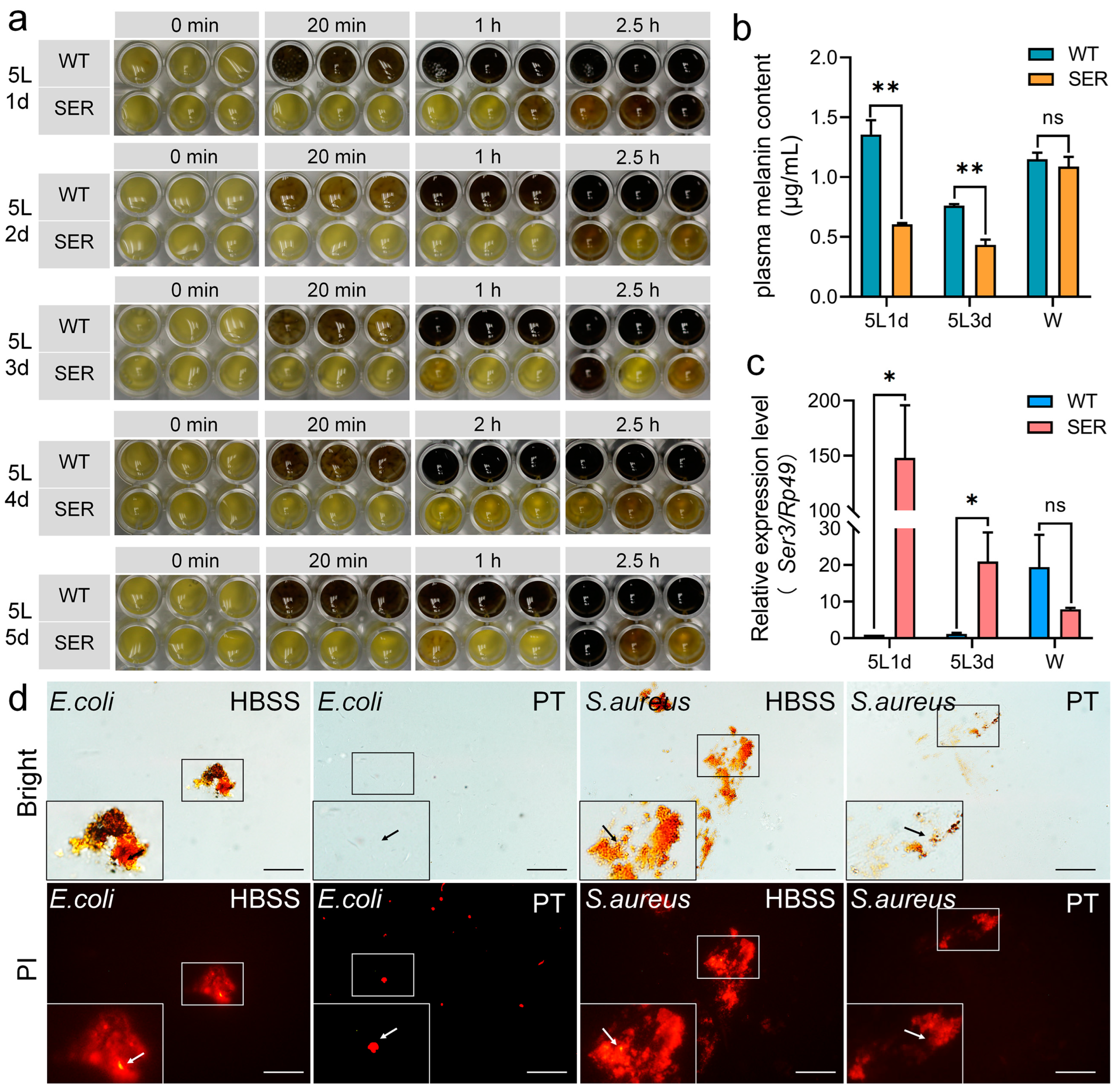
3.1. The Recombinant Ser3 Gene Expressed in the Posterior Silk Gland Affects the Hemolymph Immune Melanization Response of Silkworms

The fifth instar (5L) of silkworm larvae is a period of mass expression of the silk protein gene, rapid synthesis of silk protein, and secretion into the silk gland cavity. In order to study the effect of the recombinant Ser3 gene expression in the posterior silk gland (PSG) on the immune resistance of silkworms, the melanization of silkworm hemolymph, which is closely related to humoral and cellular immunity, was investigated. From the early stage (5L1d) to the late stage (5L5d) of the 5L of the larva, the hemolymph of the WT silkworm began melanization after being exposed to the air for about 20 min. The starting time of melanization had little correlation with the age of the 5L (Figure 1a). The hemolymph of the SER silkworm took more than 2.5 h in vitro to approach the melanization start of the WT group, and the melanization rate was about five times longer.
By comparing the difference in the rate of hemolymph melanization among different silkworm strains, melanization was a common phenomenon. Although there were differences in the time of hemolymph melanization in vitro of 11 representative silkworm varieties or germplasm resources in 5L3d larvae, they all initiated melanization within 5–10 min; only one PSG transgenic silkworm strain TBH started melanization after 30 min (Figure S1), proving that the transgenic expression of exogenous proteins in PSG could significantly reduce the rate of melanization in the hemolymph of larvae.
The changes in melanin content in the hemolymph of transgenic silkworms were further investigated. The results showed that the melanin content of mutant SER was significantly lower than that of WT hemolymph in 5L1d and 5L3d, but there was no statistically significant difference between SER and WT at the wandering stage (W) (Figure 1b); meanwhile, the plasma melanization speed was still significantly slower than that of WT (Figure 1a). It is worth noting that the melanin content in hemolymph of the fifth instar larvae of SER was reduced, which was consistent with the very high expression period of the silk gland Ser3 gene (PSG + MSG) superposed by PSG cells caused by transgenic change (Figure 1c). In the W stage, the transcription level of the Ser3 gene in the SER silk gland was reduced to a level lower than that of WT (Figure 1c), and the melanin content in hemolymph also recovered to that of the WT level. The results indicated that the expression of the recombinant Ser3 gene in the PSG of the mutant in the early and middle stages of the 5L affected the melanin production in hemolymph.
To identify the germicidal efficacy of melanization of WT silkworm hemolymph, E. coli and S. aureus were co-cultured with normal plasma (HBSS) and non-melanized plasma with added phenylthiourea (PT), as shown in Figure 1d. The HBSS group was accompanied by rapid plasma melanization, and propidium iodide (PI) staining showed that bacteria were dead in large numbers and highly clustered. However, the plasma in the PT group did not darken for a long time, and there were few dead bacteria and no serious bacterial aggregation. The results showed that melanization played an important role in the immune response of silkworm after the invasion of a pathogen.
To further study the effect of recombinant Ser3 expression in PSG on the melanin synthesis pathway, the study focused on four key enzymes, including phenylalanine hydroxylase (PAH), tyrosine hydroxylase (TH), dopa decarboxylase (DDC) and phenol oxidase (PO). PAH converts phenylalanine into tyrosine, TH hydroxylates tyrosine into dopamine hydroxylase (TH), DDC converts dopamine into dopamine decarboxylase and PO converts dopamine into dopamine pigment. An enzyme activity survey showed that the activities of PAH, TH and DDC in the hemolymph of SER were higher in 5L1d and 5L3d than in the WT group. However, at the W stage, although PAH was still higher than WT, DDC was lower than WT (Figure 2a–c). It is noteworthy that the PO activity of the mutant SER remained at a very low level throughout the period, maintaining around 10% of WT (Figure 2d); meanwhile, the activities of the other three enzymes in the mutant showed an increasing trend compared with those in the WT group, but were less than one times higher than those in the WT group (Figure 2a–c). Gene transcription levels also showed that that the mRNA level changes of four enzyme genes in the 5L larvae were consistent with their enzyme activity levels (Figure 2e–h). The expressions of the PAH, TH and DDC genes in hemocytes of the SER were significantly up-regulated compared with those in the WT group, except at the W stage (Figure 2e–g), while the PPO1 and PPO2 genes were significantly down-regulated (Figure 2h and Figure S3a), showing a time-inhibitory effect. However, the expression level of the transcription factor Lozenge gene in the hemolymph of SER silkworm was significantly up-regulated (Figure S3b). It showed that the rate-limiting step of SER melanin synthesis was the key enzyme PO of the final reaction, and its gene transcription level was down-regulated with the enzyme activity significantly inhibited; this resulted in a decrease in melanin content in hemolymph and its slowed melanization.

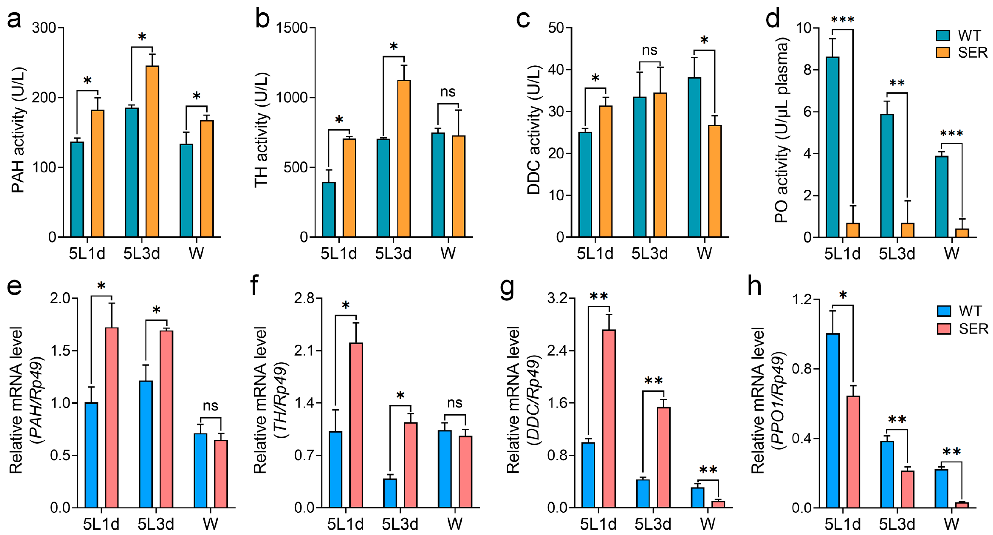
3.2. Mutant Silkworm Affects Hemolymph Melanization by Changing the Serine Protease Cascade

In order to study the reason for the decrease in PO enzyme activity in the rate-limiting step of melanin synthesis in mutant hemolymph, the gene transcription levels of serine protease 21 (SP21) and serine protease phenoloxidase-activating enzyme (PPAE) that finally activate PPO were investigated. The results showed that the mRNA levels of SP21 (Figure 3b) and PPAE (Figure 3c) in SER hemolymph were up-regulated compared with those in WT silkworm at 5L1d and 5L3d, but down-regulated at the W stage; furthermore, the up-regulated and down-regulated changes were less than one-fold. It is worth noting that the mRNA levels of these two genes (Figure S2a,b) were significantly up-regulated compared with those of WT in the main synthetic tissue fat body during the whole 5L period. The PPAE gene was up-regulated more than 200 times in 5L3d, and also significantly up-regulated in the W stage.
Further investigation of the changes in the transcription levels of serine protease inhibitor family genes showed that the Spn5, Spn6 and Spn32 genes were significantly up-regulated in SER hemolymph (Figure 3) and fat bodies (Figure S2) in 5L1d and 5L3d, similar to PPAE and SP21. The extent of up-regulation was greater in fat bodies, which were four-, 550- and 100-times higher than the WT group in 5L3d, respectively, and the Spn32 gene was still up-regulated nearly eight times compared with the WT group in the W stage. The transcription levels of the Spn6 and Spn32 genes in SER fat bodies were sharply up-regulated by more than 100 times during the critical period of Ser3 gene transition from high to low expression in the silk gland of 5L3d mutant (Figure S2d,e), but the PPAE gene that ultimately activated PPO was also up-regulated by 200 times (Figure S2a), showing a two-way extreme abnormal consumptive regulation of proenzyme PPO activation and negative regulation of a key rate-limiting enzyme in melanin synthesis.
Staining analysis of the inhibitory activity of serine protease inhibitors in hemolymph showed that both WT and SER hemolymph had stable inhibitory activity bands for α-chymotrypsin, subtilisin A and proteinase K (Figure 3g–i), but no inhibitory activity bands for trypsin (Figure 3j). Compared with the difference in activity represented by the area of stained bands, SER hemolymph showed stronger inhibitory activity against protease K than WT, especially in 5L1d (Figure 3i). It was suggested that the expression levels of serine protease inhibitor genes Spn5, Spn6 and Spn32 were significantly up-regulated in the mutant, which may have been mainly through the enhanced inhibitory activity of their products on proteinase K, which further affected the expression of PPO cascade genes.

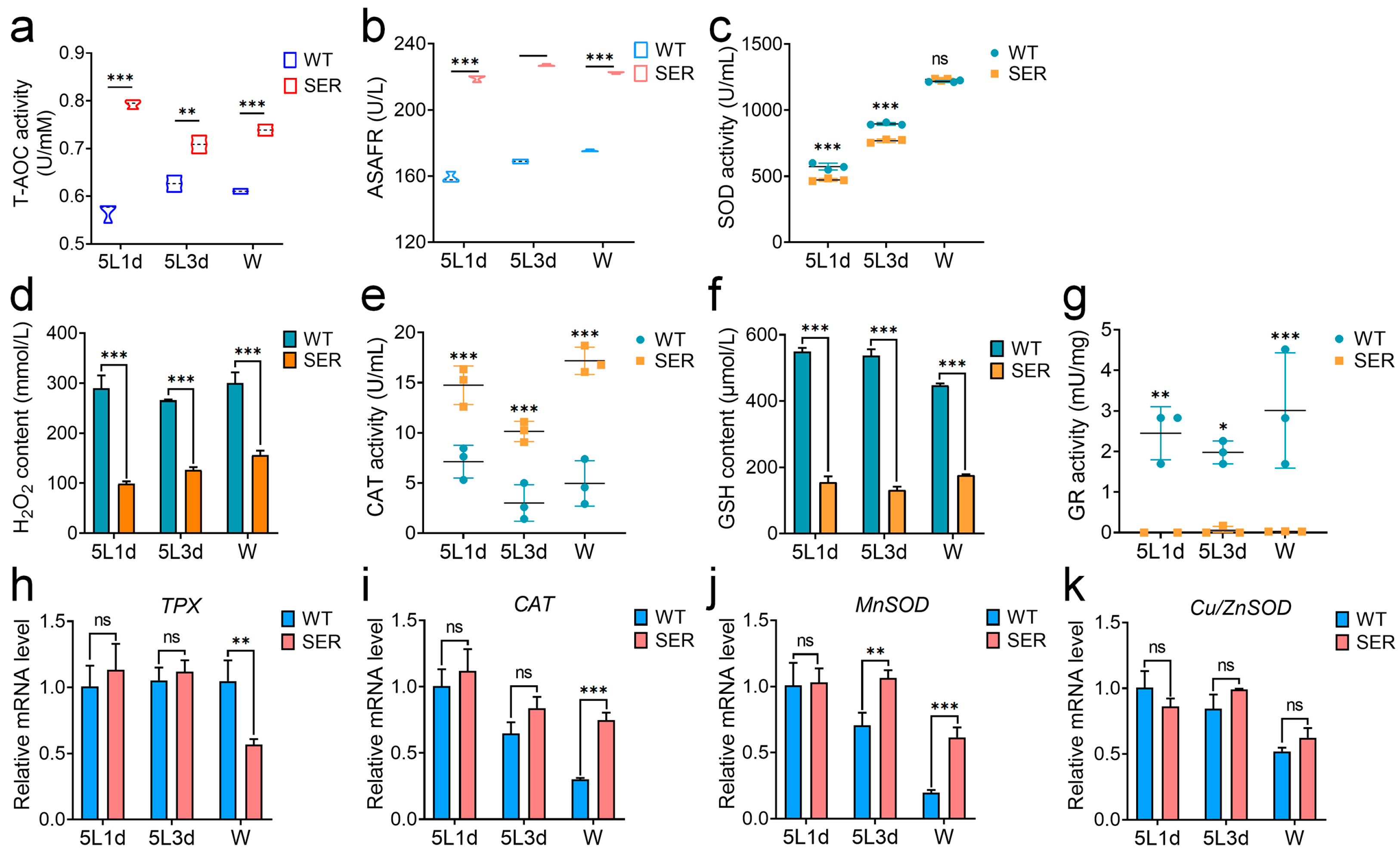
3.3. Mutant Silkworm Affects Hemolymph Melanization by Changing the Level of Hemolymph Redox Metabolism

In order to further explore the relationship between the changes in hemolymph melanization ability and redox metabolism of SER silkworms, the redox ability of hemolymph and the expression levels of its related genes were investigated. The results showed that the hemolymph total antioxidant capacity (T-AOC) of SER silkworms during the whole 5L larval stage was significantly increased (Figure 4a), and inhibited the ability of anti-superoxide anion free radicals (ASAFR) that were also 30 to 40% higher than those of WT silkworms over the same period (Figure 4b). The content of H2O2 in hemolymph of mutant silkworm (Figure 4d) was always significantly lower than that of WT silkworm, but it kept rising in the 5th instar, and increased by nearly 50% in the W stage compared with 5L1d. It is worth noting that the enzyme activity level of SOD involved in oxygen free radical scavenging in the hemolymph of SER silkworms was lower than that of WT (Figure 4c); moreover, the content of glutathione (GSH), which both scavenges oxygen free radicals and H2O2, was significantly lower than that of WT silkworm in the same period (Figure 4f). The activity of glutathione reductase (GR), which generates GSH, had only a trace amount (Figure 4g), but the activity of the CAT enzyme was significantly higher than that of WT silkworms over the same period (Figure 4e).
The investigation results of gene mRNA levels showed that although the TPX (Figure 4h) and CAT (Figure 4i) genes that cleared H2O2 in SER showed no significant differences from those in WT silkworms in the early and middle stages of the 5L, they showed opposite changes at the W stage; the down-regulated expression amplitude of the TPX gene was close to the up-regulated expression amplitude of the CAT gene. It is worth noting that the transcription level of Mn-SOD of the SER silkworm was significantly up-regulated compared with that of WT in the middle and late stages of the 5L larvae (Figure 4j), while Cu/Zn-SOD was always maintained at a similar level to that of WT (Figure 4k), which was inconsistent with the enzyme activity level. It is hypothesized that there may be inhibitory molecules affecting the activity of SOD in hemolymph. These results indicated that the ectopic expression of the recombinant Ser3 gene in PSG led to an increased oxidative stress response level in hemolymph, and increased the antioxidant capacity in body fluids.

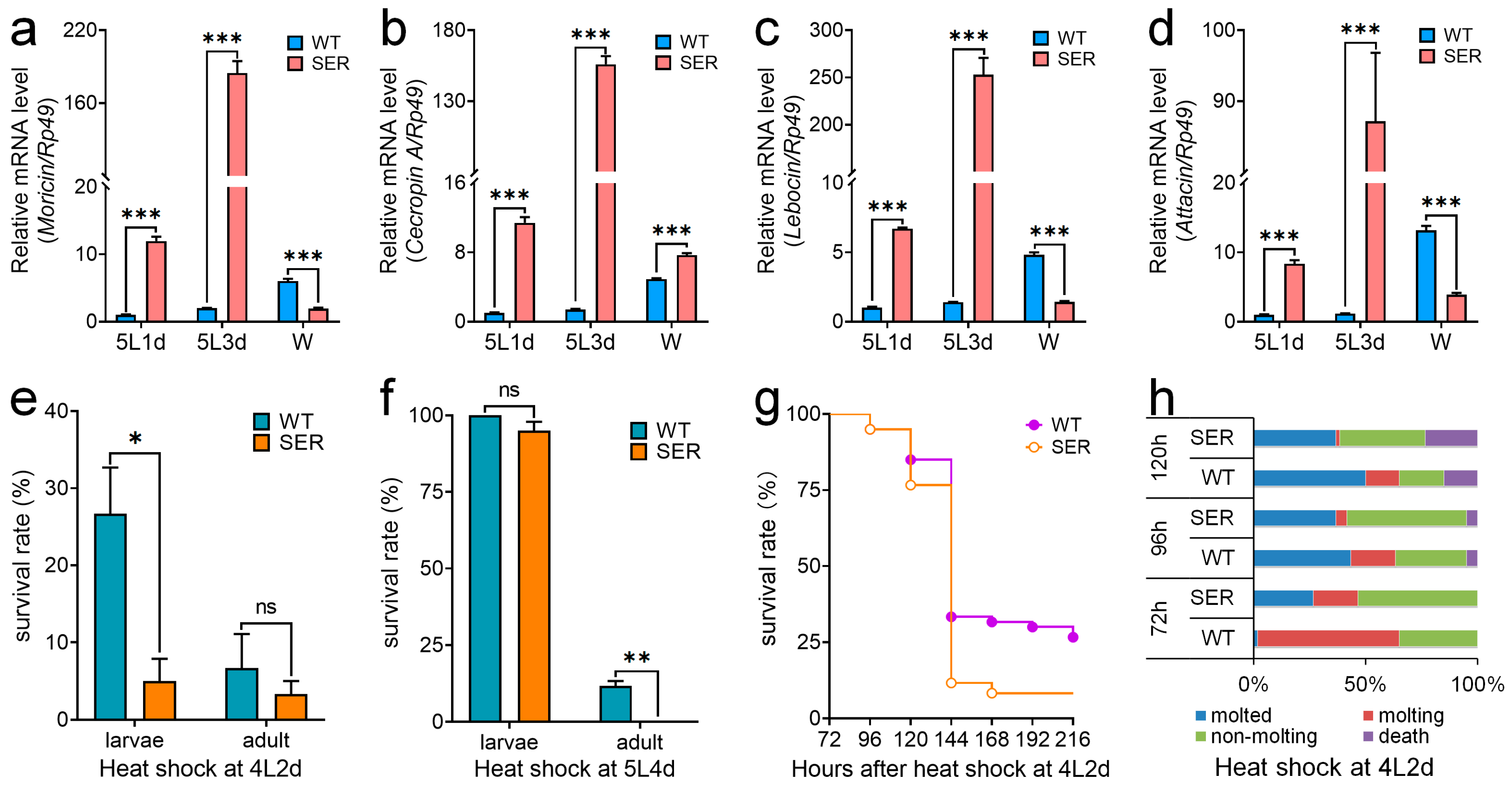
3.4. Mutant Silkworm Affects the Immune Resistance of Silkworms

In order to verify whether humoral immunity was affected in transgenic mutants, we further investigated transcription levels of antimicrobial peptides (AMPs) genes in fat bodies. The results showed that the mRNA levels of moricin, cecropin A, lebocin and attacin were significantly up-regulated in 5L1d and 5L3d (Figure 5a–d). At the W stage, moricin, lebocin and attacin were down-regulated, except cecropin A, which was still up-regulated, presumably due to autoimmune regulation. These results showed that the ectopic expression of the Ser3 gene in the PSG induced the expression of AMPs.
In our previous study, we found that there was no significant difference between the growth and development of SER silkworms and WT under normal conditions [1]. In order to explore whether there were differences under stress conditions, we investigated the growth and development of 4L2d and 5L4d silkworm larvae after 40 °C high temperature shock for 3 h (Figure 5e–h). The results showed that the survival rate of SER larvae significantly decreased after high temperature shock at 4L2d, and the number of SER individuals that developed into adults decreased, but there was no significant difference (Figure 5e). After high temperature shock at 5L4d, the survival rate of SER larvae had no significant effect, but all died in the pupal stage, while 11.67% of WT pupae successfully developed into adults (Figure 5f). The survival curve showed that both WT and SER silkworms began to die at 96 h after high temperature shock at 4L2d; the number of dead individuals increased rapidly from 120 h, and the mortality rate of the SER group was higher than that of the WT group. Finally, 26.67% and 8.33% silkworms in the WT and SER groups spun cocoons, respectively (Figure 5g). At the same time, it was found that after the high temperature shock at 4L2d, the proportion of SER silkworms from molted and molting larvae, and the proportion of non-molting and dead larvae increased compared with WT (Figure 5h). The results showed that high temperature stress significantly affected the growth and development of SER silkworms, suggesting that the basic resistance of SER silkworms was different from that of WT silkworms.

4. Discussion

4.1. The Recombinant Ser3 Expression in Posterior Silk Glands Affects Hemolymph Metabolism

Insect melanization, including that in the silkworm, is activated by the prophenoloxidase (PPO) cascade, and mediated and regulated by serine proteinase (SP) and serine protease inhibitors (serpins) [32,33,34]. Phenoloxidase (PO) is a key rate-limiting enzyme [35], which is mainly secreted by hemocytes called oenocytoids in the hemolymph of the silkworm [36]; it exists in the form of PPO1/PPO2, is activated by SP [37,38] and is negatively regulated by serpins such as Spn5, Spn6 and Spn32 [32,39,40]. The expression of serpin genes in fat bodies was higher than that in other tissues [40]; our study also shows consistent results. In addition to serpins, PO activity is also regulated by clustering proteins and transcription factors [41,42,43,44]. After an overexpression of transcription factor Lozenge in silkworm, the expressions of PPAE, PPO1 and PPO2 genes were up-regulated, and the melanization rate of hemolymph was accelerated when exposed to air, while the melanization rate of hemolymph was significantly slowed down after interference with Lozenge [42]. In this study, the transcription levels of PPO1 and PPO2 were significantly down-regulated, but the expression level of the Lozenge gene was significantly up-regulated. These contradictory changes in a series of key enzyme genes and enzyme activities of melanin synthesis, accompanied by the simultaneous high expression of the exogenous gene Ser3 of the silk gland transgene, showed that the production of melanin in the silk gland transgene mutant was affected by many complex aspects, and ultimately led to the reduction in melanin level in hemolymph.
The production of melanin and the rate of hemolymph melanization in silkworm were also closely related to redox metabolism in vivo. Previous studies have shown that eliminating ROS and improving the antioxidant level of cells can effectively inhibit UV-induced melanin formation [45,46,47], and the excessive production of ROS under oxidative stress leads to the massive production and abnormal metabolism of melanin [48,49]. The results of this study showed that the antioxidant capacity of hemolymph in the SER silkworm was enhanced, and the content of H2O2, which is one of the ROS, was also significantly decreased; this suggests that the decrease in melanin content may have been related to ROS levels. In addition, the results of mRNA levels and enzyme activity for CAT and SOD were not consistent. We speculate that the inconsistency may have been due to the fluctuation of weak difference values on the one hand, while on the other hand, the oxygen free radical scavenging function of SOD had other means of compensating.

4.2. The Recombinant Ser3 Expression in the Posterior Silk Gland Affects the Immune Ability of Silkworms

Studies have shown that insect melanization can isolate and wrap pathogens, and promote wound healing [50]. The melanization reaction is thought to strengthen nodules, provide ROS to kill microbes and to conduct immune defense together with antimicrobial peptides (AMPs) [51]. The PPO cascade regulating melanin production is a unique immune defense response of arthropods, including silkworm. The immune protein PO is involved in the immune defense response of the body, and the serpin gene family is also involved in regulating the immune response [27,28,29,33,39,40,52], where Spn5 can inhibit its activity by combining with serine proteases HP6 and HP21, and down-regulate AMPs expression and PPO activation [32]. However, AMPs expression is not only regulated by serpins, but also by Imd and Toll signaling pathways [27]. The down-regulation of AMPs gene expression levels downstream of the Imd pathway in Drosophila can significantly enhance heat tolerance and prolong life span [53]. When the ROS levels of silkworms decreased, it also improved their high temperature resistance [54].
In our previous study, it was found that the survival ability of SER larvae infected with E. coli and S. aureus was higher than that of WT [1], which may be related to the significantly increased AMPs level of SER silkworms. In addition to the survival and development of insects being closely related to bacteria and other microorganisms, their physiology and metabolism are also very sensitive to temperature. High temperatures can cause mitochondrial disorder, cell protein denaturation, and eventually lead to cell dysfunction and death [55,56]. Studies have shown that insect resistance to high temperature is regulated by redox signaling pathways [57,58]. A moderate dose of H2O2 injected into Mythimna separata larvae consistently increases ROS and antioxidant activity in hemolymph, leading to a significant increase in survival after lethal heat stress [58]. In this study, the redox metabolism of the mutant was significantly affected, the total antioxidant capacity was increased, but the contents of H2O2 and GSH were significantly reduced. Therefore, we speculated that the reduction in high temperature resistance was closely related to disorder in redox metabolism.
It can be seen that the expression of the recombinant Ser3 gene specifically expressed by autologous MSG in PSG transgenic silkworms can avoid the poor growth and development of silk gland tissues and individuals that may occur in PSG transgenic silkworms [1]; however, the basic stress response still appeared, which led to the decline in melanin synthesis and melanization immune ability in hemolymph (Figure 6). We speculate that there may be the following reasons for this phenomenon. Firstly, SER3 protein is an exogenous water-soluble protein that is different from the original fibrin such as Fib-H for PSG, and directly affects the silk protein interaction and secretory transport of silk gland cells and tissues. On the other hand, the recombinant Ser3 gene is highly activated by the Fib-H promoter in PSG, which changes the original supply ratio of amino acids of silk fibroin protein synthesis raw materials in PSG cells [1], even if the change is weak. The nutrients and energy of silk gland cells come from hemolymph, which is also the source of raw materials for silk protein synthesis. Therefore, the ectopic high expression of SER3 in PSG affects the hemolymph immune melanization response, which does not refer to the effect of Ser3 fusion protein products expressed in the posterior silk gland itself, nor is the effect on immunity simply reduced or increased, but there is a change in the immune balance state. These results suggest that it is necessary to study the in-depth biological safety of transgenic exogenous protein mutants for the production of target silk glands and other highly specialized tissues.

5. Conclusions

The recombinant sericin SER3 specific to the middle silk gland was significantly expressed in the posterior silk gland of SER silkworm larvae, which also significantly affected the expressions of antimicrobial peptides and melanization. Meanwhile, it affected the temporal and spatial expression profile of the silkworm genome, resulting in changes in transcription factor regulation, cleavage of proenzyme activity, and oxidative metabolism during melanin synthesis, further leading to significant changes in hemolymph metabolism, and ultimately reducing the melanin synthesis capacity of hemolymph.

Supplementary Materials

The following supporting information can be downloaded at: https://www.mdpi.com/article/10.3390/insects14030245/s1, Figure S1: In vitro melanization of hemolymphs of multiple silkworm varieties; Figure S2: Changes in serine protease inhibitor gene transcription levels in the fat body of transgenic silkworm; Figure S3: Changes in PPO2 and Lozenge gene transcription levels in hemolymphs of transgenic silkworm. Table S1: The primers used in the study.

Author Contributions

Conceptualization and methodology, S.X., Y.W., M.S. and X.C.; investigation, M.S., Y.W., J.Y., L.M., M.X., R.P., G.W. and Z.P.; Validation, J.Y., L.M., M.X., R.P., G.W. and Z.P.; data curation, S.X. and Y.W.; resources, Y.S.; writing—original draft preparation, S.X., M.S. and Y.W.; writing—review and editing, S.X., Y.S. and Y.W.; funding acquisition, S.X. and Y.W. All authors have read and agreed to the published version of the manuscript.

Funding

This research was funded by the National Natural Science Foundation of China (Grant No. 32102608 to Y.W., and 31972625 to S.X.), the Priority Academic Program Development of Jiangsu Higher Education Institutions (PAPD to S.X.), Jiangsu Planned Projects for Postdoctoral Research Funds (2021K321C to Y.W.) and the Nantong science and technology project (JC2021010 to Y.W.).

Data Availability Statement

All of the available data are produced or analyzed with this manuscript.

Conflicts of Interest

The authors declare no conflict of interest.

References

  1. Chen, X.; Wang, Y.; Wang, Y.; Li, Q.; Liang, X.; Wang, G.; Li, J.; Peng, R.; Sima, Y.; Xu, S. Ectopic expression of sericin enables efficient production of ancient silk with structural changes in silkworm. Nat. Commun. 2022, 13, 6295. [Google Scholar] [CrossRef]
  2. Omenetto, F.G.; Kaplan, D.L. New opportunities for an ancient material. Science 2010, 329, 528–531. [Google Scholar] [CrossRef] [PubMed] [Green Version]
  3. Lian, A.A.; Yamaji, Y.; Kajiwara, K.; Takaki, K.; Mori, H.; Liew, M.W.O.; Kotani, E.; Maruta, R. A Bioengineering Approach for the Development of Fibroblast Growth Factor-7-Functionalized Sericin Biomaterial Applicable for the Cultivation of Keratinocytes. Int. J. Mol. Sci. 2022, 23, 9953. [Google Scholar] [CrossRef] [PubMed]
  4. Yamada, K.; Masuda, K.; Ida, S.; Tada, H.; Bando, M.; Abe, K.; Tatematsu, K.I.; Sezutsu, H.; Oyama, T.; Chikamatsu, K.; et al. In vitro assessment of antitumor immune responses using tumor antigen proteins produced by transgenic silkworms. J. Mater. Sci. Mater. Med. 2021, 32, 58. [Google Scholar] [CrossRef] [PubMed]
  5. Kurihara, H.; Sezutsu, H.; Tamura, T.; Yamada, K. Production of an active feline interferon in the cocoon of transgenic silkworms using the fibroin H-chain expression system. Biochem. Biophys. Res. Commun. 2007, 355, 976–980. [Google Scholar] [CrossRef]
  6. Hino, R.; Tomita, M.; Yoshizato, K. The generation of germline transgenic silkworms for the production of biologically active recombinant fusion proteins of fibroin and human basic fibroblast growth factor. Biomaterials 2006, 27, 5715–5724. [Google Scholar] [CrossRef]
  7. Teulé, F.; Miao, Y.G.; Sohn, B.H.; Kim, Y.S.; Hull, J.J.; Fraser, M.J., Jr.; Lewis, R.V.; Jarvis, D.L. Silkworms transformed with chimeric silkworm/spider silk genes spin composite silk fibers with improved mechanical properties. Proc. Natl. Acad. Sci. USA 2012, 109, 923–928. [Google Scholar] [CrossRef] [PubMed] [Green Version]
  8. Xu, J.; Dong, Q.; Yu, Y.; Niu, B.; Ji, D.; Li, M.; Huang, Y.; Chen, X.; Tan, A. Mass spider silk production through targeted gene replacement in Bombyx mori. Proc. Natl. Acad. Sci. USA 2018, 115, 8757–8762. [Google Scholar] [CrossRef] [Green Version]
  9. Otsuki, R.; Yamamoto, M.; Matsumoto, E.; Iwamoto, S.I.; Sezutsu, H.; Suzui, M.; Takaki, K.; Wakabayashi, K.; Mori, H.; Kotani, E. Bioengineered silkworms with butterfly cytotoxin-modified silk glands produce sericin cocoons with a utility for a new biomaterial. Proc. Natl. Acad. Sci. USA 2017, 114, 6740–6745. [Google Scholar] [CrossRef] [PubMed] [Green Version]
  10. Wang, H.; Wang, L.; Wang, Y.; Tao, H.; Yin, W.; Sima, Y.; Wang, Y.; Xu, S.Q. High yield exogenous HPL protein production in the Bombyx mori silk gland provides novel insight into recombinant expression systems. Sci. Rep. 2015, 5, 13839. [Google Scholar] [CrossRef] [Green Version]
  11. Tomita, M.; Munetsuna, H.; Sato, T.; Adachi, T.; Hino, R.; Hayashi, M.; Shimizu, K.; Nakamura, N.; Tamura, T.; Yoshizato, K. Transgenic silkworms produce recombinant human type III procollagen in cocoons. Nat. Biotechnol. 2003, 21, 52–56. [Google Scholar] [CrossRef] [PubMed]
  12. Tamura, T.; Thibert, C.; Royer, C.; Kanda, T.; Abraham, E.; Kamba, M.; Komoto, N.; Thomas, J.L.; Mauchamp, B.; Chavancy, G.; et al. Germline transformation of the silkworm Bombyx mori L. using a piggyBac transposon-derived vector. Nat. Biotechnol. 2000, 18, 81–84. [Google Scholar] [CrossRef]
  13. Uchino, K.; Sezutsu, H.; Imamura, M.; Kobayashi, I.; Tatematsu, K.; Iizuka, T.; Yonemura, N.; Mita, K.; Tamura, T. Construction of a piggyBac-based enhancer trap system for the analysis of gene function in silkworm Bombyx mori. Insect Biochem. Mol. Biol. 2008, 38, 1165–1173. [Google Scholar] [CrossRef] [PubMed]
  14. Horn, C.; Offen, N.; Nystedt, S.; Häcker, U.; Wimmer, E.A. piggyBac-based insertional mutagenesis and enhancer detection as a tool for functional insect genomics. Genetics 2003, 163, 647–661. [Google Scholar] [CrossRef] [PubMed]
  15. Nolan, T.; Bower, T.M.; Brown, A.E.; Crisanti, A.; Catteruccia, F. piggyBac-mediated germline transformation of the malaria mosquito Anopheles stephensi using the red fluorescent protein dsRED as a selectable marker. J. Biol. Chem. 2002, 277, 8759–8762. [Google Scholar] [CrossRef] [Green Version]
  16. Yonemura, N.; Tamura, T.; Uchino, K.; Kobayashi, I.; Tatematsu, K.; Iizuka, T.; Sezutsu, H.; Muthulakshmi, M.; Nagaraju, J.; Kusakabe, T. PhiC31 integrase-mediated cassette exchange in silkworm embryos. Mol. Genet. Genom. 2012, 287, 731–739. [Google Scholar] [CrossRef]
  17. Nakayama, G.; Kawaguchi, Y.; Koga, K.; Kusakabe, T. Site-specific gene integration in cultured silkworm cells mediated by phiC31 integrase. Mol. Genet. Genom. 2006, 275, 1–8. [Google Scholar] [CrossRef]
  18. Imamura, M.; Nakai, J.; Inoue, S.; Quan, G.X.; Kanda, T.; Tamura, T. Targeted gene expression using the GAL4/UAS system in the silkworm Bombyx mori. Genetics 2003, 165, 1329–1340. [Google Scholar] [CrossRef]
  19. Tatematsu, K.; Kobayashi, I.; Uchino, K.; Sezutsu, H.; Iizuka, T.; Yonemura, N.; Tamura, T. Construction of a binary transgenic gene expression system for recombinant protein production in the middle silk gland of the silkworm Bombyx mori. Transgenic Res. 2010, 19, 473–487. [Google Scholar] [CrossRef]
  20. Baci, G.M.; Cucu, A.A.; Giurgiu, A.I.; Muscă, A.S.; Bagameri, L.; Moise, A.R.; Bobiș, O.; Rațiu, A.C.; Dezmirean, D.S. Advances in Editing Silkworms (Bombyx mori) Genome by Using the CRISPR-Cas System. Insects 2021, 13, 28. [Google Scholar] [CrossRef]
  21. Brady, D.; Saviane, A.; Cappellozza, S.; Sandrelli, F. An Efficient Workflow for Screening and Stabilizing CRISPR/Cas9-Mediated Mutant Lines in Bombyx mori. Methods Protoc. 2020, 4, 4. [Google Scholar] [CrossRef] [PubMed]
  22. Tomihara, K.; Andolfatto, P.; Kiuchi, T. Allele-specific knockouts reveal a role for apontic-like in the evolutionary loss of larval melanin pigmentation in the domesticated silkworm, Bombyx mori. Insect Mol. Biol. 2022, 31, 701–710. [Google Scholar] [CrossRef]
  23. Jiang, L.; Xia, Q. The progress and future of enhancing antiviral capacity by transgenic technology in the silkworm Bombyx mori. Insect Biochem. Mol. Biol. 2014, 48, 1–7. [Google Scholar] [CrossRef] [Green Version]
  24. Yashwant, R.S.; Thomas, D.S.; Manoharan, C.; Roy, G.; Kunjupillai, V.; Mishra, R.K.; Nongthomba, U.; Gopalapillai, R. Transgenic Silkworms Overexpressing Relish and Expressing Drosomycin Confer Enhanced Immunity to Multiple Pathogens. Mol. Biotechnol. 2022, 64, 711–724. [Google Scholar] [CrossRef] [PubMed]
  25. Zhao, P.; Xia, F.; Jiang, L.; Guo, H.; Xu, G.; Sun, Q.; Wang, B.; Wang, Y.; Lu, Z.; Xia, Q. Enhanced antiviral immunity against Bombyx mori cytoplasmic polyhedrosis virus via overexpression of peptidoglycan recognition protein S2 in transgenic silkworms. Dev. Comp. Immunol. 2018, 87, 84–89. [Google Scholar] [CrossRef]
  26. Jiang, L.; Cheng, T.; Zhao, P.; Yang, Q.; Wang, G.; Jin, S.; Lin, P.; Xiao, Y.; Xia, Q. Resistance to BmNPV via overexpression of an exogenous gene controlled by an inducible promoter and enhancer in transgenic silkworm, Bombyx mori. PLoS ONE 2012, 7, e41838. [Google Scholar] [CrossRef] [PubMed] [Green Version]
  27. Wang, Y.F.; Wang, G.; Li, J.L.; Qu, Y.X.; Liang, X.Y.; Chen, X.D.; Sima, Y.H.; Xu, S.Q. Influence of Hyperproteinemia on Insect Innate Immune Function of the Circulatory System in Bombyx mori. Biology 2021, 10, 112. [Google Scholar] [CrossRef] [PubMed]
  28. Cooper, D.; Wuebbolt, C.; Heryanto, C.; Eleftherianos, I. The prophenoloxidase system in Drosophila participates in the anti-nematode immune response. Mol. Immunol. 2019, 109, 88–98. [Google Scholar] [CrossRef]
  29. Hillyer, J.F. Insect immunology and hematopoiesis. Dev. Comp. Immunol. 2016, 58, 102–118. [Google Scholar] [CrossRef] [Green Version]
  30. Zhou, L.; Li, H.; Hao, F.; Li, N.; Liu, X.; Wang, G.; Wang, Y.; Tang, H. Developmental Changes for the Hemolymph Metabolome of Silkworm (Bombyx mori L.). J. Proteome Res. 2015, 14, 2331–2347. [Google Scholar] [CrossRef] [Green Version]
  31. Peng, L.H.; Liu, S.; Xu, S.Y.; Chen, L.; Shan, Y.H.; Wei, W.; Liang, W.Q.; Gao, J.Q. Inhibitory effects of salidroside and paeonol on tyrosinase activity and melanin synthesis in mouse B16F10 melanoma cells and ultraviolet B-induced pigmentation in guinea pig skin. Phytomedicine 2013, 20, 1082–1087. [Google Scholar] [CrossRef] [PubMed]
  32. Li, J.; Ma, L.; Lin, Z.; Zou, Z.; Lu, Z. Serpin-5 regulates prophenoloxidase activation and antimicrobial peptide pathways in the silkworm, Bombyx mori. Insect Biochem. Mol. Biol. 2016, 73, 27–37. [Google Scholar] [CrossRef] [PubMed] [Green Version]
  33. Suwanchaichinda, C.; Ochieng, R.; Zhuang, S.; Kanost, M.R. Manduca sexta serpin-7, a putative regulator of hemolymph prophenoloxidase activation. Insect Biochem. Mol. Biol. 2013, 43, 555–561. [Google Scholar] [CrossRef] [PubMed] [Green Version]
  34. Nappi, A.; Poirié, M.; Carton, Y. The role of melanization and cytotoxic by-products in the cellular immune responses of Drosophila against parasitic wasps. Adv. Parasitol. 2009, 70, 99–121. [Google Scholar] [CrossRef] [PubMed]
  35. Sugumaran, M.; Barek, H. Critical Analysis of the Melanogenic Pathway in Insects and Higher Animals. Int. J. Mol. Sci. 2016, 17, 1753. [Google Scholar] [CrossRef] [PubMed] [Green Version]
  36. Iwama, R.; Ashida, M. Biosynthesis of prophenoloxidase in hemocytes of larval hemolymph of the silkworm, Bombyx mori. Insect Biochem. 1986, 16, 547–555. [Google Scholar] [CrossRef]
  37. Tokura, A.; Fu, G.S.; Sakamoto, M.; Endo, H.; Tanaka, S.; Kikuta, S.; Tabunoki, H.; Sato, R. Factors functioning in nodule melanization of insects and their mechanisms of accumulation in nodules. J. Insect Physiol. 2014, 60, 40–49. [Google Scholar] [CrossRef]
  38. Satoh, D.; Horii, A.; Ochiai, M.; Ashida, M. Prophenoloxidase-activating enzyme of the silkworm, Bombyx mori. Purification, characterization, and cDNA cloning. J. Biol. Chem. 1999, 274, 7441–7453. [Google Scholar] [CrossRef] [Green Version]
  39. Wang, L.; Liu, H.; Fu, H.; Zhang, L.; Guo, P.; Xia, Q.; Zhao, P. Silkworm serpin32 functions as a negative-regulator in prophenoloxidase activation. Dev. Comp. Immunol. 2019, 91, 123–131. [Google Scholar] [CrossRef]
  40. Li, B.; Yu, H.Z.; Ye, C.J.; Ma, Y.; Li, X.; Fan, T.; Chen, F.S.; Xu, J.P. Bombyx mori Serpin6 regulates prophenoloxidase activity and the expression of antimicrobial proteins. Gene 2017, 610, 64–70. [Google Scholar] [CrossRef]
  41. Zhou, X.S.; Chen, C.; Li, T.H.; Tang, J.J.; Zhu, B.J.; Wei, G.Q.; Qian, C.; Liu, C.L.; Wang, L. A QM protein from Bombyx mori negatively regulates prophenoloxidase activation and melanization by interacting with Jun protein. Insect Mol. Biol. 2019, 28, 578–590. [Google Scholar] [CrossRef] [PubMed]
  42. Xu, M.; Wang, X.; Tan, J.; Zhang, K.; Guan, X.; Patterson, L.H.; Ding, H.; Cui, H. A novel Lozenge gene in silkworm, Bombyx mori regulates the melanization response of hemolymph. Dev. Comp. Immunol. 2015, 53, 191–198. [Google Scholar] [CrossRef]
  43. Bao, Y.Y.; Xue, J.; Wu, W.J.; Wang, Y.; Lv, Z.Y.; Zhang, C.X. An immune-induced reeler protein is involved in the Bombyx mori melanization cascade. Insect Biochem. Mol. Biol. 2011, 41, 696–706. [Google Scholar] [CrossRef]
  44. Terenius, O.; Bettencourt, R.; Lee, S.Y.; Li, W.; Söderhäll, K.; Faye, I. RNA interference of Hemolin causes depletion of phenoloxidase activity in Hyalophora cecropia. Dev. Comp. Immunol. 2007, 31, 571–575. [Google Scholar] [CrossRef]
  45. Bae, J.S.; Han, M.; Yao, C.; Chung, J.H. Chaetocin inhibits IBMX-induced melanogenesis in B16F10 mouse melanoma cells through activation of ERK. Chem. Biol. Interact. 2016, 245, 66–71. [Google Scholar] [CrossRef] [PubMed]
  46. Jin, Y.J.; Lin, C.C.; Lu, T.M.; Li, J.H.; Chen, I.S.; Kuo, Y.H.; Ko, H.H. Chemical constituents derived from Artocarpus xanthocarpus as inhibitors of melanin biosynthesis. Phytochemistry 2015, 117, 424–435. [Google Scholar] [CrossRef]
  47. Funasaka, Y.; Komoto, M.; Ichihashi, M. Depigmenting effect of alpha-tocopheryl ferulate on normal human melanocytes. Pigment Cell Res. 2000, 13, 170–174. [Google Scholar] [CrossRef]
  48. Fu, W.; Wu, Z.; Zheng, R.; Yin, N.; Han, F.; Zhao, Z.; Dai, M.; Han, D.; Wang, W.; Niu, L. Inhibition mechanism of melanin formation based on antioxidant scavenging of reactive oxygen species. Analyst 2022, 147, 2703–2711. [Google Scholar] [CrossRef] [PubMed]
  49. Gebicki, J.M. Oxidative stress, free radicals and protein peroxides. Arch. Biochem. Biophys. 2016, 595, 33–39. [Google Scholar] [CrossRef]
  50. Cerenius, L.; Lee, B.L.; Söderhäll, K. The proPO-system: Pros and cons for its role in invertebrate immunity. Trends. Immunol. 2008, 29, 263–271. [Google Scholar] [CrossRef]
  51. Kan, H.; Kim, C.H.; Kwon, H.M.; Park, J.W.; Roh, K.B.; Lee, H.; Park, B.J.; Zhang, R.; Zhang, J.; Söderhäll, K.; et al. Molecular control of phenoloxidase-induced melanin synthesis in an insect. J. Biol. Chem. 2008, 283, 25316–25323. [Google Scholar] [CrossRef] [PubMed] [Green Version]
  52. Marieshwari, B.N.; Bhuvaragavan, S.; Sruthi, K.; Mullainadhan, P.; Janarthanan, S. Insect phenoloxidase and its diverse roles: Melanogenesis and beyond. J. Comp. Physiol. B 2023, 193, 1–23. [Google Scholar] [CrossRef]
  53. Lin, Y.R.; Parikh, H.; Park, Y. Stress resistance and lifespan enhanced by downregulation of antimicrobial peptide genes in the Imd pathway. Aging 2018, 10, 622–631. [Google Scholar] [CrossRef] [PubMed] [Green Version]
  54. Jiang, L.; Huang, C.; Wang, B.; Guo, H.; Sun, Q.; Xia, F.; Xu, G.; Xia, Q. Enhanced heat tolerance in transgenic silkworm via overexpression of Pyrococcus furiosus superoxide reductase. Insect Biochem. Mol. Biol. 2018, 92, 40–44. [Google Scholar] [CrossRef] [PubMed]
  55. Kültz, D. DNA damage signals facilitate osmotic stress adaptation. Am. J. Physiol. Ren. Physiol. 2005, 289, F504–F505. [Google Scholar] [CrossRef] [Green Version]
  56. Tower, J. Heat shock proteins and Drosophila aging. Exp. Gerontol. 2011, 46, 355–362. [Google Scholar] [CrossRef] [Green Version]
  57. Hayakawa, Y. N-acetyltyrosine-induced redox signaling in hormesis. Biochim. Biophys. Acta Mol. Cell. Res. 2021, 1868, 118990. [Google Scholar] [CrossRef]
  58. Matsumura, T.; Matsumoto, H.; Hayakawa, Y. Heat stress hardening of oriental armyworms is induced by a transient elevation of reactive oxygen species during sublethal stress. Arch. Insect Biochem. Physiol. 2017, 96, e21421. [Google Scholar] [CrossRef]
Figure 1. Reduced hemolymph immune melanization response of transgenic silkworm SER. (a) Melanization rate in vitro and (b) melanin content in hemolymph of 5th instar larvae. (c) Transcription levels of the Ser3 gene in silk glands. Rp49 was the internal reference gene. SER, a transgenic mutant. WT, wild type. 5L1d and 5L3d represent the 5th instar 1d and 3d larvae, respectively. W, wandering stage. * p < 0.05; ** p < 0.01; ns, no significant difference between the two groups. n = 3 biological repeats. (d) PI staining was used to investigate the germicidal efficacy of plasma melanization of 5L3d larvae. PI, propidium iodide, dyes the dead bacterial nucleus red. HBSS, Hank’s balanced salt solution. PT, phenylthiourea. Bar = 200 μm.
Insects 14 00245 g001
Figure 2. Changes in key enzyme activities and gene levels for melanin synthesis in hemolymph of transgenic silkworms. (ad) Enzyme activity levels. (eh) Gene transcription levels. PAH, phenylalanine hydroxylase. TH, tyrosine hydroxylase. DDC, dopa decarboxylase. PO, phenoloxidase. PPO1, prophenoloxidase 1. Rp49 was the internal reference gene. * p < 0.05; ** p < 0.01; *** p < 0.001; ns, no significant difference between the two groups. n = 3 biological repeats.
Insects 14 00245 g002
Figure 3. Changes in transcription levels of genes related to the serine protease cascade pathway in hemolymph of transgenic silkworms. (a) Schematic diagram of melanization regulation pathway. (bf) qPCR was used to investigate the relative transcription levels of genes. PPAE and SP21 are the positive regulatory genes that eventually activate PPO into PO; Spn5, Spn6 and Spn32 are the main member genes of the serine protease inhibitor family that are reversely regulated; and Rp49 was the internal reference gene. (gj) Activities of serine protease inhibitors in hemolymph. (g) α-chymotrypsin. (h) Subtilisin A. (i) Proteinase K. (j) Trypsin. * p < 0.05; ** p < 0.01; *** p < 0.001; ns, no significant difference between the two groups. n = 3 biological repeats.
Insects 14 00245 g003
Figure 4. Changes in antioxidant capacity of SER hemolymph of transgenic silkworms. (a) T-AOC, total antioxidant capacity. (b) ASAFR, inhibition ability of anti-superoxide anion free radicals. (cg) Changes in substance content and enzyme activities related to oxidative metabolism in hemolymph. (c) SOD activity. (d) H2O2 content. (e) CAT activity. (f) GSH content. (g) Glutathione reductase (GR) activity. (hk) Changes in transcription levels of redox metabolism-related genes in hemolymph. (h) TPX. (i) CAT. (j) Mn-SOD. (k) Cu/Zn SOD. Rp49 was the internal reference gene. * p < 0.05; ** p < 0.01; *** p < 0.001; ns, no significant difference between the two groups. n = 3 biological repeats.
Insects 14 00245 g004
Figure 5. Recombinant Ser3 expression in the posterior silk gland affected the immune resistance of silkworms. (ad) The transcription level of antimicrobial peptide gene in fat body. (a) Moricin. (b) Cecropin A. (c) Lebocin. (d) Attacin. Rp49 was the internal reference gene. (e,f) Survival rates of silkworms in 4L2d and 5L4d after high temperature shock. (g) Survival curve of silkworms in 4L2d after high temperature shock. (h) The growth and development of 4L2d silkworms after high temperature shock. * p < 0.05; ** p < 0.01; *** p < 0.001; ns, no significant difference between the two groups. n = 3 biological repeats.
Insects 14 00245 g005
Figure 6. Impacts of the recombinant Ser3 expression in posterior silk glands on the hemolymph immune melanization response of silkworms. SG, silk gland. FB, fat body. HL, hemolymph. Red means up-regulation and green means down-regulation.
Insects 14 00245 g006
Disclaimer/Publisher’s Note: The statements, opinions and data contained in all publications are solely those of the individual author(s) and contributor(s) and not of MDPI and/or the editor(s). MDPI and/or the editor(s) disclaim responsibility for any injury to people or property resulting from any ideas, methods, instructions or products referred to in the content.

