Article

DNA Barcoding of Morphologically Characterized Mosquitoes Belonging to the Genus Mansonia from the Atlantic Forest and Brazilian Savanna

by
Karin Kirchgatter
1,2,*,
Lilian de Oliveira Guimarães
1,
Eliana Ferreira Monteiro
1,
Vanessa Christe Helfstein
1,2,
Juliana Telles-de-Deus
1,
Regiane Maria Tironi de Menezes
1,
Simone Liuchetta Reginato
1,
Carolina Romeiro Fernandes Chagas
3,4 and
Vera Lucia Fonseca de Camargo-Neves
1
1
Pasteur Institute, São Paulo 01027-000, SP, Brazil
2
Postgraduate Program in Tropical Medicine, School of Medicine, University of São Paulo, São Paulo 05403-000, SP, Brazil
3
Nature Research Centre, 08412 Vilnius, Lithuania
4
Applied Research Department, São Paulo Zoological Foundation, São Paulo 04301-905, SP, Brazil
*
Author to whom correspondence should be addressed.
Insects 2023, 14(2), 109; https://doi.org/10.3390/insects14020109
Submission received: 23 December 2022 / Revised: 16 January 2023 / Accepted: 18 January 2023 / Published: 20 January 2023
(This article belongs to the Special Issue Insect Vector-Focused Approaches for Disease Control)

Abstract

:

Simple Summary

This study aimed to provide genetic data of Mansonia, a mosquito species that is an important vector of viruses and other parasites to both humans and animals. The morphological identification of this species, is quite difficult, even for experienced entomologists, and often requires the assembly of male genitalia, whose structural characters allow for accurate identification of most species, which is not always possible. The DNA sequences obtained in this study can be used for future molecular identifications of this species (DNA barcoding).

Abstract

The identification of mosquito species is necessary for determining the entomological components of disease transmission. However, identification can be difficult in species that are morphologically similar. The cytochrome c oxidase subunit I (COI) DNA barcode region is considered a valuable and reliable diagnostic tool for mosquito species recognition, including those that belong to species complexes. Mansonia mosquitoes are found in forests near swampy areas. They are nocturnal and are highly attracted to light. Hematophagous adult females exhibit aggressive biting behavior and can become infected with and transmit pathogens during their feeding, including some epizootic viruses and avian malaria. In Brazil, twelve Mansonia species have been reported. In a recent study from the São Paulo Zoo in Brazil, three morphologically distinct species were collected and identified, namely: Mansonia (Mansonia) indubitans, Ma. (Man.) pseudotitillans and Ma. (Man.) titillans. However, confirmation of these species by molecular identification was unsuccessful due to a lack of COI sequences in the GenBank database. Thus, this research aimed to describe the COI DNA barcode sequences of some morphologically characterized Mansonia (Man.) species from Brazil and to determine their utility in delimiting species collected from the Atlantic Forest and Brazilian Savanna. Accordingly, we provide tools for the genetic identification of species that play a significant role in pathogen transmission in wildlife and potentially humans. We show that the delimitation of Mansonia species via five different approaches based on COI DNA sequences (BI, NJ, ASAP, bPTP and GMYC) yield basically the same groups identified by traditional taxonomy, and we provide the identification of specimens that were previously identified only up to the subgenus level. We also provide COI sequences from two Mansonia species that were not previously available in sequence databases, Ma. wilsoni and Ma. pseudotitillans, and thus contribute to the ongoing global effort to standardize DNA barcoding as a molecular means of species identification.

1. Introduction

Mosquito species identification is crucial for determining the entomological components of disease transmission, but it can be difficult in species that are morphologically similar. Moreover, the successful identification of these challenging species groups using morphology is time-consuming, requires rare taxonomic expertise, and is dependent on the integrity of the external characteristics of the specimens (e.g., scales can be lost during collection) [1].
Taxonomic keys are the most common method used for the identification of adult mosquitoes. They involve a stepwise comparison of morphological features, selecting among the ones that fit the described characteristics and eliminating species that do not fit the description, until a conclusion is reached. However, taxonomic keys are complex and have several limitations. For instance, there may be some natural variation between different populations of the same species, or it is simply not possible to distinguish between some species based on external appearance alone [1].
DNA sequences are used as an additional tool for species recognition, including those that belong to species complexes [2]. Herbert and co-workers proposed the use of a 658 base pair (bp) region of the cytochrome c oxidase subunit 1 (COI) gene as a universal marker to barcode animal life [3]. The COI DNA barcode region is considered a valuable and reliable diagnostic tool for studying the genetic structure and diversity of mosquitoes (Diptera: Culicidae) [4,5,6,7].
The Culicidae is divided into two subfamilies, the Anophelinae and the Culicinae. Within the Culicinae, ten tribes are recognized: Aedeomyiini, Aedini, Culicini, Culisetini, Ficalbiini, Hodgesiini, Mansoniini, Orthopodomyiini, Sabethini, and Uranotaeniini [8].
The Mansoniini comprises two genera: Mansonia Blanchard, 1901 and Coquillettidia Dyar, 1905. These genera usually deposit their eggs directly on the surface of the water or in aquatic vegetation, where the larvae are fixed by their respiratory siphons. Immature forms of these two genera have a spiracular apparatus adapted to perforate the submerged vegetation and obtain oxygen from the tissues of plants [9]. Thus, an abundance of aquatic plants and a reduction in water flow can facilitate the proliferation of these mosquitoes, which are accordingly found in forests near swampy areas. Mansonia mosquitoes are nocturnal and are highly attracted to light. Hematophagous adult females exhibit aggressive biting behavior and are often a nuisance to humans [10].
The Mansonia genus comprises 27 species distributed in two subgenera. The subgenus Mansonioides Theobald, 1907 consists of 12 species, ten distributed in Asia and two in Ethiopia [11]. The subgenus Mansonia contains 15 neotropical species [10,12,13,14], namely: Mansonia amazonensis Theobald, 1901, Mansonia cerqueirai Barreto & Coutinho, 1944, Mansonia chagasi da Costa Lima, 1935, Mansonia dyari Belkin, Heinemann & Page, 1970, Mansonia flaveola Coquillett, 1906, Mansonia fonsecai Pinto, 1932, Mansonia humeralis Dyar and Knab, 1916, Mansonia iguassuensis Barbosa, Navarro da Silva & Sallum, 2007, Mansonia indubitans Dyar & Shannon, 1925, Mansonia leberi Boreham, 1970, Mansonia pessoai Barreto & Coutinho, 1944, Mansonia pseudotitillans Theobald, 1901, Mansonia suarezi Cova Garcia & Sutil Oramas, 1976, Mansonia titillans Walker, 1848, and Mansonia wilsoni Barreto & Coutinho, 1944. Only Ma. titillans and Ma. indubitans have a distribution that reaches the southern tip of the Nearctic Region. Mansonia titillans is the species with the widest geographical distribution, being found in the United States (Florida, Texas), Mexico, South and Central America, and the Antilles [12].
In Brazil, the Mansonia genus is near ubiquitously distributed [12,13], with twelve species currently known in the country: Ma. amazonensis, Ma. cerqueirai, Ma. chagasi, Ma. flaveola, Ma. fonsecai, Ma. humeralis, Ma. iguassuensis, Ma. indubitans, Ma. pessoai, Ma. pseudotitillans, Ma. titillans, and Ma. wilsoni [15].
A recent study conducted at the São Paulo Zoo aimed to identify potential vectors of avian Plasmodium. One hundred and eight specimens of Mansonia were found and were morphologically identified as three distinct species: Ma. indubitans, Ma. pseudotitillans, and Ma. titillans [16]. To confirm the mosquito species infected with hemosporidian parasites, we conducted molecular identification using DNA barcoding. However, for mosquitoes of the Mansonia genus, species identification based on the best close match (BCM) approach was unsuccessful since queries in the GenBank database (hereafter GenBank) returned BCM values far below the threshold (90%) for a successful identification (>99%) [16]. A screening for COI sequences of Mansonia species in GenBank revealed sequences for Ma. titillans (Colombia and Mexico), Ma. indubitans (Brazil and Colombia), Ma. flaveola (Puerto Rico), Ma. humeralis (Argentina), Ma. dyari (Mexico and Virgin Islands), and Ma. amazonensis (Brazil). Therefore, of the sequences available in GenBank, only two sequences were from Brazil and not even half of the species described in Brazil had sequences deposited in GenBank; no sequences were found for Ma. cerqueirai, Ma. chagasi, Ma. fonsecai, Ma. iguassuensis, Ma. pessoai, Ma. pseudotitillans, and Ma. wilsoni.
Although there are few studies exploring the vector potential of Mansonia (Man.) species, some specimens have been found to be infected with arboviruses and other pathogens. Mansonia indubitans is moderately susceptible to infection with four strains of Venezuelan equine encephalitis virus (VEEV) [17,18,19]. Mansonia titillans is a species from which both epizootic [20] and enzootic [21] VEEV was isolated and shows an intermediate capacity to become infected with and transmit epizootic viruses [17,22]. Saint Louis encephalitis virus (SLEV) was detected in Ma. titillans for the first time in Colombia [23] but was also reported in Argentina [24]. The Bunyamwera serogroup, one of the most important serogroups in the Orthobunyavirus genus [25], was also reported in Ma. titillans from Argentina [24]. Lastly, the occurrence of avian Plasmodium lineages in Mansonia mosquitoes from Brazil has been reported in two species (Ma. titillans and Ma. pseudotitillans) [26]. More recently and as mentioned before, the avian pathogens Plasmodium nucleophilum and Haemoproteus (Parahaemoproteus) sp. were observed in two specimens in Brazil, Mansonia indubitans and Mansonia (Man.) sp. [16].
Studies on insect genetics are important for improving vector control measures and aim to prevent or reduce epidemic impacts. Thus, this research aimed to describe the COI DNA barcode sequences of some morphologically characterized Mansonia (Man.) species from Brazil and to determine their utility in delimiting species collected in the Atlantic Forest and Brazilian Savanna. Accordingly, we provide tools for the genetic identification of species that play a significant role in pathogen transmission in wildlife and potentially humans.

2. Materials and Methods

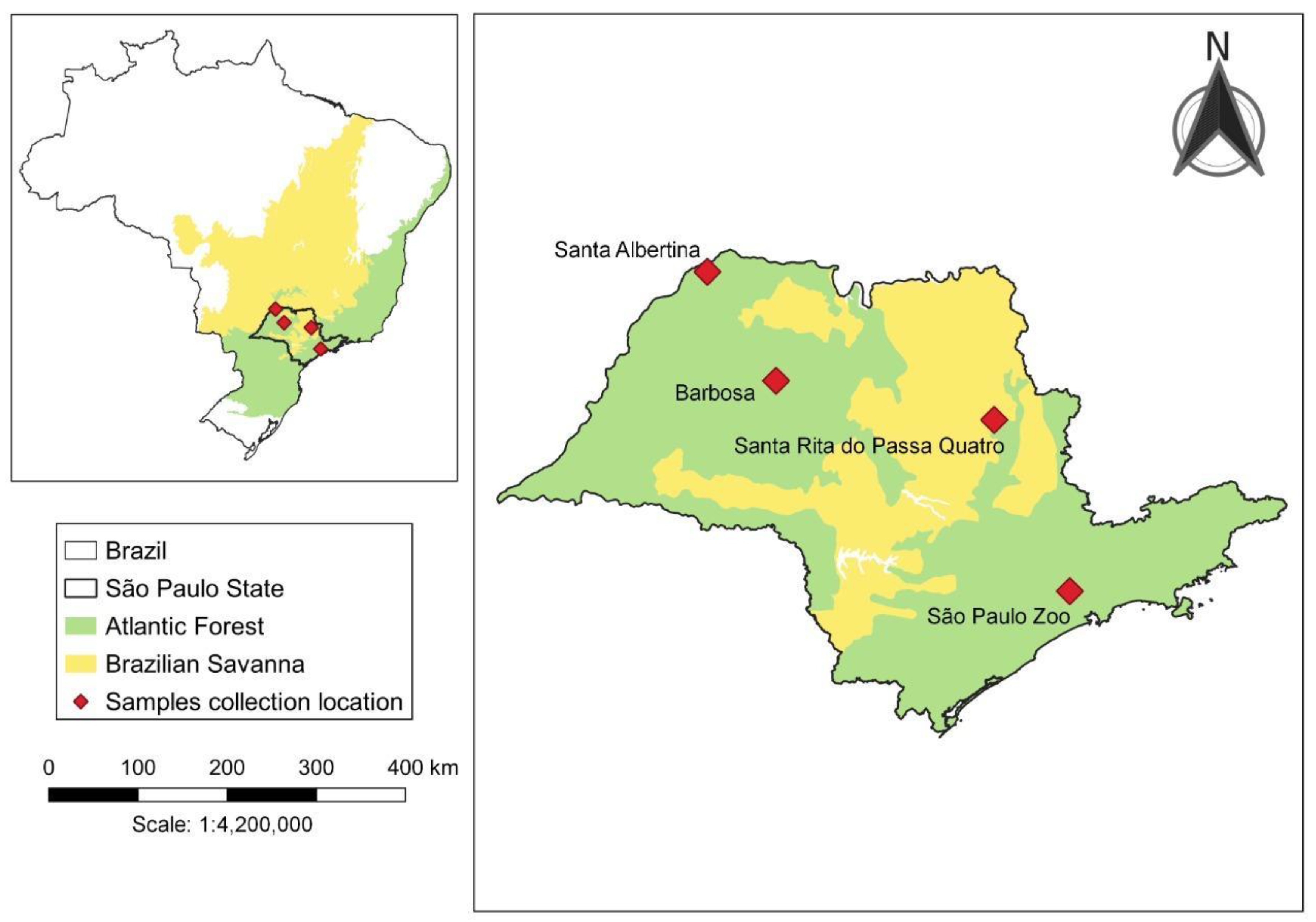
2.1. Mosquito Sampling and Handling

Female mosquitoes were collected at four study sites in the State of São Paulo, Brazil. Study sites and years are shown in Figure 1 and Table 1. Santa Albertina (20°01′55″ S, 50°43′40″ W), Barbosa (21°16′00″ S, 49°56′57″ W), and Santa Rita do Passa Quatro (21°42′36″ S, 47°28′40″ W) are municipalities located in the northwest, north-northwest, and northeast regions of the State of São Paulo, respectively. The São Paulo Zoo (23°39′03″ S, 46°37′14″ W) is in the city of São Paulo, the capital of the State with the same name, and situated in its southeast region (Figure 1).
Mosquitoes were collected using CDC (Center for Disease Control) light traps [27] baited with CO2 (dry ice) for 12 h. The traps were set at dusk and removed a few hours after sunset or with Nasci aspirator for 2 h, from 9:00 to 11:00 a.m. [28]. Part of the mosquitoes were killed with chloroform vapor and part were killed in liquid nitrogen and transported to the laboratory for taxonomic identification using morphological taxonomic keys [10,29,30]. Mosquitoes were individually stored at −20 °C in 1.5 mL plastic tubes sealed with parafilm before molecular processing.
The photomicrographs were performed under a Leica M205C stereomicroscope, with images being captured with an attached Leica DFC320 digital camera and processed with the FusionOptics technology that provides a 3D image in Leica Application Suite 3.7 (Leica Microsystems, Wetzlar, Germany). The maxillary palpus and proboscis lengths were measured using this system.

2.2. Genomic DNA Extraction and PCR Amplification of Mitochondrial Gene Fragments

Collected mosquitoes were transferred to a Master Mix lysis buffer [200 µL Nuclei Lysis Solution, 50 µL EDTA (Ethylene Diamine Tetraacetic Acid) 0.5 M (pH 8.0), 20 µL proteinase K (20 mg/mL), and 5 µL RNase A Solution] and thereafter triturated using FastPrep-96 (MP Biomedicals, Solon, OH, USA) in combination with two 1.4 mm ceramic beads (MagNA Lyser Green Beads-Roche Molecular Systems) coated with 6.35 mm zirconium oxide (MP Biomedicals). The trituration process was conducted for 3 min at 1800 rpm. Samples were then centrifuged at room temperature for 5 min, at 14,000 rpm. DNA was extracted using the Wizard SV 96 Genomic DNA Purification System (Promega) according to manufacturer instructions. Finally, extracted DNA was eluted in 100 µL of nuclease-free water and stored at −20 °C until analysis.
A fragment of 710 base pairs (bp) of the barcode region of the mitochondrial COI gene was amplified by PCR using the primers LCO1490/HCO2198 [31], following the protocol proposed by Ruiz et al. [32].

2.3. Sequencing, Alignment, and Sequence Analysis

PCR products were directly sequenced in both directions by means of a BigDye Terminator v3.0 Cycle Sequencing Kit in an ABI Genetic Analyzer (Applied Biosystems®, Foster City, CA, USA); corresponding flanking primers were used. Sequences were aligned with reference sequences (Table 2) using Clustal W [33], inspected, and edited within MEGA version X [34]. Obtained sequences were deposited within the GenBank database (OQ120978-OQ121013).
For the phylogenetic analysis, an alignment matrix was prepared. The matrix consisted of 36 COI sequences from the collected specimens morphologically identified as: Ma. humeralis, Ma. pseudotitillans, Ma. titillans, Ma. wilsoni, Ma. indubitans, and Mansonia (Man.) sp. (Table 1). Additionally, the matrix included another 41 COI sequences of Mansonia (Man.) species from other Neotropical areas that were retrieved from GenBank (Table 2), and only sequences with >609 bp were used.
Table 1. Collected female Mansonia (Man.) spp., according to collection year, period, method, strata, and sites.
Sample IDCollection
Year
PeriodMethodStrataCollection SiteSpecies (According to Taxonomic Keys)
A290E2019DNasci GSanta AlbertinaMansonia humeralis
A290K2019DNasciGSanta AlbertinaMansonia humeralis
A290L2019DNasciGSanta AlbertinaMansonia humeralis
A290M2019DNasciGSanta AlbertinaMansonia humeralis
A290W2019DNasciGSanta AlbertinaMansonia titillans
A57252020NCDCGSanta Rita do Passa QuatroMansonia (Man.) sp.
A6132019DNasci GBarbosaMansonia humeralis
A613B2019DNasciGBarbosaMansonia humeralis
B1732020NCDCCSão Paulo ZooMansonia wilsoni
B1892020NCDCCSão Paulo ZooMansonia (Man.) sp.
B2402020NCDCCSão Paulo ZooMansonia (Man.) sp.
B2452020NCDCCSão Paulo ZooMansonia (Man.) sp.
B3782020NCDCCSão Paulo ZooMansonia wilsoni aff
B5562020NCDCCSão Paulo ZooMansonia wilsoni aff
B6152020NCDCGSão Paulo ZooMansonia (Man.) sp.
B832020NCDCCSão Paulo ZooMansonia (Man.) sp.
B926 2020NCDCCSão Paulo ZooMansonia wilsoni aff
Zoo0442015NCDCGSão Paulo ZooMansonia (Man.) sp.
Zoo2522015NCDCGSão Paulo ZooMansonia (Man.) sp.
Zoo5552015NCDCGSão Paulo ZooMansonia (Man.) sp.
Zoo6342015NCDCGSão Paulo ZooMansonia (Man.) sp.
Zoo6832015NCDCGSão Paulo ZooMansonia (Man.) sp.
Zoo6842015NCDCGSão Paulo ZooMansonia (Man.) sp.
Zoo6852015NCDCGSão Paulo ZooMansonia (Man.) sp.
Zoo6862015NCDCGSão Paulo ZooMansonia (Man.) sp.
Zoo7982015NCDCGSão Paulo ZooMansonia (Man.) sp.
Zoo7992015NCDCGSão Paulo ZooMansonia indubitans
Zoo8002015NCDCGSão Paulo ZooMansonia (Man.) sp.
ZooB0502015NCDCGSão Paulo ZooMansonia pseudotitillans
ZooB2522015NCDCGSão Paulo ZooMansonia (Man.) sp.
ZooB2532015NCDCGSão Paulo ZooMansonia pseudotitillans
ZooB3652015NCDCGSão Paulo ZooMansonia pseudotitillans
ZooB3832015NCDCGSão Paulo ZooMansonia pseudotitillans
ZooB5912015NCDCGSão Paulo ZooMansonia (Man.) sp.
ZooB5922015NCDCGSão Paulo ZooMansonia pseudotitillans
ZooB7972015NCDCGSão Paulo ZooMansonia (Man.) sp.
Note: Period: D = day and N = night; Strata: C = canopy and G = ground. (Man.) = abbreviation of the subgenus Mansonia according to Reinert [35].
Table 2. GenBank accession numbers of the reference Mansonia (Man.) sequences used in the study.
Species Country Source#GenBank
Mansonia indubitansBrazil (Caatinga)MH118158
Mansonia indubitansColombia MN997669-MN997672
Mansonia flaveolaPuerto RicoJX260065
Mansonia humeralisArgentina MW363430-MW363432
Mansonia titillansColombiaKT766533
Mansonia titillansColombia KY859898-KY859902
MN997665-MN997667
Mansonia titillansMexico MN968219, MN968225, MN968231, MN968233, MN968240, MN968241, MN968244, MN968259, MN968266,
MN968270, MT999303
Mansonia dyariMexico MN968222, MN968243, MN968246,
MN968251, MN968254, MN968264, MN968268, MN968272, MN968273,
MN968274
Mansonia dyariVirgin IslandsMN129182
Mansonia amazonensisBrazilMK575483
Phylogenetic tree reconstruction was performed using the Bayesian approach implemented in MrBayes v3.2.2 [36]. This phylogenetic tree was built with the aim of obtaining support values for the taxa where genetic clusters may represent new or cryptic species; it was not our objective to infer phylogenetic relationships between the species analyzed. This Bayesian inference (BI) was conducted with two Markov Chain Monte Carlo (MCMC) chains run simultaneously for 3 million generations, sampling 1 in every 300 trees. After a burn-in of 25%, the remaining 15,002 trees were used to generate a 50% majority-rule consensus tree. The standard deviation of the split frequencies between runs (<0.01) and the effective sample size was monitored to ensure stability, convergence, and correct mixing of the chains. The result of the analysis was visualized using FigTree version 1.4.4 [37]. The topology was rooted using a sequence obtained for Aedes aegypti (KX420454). Another tree reconstruction was performed by the Neighbor-Joining (NJ) method using MEGA version X [34] and Kimura-2 Parameter distances. Branch supports of NJ trees were assessed by bootstrapping with 1000 replicates. MEGA version X [34] was also used to compute intraspecific (mean distance within group) and interspecific (mean distance between groups) sequence divergence using the Kimura-2 parameter distance model [38].

2.4. Species Delimitation

The use of DNA barcodes for delimiting species into molecular operational taxonomic units (MOTUs) includes a series of strategies that use a combination of laboratory and bioinformatics methods [39]. Beside BI and NJ, three more approaches were used to establish the species delimitation. First, the Assemble Species by Automatic Partitioning (ASAP) was run on a web server (https://bioinfo.mnhn.fr/abi/public/asap/) (accessed on 3 October 2022) using Kimura (K80) ts/tv 2.0; the lower score (1.5) was considered the better partition [40]. Second, the Poisson Tree Processes method (bPTP) [41] based on the unrooted binary maximum likelihood (ML) tree (GTR+G) was used and obtained with MEGA version X [34] and implemented on a web server (https://species.h-its.org/ (accessed on 3 October 2022)) applying default settings. The bPTP adds Bayesian support (BS) values to delimited species on the input tree; the higher the BS, the more likely it is that the taxa forming the node belong to the same species. Third, the Generalized Mixed Yule Coalescent model (GMYC) [42] based on an ultrametric tree resulting from a single locus was used and was run on a web server (http://species.h-its.org/gmyc/ (accessed on 3 October 2022)) using default parameters.

3. Results

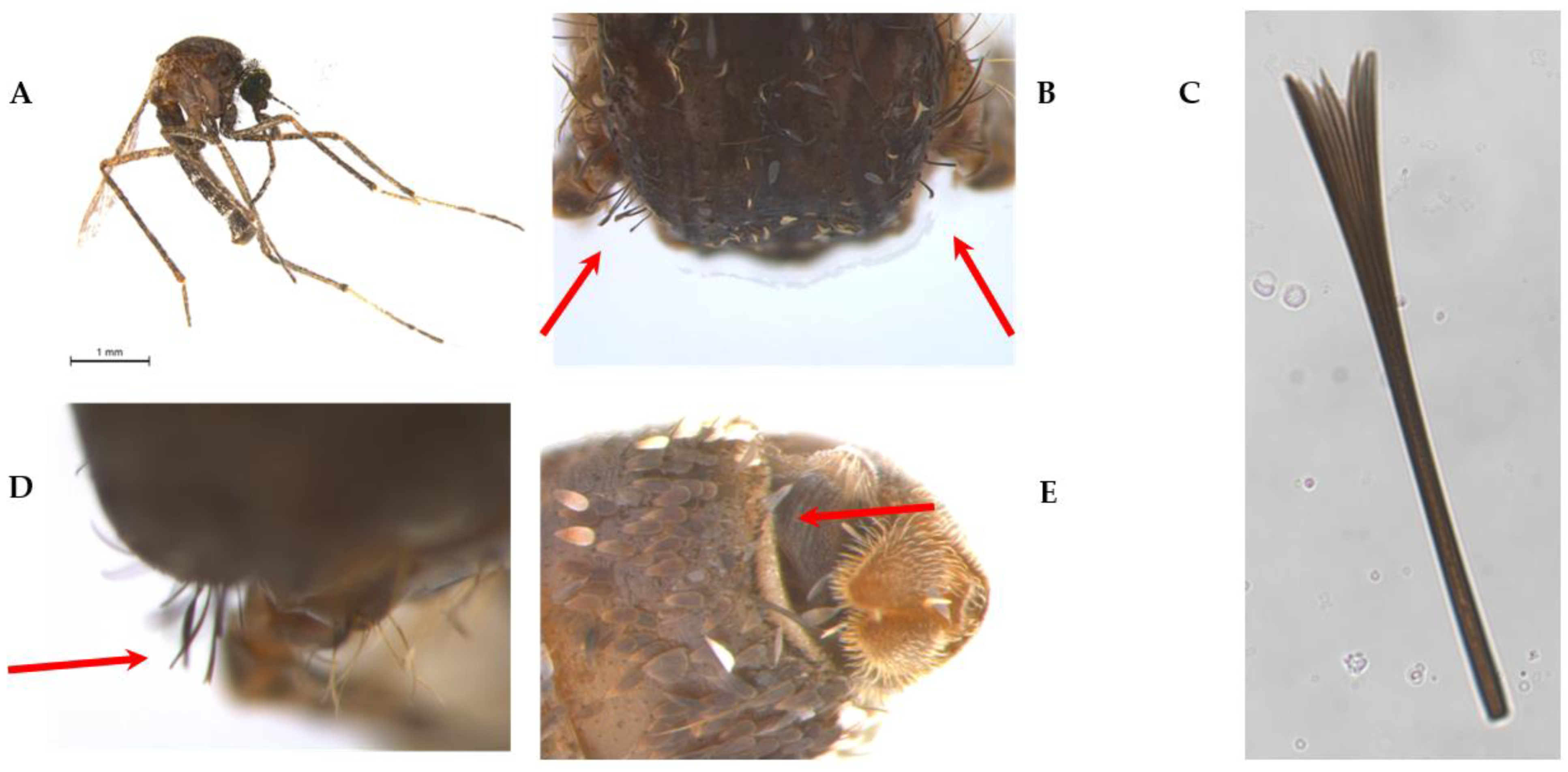
3.1. Morphological Assessment

We identified a total of 17 specimens to the species level: six Ma. humeralis, five Ma. pseudotitillans, one Ma. titillans, four Ma. wilsoni, and one Ma. indubitans. Nineteen specimens were identified only up to the subgenus level (Table 1). Below, we list and provide photographs of some characteristics used in the identification of Mansonia species analyzed in this study that we consider decisive, subjective, or difficult to visualize.

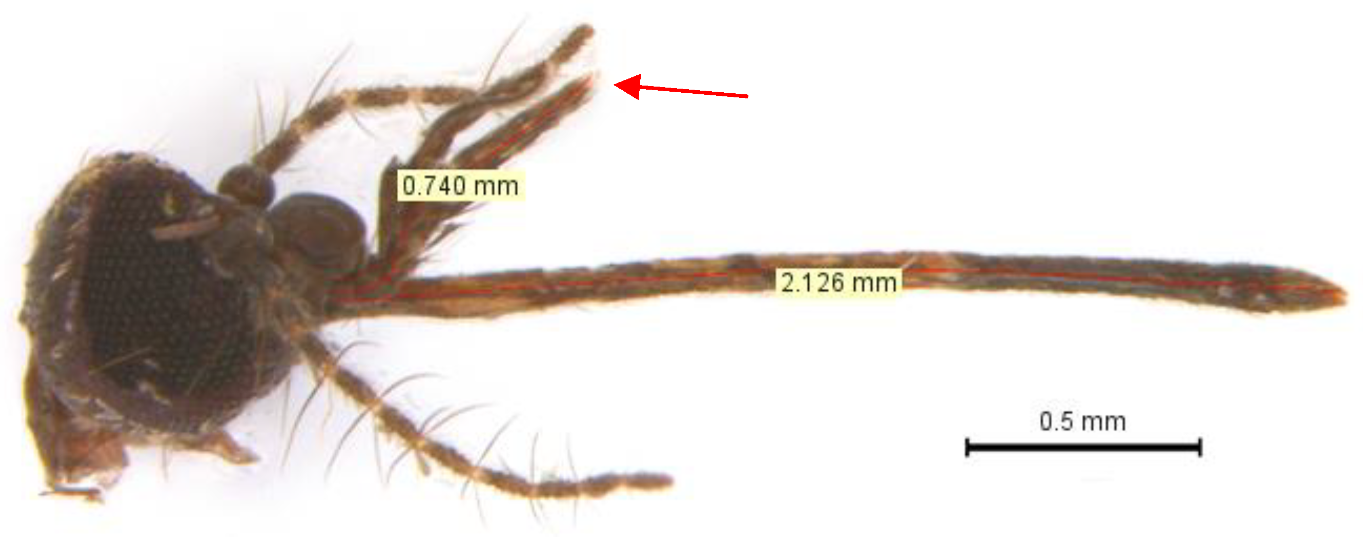
3.1.1. Maxillary Palpus

Mansonia indubitans and Ma. pseudotitillans are difficult to differentiate due to the similarity of their maxillary palpus. A maxillary palpus with less than one fourth proboscis length (0.25) are characteristics of Ma. indubitans, while a maxillary palpus with more than 0.3 of the proboscis length are attributed to Ma. pseudotitillans [10,43]. Using morphometry, we were able to confirm this feature in the identification of different Ma. pseudotitillans individuals, although in some cases the measure was slightly below 0.3 (Figure 2 and Figure 3).

3.1.2. Spines on the Abdominal Tergite VII and Suprawing Scale

Two further morphological characters that were examined to identify the female Mansonia specimens are the suprawing scale and the potential presence of spines on the abdominal tergite VII (Figure 4).
The taxonomic key of Forattini 2002 [10] uses the following as a differentiating criterion of Mansonia titillans (Figure 4A): the presence of suprawing scales with a simple apex rather than suprawing scales with a forked apex, for Ma. pseudotitillans, Ma. indubitans, or Ma. dyari. However, we consider this characteristic to be difficult to identify (Figure 4B–D), even using magnification of a suprawing scale (Figure 4C).
According to the taxonomic keys of Forattini 2002 [10] and Assumpção 2009 [30], Ma. indubitans do not present spines on the abdominal tergite VII. However, the authors disagree about the presence of the same character in Ma. pseudotitillans: the key by Forattini 2002 [10] states that this species does not have spines in the abdominal segment VII and both the key and the description by Assumpção 2009 [30] mention spines in that segment, as well as Forattini 2002 [10] for Ma. titillans (Figure 4E).

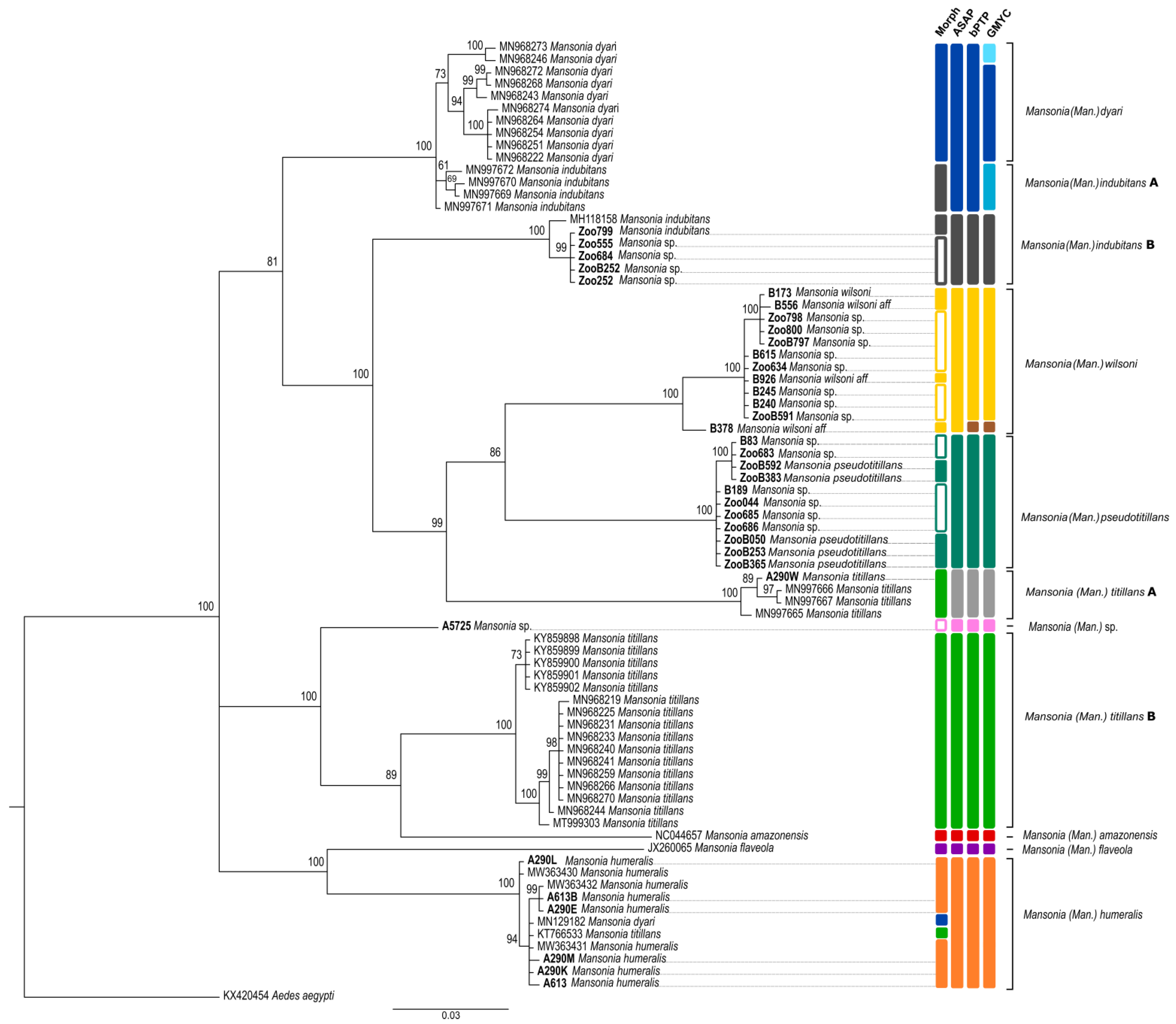
3.2. Sequence Analysis and Species Delimitation

The 36 COI sequences obtained for the present study (with sequences ranging from 559 to 658 bp) represented five different Mansonia species. Of these, two sequences were, for the first time, linked to morphologically identified specimens: Ma. wilsoni and Ma. pseudotitillans. The results of the analyses using these DNA sequences are shown in Figure 5. Bayesian analysis of the obtained COI sequences resulted in clade topologies that corroborate with the morphologically identified species (except for MN129182 and KT766533). However, for some species, the tree did not group all sequences into the same clade. For example, Ma. indubitans appears polyphyletic and is separated into two different clades (Figure 5). Further, the Ma. wilsoni and Ma. pseudotitillans clades were split into subclades, indicating a cryptic diversity in this species collected in the São Paulo State.
The BI tree shows that sequences collected from Brazil in this study basically clustered in two strongly supported clades (Figure 5). The first one (Ma. humeralis clade) was formed by six sequences from Brazil and five different GenBank sequences (mainly Ma. humeralis sequences). The second clade of sequences from Brazil was split into four subclades encompassing: (i) sequences from specimens identified as Ma. titillans (clade Ma. titillans A); (ii) one monophyletic clade with all our Ma. pseudotitillans sequences, but without any GenBank reference sequence; (iii) one monophyletic clade with all Ma. wilsoni sequences, but again without any GenBank reference; and (iv) one sequence of the polyphyletic Ma. indubitans from GenBank that grouped with five Ma. indubitans sequences obtained in the Sao Paulo State (clade Ma. indubitans B) (Figure 5).
The NJ tree further supported the species identities from Brazil. Each species was represented by well supported clades (>98% bootstrap support), confirming the morphological identification (Figure S1 in Supplementary Material).
As shown in Table 3, the mean intraspecific K2P distances for all the species were less than 2%. The maximum distance was seen among the sequences of Ma. dyari which was 1%, while Ma. wilsoni and Ma. pseudotitillans reported the lowest mean intraspecific distance of 0.2%. The interspecific distances ranged from 9.6% between Ma. amazonensis and Ma. titillans B to 18.3% between Ma. titillans A and Ma. flaveola. Interspecific distances obtained between Ma. titillans A and B and Ma. indubitans A and B were 13.4% and 10.9%, respectively. Mansonia sp. A5725 showed 7.3% interspecific distance with Ma. titillans B, while Mansonia wilsoni aff B378 presented 2.1% interspecific distance with the Ma. wilsoni group. Aedes aegypti showed the highest values, ranging from 14.7 to 18.9% (Table 3).
Each of the species delimitation methods (ASAP, bPTP and GMYC) obtained a result that corroborates with the Bayesian and Neighbor-joining analyses, with some slight variations (Figure 5 and Figures S1–S4 in Supplementary Material). A correspondence in ASAP (Figure S2) and bPTP (Figure S3) algorithms was observed for all the species, which were split into ten and eleven MOTUs, respectively, with the only difference being that taxon B378 was considered as a different species by bPTP but grouped with Ma. wilsoni by ASAP (score 1.5) (Figure 5). On the other hand, GMYC analysis resulted in 13 clusters (Figure S4), where specimen B378 was also considered as a different species. In contrast, however, some barcodes of Ma. indubitans (clade Ma. indubitans A) and Ma. dyari were merged into a single MOTU (Figure 5) in ASAP and bPTP algorithms, while they were split into three MOTUs using GMYC (Figure S4).
Sequences of Ma. titillans formed two MOTUs using all algorithms. However, Ma. wilsoni, Ma. pseudotitillans, and Ma. humeralis were strongly supported as a monophyletic species by Bayesian posterior probability (100 BPP) (Figure 5), with a clade formed by specimens from Argentina and Brazil in the case of Ma. humeralis (Figure 5). Likewise, the ASAP, bPTP, and GMYC approaches clustered Ma. wilsoni and Ma. pseudotitillans into a single molecular species (Figure 5 and Figures S2–S4).

4. Discussion

Morphological identification of Mansonia species is challenging and individuals from this genus are notoriously difficult to distinguish unless the male genitalia—which are needed to differentiate some morphological characters—are examined under a microscope. This study sought to determine the utility of DNA barcoding in delimiting species from the genus Mansonia collected in the Atlantic Forest and Brazilian Savanna. The total number of MOTUs within the same taxon varied very little and was independent of the model used to partition the COI data, since all models recovered all eight species identified by traditional morphology. Each of the molecular delimitation methods used (ASAP, bPTP, GMYC, NJ and BI) showed highly supported clusters in the identified operational taxonomic units with few differences in their topologies. Mansonia indubitans and Ma. titillans were recovered as polyphyletic, while Ma. humeralis, Ma. wilsoni, and Ma. pseudotitillans were supported as a monophyletic species according to all methodologies.
A value of 3% in the interspecific distance between COI sequences is considered as the threshold in differentiating species [2,3] and this has been applied in many mosquito studies [1,4,5]. Here, the intraspecific distances of all the species identified was less than 3% (ranging from 0.2 to 1%), while the interspecific distances were above 3% (ranging from 9.6 to 18.3%). The interspecific distances confirmed two groups of Ma. titillans and Ma. indubitans, with values of 13.4% and 10.9%, respectively. They also pointed to a different species (among those analyzed here) in Mansonia sp. A5725 (closest interspecific distance was 7.3% with Ma. titillans B), a result shared by ASAP, bPTP, and GMYC. Moreover, the interspecific distance of 2.1% supported Mansonia wilsoni aff B378 within the Ma. wilsoni group, a result corroborated by morphology and ASAP.
Of the 58 sequences with morphological identification to species, only six were not correctly positioned by ASAP, bPTP, and GMYC. These were: two potentially morphologically misidentified GenBank sequences (MN129182 and KT766533) that grouped with Ma. humeralis sequences and four Ma. indubitans sequences from Colombia that clustered with the sequences of Ma. dyari from Mexico. It is worth mentioning that the sequences MN129182 and KT766533 were also identified as Ma. humeralis using the BOLD platform (Barcode of Life Data system) using the option “Species Level Barcode Records” (http://www.boldsystems.org/index.php/IDS_OpenIdEngine) (accessed on 3 October 2022).
In fact, in the original description, Ma. dyari was considered a taxon morphologically close to Ma. indubitans due to the absence of spines on the posterior margin of tergite VII and by the presence of evenly spaced spines on tergite VIII [44]. However, the authors insisted that the “indubitans” of the Caribbean area are different from those in South America and named them as Ma. dyari [44]. Based on morphologic and phylogenetic data, Ma. dyari and Ma. indubitans were also grouped into a clade (with 76% bootstrap) [14]. More recently, this cluster was also found by DNA barcoding analysis, pointing to the occurrence of cryptic speciation within Ma. dyari [45]. Nevertheless, in our analysis, the sequences of Ma. indubitans collected from Brazil clustered in a clade distinct from the one mentioned above (Ma. dyari/indubitans A). The clade named Ma. indubitans B comprised one Ma. indubitans sequence from São Paulo Zoo and four Mansonia sp. sequences that were clustered with a sequence from the Ma. indubitans collected in the Brazilian Caatinga (MH118158). Thus, we believe these unidentified specimens from São Paulo Zoo were Ma. indubitans, although some characteristics necessary to confirm morphological identification were missing. It is important to note that our GMYC analysis positioned these two clades (Ma. indubitans A and B) as a monophyletic group, including the Ma. dyari sequences.
Adult female specimens of Ma. indubitans and Ma. pseudotitillans are also difficult to differentiate because one important character that distinguishes them, according to the key of Forattini 2002 [10], may vary between individuals of the same species: the proportion of maxillary palpus in relation to the proboscis. However, the divergence between the keys of Forattini 2002 [10] and Assumpção 2009 [30] regarding the presence of spines on the abdominal tergite VII may result in misidentification. Here, using morphometry and high-magnification photomicrographs, we were able to identify Ma. pseudotitillans according to Forattini 2002 [10], using the proportion of maxillary palpus in relation to the proboscis and the absence of spines on the abdominal tergite VII. Additionally, we have generated the first COI sequences of this species.
In contrast, another important distinctive characteristic is the presence of suprawing scales with a simple apex in Ma. titillans rather than suprawing scales with a forked apex in Ma. pseudotitillans, Ma. indubitans, and Ma. Dyari [10]. In this case, even using high magnification photomicrographs, we were unable to confirm this characteristic. However, using the COI sequences, these species were separated into strongly supported clades and additionally grouped with individuals of the same species previously identified in other studies.
Mansonia titillans specimens were recovered as polyphyletic groups according to all methodologies used, where two clades were obtained, named here as Ma. titillans A and Ma. titillans B. Comparable results have been found in other studies [45]. However, the position of these two clades was different among the methods, positioning as sister clades in GMYC and bPTP approaches. In this study, taxon A5725 was identified as a different species of Ma. titillans according to all the approaches but, using the BOLD platform and the option “Species Level Barcode Records”, the COI sequence was matched to Ma. titillans and was considered as a reliable identification.
Finally, it is important to mention that there are no Ma. iguassuensis or Ma. fonsecai sequences in the available databases and, despite this species having already been reported in the State of São Paulo [13,46], we did not morphologically identify specimens of this species from our sampling sites. Thus, it is possible that one of the two clades of Ma. titillans and one of the two clades of Ma. indubitans are, in fact, Ma. iguassuensis and Ma. fonsecai, respectively. Mansonia iguassuensis has often been misidentified as Ma. titillans and, due to the morphological similarities between the adults [13,46], Ma. fonsecai has been considered a junior synonym of Ma. indubitans. New integrative studies combining morphological and molecular analyses are needed to identify the COI sequences of these species.
To know the culicid fauna of a given locality, it is also important to identify male specimens through the structure of their genitalia, as well as immature forms [47]. However, it is not always possible to collect male specimens or immature forms. For this reason, in our work, we use COI sequences and thus show that these species can be separated into strongly supported clades. Our Bayesian analysis yielded trees with well-supported internal branches (≥90), resolving six out of the eight taxa as monophyletic groups. We provided sequences of the COI gene from two Mansonia species that were not previously available in sequence databases, Ma. wilsoni and Ma. pseudotitillans, thereby contributing to the ongoing global effort to standardize DNA barcoding as a molecular means tool for species identification by the Consortium for the Barcode of Life (CBOL). The use of integrated systematics, combining DNA barcodes and phylogenetic inferences, allows us to refine taxonomic identification and better understand the genetics and distribution of mosquito species.

5. Conclusions

Our findings show that Mansonia species delimitation via the BI, NJ, ASAP, bPTP, and GMYC approaches yields basically the same groups as those identified by traditional taxonomy, and additionally provides the species identification of specimens classified only up to the subgenus level. Morphological taxonomy combined with molecular taxonomy, as performed for many specimens in this study, makes species identification more consistent. However, morphological identification requires specialist taxonomic knowledge, is time consuming, needs well-preserved specimens, and depends on the subjective interpretation of measures in terms of relative sizes. Here, we show that the use of COI sequences is a useful tool to identify morphologically similar species of the subgenus Mansonia in the State of São Paulo, overcoming the difficulties encountered when using traditional taxonomy alone.

Supplementary Materials

The following supporting information can be downloaded at: https://www.mdpi.com/article/10.3390/insects14020109/s1, Figure S1: Neighbor-Joining tree based on the COI barcoding region of Mansonia species; Figure S2: COI gene species delimitation of Mansonia species by ASAP; Figure S3: COI gene species delimitation of Mansonia species by bPTP; Figure S4: COI gene species delimitation of Mansonia species by GMYC.

Author Contributions

Conceptualization, K.K. and J.T.-d.-D.; Data curation, K.K., L.d.O.G., E.F.M., V.C.H., J.T.-d.-D. and V.L.F.d.C.-N.; Formal analysis, K.K.; Funding acquisition, V.L.F.d.C.-N.; Investigation, K.K., L.d.O.G., E.F.M., V.C.H., J.T.-d.-D., R.M.T.d.M., S.L.R. and C.R.F.C.; Methodology, K.K.; Project administration, V.L.F.d.C.-N.; Resources, K.K. and V.L.F.d.C.-N.; Supervision, J.T.-d.-D., R.M.T.d.M., C.R.F.C. and V.L.F.d.C.-N.; Validation, K.K. and R.M.T.d.M.; Visualization, E.F.M., V.C.H. and J.T.-d.-D.; Writing—original draft, K.K., E.F.M., J.T.-d.-D., R.M.T.d.M. and C.R.F.C.; Writing—review & editing, K.K., L.d.O.G., E.F.M., V.C.H., J.T.-d.-D., R.M.T.d.M., S.L.R., C.R.F.C. and V.L.F.d.C.-N. All authors have read and agreed to the published version of the manuscript.

Funding

This research was funded by Fundação de Amparo à Pesquisa do Estado de São Paulo (FAPESP 2012/51427-1 and 2017/50345-5). L.d.O.G. is supported by a postdoctoral fellowship (FAPESP 2018/16232-1). K.K. is a CNPq research fellow (process number 309396/2021-2).

Institutional Review Board Statement

This study was performed according to the Ethical Principles in Animal Research. It was approved by the Ethics Committee of Institute of Tropical Medicine, University of Sao Paulo (CPE-IMT/193 and CPE-IMT/371A, 4 October 2019), and the Brazilian Ministry of Environment (SISBIO 34605-7, 27 October 2016). (CPE-IMT/294A, 30 October 2014, and CPE-IMT/371A).

Informed Consent Statement

Not applicable.

Data Availability Statement

The data presented in this study are available in Supplementary Materials and the GenBank database (https://www.ncbi.nlm.nih.gov/genbank/ (accessed on 3 October 2022)) (GenBank #OQ120978-OQ121013).

Acknowledgments

We thank the São Paulo Zoological Foundation (Fundação Zoológico de São Paulo) for the support provided to this research. We thank Bruno Mathias and Carolina Anjos for their help in editing Figure 1 and Figure 5, respectively.

Conflicts of Interest

The authors declare no conflict of interest.

References

  1. Chan, A.; Chiang, L.-P.; Hapuarachchi, H.C.; Tan, C.-H.; Pang, S.-C.; Lee, R.; Lee, K.-S.; Ng, L.-C.; Lam-Phua, S.-G. DNA Barcoding: Complementing Morphological Identification of Mosquito Species in Singapore. Parasites Vectors 2014, 7, 569. [Google Scholar] [CrossRef] [PubMed]
  2. Hebert, P.D.N.; Cywinska, A.; Ball, S.L.; deWaard, J.R. Biological Identifications through DNA Barcodes. Proc. R. Soc. Lond. B 2003, 270, 313–321. [Google Scholar] [CrossRef] [PubMed] [Green Version]
  3. Hebert, P.D.N.; Ratnasingham, S.; de Waard, J.R. Barcoding Animal Life: Cytochrome c Oxidase Subunit 1 Divergences among Closely Related Species. Proc. R. Soc. Lond. B 2003, 270, S96–S99. [Google Scholar] [CrossRef] [Green Version]
  4. Laurito, M.; de Oliveira, T.M.; Almiron, W.R.; Sallum, M.A.M. COI Barcode versus Morphological Identification of Culex (Culex) (Diptera: Culicidae) Species: A Case Study Using Samples from Argentina and Brazil. Mem. Inst. Oswaldo Cruz 2013, 108, 110–122. [Google Scholar] [CrossRef]
  5. Laurito, M.; Ayala, A.M.; Arias-Builes, D.L.; Almirón, W.R. Improving the DNA Barcode Library of Mosquito Species with New Identifications and Discoveries in North-Central Argentina. J. Med. Entomol. 2022, 59, 173–183. [Google Scholar] [CrossRef]
  6. Kirchgatter, K.; de Oliveira Guimarães, L.; Hugo Yañez Trujillano, H.; Rafael Arias, F.; Cáceres, A.; de Castro Duarte, A.; dos Santos Malafronte, R.; Tubaki, R.; Mureb Sallum, M. Phylogeny of Anopheles (Kerteszia) (Diptera: Culicidae) Using Mitochondrial Genes. Insects 2020, 11, 324. [Google Scholar] [CrossRef] [PubMed]
  7. Zouré, A.A.; Noël, G.; Sombié, A.; Somda, Z.; Badolo, A.; Francis, F. Genetic Analysis and Population Structure of the Anopheles gambiae Complex from Different Ecological Zones of Burkina Faso. Infect. Genet. Evol. 2020, 81, 104261. [Google Scholar] [CrossRef] [PubMed]
  8. Harbach, R.E.; Kitching, I.J. Phylogeny and Classification of the Culicidae (Diptera). Syst. Entomol. 1998, 23, 327–370. [Google Scholar] [CrossRef]
  9. Silver, J.B. Mosquito Ecology; Springer: Dordrecht, The Netherlands, 2008; ISBN 978-1-4020-6665-8. [Google Scholar]
  10. Forattini, O.P. Culicidologia Médica; Publisher of the University of São Paulo: São Paulo, Brazil, 2002; Volume 2. [Google Scholar]
  11. Ronderos, R.A.; Bachmann, A.O. A proposito del complejo Mansonia (Diptera-Culicidae). Rev. Soc. Entomol. Argent. 1963, 25, 43–51. [Google Scholar]
  12. Guimarães, J.H. Systematic Database of Diptera of the Americas South of the United States (Family Culicidae); Plêiade/FAPESP: São Paulo, Brazil, 1997. [Google Scholar]
  13. Barbosa, A.A.; Silva, M.A.N.D.; Sallum, M.A.M. Description and Revalidation of Mansonia (Mansonia) fonsecai (Pinto) (Diptera: Culicidae). Zootaxa 2005, 905, 1–11. [Google Scholar] [CrossRef]
  14. Barbosa, A.A. Revisão do Subgênero Mansonia Blanchard, 1901 (Diptera, Culicidae) e Estudo Filogenético de Mansoniini. Ph.D. Thesis, Federal University of Paraná, Biological Sciences Sector, Curitiba, Brazil, 2007. [Google Scholar]
  15. Gaffigan, T.V.; Wilkerson, R.C.; Pecor, J.E.; Stoffer, J.A.; Anderson, T. Systematic Catalog of Culicidae. Walter Reed Biosystematics Unit (WRBU), Division of Entomology, Walter Reed Army Institute of Research (WRAIR), Silver Spring, Maryland, USA. Available online: https://www.mosquitocatalog.org/ (accessed on 3 October 2022).
  16. Guimarães, L.d.O.; Simões, R.F.; Chagas, C.R.F.; de Menezes, R.M.T.; Silva, F.S.; Monteiro, E.F.; Holcman, M.M.; Bajay, M.M.; Pinter, A.; de Camargo-Neves, V.L.F.; et al. Assessing Diversity, Plasmodium Infection and Blood Meal Sources in Mosquitoes (Diptera: Culicidae) from a Brazilian Zoological Park with Avian Malaria Transmission. Insects 2021, 12, 215. [Google Scholar] [CrossRef] [PubMed]
  17. Turell, M.J.; Jones, J.W.; Sardelis, M.R.; Dohm, D.J.; Coleman, R.E.; Watts, D.M.; Fernandez, R.; Calampa, C.; Klein, T.A. Vector Competence of Peruvian Mosquitoes (Diptera: Culicidae) for Epizootic and Enzootic Strains of Venezuelan Equine Encephalomyelitis Virus. J. Med. Entomol. 2000, 37, 835–839. [Google Scholar] [CrossRef] [PubMed]
  18. Unlu, I.; Kramer, W.L.; Roy, A.F.; Foil, L.D. Detection of West Nile Virus RNA in Mosquitoes and Identification of Mosquito Blood Meals Collected at Alligator Farms in Louisiana. J. Med. Entomol. 2010, 47, 625–633. [Google Scholar] [CrossRef] [PubMed]
  19. Méndez, W.; Liria, J.; Navarro, J.-C.; García, C.Z.; Freier, J.E.; Salas, R.; Weaver, S.C.; Barrera, R. Spatial Dispersion of Adult Mosquitoes (Diptera: Culicidae) in a Sylvatic Focus of Venezuelan Equine Encephalitis Virus. J. Med. Entomol. 2001, 38, 813–821. [Google Scholar] [CrossRef] [Green Version]
  20. Sudia, W.D.; McLEAN, R.G.; Newhouse, V.F.; Johnston, J.G.; Miller, D.L.; Trevino, H.; Bowen, G.S.; Sather, G. Epidemic Venezuelan Equine Encephalitis in North America in 1971: Vertebrate Field Studies. Am. J. Epidemiol. 1975, 101, 36–50. [Google Scholar] [CrossRef]
  21. Aitken, T.H.G. Habits of Some Mosquito Hosts of VEE (Mucambo) Virus from Northeastern South America, Including Trinidad. In Venezuelan encephalitis; Science Publication 243; Pan American Health Organization: Washington, DC, USA, 1972; pp. 254–256. [Google Scholar]
  22. Turell, M.J. Vector Competence of Three Venezuelan Mosquitoes (Diptera: Culicidae) for an Epizootic IC Strain of Venezuelan Equine Encephalitis Virus: Table 1. J. Med. Entomol. 1999, 36, 407–409. [Google Scholar] [CrossRef]
  23. Hoyos-López, R.; Soto, S.U.; Rúa-Uribe, G.; Gallego-Gómez, J.C. Molecular Identification of Saint Louis Encephalitis Virus Genotype IV in Colombia. Mem. Inst. Oswaldo Cruz 2015, 110, 719–725. [Google Scholar] [CrossRef] [Green Version]
  24. Beranek, M.D.; Gallardo, R.; Almirón, W.R.; Contigiani, M.S. First Detection of Mansonia titillans (Diptera: Culicidae) Infected with St. Louis Encephalitis Virus (Flaviviridae: Flavivirus) and Bunyamwera Serogroup (Peribunyaviridae: Orthobunyavirus) in Argentina. J. Vector Ecol. 2018, 43, 340–343. [Google Scholar] [CrossRef] [Green Version]
  25. Maes, P.; Alkhovsky, S.V.; Bào, Y.; Beer, M.; Birkhead, M.; Briese, T.; Buchmeier, M.J.; Calisher, C.H.; Charrel, R.N.; Choi, I.R.; et al. Taxonomy of the Family Arenaviridae and the order Bunyavirales: Update 2018. Arch. Virol. 2018, 163, 2295–2310. [Google Scholar] [CrossRef] [Green Version]
  26. Ferreira, F.C.; Rodrigues, R.A.; Sato, Y.; Borges, M.A.Z.; Braga, É.M. Searching for Putative Avian Malaria Vectors in a Seasonally Dry Tropical Forest in Brazil. Parasites Vectors 2016, 9, 587. [Google Scholar] [CrossRef] [Green Version]
  27. Sudia, W.D.; Chamberlain, R.W. Battery-Operated Light Trap, an Improved Model. By W. D. Sudia and R. W. Chamberlain, 1962. J. Am. Mosq. Control Assoc. 1988, 4, 536–538. [Google Scholar]
  28. Nasci, R.S. A Lightweight Battery-Powered Aspirator for Collecting Resting Mosquitoes in the Field. Mosq. News 1981, 41, 808–811. [Google Scholar]
  29. Consoli, R.A.G.B.; Lourenço-de-Oliveira, R. Principais Mosquitos de Importância Sanitária No Brasil; Cadernos de Saúde Pública; FIOCRUZ: Rio de Janeiro, Brazil, 1994. [Google Scholar]
  30. Assumpção, I.C.D. Chave de identificação pictórica para o subgênero Mansonia Blanchard, 1901 (Diptera, Culicidae) da região neotropical. In Biological Sciences Sector; Federal University of Paraná: Curitiba, Brazil, 2009; Volume 41, p. 34. [Google Scholar]
  31. Folmer, O.; Black, M.; Hoeh, W.; Lutz, R.; Vrijenhoek, R. DNA Primers for Amplification of Mitochondrial Cytochrome c Oxidase Subunit I from Diverse Metazoan Invertebrates. Mol. Mar. Biol. Biotechnol. 1994, 3, 294–299. [Google Scholar] [PubMed]
  32. Ruiz, F.; Linton, Y.-M.; Ponsonby, D.J.; Conn, J.E.; Herrera, M.; Quiñones, M.L.; Vélez, I.D.; Wilkerson, R.C. Molecular Comparison of Topotypic Specimens Confirms Anopheles (Nyssorhynchus) dunhami Causey (Diptera: Culicidae) in the Colombian Amazon. Mem. Inst. Oswaldo Cruz 2010, 105, 899–903. [Google Scholar] [CrossRef] [PubMed]
  33. Thompson, J. The CLUSTAL_X Windows Interface: Flexible Strategies for Multiple Sequence Alignment Aided by Quality Analysis Tools. Nucleic Acids Res. 1997, 25, 4876–4882. [Google Scholar] [CrossRef] [PubMed] [Green Version]
  34. Kumar, S.; Stecher, G.; Li, M.; Knyaz, C.; Tamura, K. MEGA X: Molecular Evolutionary Genetics Analysis across Computing Platforms. Mol. Biol. Evol. 2018, 35, 1547–1549. [Google Scholar] [CrossRef]
  35. Reinert, J.E. Revised List of Abbreviations for Genera and Subgenera of Culicidae (Diptera) and Notes on Generic and Subgeneric Changes. J. Am. Mosq. Control Assoc. 2001, 17, 51–55. [Google Scholar]
  36. Huelsenbeck, J.P.; Ronquist, F. MRBAYES: Bayesian Inference of Phylogenetic Trees. Bioinformatics 2001, 17, 754–755. [Google Scholar] [CrossRef] [Green Version]
  37. Rambaut, A. FigTree: Tree Figure Drawing Tool. Version 1.4.0. Institute of Evolutionary Biology, University of Edinburgh. 2006–2012. Available online: http://tree.bio.ed.ac.uk/software/figtree (accessed on 22 May 2019).
  38. Kimura, M. A simple method for estimating evolutionary rate of base substitutions through comparative studies of nucleotide sequences. J. Mol. Evol. 1980, 16, 111–120. [Google Scholar] [CrossRef]
  39. Fontaneto, D.; Hortal, J. At Least Some Protist Species Are Not Ubiquitous. Mol. Ecol. 2013, 22, 5053–5055. [Google Scholar] [CrossRef] [Green Version]
  40. Puillandre, N.; Brouillet, S.; Achaz, G. ASAP: Assemble Species by Automatic Partitioning. Mol. Ecol. Resour. 2021, 21, 609–620. [Google Scholar] [CrossRef] [PubMed]
  41. Zhang, J.; Kapli, P.; Pavlidis, P.; Stamatakis, A. A General Species Delimitation Method with Applications to Phylogenetic Placements. Bioinformatics 2013, 29, 2869–2876. [Google Scholar] [CrossRef] [PubMed] [Green Version]
  42. Fujisawa, T.; Barraclough, T.G. Delimiting Species Using Single-Locus Data and the Generalized Mixed Yule Coalescent Approach: A Revised Method and Evaluation on Simulated Data Sets. Syst. Biol. 2013, 62, 707–724. [Google Scholar] [CrossRef] [PubMed] [Green Version]
  43. Forattini, O.P. Entomologia Médica. Culicini: Haemagogus, Mansonia, Culiseta, Sabethini, Toxorhynchitini, Arboviroses, Filariose Bancroftiana. Genética; Publisher of the University of São Paulo: São Paulo, Brazil, 1965; Volume 3. [Google Scholar]
  44. Belkin, J.N.; Heinemann, S.J.; Page, W.A. Mosquito studies (Diptera: Culicidae) XXI. The Culicidae of Jamaica. Contrib. Am. Entomol. Inst. 1970, 6, 1–458. [Google Scholar]
  45. Scarpassa, V.M.; Batista, E.T.; Ferreira, V.C.; Santos Neto, V.A.; Roque, R.A.; Tadei, W.P.; Ferreira, F.A.S.; Costa, F.M. DNA Barcoding Suggests New Species for the Mansonia Subgenus (Mansonia, Mansoniini, Culicidae, Diptera) in the Area Surrounding the Jirau Hydroelectric Dam, Porto Velho Municipality, Rondônia State, Brazil. Acta Trop. 2022, 233, 106574. [Google Scholar] [CrossRef]
  46. Barbosa, A.A.; Da Silva, M.A.N.; Sallum, M.A.M. Mansonia (Mansonia) iguassuensis Sp. Nov. (Diptera: Culicidae) from Brasil. Zootaxa 2007, 1527, 45–52. [Google Scholar] [CrossRef]
  47. LANE, C.J. On a Collection of Culicinae (Diptera: Culicidae) from Brazil. Mosq. Syst. 1992, 24, 16–22. [Google Scholar]
Figure 1. Location of municipalities in the State of São Paulo, Brazil, where Mansonia specimens were collected.
Insects 14 00109 g001
Figure 2. Head of Mansonia pseudotitillans (ID ZooB592) showing (arrow) maxillary palpus (0.740 mm) about 0.29 of the proboscis total length (2.126 mm).
Insects 14 00109 g002
Figure 3. Head of three specimens of Mansonia pseudotitillans (IDs A7206A, A7206B e A7208) showing maxillary palpus with 0.33, 0.36 and 0.29 of the total proboscis length.
Insects 14 00109 g003
Figure 4. (A) Mansonia titillans (ID A290W). (BD) Magnifications showing suprawing scale (arrows). (E) Abdominal tergite VII with spines.
Insects 14 00109 g004
Figure 5. Bayesian inference tree based on the cytochrome c oxidase I (COI) barcoding region of Mansonia (Man.) species. This analysis involved 77 nucleotide sequences. Aedes aegypti (KX420454) was used as an outgroup. There was a total of 641 positions in the final dataset. Average standard deviation of the split frequencies was 0.004342. Posterior probability values are shown for each clade. Specimens colored blank in the morphology column were not morphologically identified to species level due to a lack of important characters. Sequences obtained in this study are given in bold.
Insects 14 00109 g005
Table 3. Mean interspecific (below the diagonal) and intraspecific (along the diagonal) distances for COI sequences. Distances were calculated using the Kimura 2-parameter distance algorithm.
Species12345678910111213
1Ma. wilsoni0.002
2Ma. humeralis0.1610.003
3Ma. titillans A0.1480.1580.005
4Mansonia sp. A57250.1480.1260.142
5Ma. pseudotitillans0.1120.1490.1320.1530.002
6Ma. wilsoni aff B3780.0210.1490.1350.1380.103
7Ma. flaveola0.1600.1360.1830.1490.1510.155
8Aedes aegypti0.1520.1650.1890.1470.1720.1500.174
9Ma. titillans B0.1430.1400.1340.0730.1420.1340.1510.1640.005
10Ma. indubitans B0.1240.1340.1330.1200.1120.1130.1550.1550.1260.003
11Ma. dyari0.1240.1390.1230.1080.1180.1110.1440.1370.1070.1100.010
12Ma. indubitans A0.1240.1330.1200.1050.1190.1110.1480.1390.1080.1090.0130.004
13Ma. amazonensis0.1400.1540.1480.1040.1390.1330.1620.1620.0960.1280.1270.122
Disclaimer/Publisher’s Note: The statements, opinions and data contained in all publications are solely those of the individual author(s) and contributor(s) and not of MDPI and/or the editor(s). MDPI and/or the editor(s) disclaim responsibility for any injury to people or property resulting from any ideas, methods, instructions or products referred to in the content.

