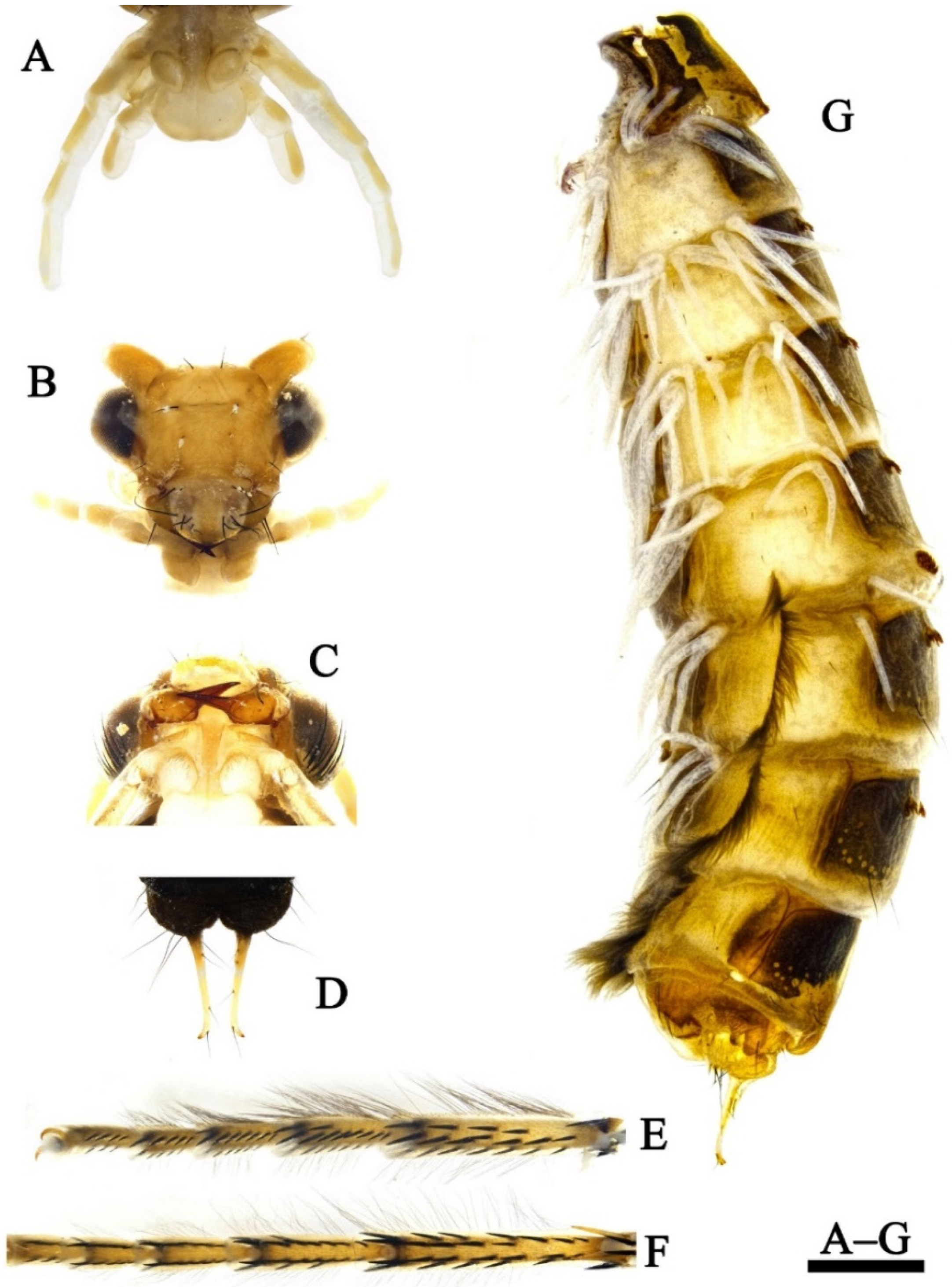
Article

A New Species of Limnephilus (Insecta: Trichoptera: Limnephilidae) from China, with Revision of the Genus Limnephilus on the Chinese Mainland †

Lab of Insect Taxonomy & Aquatic Insects, College of Plant Protection, Nanjing Agricultural University, Nanjing 210095, China
*
Authors to whom correspondence should be addressed.
This published work and the nomenclatural acts it contains have been registered in ZooBank, the online registration system for the ICZN (International Code of Zoological Nomenclature). The LSID (Life Science Identifier) for this publication is: urn:lsid:zoobank.org:pub:BDA500DA-E3CD-469F-9648-1C2D883E11DE.
Insects 2022, 13(7), 653; https://doi.org/10.3390/insects13070653
Submission received: 25 April 2022 / Revised: 13 July 2022 / Accepted: 14 July 2022 / Published: 19 July 2022
(This article belongs to the Topic Arthropod Biodiversity: Ecological and Functional Aspects)

Abstract

:

Simple Summary

As the most diverse genus in the family Limnephilidae, Limnephilus Leach, 1815 (194 extant species and 9 fossil species), has a wide distribution and a broad range of ecological tolerances among its species. Although it has a high species diversity in the Holarctic region, only 16 species have been recognized in China until now, of which 7 are endemic to China. In this study, a new species of the genus Limnephilus was found. The adult females, larvae, and pupae of this new species were associated with the adult males based on COI sequencing, and all the life stages except for the egg stage are described and illustrated. In addition, the biological and habitat information for this new species is provided. Furthermore, all of the Limnephilus species from the Chinese mainland are revised at the species group level, and diagnoses for species groups and species (keys of groups and species) are given, along with a distribution map.

Abstract

Fifty individuals of Limnephilus from the Qinghai Province, China, were examined, and their COI barcode sequences were extracted and analyzed. Forty individuals of Limnephilus from the Insect Collection of Nanjing Agricultural University (ICNAU), China, were examined, and photos of the male genitalia of four Limnephilus species are here presented. The males, females, larvae, and pupae of a new species, Limnephilus deqianensis n. sp., associated via COI barcode sequences, are described and illustrated. Ecological photos of the male, pupal case, and the habitat of the new species L. deqianensis n. sp. are also provided. Five species groups containing all seventeen Chinese Limnephilus species are revised. Diagnoses, keys, and a distribution map of them are provided. All of the sequences have been uploaded to GenBank. All specimens are deposited in the ICNAU, Nanjing, Jiangsu Province, P. R. China.

1. Introduction

Limnephilidae Kolenati, 1848, is one of the most diverse caddis families of the Northern Hemisphere, containing 4 subfamilies and 101 genera [1].
Limnephilus Leach, 1815, is the most diverse genus in the family Limnephilidae [2], containing 194 extant species and 9 fossil species [1,3,4]. Out of all known species, 82 are endemic to the Nearctic region; 44 to the West Palearctic region; 20 to the East Palearctic region; 9 to the Oriental region; 3 to the Neotropical region; and 29 of them are distributed in 2 regions, with 16 of them distributed in 3 or more regions.
Schmid [5] established 37 species groups to accommodate all 114 species known at that time, 14 of which were monotypic; 19 species were isolated, and 7 species were treated as incertae sedis. However, he did not present the diagnostic characteristics for each group. Afterwards, Nimmo [6,7] reviewed 36 Limnephilus species found in Alberta and eastern British Columbia (including his 3 new species, but these were synonymized by various authors later). He followed Schmid’s arrangement, ascribing 33 species from that area to 16 species groups (actually 15 species groups; among them, L. alvatus Denning, 1968, was considered to be a synonym of L. major Martynov, 1909, from the L. incisus Species Group; L. minusculus, Banks, 1907, was synonymized as L. janus Ross, 1938, and was placed into the L. incisus Species Group by Schmid [5], but was considered as a valid species and was transferred into the L. fenestratus Species Group by Nimmo [6]; L. alvatus Denning, 1968, was placed into the L. incisus Species Group by Nimmo, then it was synonymized as L. major, Martynov, 1909, by Ruiter in 1995 [8]), and established 3 new monotypic groups for the 3 remaining species. Furthermore, Nimmo presented the diagnoses for these 15 species groups and illustrations for all 36 species. Ruiter [8] organized the Nearctic region species into species groups that were similar to those of Schmid and Nimmo. He also provided diagnostic characteristics for these species’ groups and keys with figures for nearly all New World species. His species group system mostly reflects changes in new data from females and from variation discovered among newly available specimens [9]. However, in recent years, newly published species have rarely been ascribed to species groups by their authors.
Despite the large number of known species in the world, only 16 known species (Table 1) have been recorded in the Chinese mainland [3,10,11]. Among them, 7 are endemic to China [3,12,13,14,15,16]. Moreover, most species of Limnephilus were recognized before the 21st century and only 10 new species of Limnephilus have been recorded in the last 20 years [3,4,14,15,16,17,18,19].
According to Schmid, among the Limnephilus species from the Chinese mainland, L. fuscovittatus Matsumura, 1904, and L. sibiricus Martynov, 1935, were isolated species, and L. incertus Martynov, 1909, and L. mclachlani Martynov, 1909, were incertae sedis. An amount of 7 out of the remaining 12 species were placed into 5 species groups by Schmid [5], i.e., L. correptus McLachlan, 1880, was placed in the L. flavicornis Species Group, and L. politus McLachlan, 1865, and L. abstrusus McLachlan, 1872, were placed in the L. stigma Species Group; L. externus, Hagen, 1861, was placed in the L. externus Species Group; and L. alaicus, Martynov, 1915, L. tricalcaratus, Mosely, 1936, and L. samoedus, McLachlan, 1880, were placed in the L. asiaticus Species Group; another 5 species that were published after 1955 were not ascribed to the proper species groups by their various authors.
The different members of Limnephilus have broad ranges of different ecological tolerance and a strong affinity for lentic habitats [20,21,22]. They inhabit lakes [23,24], permanent or temporary ponds [25,26,27,28], pools [29], and even salt marshes [21]; some species also inhabit lotic habitats such as free-flowing rivers [30], streams, and springs [31]. The genus is particularly well represented at low altitudes and high latitudes, with many subarctic species [32]. Some species, however, are found at high altitudes [19,25,31].
In this study, 50 individuals of Limnephilus, collected from the Qinghai Province, China, were examined, and their barcode gene sequences were generated and analyzed. One species, L. deqianensis n. sp., was found to be new to the Limnephilus fauna; additionally, 5 females, 5 pupae, and 2 larvae were associated with its males by using molecular analysis. We described the larva, pupa, adult male, and female of L. deqianensis n. sp. We also provide biological information and ecological photos of them. Furthermore, we ascribed 6 other Chinese Limnephilus species to 3 species groups; provided diagnoses for 5 species groups and species within 5 species groups, and the keys to 5 species groups and to species within 2 species groups, with a distribution map of all 17 Chinese mainland Limnephilus species.

2. Materials and Methods

Specimens of the new species studied in this research were collected in Qinghai Province, China. A total of 50 specimens, including 20 adult males and 20 females, 2 larvae, and 8 pupae, were examined in this research. Additional specimens studied in this research were from ICNAU.

2.1. Sample Collection

Larvae and pupae were collected by handpicking specimens off of stones in a high-altitude stream (Figure 1C). Only one male adult (Figure 1A) was captured by hand during collection; the other 40 adults were reared from pupae collected at the site. The adult male, larvae and some pupae were preserved in 95% ethanol in the field directly after collection. Other collected pupae were returned to the temporary residence for rearing and the adults that emerged from them were stored in 95% ethanol after they were completely sclerotized about 3–5 days later. Two larvae were found in their pupal cases. We determined that the 2 larvae are the final instar when comparing the metamorphotype larval frontoclypeal apotome remaining in the pupal cases to our reared pupae. Ecological photos of the habitat, the collected male, and pupal case (Figure 1B) were taken with a VIVO X60Pro smartphone. Types and voucher specimens were deposited at the Insect Collection of Nanjing Agricultural University (ICNAU), Nanjing, Jiangsu Province, P. R. China.

2.2. Morphological Study

The following methods were used for specimen preparation: abdomens of the adults were cut from their bodies, transferred into 10% NaOH solution, and heated to 90 °C for 2 h to remove all of the non-chitinous tissues. Then, the cleaned abdomens were rinsed in distilled water and mounted on a depression slide with 75% ethanol for examination. Photographs of the male and female genitalia, larvae, and pupae were taken by using a stereomicroscope (Nikon SMZ18), and photos of the female spermathecal structure were taken by using a compound microscope (Olympus BX41). Each of these pieces of equipment had a built-in camera and Nis-Element D® software (Version 3.22.14, Nikon, Shanghai, China). A series of photos at different focal depths were taken and stitched together with Zerene Stacker® software (Zerene Systems LLC, Richland, WA, USA) and then arranged using Adobe Photoshop® (version 2017.0.0, Adobe, San Jose, CA, USA). Subsequently, each abdomen was stored in a microvial together with the remainder of the specimen in 75% ethanol.
The terminology for the male genitalia follows that presented by Schmid [32]; the terminology for the female genitalia was adapted from that of Schmid [32], and for the female spermatheca, the terminology was adapted from that of Nielson [33]. The terminology for the larva corresponds with that of Wiggins [34], while for the pupae, it conforms to that of Denning [35].

2.3. Molecular Analysis

The left hind legs of 12 individuals (males, females, and larvae) and left forelegs of 5 individuals (pupae) were taken from the bodies for DNA extractions. Extraction followed the animal tissue protocols of the Ezup Column Animal Genomic DNA Purification kit (Sangon Biotech, Shanghai, China). The PCR amplification, fragment sequencing, and analyses followed the procedures of Xu [36]. The primers [37] (LCO1490/HCO2198) are listed in Table 2. Neighbor-joining (NJ) trees were constructed using Mega v10.2.6 [38]. Calculation parameters were set as follows: Kimura 2-parameter substitution model, pairwise gap deletion and others as defaults. The NJ tree was plugged into ITOL v5 [39]. Female, larval, and pupal specimens were associated with adult males for L. deqianensis n. sp. according to the criteria outlined by Zhou [40]. Their COI sequences were uploaded to GenBank and their accession numbers are included in Table 3.

3. Results

Holotype male: CHINA, Qinghai Province, Men-yuan County, De-qian Town, 37°21′83″ N, 102°20′37″ E, alt. 2930.0 m, 2 July 2021, from a pupa hand-picked from a stone in the stream and raised indoors, Zang Hao-ming. Paratypes: same data as holotype, 19 males, 20 females, 2 larvae, and 8 pupae.
Etymology: The specific epithet is from the Chinese name of the type locality, De-qian Town, Qinghai Province.
Distributions: China (Qinghai).

3.1. Description

Adult: Body brown, 9.5–10.1 mm long for males (n = 20), and 10.8–11.9 mm long for females (n = 20). Head brown, antennae brown, 13.6–13.6 mm long for males (n = 20) and 14.3–14.8 mm for females (
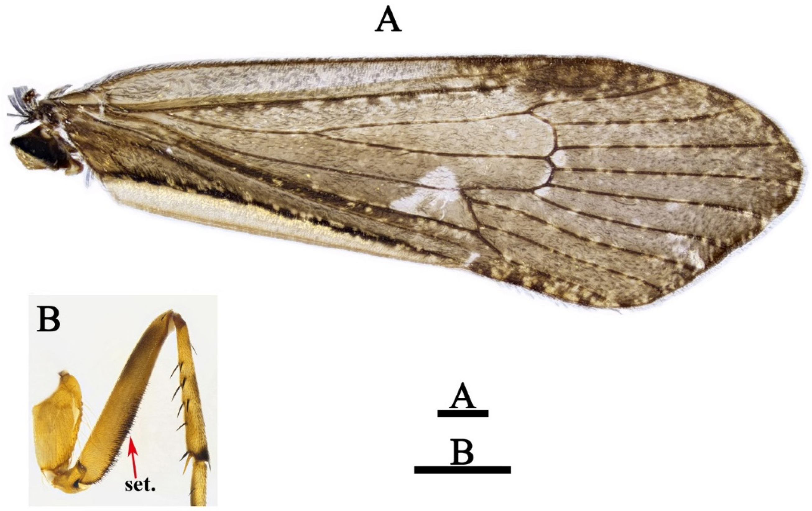
n = 20). Forewings (Figure 2A) 14.0–14.2 mm long for males (n = 20) and 14.7–15.8 mm long for females (n = 20), brown, densely covered with dark spots, each with dark stigma, huge pale spot near apex of Rs cell and thyridial cell, small spot in each base of fork II and III. Hind wings wider than forewings, transparent, without any dark brown spots. Spur formula 1-3-4; forefemora of male, each with lower margin having black setal brush (Figure 2B).
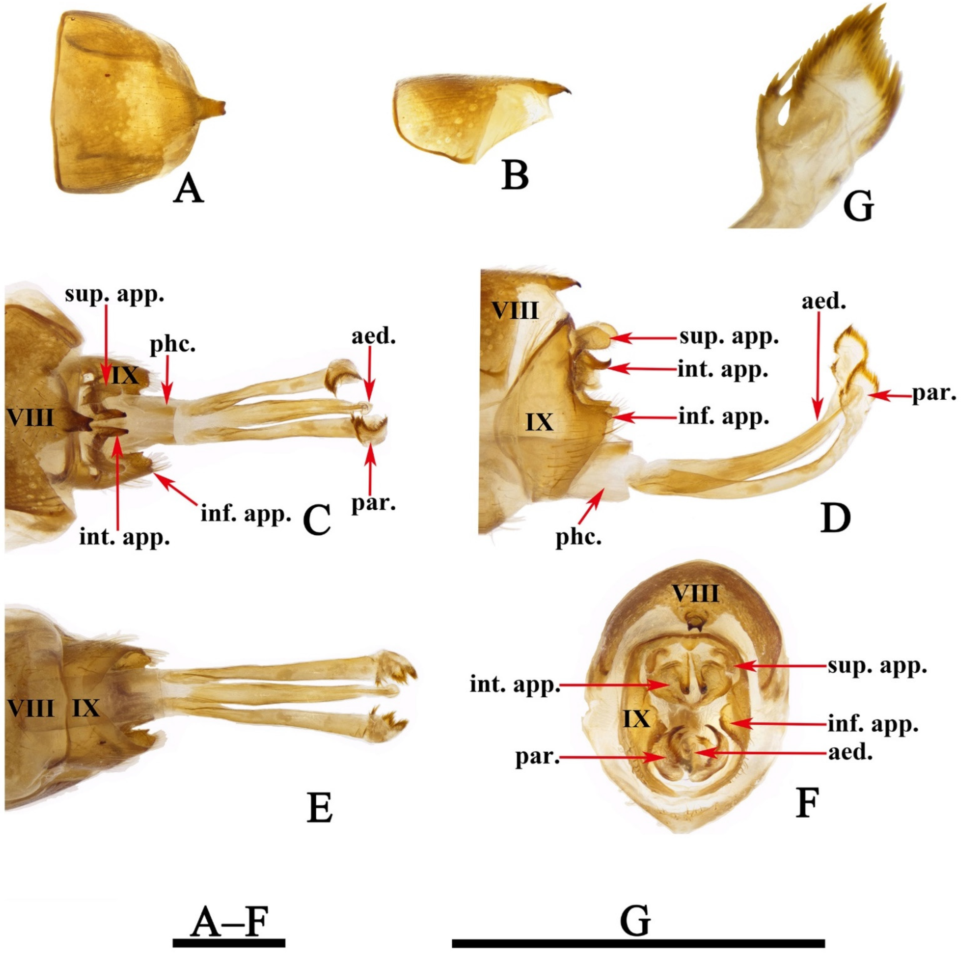
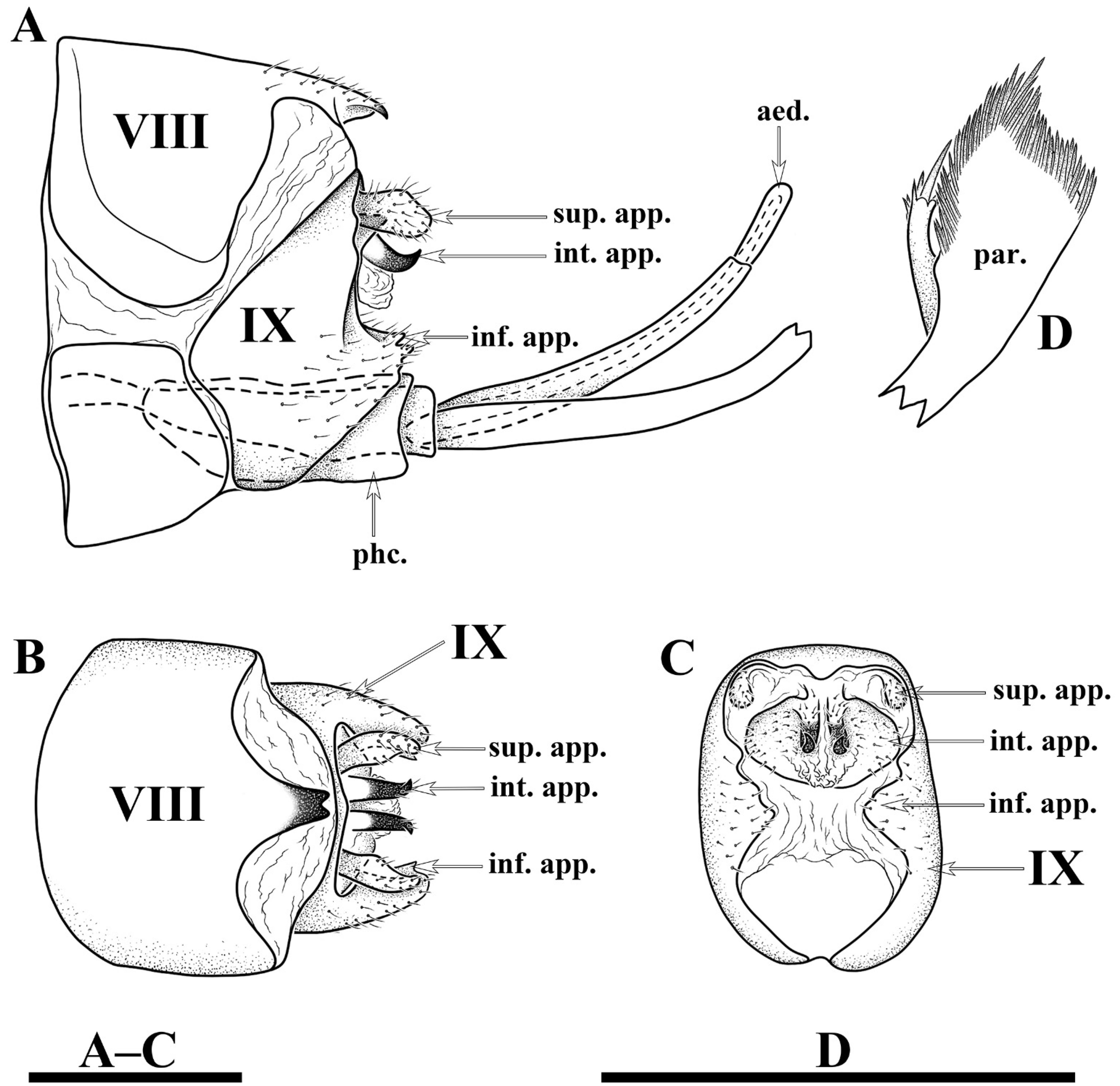
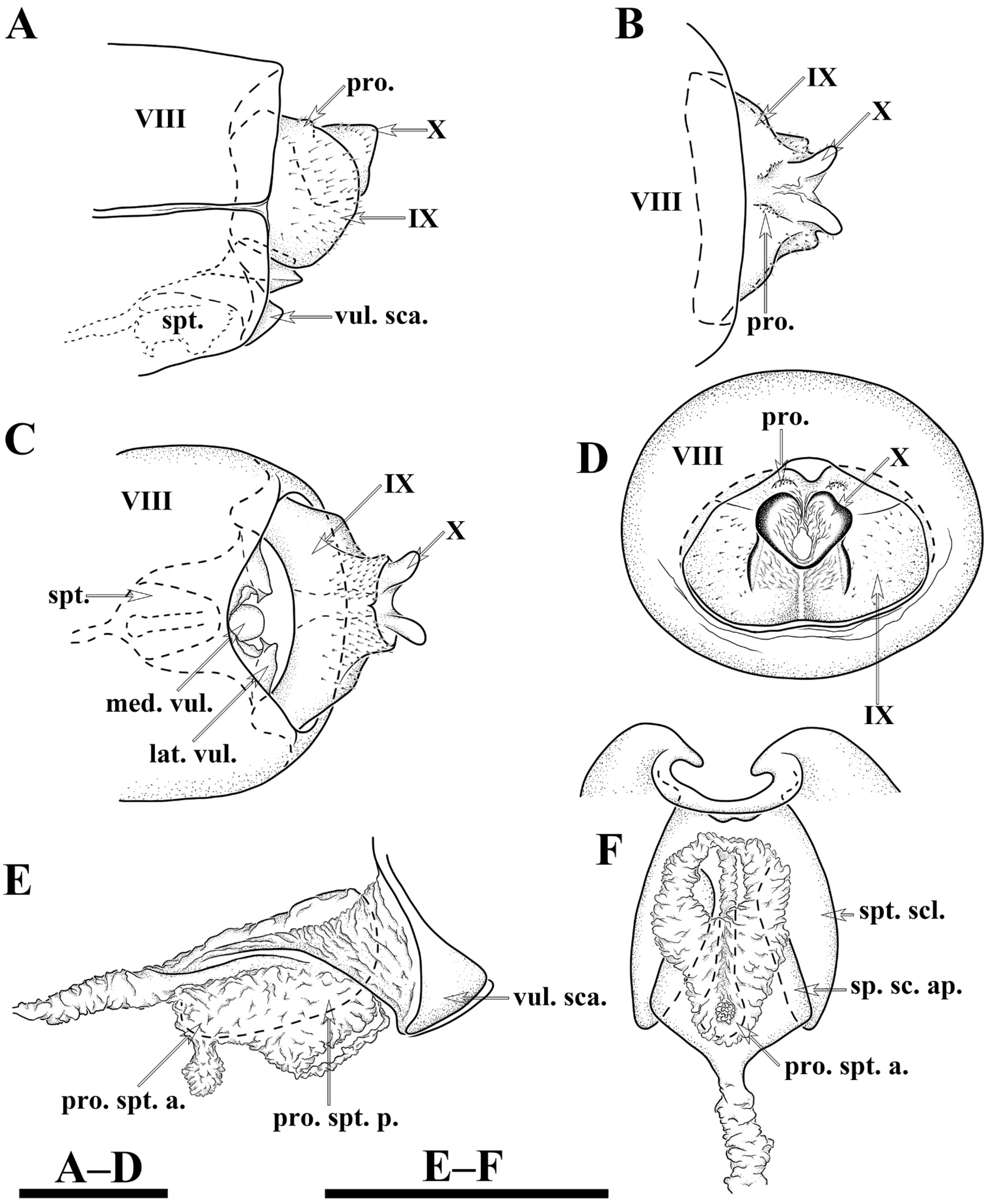
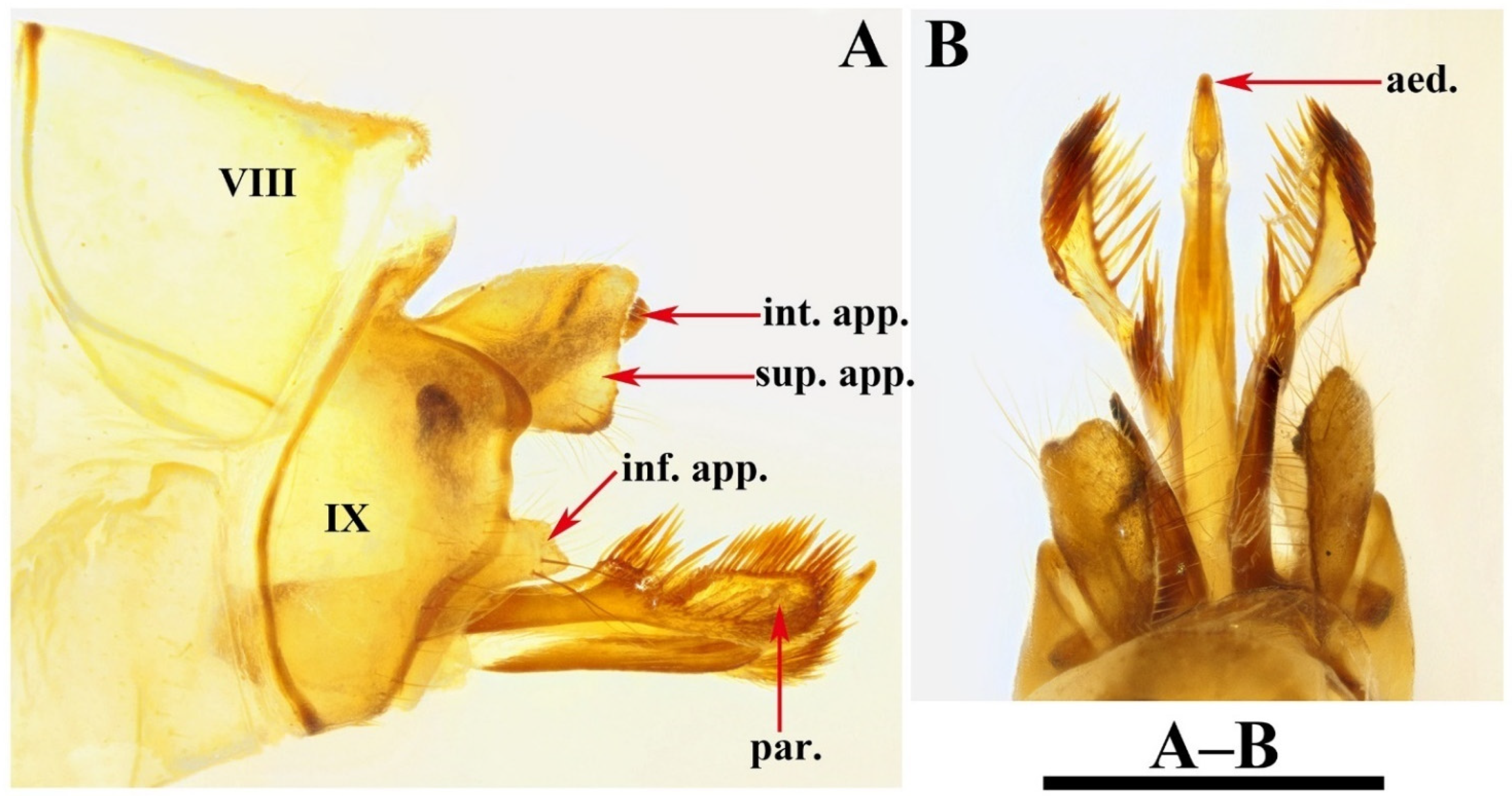
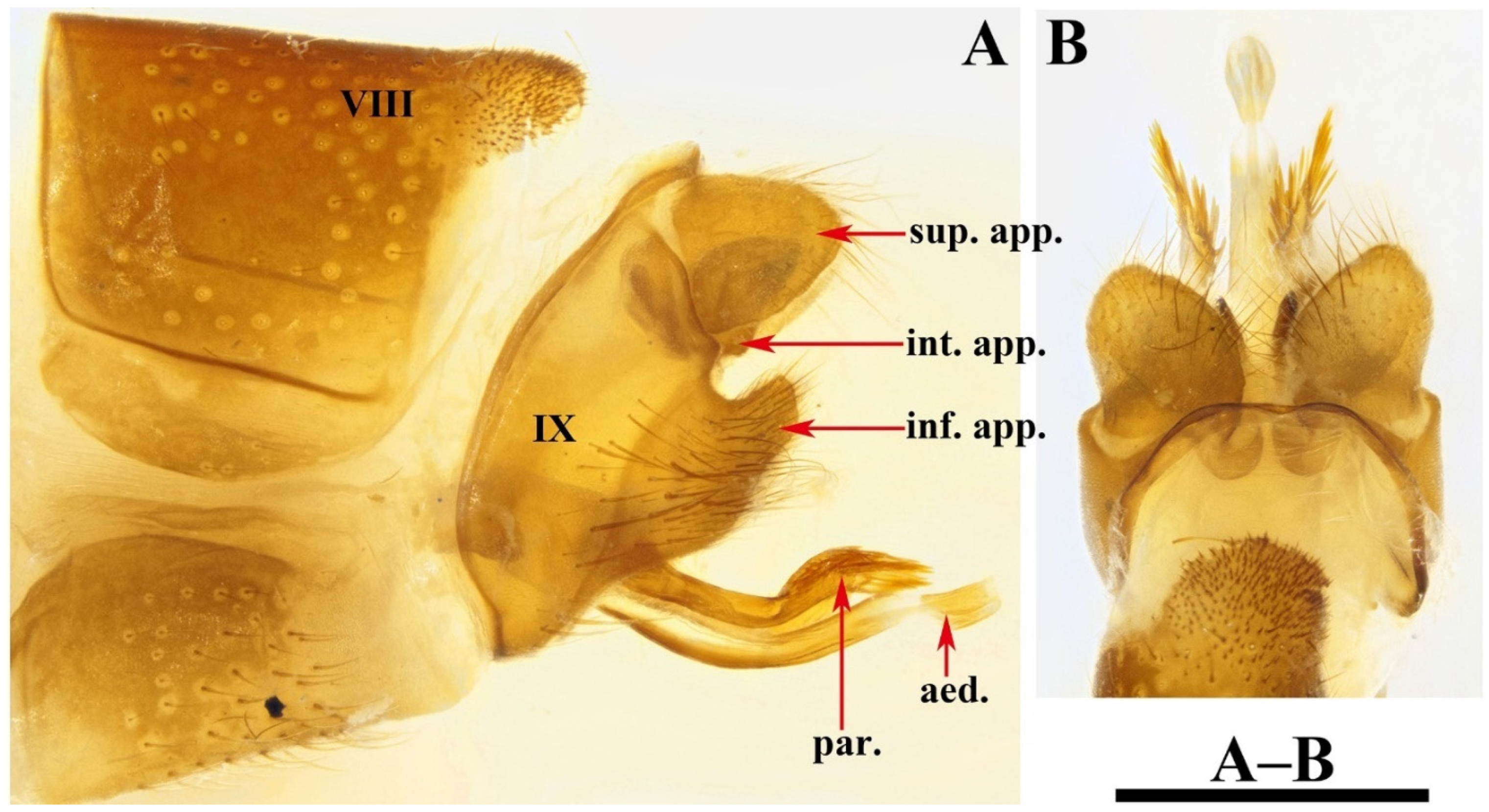
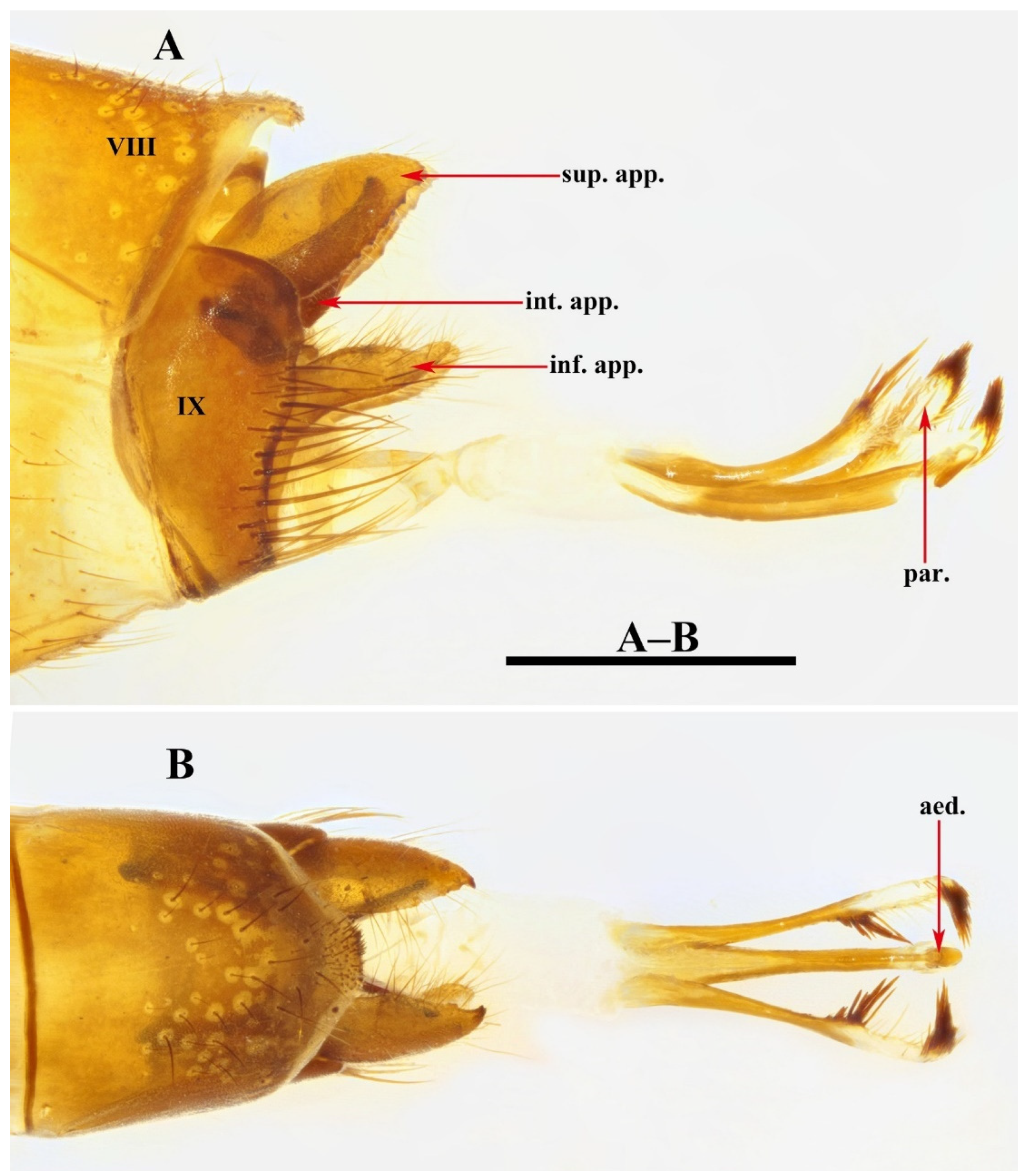
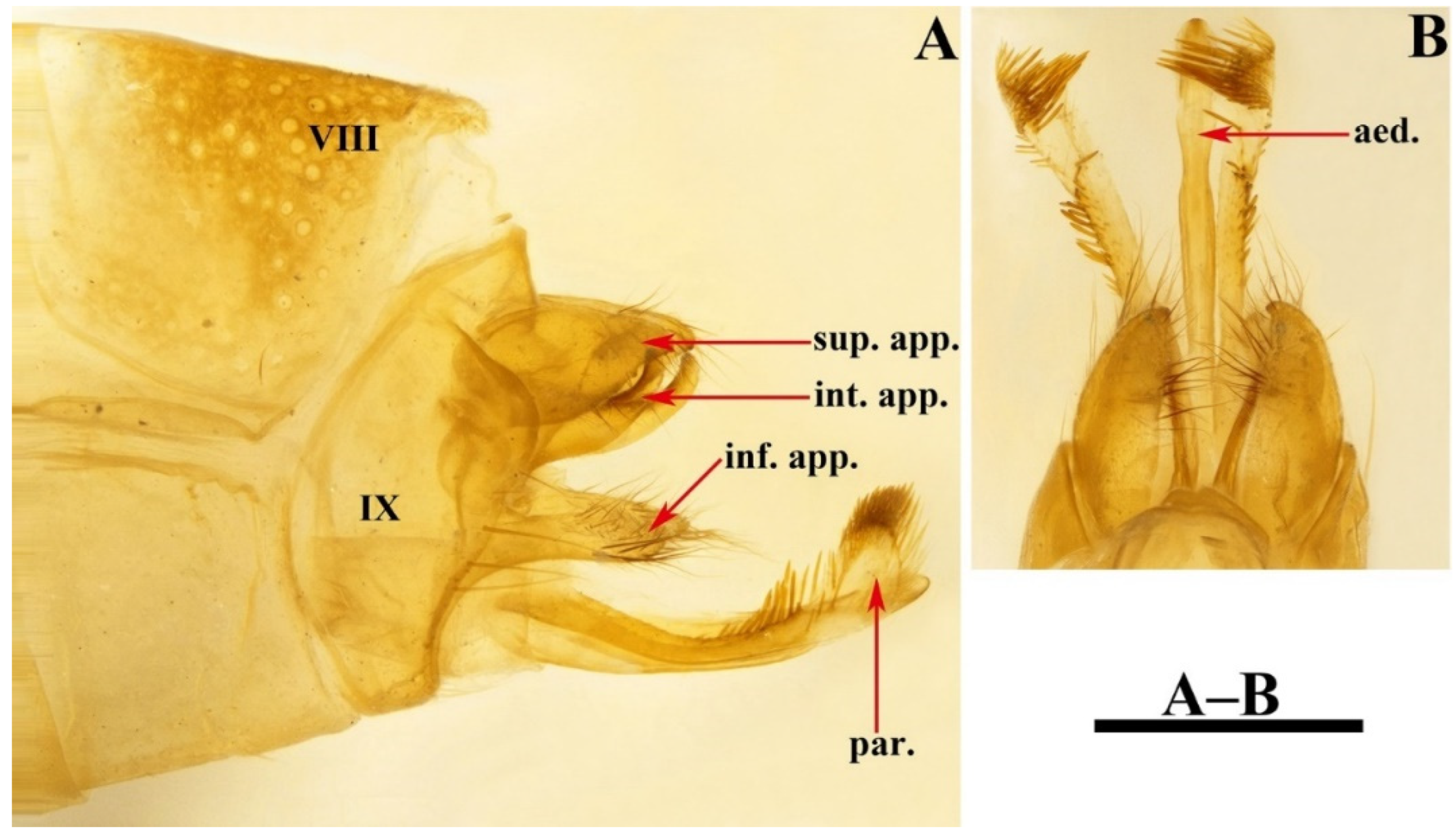
Male genitalia. Tergum VIII in dorsal view (Figure 3A) with basal portion subrectangular and distal portion triangular and produced into a bifid apicodorsal lobe; in lateral view (Figure 3B), basal portion subrectangular, apicodorsal lobe with apex beak-shaped. Segment IX in dorsal view (Figure 3C), with anterior margin straight and posterolateral corners each produced into small process directed posteromesad (Figure 3F); in lateral view (Figure 3D and Figure 4A), with anterior margin convex anteriad, upper portion triangular, lower portion rhomboid and narrowing ventrally. Superior appendages flaplike, oval in lateral view, spatulate in dorsal view (Figure 3C,D and Figure 4A–C). Intermediate appendages each divided into basal plate and apical lobe; basal plates in lateral view (Figure 3D and Figure 4A), each somewhat triangular, with lower portion of each inner margin produced into small membranous process, in caudal view (Figure 3F and Figure 4C), each somewhat semicircular; apical lobes in lateral view (Figure 3D and Figure 4A), each tapered to apex and with apex slightly curved upwards; in dorsal view (Figure 3C), each digitate. Inferior appendages fused with segment IX in lower portion of posterior margins; in lateral view (Figure 3D and Figure 4A), higher than long, each with upper portion thicker than lower portion, upper portion with posterior margins sinuate, and produced into the free portion. Phallic apparatus slender, phallocrypt cylindrical tube (Figure 3C,D), endotheca membranous. Aedeagus (Figure 3C–E) slender, tube-like, tapering from base to apex, slightly curved upwards. Parameres (Figure 3D,G and Figure 4D) each slightly longer than aedeagus, in lateral view tubular with flattened setose apex, and having a small setose lobe on inner side.
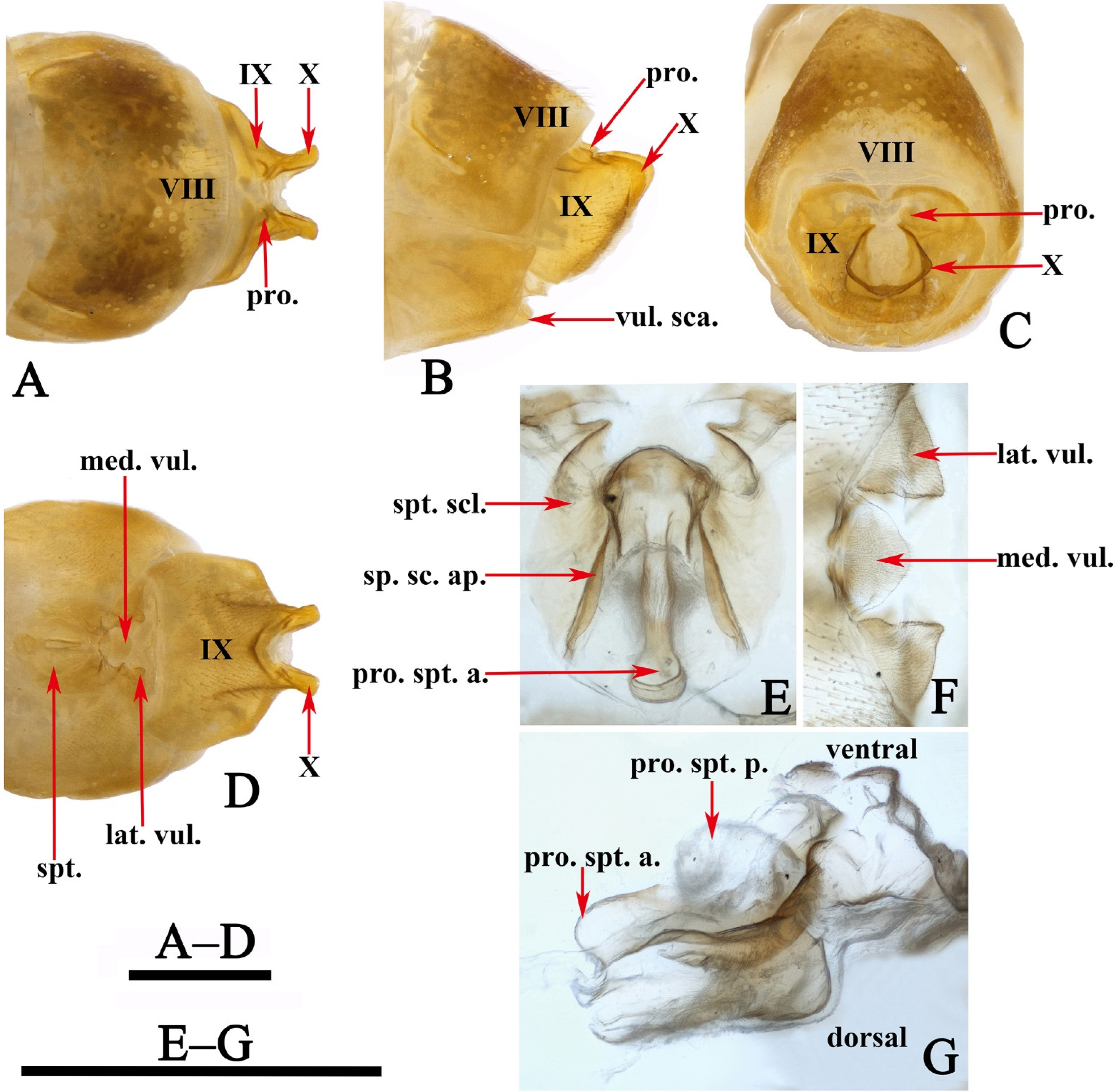
Female genitalia. Tergum VIII subrectangular in dorsal and lateral views (Figure 5A,B). Sternum VIII in lateral view (Figure 5B), subrectangular. Segment IX in dorsal view (Figure 5A and Figure 6B), trapezoid, in lateral view (Figure 5B and Figure 6A), somewhat subrectangular, with paired button-like processus. Segment X fused with segment IX, tubular in both caudal and lateral views (Figure 5B,C and Figure 6A,D), with upper portion deeply incised anteriad. Vulvar scales (Figure 5F and Figure 6C), with median vulvar lobe semicircular and paired lateral vulvar lobes each triangular. Spermatheca (Figure 5E,G and Figure 6E,F) somewhat elliptic, spermathecal sclerites slightly sclerotized, each with lateral margin arc-shaped, with apodeme straight and strongly scerotized, somewhat calvate; processus spermatheca obviously divided into 2 sections, anterior section slightly sclerotized, saccular in lateral view (Figure 5G and Figure 6E), and vase-like in dorsal view (Figure 5E); posterior section membranous, elliptic in lateral view (Figure 5G) and somewhat circular in dorsal view (Figure 5G).
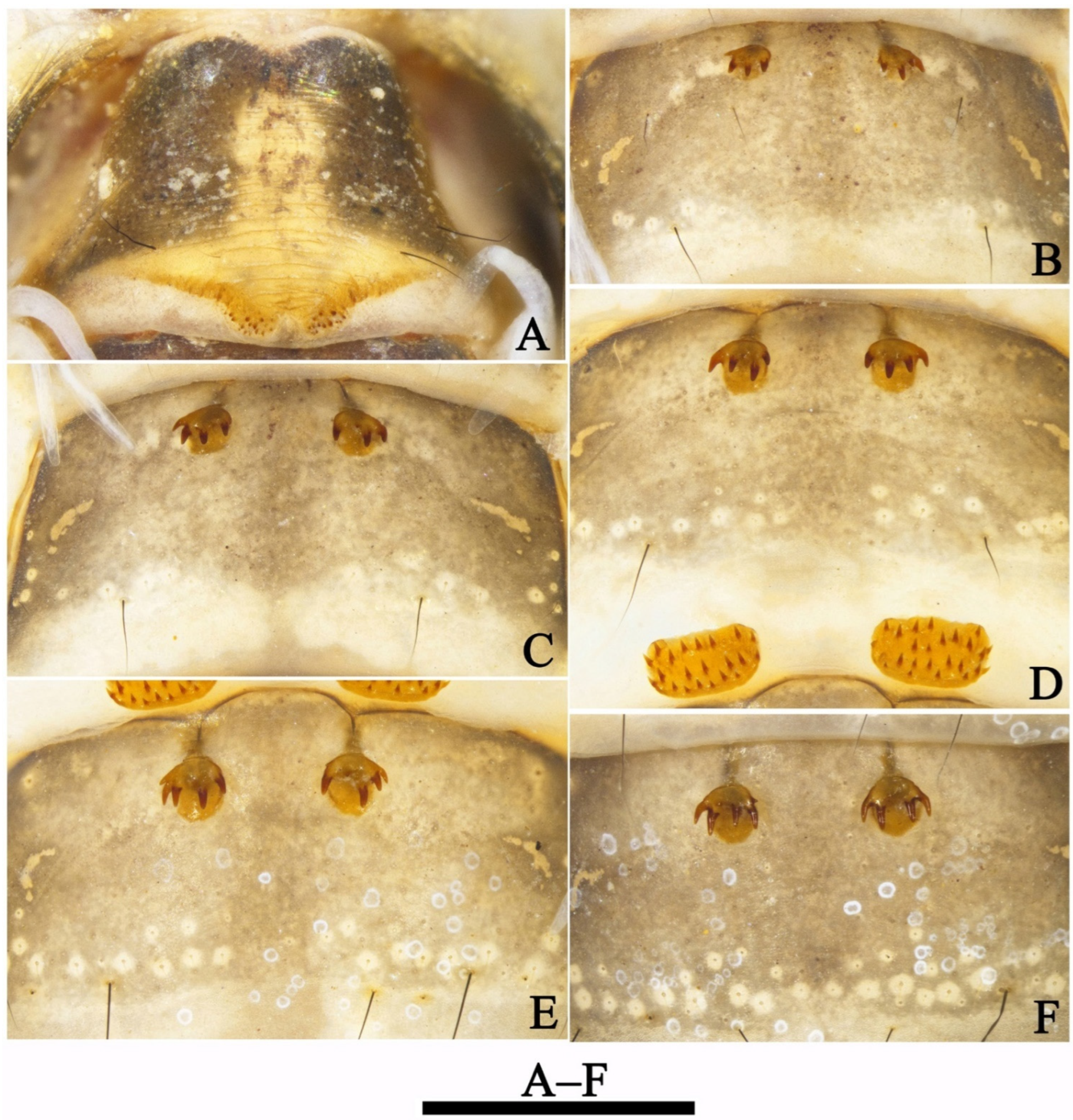
Larva (final instar): Body length 15.7–16.5 mm (n = 2). Head, pronotum and mesonotum strongly sclerotized.
Head. Head capsule nearly ovoid, dark brown with irregular yellow patches (Figure 7A–D), 2.4–2.6 mm long and 1.7–1.9 mm wide in dorsal view (n = 2). Some muscle scars longitudinally arranged on posteromedial portion of frontoclypeal apotome (Figure 7A,C) and randomly arranged on each of parietal apotome (Figure 7A,C). Frontoclypeal suture (Figure 7A,C) Ʊ-shaped with frontoclypeal apotome posterior angle in about 100°, each frontoclypeal suture at seta 5 curved at about 120°. In lateral view (Figure 7E), eyes oval, white (color probably due to speciemens being prepupae, having already begun to molt into pupae). Some muscle scars randomly arranged on each side from postgena to parietal. In ventral view (Figure 7B), anterior ventral apotome (avp) narrowly subpentagonal, with anterior margin dark brown; ventral ecdysial line on posterior meson about half as long as anterior ventral apotome, posterior ventral apotome absent. Pairs of primary setae (Figure 7B–D) 1–7, 9–17 arising on dorsal surface of head; setae 8 and 18 arising on ventral surface of the head. Setae 1, 4, 10, and 16 golden, hair-like; setae 10 thinner and shorter than 1 and 16; other setae black, spike-like; seta 14 longest and thickest; setae 2, 3, 5, 7, and 17 short and thick; setae 15 long and slender; setae 6 and 11 short and fine; setae 8, 9, 12, and 13 of medium size, setae 18 broken away, leaving only alveoli. Labrum (Figure 7A,C) brown, with middle portion of anterior margin slightly concave, and a setal brush at each anterolateral corner.
Mandibles (Figure 8A,C) shaped for scraping, black, asymmetrical in shape and arrangement of hairs; only left mandible with stiff hairs on subbasomesal edge of dorsal margin. Cardo (Figure 7B) subpentagonal, with inner and outer ends black, and intermediate portion yellow to brown.
Thorax. Pronotum (Figure 9A) subrectangular, brown in dorsal view, subdivided longitudinally by mesal suture, with several black, spike-like setae; shallow transverse depression about 1/3 distance from anterior margin; posterior ridge dark brown; Prosternal horn (Figure 9D) present and apical half sclerotized. Prosternite (Figure 9D) subpentagonal, convex anteriorly, slender laterally, and posteromesal margin slightly concave. Paired lateral posterior prosternites (Figure 9D) with irregular shape.
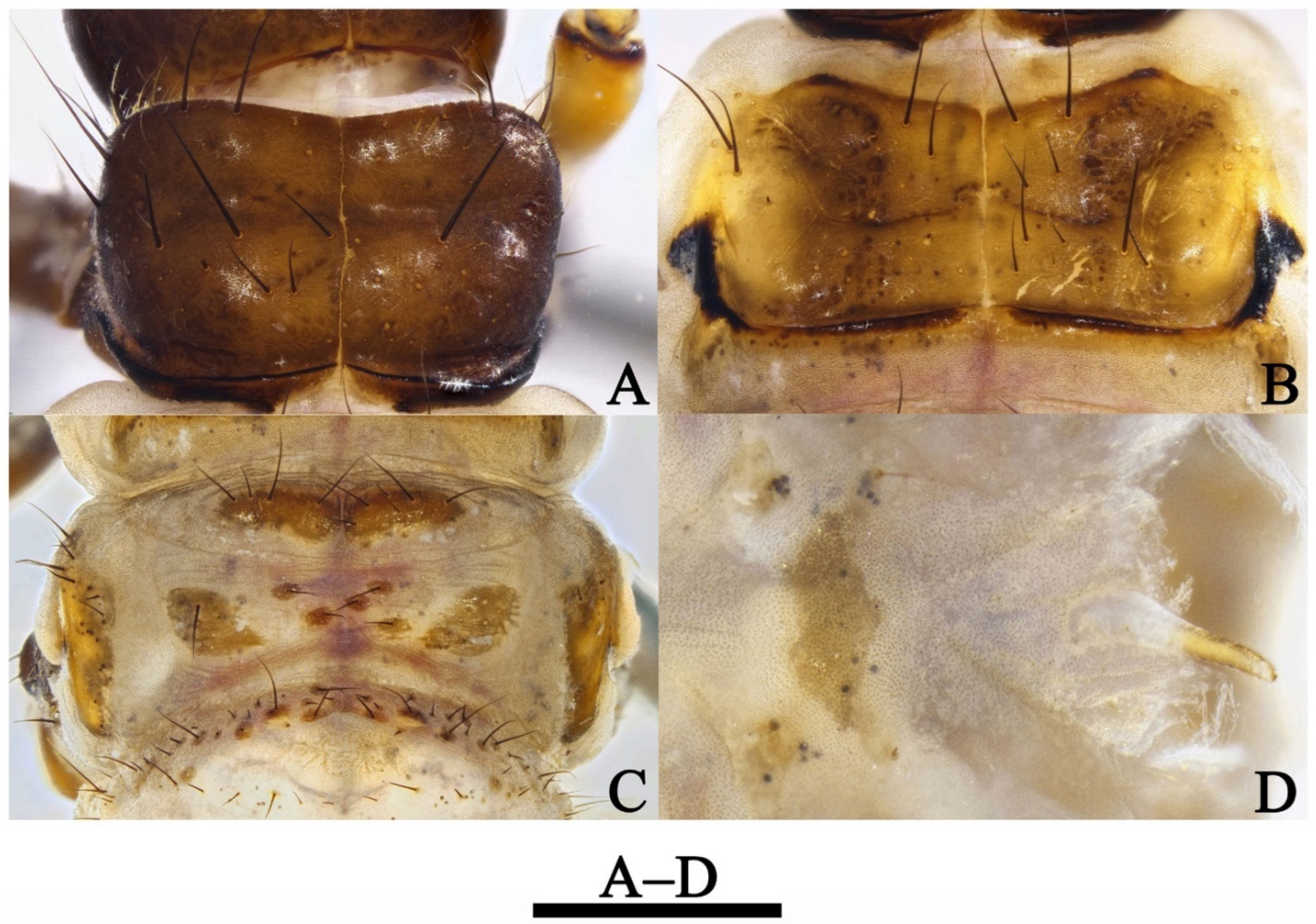
Mesonotum (Figure 9B) subrectangular dorsally, subdivided longitudinally by mesal suture, each part yellowish brown, with scattered long spike-like setae and random muscle scars; each anterior margin with convex black spot mesal of anterolateral corner, dark spots forming transverse W-shaped mark across middle of pair of nota, posterolateral margins each black and produced ventrolaterad as small triangular projection, posterior margins black except for pair of narrow yellowish brown transverse bands mesal of posterolateral corners.
Metanotum (Figure 9C) with sa1 sclerites large, elongate triangular, close to each other, each having 4–6 setae; sa2 sclerites each large, subrectangular-oval, widely separated, with 2–3 pairs of small rounded sclerites mesally between them. Sa3 sclerites each subrectangular, with mesal margin irregular, with some scattered, strong, short and long setae.
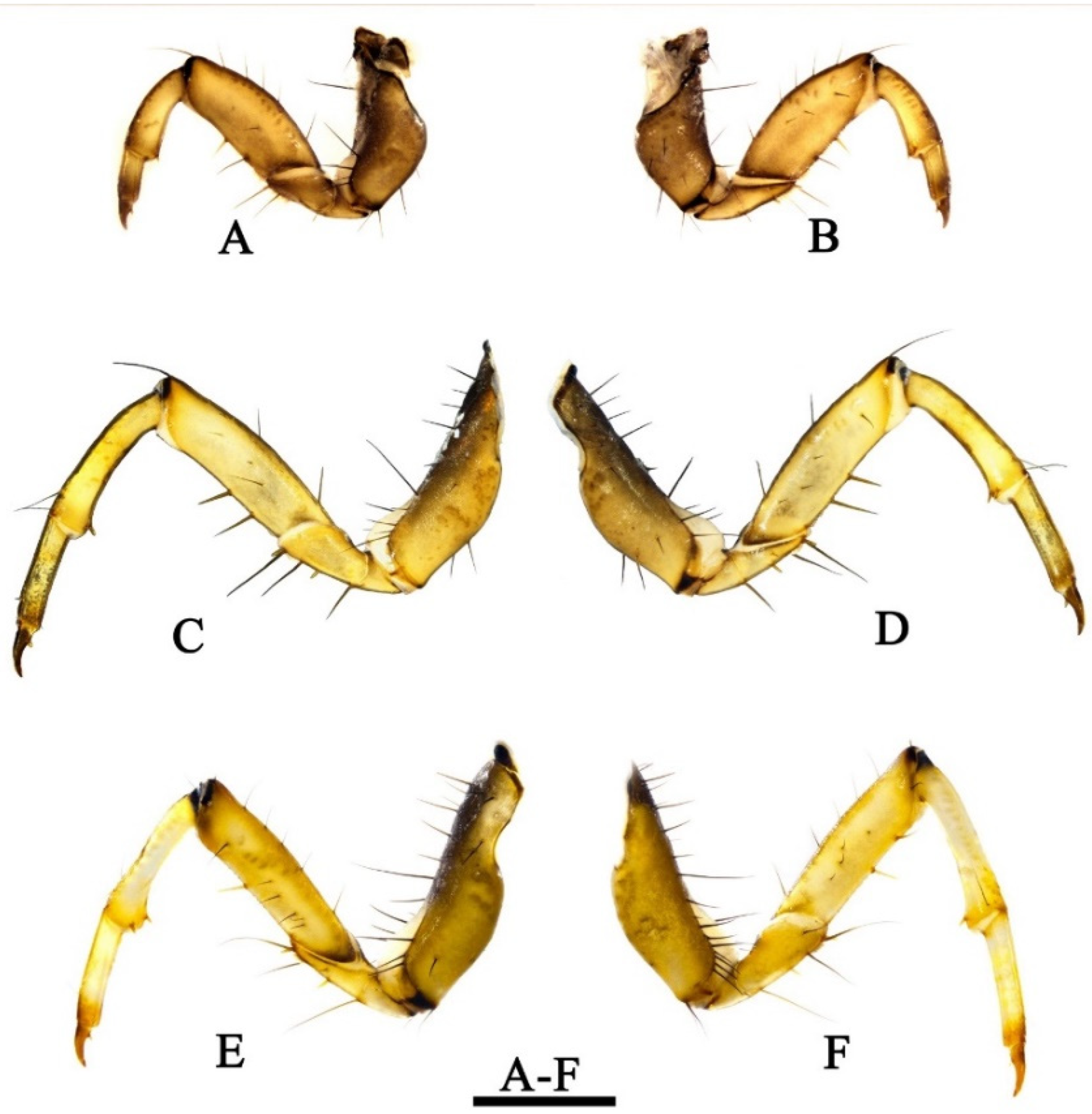
Legs. Legs yellowish to light brown. Forelegs (Figure 10A,B) shorter and slightly thicker than mid- and hind-legs. Forecoxae (Figure 10A,B) shorter than mid- and hind- coxae and conical in lateral view, each anterior side (Figure 10B) with 10–12 black spike-like setae. Foretrochanters (Figure 10A,B) each 2-segmented, both segments subtriangular, basal joint slightly shorter than apical one; apical joint with anterior side (Figure 10B) bearing 3 light-brown spur-like major setae. Forefemora (Figure 10A,B) each cylindrical with upper margin arc-shaped, with 6 brown spike-like seta dorsally; lower margin straight, with 2–3 brown spike-like setae; anterior side (Figure 10B) with 3 short black setae mesally. Foretibiae (Figure 10A,B) each cylindrical, slightly curved, with apex slightly enlarged, with 2 light brown apical spurs on anterior side and ventral margin. Foretarsi cylindrical, without setae. Foretarsal claws (Figure 10A,B) stouter and shorter than on mid- and hind legs, somewhat conical, brown and slightly curved downwards.
Mid- and hind-coxae (Figure 10C–F) longer and slenderer, each bearing a row of black spike-like setae on upper margins. Mid- and hind-trochanters (Figure 10C–F) each 2-segmented with basal joint subtriangular and shorter than apical joint; apical joint trapezoidal with anterior side (Figure 10D,E) bearing 3 light brown spur-like major setae, with ventral margin having trochanteral brush. Each mid-femur (Figure 10C,D) with anterior side (Figure 10D) bearing 4 black short setae and posterior side (Figure 10C) 2 setae. Each hind femur (Figure 10E,F) with upper margin bearing 5 brown spike-like setae and with anterior side (Figure 10E) having 6 short black short setae and posterior side (Figure 10F) having 5.
Light brown major femoral setae present in all legs. All femora with lower margins bearing inconspicuous setal buds. Mid- and hind-tibiae (Figure 10C–F) are same shape as foretibiae, but slightly more elongate; tibial spurs present same as foretibiae. Light brown spur-like basal setae present in all legs.
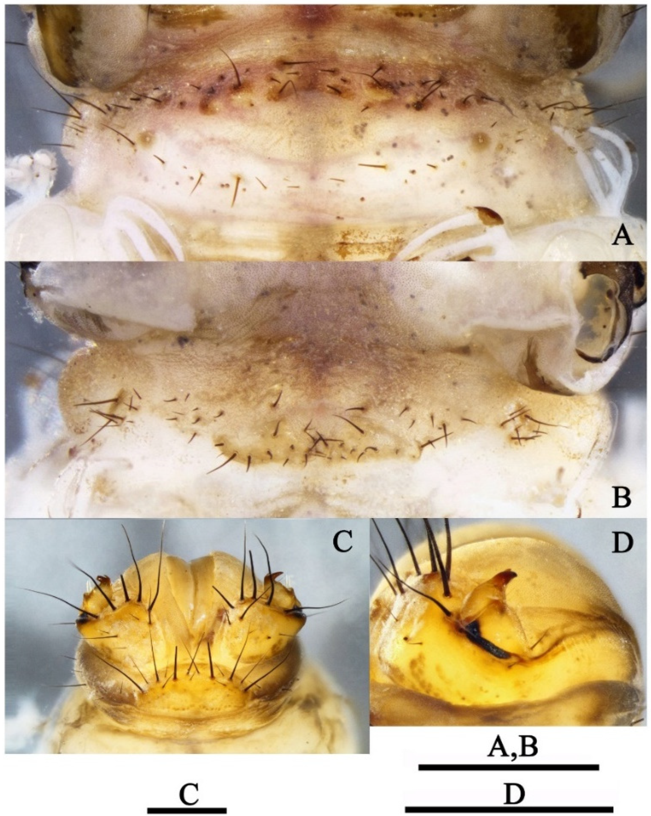
Abdomen. Abdominal segment I (Figure 11A,B) with 1 dorsal and 2 lateral humps. About 60 black setae arranged in two rows on tergum I (Figure 11A); anterior row with about 40 setae and most of them each with basal sclerite; posterior row with about 20 setae and most with basal sclerites smaller than in anterior row, only 2 of them same size as anterior sclerites. Sternum I (Figure 11B) with 2 triangular median sa2 sclerites and 2 irregular lateral sa3 sclerites; each median sclerite with about 20 setae and lateral sclerites each with about 9 setae.
Tracheal gills trifid, bifid or unbranched. Gill forms and their distributions from segments II–VII as in Table 4. Lateral fringe present on each side from segment II to segment VIII.
Dorsal sclerite of segment IX (Figure 11C) semicircular and yellowish, with about 10 long black spike-like setae arranged in arc, intermingled with 5 short fine black setae. Lateral sclerites (Figure 11C,D) each with 7 long, dark spike-like setae apically, 1 long black seta subapically, and 1 long seta and about 3 short black setae mesally. Ventral sole plate (Figure 11D) triangular with anterior margin black in lateral view. Anal proleg claws (Figure 11C,D) brown and strongly sclerotized, each with about 6 fine yellowish basal setae, dorsal accessory hook present, shorter and finer than main claw.
Pupa: Length 13.7–14.6 mm (n = 10). Antennae slightly shorter than body, each straight at the end; scape longer and thicker than remaining joints. Each eye with entire posterior margin having 17–19 curved hair-like setae (Figure 12C) overlying eye surface. Labrum (Figure 12B) wide, brown with 14 strong and slender setae on surface. Mandibles (Figure 12C) blackish brown and crossing each other apically, distal parts relatively slender; median edges of blades slightly serrated; basal parts yellowish and each with 2 strong black hair-like setae. Wing sheaths reaching end of abdominal segment IV. Mid- and hind-legs natatorial; tarsal segments I–IV (Figure 12E,G) each with dark fringe. Abdominal segments V–VIII fringed (Figure 12G).
Anterior pair of dorsal hook plates on abdominal terga III–VII, each with 4–6 strong and curved hooks. IIIa (Figure 13B) relatively small, suboval; IVa (Figure 13C), Va (Figure 13D), VIa (Figure 13E) and VIIa (Figure 13F) round and subequal in size, each with strong and fine teeth more strongly sclerotized than supporting hook plate.
Posterior pair of dorsal hook plates on abdominal terga I and V. Ip (Figure 13A) with each anterior margin indiscernible, with stubby teeth arranged mesally. Vp (Figure 13D) subrectangular with 21–23 strong and short hooks more strongly sclerotized than supporting hook plate. Hook numbers on each hook plate as follows: Ip 13–15; IIIa 4; IVa 3–4; Va 4–5; Vp 21–23; VIa 4–5, VIIa 4–5. Gill forms and their distributions from segments II–VII are list as in Table 5.
Anal appendages (Figure 12D) long and apically sclerotized, in dorsal and ventral views each inner margin bearing row of black spike-like setae; apex bent outward and concave laterally.

3.2. Diagnosis

Male: The male genitalia of this new species are similar to those of Limnephilus kaumarajiva Schmid, 1961, from Pakistan, but differ from those of the latter in that (1) the superior appendages are small and slightly shorter than the intermediate appendages in the lateral view (large and significantly longer than the intermediate appendages in lateral view in L. kaumarajiva); (2) the intermediate appendages each have their basal plate semicircular in the caudal view (subtriangular in caudal view in L. kaumarajiva); (3) the inferior appendages have the apices each bifid (the apices are acute in L. kaumarajiva); (4) the parameres each have the single setose subapex flattened and with 1 small setose subapical lobe on its inner side (the parameres each have 2 flattened setose apices and 1 small setose subapical lobe on its inner side in L. kaumarajiva).
Female: The female genitalia of this new species are similar to those of Limnephilus primoryensis Nimmo, 1995, from Russia (Primorye). This female was described by Nimmo, 1995 [41], but L. deqianensis differs from the latter in that (1) segment IX is subtriangular and broad in the middle in the lateral view (the upper portion of the posterior margin is concave anteriorly, narrow in the upper portion in L. primoryensis); (2) segment IX has paired button-like processus (this processus is absent in L. primoryensis); (3) segment X is narrow in the lateral view (segment X is broad and triangular laterally in L. primoryensis).
Larva: The larva of this new species is similar to that of Limnephilus ademus Ross, 1941, from America; this larva was described by Ruiter [42], but differs from the latter in that (1) the head capsule is without blotches outside the anterior constriction of the frontoclypeal suture (with two blotches primarily outside the anterior constriction of the frontoclypeal suture in L. ademus); (2) the meso and metafemora are without accessory setae on the lateral margins in the new species (accessory setae are present on the lateral margins of the meso and metafemora in L. ademus); (3) 5 small brown sclerites are arranged randomly between the metanotal sa2 sclerites (without such 5 small sclerites in this position in L. ademus).
Pupa: The pupa of the new species is far from any known described pupa and can be distinguished from other species by the numbers of hooks on each hook plate.

3.3. Biology and Habitat

Limnephilus deqianensis n. sp. larvae and pupae were found in a small high-altitude stream (2930 m) in open areas. The riparian vegetation is mainly alpine meadow. Stream margins were covered with detritus (mainly grass blades). The water in the stream was clear and transparent at the collection site. The anterior ends of each pupal case was attached under a flat stone at the bottom of the stream. The species has the habit of aggregating to pupate. A newly emerged male was found during collection, and the pupae in the pupal cases were well developed.

3.4. Molecular Analysis

Nineteen mitochondrial COI sequences, including seventeen from Limnephilus deqianensis n. sp., and two downloaded from GenBank were pruned to 658bp to construct the NJ tree (Figure 14).
The genetic distances are displayed at the node and rounded to four decimal places. Larvae, pupae, and female adults of Limnephilus deqianensis n. sp. were clustered with the male adults of L. deqianensis n. sp. The maximum genetic distances of the L. deqianensis n. sp. cluster were lower than 2.43%. Therefore, we concluded that the association is successful.

3.5. Revision of Limnephilus Species from the Chinese Mainland

  • Key to the males of 5 species groups of Limnephilus species occurring on the Chinese mainland:
1. Superior appendages, each with posterior margin having incision (as figures on p. 34 in Malicky, 2011)…………
……………………………………………………………………………………………L. perpusillus Species Group nomen novum
-Superior appendages without incision……………………………………………………………………………………2
2.Superior appendages, each triangular with apical corner acute and inferior appendages, each with free portion bearing an apicodorsal angle produced upwards in lateral view (as Figure 320 in Nimmo, 1971)…………………
………………………………………………………………………………………………………………...L. externus Species Group
-Superior appendages, each trapezoidal, pentagonal, ligulate, or irregular, free portion of each inferior appendage with apicodorsal angle not produced upwards in lateral view…………………………………………………………3
3.Superior appendages trapezoidal and intermediate appendages straight in lateral view, setose portion of each paramere about half as long as whole length (Figure 15)…………………………………L. flavicornis Species Group
-Superior appendages ligulate, pentagonal, or irregular, intermediate appendages curved obviously in lateral view, setose portion of each parameres shorter than half of whole length…..…………………………………………4
4.Superior appendages shorter than 1/2 height of segment IX in lateral view (Figure 3 and Figure 4)……………………………
………………………………………………………………………………………………………………...L. asiaticus Species Group
-Superior appendages longer than 1/2 height of segment IX in lateral view (Figures 17 and 18)………………………
…………………………………………………………………………………………………………………...L. stigma Species Group

3.5.1. Limnephilus perpusillus Species Group nomen novum (L. incisus Species Group of Schmid, 1955)

We proposed renaming this species group to the “L. perpusillus Species Group”, from the species L. incisus Curtis, 1834, which was used to name the group when it had been transferred to the genus Colpotaulius by Vshivkova et al. [43], and L. perpusillus Walker, 1852, which was the oldest species in the group. Originally, this species group included the following 7 species: L. hyalinus Hagen, 1861; L. acnestus Ross, 1938; L. ademus Ross, 1941; L. janus Ross, 1938; L. major Martynov, 1909; L. perpusillus Walker, 1852; and L. secludens Banks, 1914. The species L. homeros Malicky, 2011, was distributed in China and was not ascribed to any species group by the author [16]. Here, we place L. homeros Malicky, 2011, into this species group based on its male genitalia characteristics. The group can be diagnosed by following characteristics; (1) superior appendages, each with a posterior margin having an incision, as in L. major Martynov, 1909, L. secludens Banks, 1914, L. ademus Ross, 1941, L. acnestus Ross, 1938, and L. homeros Malicky, 2011; or subtriangular and curved and extended toward intermediate appendages in the caudal view as in L. hyalinus Hagen, 1861, L. janus Ross, 1938, and L. perpusillus Walker, 1852 (Table 6); (2) inferior appendages, each with the fused portion narrowed, as in L. major Martynov, 1909, L. secludens Banks, 1914, L. homeros Malicky, 2011, L. hyalinus Hagen, 1861, L. ademus Ross, 1941 and L. acnestus Ross, 1938; or almost invisible, as in. L. janus Ross, 1938, and L. perpusillus Walker, 1852 (Table 6); (3) aedeagus with a thick extensible extremity.
  • Limnephilus homeros Malicky, 2011
Limnephilus homeros Malicky, 2011, 35, figure p34, male, China.
Diagnosis: (1) Superior appendages, each with the posterior margin having a strong incision; (2) intermediate appendages are subrectangular, with the upper and lateral margins concave in the caudal view; (3) inferior appendages, each with the free portion forming an apicodorsal lobe and with the fused portion broader than the free portion; (4) parameres slightly longer than the aedeagus, each with setose lobe subapically.
Type country: China.
Distribution: China (Sichuan).

3.5.2. Limnephilus externus Species Group of Schmid, 1955

This species group contains the following two species: L. externus Hagen, 1861, and L. thorus Ross, 1938, of which L. externus Hagen, 1861, was distributed in China. The group can be diagnosed by following characteristics: (1) superior appendages are triangular, each with the upper margin straight and the apical corner acute in the lateral view; (2) inferior appendages, each with the free portion bearing an apicodorsal angle produced upwards; (3) parameres, each with flattened setose subapex, and each setose portion shorter than 1/2 of the whole length.
  • Limnephilus externus Hagen, 1861
Limnophilus externus Hagen, 1861, 257, female, Canada. 477.
Limnophilus externus Tjeder, 1930, 201–203, Figure 1, Figure 2, Figure 3, Figure 4, Figure 5, Figure 6, Figure 7 and Figure 8.
Limnophilus congener McLachlan, 1875, 56–57, pl. 8, Figure 1, Figure 2, Figure 3, Figure 4, Figure 5, Figure 6, Figure 7 and Figure 8, Russia, Finland. 479 (synonymized by Ulmer 1907 [44]).
Limnephilus luteolus Banks, 1899, 207–208. 480 (synonymized by Ross 1944 [45]).
Limnephilus oslari Banks, 1907, 121–122, pl. 9, Figure 19. 481 (synonymized by Milne 1935 [46]).
Limnephilus tersus Betten, 1934, 334, pl. 46, Figure 6, Figure 7 and Figure 8, pl. 47, Figure 1, Figure 2, Figure 3, Figure 4 and Figure 5 (synonymized by Ross 1944 [45]).
Diagnosis: (1) Superior appendages are triangular, each with the inner side of posterior margin bearing a row of black teeth; (2) intermediate appendages are curved downward in the lateral view; (3) inferior appendages, each with the free portion bearing an apicodorsal angle produced upwards; (4) parameres, each with the apicodorsal margin having a row of fringe directed anterad.
Type country: Canada.
Distribution: China (Shanghai); America; Canada; Finland; Norway; Russia; Sweden.

3.5.3. Limnephilus flavicornis Species Group of Schmid, 1955

This species group includes the following 3 species: L. correptus McLachlan, 1880 (East Palearctic and Oriental, including China), L. ectus Ross, 1941 (Nearctic), and L. flavicornis (Fabricius, 1787) (East and West Palearctic). The group can be diagnosed by the following characteristics: (1) superior appendages are trapezoidal; (2) posteromedial portion of each superior appendage bears an arc of black teeth on the inner side; (3) intermediate appendages are slender and straight, and are as long as the superior appendages, as in L. correptus McLachlan, 1880, and L. flavicornis Fabricius, 1787, or slightly shorter than superior appendages, as in L. ectus Ross, 1941; (4) setose portion of each paramere is about half as long as the whole length.
  • Limnephilus correptus McLachlan, 1880 (Figure 15)
Limnophilus correptus McLachlan, 1880, 18, pl. 53, Figure 1 and Figure 2, Russia.
Diagnosis: (1) Superior appendages are trapezoidal, with the posteromedial portion of the inner side bearing an arc of black teeth; (2) intermediate appendages are straight and slightly longer than the superior appendages; (3) parameres, each with a trapezoidal setose apex and a stubby setose lobe in the lateral view.
Type country: Russia.
Distribution: China (Sichuan, Heilongjiang); Japan; Mongolia; North Korea; Russia.
Material examined: CHINA, Hei Long-jiang Province, Hei-he City, Wudalianchi County, 8 August 1987, leg. Xue Yin-gen.

3.5.4. Limnephilus asiaticus Species Group of Schmid, 1955

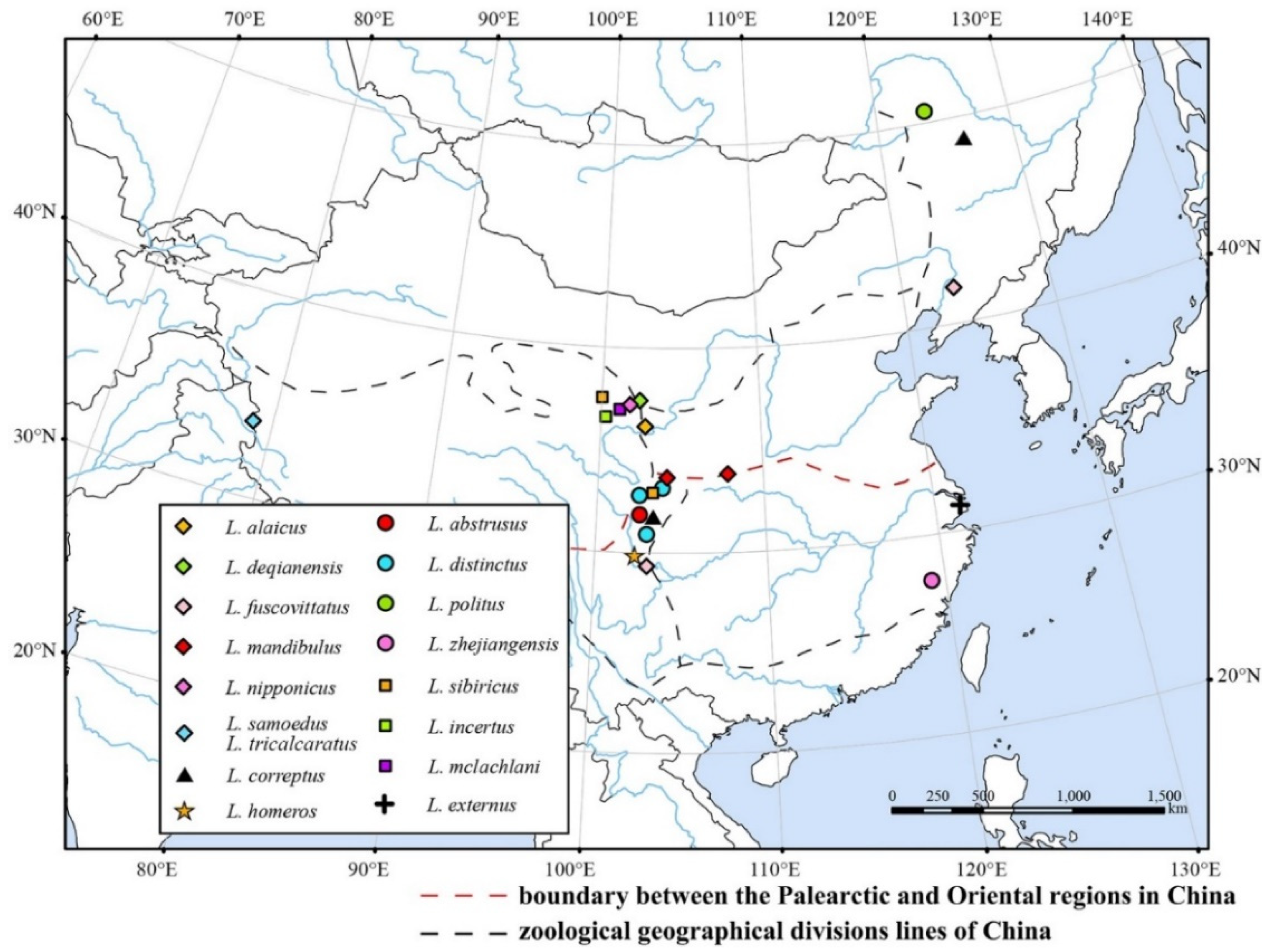
Originally, this species group included the following 11 species: L. alaicus Martynov, 1915, (East and West Palearctic, including China); L. asiaticus McLachlan, 1874, (East and West Palearctic and Oriental); L. centralis Curtis, 1834, (West Palearctic); L. chereshnevi Nimmo, 1995, (East Palearctic), L. frijole Ross, 1944 (Nearctic); L. hirsutus Pictet, 1834, (East and West Palearctic); L. labus Ross, 1941, (Nearctic); L. pallens Banks, 1920, (Nearctic); L. quadratus Martynov, 1914, (East and West Palearctic); L. samoedus McLachlan, 1880, (East Palearctic, Nearctic, and Oriental, including China); and L. tricalcaratus Mosely, 1936, (East Palearctic and Oriental, including China). The isolated species in Schmid’s system, L. fuscovittatus Matsumura, 1904; the new species described in this article, L. deqianensis n. sp.; L. mandibullus Yang & Yang, 2005, and L. nipponicus Schmid, 1964, published after 1955, show the diagnostic characteristics of the L. asiaticus Species Group, so we ascribe them to this species group. The group can be diagnosed by following characteristics: (1) Superior appendages, each ligulate and shorter than half of the height of segment IX in the lateral view; (2) intermediate appendages, each with a braod basal portion.
Key to the males of seven species of the L. asiaticus Species Group from Chinese mainland:
1Parameres obviously bifid……………………………………………………………………………………L. mandibullus
-Parameres unbranched or having only small setose lobe………………………………………………………………..2
2Parameres with flattened apices………………………………………………………………………………L. deqianensis
-Parameres with apices not flattened………………………………………………………………………………………..3
3Aedeagus with apical hook longer than extensible extremity……………………………………………………………4
-Aedeagus without apical hook or with apical hook shorter than extensible extremity………………………………5
4Intermediate appendages with basal portion as wide as superior appendages………………………………L. alaicus
-Intermediate appendages with basal portion wider than superior appendages……………………….L. tricalcaratus
5Superior appendages shorter than 1/3 height of segment IX in lateral view……………………………….L. samoedus
-Superior appendages longer than 1/3 height of segment IX in lateral view……………………………………………5
6Inferior appendages each with narrow fused portion and stout free portion……………………………L. nipponicus
-Inferior appendages each with broad fused portion and stout free portion…….………………………L. fuscovittatus
  • Limnephilus mandibulus Yang & Yang, 2005
Limnephilus mandibulus Yang & Yang, 2005, 493, Figure 2, male, female, China.
Diagnosis: (1) Segment IX is trapezoidal in the lateral view; (2) superior appendages curved downwards; (3) inferior appendages are subtriangular with the apical corner acute; (4) parameres are bifid and setose.
Type country: China.
Distribution: China (Shaanxi, Gansu).
Diagnosis: (1) Distal portion of tergum VIII is triangular and produces into bifid apicodorsal lobe in the dorsal view; (2) intermediate appendages, each divided into the basal plate and apical lobe; (3) parameres, each tubular with a flattened, setose apex, and having a small setose lobe on the inner side in the lateral view.
Type country: China.
Distribution: China (Qinghai)
  • Limnephilus alaicus (Martynov, 1915)
Astratus alaicus Martynov, 1915, 417–421, Figure 12, Figure 13, Figure 14, Figure 15, Figure 16, Figure 17, Figure 18 and Figure 19, Tajikistan.
Diagnosis: (1) Aedeagus long and thick with the extensible extremity bearing an external hook; (2) parameres, each slender, with triplet apices in the dorsal view.
Type country: Tajikistan.
Distribution: China (Qinghai); Kazakhstan; Mongolia; Russia; Turkey.
  • Limnephilus tricalcaratus (Mosely, 1936)
Astratus tricalcaratus Mosely, 1936, 453–454, pl. III, Figure 1, Figure 2, Figure 3, Figure 4, Figure 5, Figure 6, Figure 7 and Figure 8, Western Tibet.
Limnephilus tricalcaratus (Mosely, 1936), Schmid, 1955: 140.
Limnephilus tricalcaratus (Mosely, 1936), Grigorenko 2002: 110, synonymized with Limnephilus samoedus (McLachlan, 1880).
Limnephilus tricalcaratus (Mosely, 1936), Oláh, 2019, 36–37, Figures 109–111, China (Tibet), resurrected from synonymy, as Limnephilus tricalcaratus (Mosely, 1936).
Diagnosis: (1) Aedeagus long and thick with extensible extremity bearing an external hook; (2) parameres, each slender, glabrous, and tapered to an apex and bearing 3 additional spines on the apex; (3) Intermediate appendages, with the basal portion wider than the superior appendages.
Type country: China.
Distribution: China (Tibet).
  • Limnephilus samoedus (McLachlan, 1880)
Astratus samoedus McLachlan, 1880, 16, pl. 53, Figure 1, Figure 2, Figure 3, Figure 4, Figure 5, Figure 6, Figure 7, Figure 8 and Figure 9, Russia.
Previously, Schmid placed this species in the L. asaticus Species Group. However, based on the morphological phylogenetic analysis, Vshivkova and her co-authors showed that the species belonged to the genus incertae sedis, not Limnephilus [43]. Studies conducted after that (e.g., Oláh et al. 2019) still treat it as a member of Limnephilus; therefore, we here also place it in the L. asiaticus Species Group.
Diagnosis: The male genitalia of this species are similar to those of L. alaicus and L. tricalcaratus but differ from L. alaicus and L. tricalcaratus in that the apical hook on the aedeagus is shorter than the extensible extremity.
Type country: Russia.
Distribution: China (Tibet); America; Mongolia; Russia.
  • Limnephilus nipponicus Schmid, 1964
Limnephilus nipponicus Schmid, 1964, 834–836, Figures 34–38, male, female, Japan.
Diagnoses: (1) Superior appendages are subtriangular in the lateral view; (2) intermediate appendages curve upwards with the basal portion broad, and slightly longer than the superior appendages in the lateral view; (3) inferior appendages, each with the fused portion narrow; (4) parameres, each slightly curved and tapered to the apex with the apical portion setose.
Type country: Japan.
Distribution: China (Qinghai); Japan; Russia.
  • Limnephilus fuscovittatus Matsumura, 1904 (Figure 16)
Limnophilus fuscovittatus Matsumura, 1904, 171, pl. 12, Figure 13, Japan.
Limnophilus subfuscus Ulmer, 1907, 20–21, Figures 32–35, Japan; synonymized by Nakahara (1914) and Nozaki and Tanida (1996).
Diagnosis: (1) The male genitalia of this species are similar to those of L. nipponicus but differ from the latter in that: (1) the intermediate appendages slightly curved downward and shorter than the superior appendages in the lateral view (curved upward and slightly longer than the superior appendages in the lateral view in L. nipponicus); (2) inferior appendages with the fused portion broad (with the fused portion narrow in L. nipponicus); (3) parameres, each with the apical portion slightly bulging (tapered to the apical portion in L. nipponicus).
Type country: Japan.
Distribution: China (Liaoning, Sichuan); Japan; Mongolia; North Korea; Russia.
Material examined: One male, CHINA, Liaoning Province, Shen-yang City, 25 May 1957.

3.5.5. Limnephilus stigma Species Group of Schmid, 1955

Originally, this species group included the following 7 species: L. abstrusus McLachlan, 1872 (East Palearctic, including China); L. ademiensis Martynov, 1914 (East Palearctic); L. flavospinosus Stein, 1874 (West Palearctic); L. indivisus Walker, 1852 (Nearctic); L. infernalis Banks, 1914 (Nearctic); L. politus McLachlan, 1865 (East and West Palearctic, including China); and L. stigma Curtis, 1834 (Nearctic, East and West Palearctic). Limnephilus zhejiangensis Leng & Yang, 2004, and L. distinctus Tian & Yang, 1992, first described after 1955, shared common characteristics with other members of this species group. We here ascribed them to this group based on male genitalia characteristics. The group can be diagnosed by following characteristics: (1) tergum VIII projected well posterad from membranous connection to segment IX; (2) superior appendages, each longer than 1/2 the height of segment IX in the lateral view; (3) intermediate appendages long and slender except in L. stigma Curtis, 1834; (4) inferior appendages, each with a narrow-fused portion and a slender free portion.
Key to the males of four species of the L. stigma Species Group from China
1.Intermediate appendages each with apical portion curved slightly upwards in lateral view………L. zhejiangensis
-Intermediate appendages obviously curved upwards from middle portion to apex in lateral view………………2
2.Inferior appendages each with fused portion very narrow and almost invisible………………………L. distinctus
-Inferior appendages each with fused portion narrow but conspicuous ………………………………………………4
3.Segment IX posterior margin straight at insertion of inferior appendages……………………………….L. abstrusus
-Segment IX posterior margin concave at insertion of inferior appendages…………………………………L. politus
  • Limnephilus zhejiangensis Leng & Yang, 2004 (Figure 17)
Limnephilus zhejiangensis Leng & Yang, 2004, 520, Figures 22–24, male, female, China.
Diagnosis: (1) Tergum VIII with a rounded posterior lobe; (2) superior appendages, each subtriangular, with the inner side bearing two rows of teeth; (3) intermediate appendages slightly shorter than the superior appendages, each with the apical portion curved slightly upward; (3) inferior appendages, each digitate with the fused portion narrow; (4) parameres, each with a slightly flattened setose apex and a small subapical setose lobe.
Type country: China.
Distribution: China (Zhejiang).
Material examined: One Paratype male. CHINA, Zhejiang Province, Qing-yuan County, Baishanzu, alt. 1300.0m, 25 October 1993, by light trap, det. Wu Hong.
  • Limnephilus distinctus Tian & Yang, 1992 (Figure 18)
Limnephilus distinctus Tian & Yang, 1992, 880, Figure 9, male, female, China.
Diagnosis: (1) Superior appendages, each subtriangular with the inner side bearing two rows of teeth; (2) intermediate appendages, each curved strongly upward, and as long as the superior appendages; (3) inferior appendages digitate, each with the fused portion very narrow and almost indiscernible; (4) parameres, each with a subtriangular-flattened setose apex.
Type country: China.
Distribution: China (Sichuan).
Materal examed: One male, CHINA, Sichuan Province, Jiuzhaigou County, 23 August 1994, leg. Du Yu-zhou.
  • Limnephilus abstrusus McLachlan, 1872
Limnophilus abstrusus McLachlan, 1872, 62–63, pl. 1, Figure 13, Russia.
Diagnosis: (1) Superior appendages subtrapezoidal in the lateral view; (2) intermediate appendages curve upwards and are slightly shorter than superior appendages; (3) posterior margins of segment IX are straight at the insertion of the inferior appendages.
Type country: Russia.
Distribution: China (Sichuan); Mongolia; Russia.
  • Limnephilus politus McLachlan, 1865
Limnophilus politus McLachlan, 1865, 39, pl. 9, Figure 24, Britain.
Goniotaulius concentricus Kolenati, 1848
Diagnosis: The male genitalia of this species are similar to those of L. abstrusus but differ from the latter in that: (1) The length of the free portion of each inferior appendage is about 3/4 of the width of segment IX (inferior appendages, each with the free portion almost as long as segment IX in L. abstrusus); (2) posterior margins of segment IX are concave at the insertion of the inferior appendages (straight in L. abstrusus).
Type country: Britain.
Distribution: China (Inner Mongolia); Britain; Czech Republic; Finland; Germany; Hungary; Kazakhstan; Netherlands; Norway; Poland; Russia; Sweden; Ukraine.

3.5.6. Limnephilus Isolated Species

  • Limnephilus sibiricus Martynov, 1929
Limnophilus subfuscus sibiricus Martynov, 1929, 305–308, Figure 13, Figure 14, Figure 15 and Figure 16, female, China.
Limnephilus spurisi Grigorenko, 2002
Schmid treated this species as being isolated [5]. Information about its distribution in China was provided by Malicky. So far, we do not have any specimens from the Provinces of Qinghai and Sichuan; therefore, we could not provide a Diagnosis nor any illustrations of the male genitalia of this species.
Type country: Russia.
Distribution: China (Qinghai, Sichuan); Russia.

3.5.7. Limnephilus Species incertae sedis

Descriptions of the two Chinese species were based on females only: L. incertus Martynov, 1909, and L. mclachlani Martynov, 1909. Schmid treated them as species incertae sedis species [5]. We did not have the appropriate materials to revise them. Their systematic status remains unresolved.
  • Limnephilus incertus Martynov, 1909
Limnophilus incertus Martynov, 1909, 271–273, pl. 5, Figure 13, Figure 14 and Figure 15, female, China.
Type country: China.
Distribution: China (Qinghai).
  • Limnephilus mclachlani Martynov, 1909
Limnophilus signifer Martynov, 1909, 273–275, pl. 5, Figure 16, Figure 17 and Figure 18, female, China; preoccupied in Limnephilus by Phryganea signifer Zetterstedt, 1840
Limnephilus mclachlani Grigorenko, 2002, 114, replacement name.
Type country: China.
Distribution: China (Qinghai).

3.6. Distribution

A total of 17 Limnephilus species were recognized on the Chinese mainland, their distribution pattern is shown in Figure 19.

4. Discussion

Limnephilus larvae live predominantly in lentic habitats, but a few [21,31] have been collected in streams and cold springs, including the new species Limnephilus deqianensis n. sp., which was collected in a cold stream during this study. Unfortunately, the investigation was performed very late, and all the larvae of the new species had pupated, so the only two larvae studied in this research were prepupae, acquired from the pupal cases. For this reason, we had no opportunity to observe the larval biology of the new species, including its feeding style, stadia, case-making behavior, etc., or its phenology and adult biology, such as the number of generations per year, the development speed, the synchronization of emergence, the life span of the adults, or the dispersal ability of the females. Therefore, further investigations are needed to determine these biological characteristics. We believe that the discovery of these biological features will enrich the stream macroinvertebrate traits datasets of China, which will allow them to be used in monitoring water quality and may even be helpful for inferring phylogenetical relationships among species in the genus Limnephilus and its higher taxa.
On the other hand, the taxonomic studies of the genus Limnephilus are mainly based on the structures of the male genitalia; species descriptions based only on female genitalia were rare, and certainly, there are some species known from both the male and female genitalia. For the species diagnoses based on the female genitalia, the abdominal segments and the vulvar scales were the most important diagnostic characteristics. However, the spermatheca is a chitinous organ inside the abdomen, potentially indicating a specificity among the species, as described by Nielsen [33]. Chuluunbat [47] applied his terminology for diagnosing the females of some Apataniidae species (also in the superfamily Limnephiloidea). Combined with the external morphology of the abdominal segments, the descriptions of the vulvar scales and the internal spermatheca can greatly improve our species identification ability and enrich the knowledge of our trichopteran fauna.
Terminology for the female genitalia presented by Schmid [27] has been widely used; however, he did not provide terms for the structure of the internal spermatheca. Nielsen [33] studied the female genital chamber of 26 species in detail and formulated terms for the structure of the internal spermatheca. In this article, we combined the terms of Schmid [32] and Nielson [33].
Finally, the morphological study of the larva and pupa of this species will be helpful in diagnosing Limnephilus species larvae and pupae in China, a common benthic macroinvertebrates group, which is useful for the biomonitoring of water quality. This morphological study of the male, female, larva, and pupa of L. deqianensis n. sp. will also provide morphological data for building hypotheses of phylogenetic relationships among species of this genus and its sister taxa. Perhaps because of the spumaline that covers the eggs of most Trichoptera, very little is known about the morphology of caddisfly eggs. Therefore, it is not likely that information about caddisfly embryos and other chorionic details will be useful for Diagnosis and phylogenetic inference in the near future.

5. Conclusions

In this study, most of the life stages of a new Limnephilus species, determined by COI sequencing, are described and illustrated. Ecological information regarding this new species is also provided.
Additionally, based on the male genitalia and following Schmid’s system, we revised all of the Chinese mainland Limnephilus species at the species group level while emphasizing their morphological characteristics. However, phylogenetic homologues or synapomorphies were not determined and cannot support the grouping of these species. Further phylogenetic studies between the species groups of Limnephilus are needed.

Author Contributions

Conceptualization, all authors.; methodology, all authors.; software, H.Z. and X.G.; validation, H.Z. and L.P.; investigation, H.Z.; resources, all authors; data curation, H.Z.; writing—original draft preparation, H.Z.; writing—review and editing, C.S.; visualization, H.Z.; supervision, C.S. and B.W.; project administration, B.W.; funding acquisition, C.S. and B.W. All authors have read and agreed to the published version of the manuscript.

Funding

This research was supported by the National Natural Science Foundation of China (NSFC, no. 41771052) and the commissioned project of the Qilian Mountain National Park administration of Qinghai Province, P. R. China (QHTX-2021-006).

Institutional Review Board Statement

Not applicable.

Informed Consent Statement

Not applicable.

Data Availability Statement

The data, holotype specimen, and other voucher specimens from this research were deposited in the Insect Classification and Aquatic Insect Laboratory, College of Plant Protection, Nanjing Agricultural University, Nanjing, China and Department of Entomology and Nematology, University of California Davis, CA, USA.

Acknowledgments

We are grateful to Zhen-Ning Chen from Qinghai Normal University for organizing the investigation. We sincerely thank the three reviewers and editor for the time and effort that they have put into reviewing our manuscript. We are particularly grateful to John C. Morse from Clemson University for his long-term help and support in our research on aquatic insects. We are also grateful to Ji-Xiang Wang for being our guide and driver during this investigation. We also thank Yi Yuan for her help during this research.

Conflicts of Interest

The authors declare no conflict of interest. The funders had no role in the design of the study; in the collection, analyses, or interpretation of data; in the writing of the manuscript, or in the decision to publish the results.

References

  1. Morse, J.C. Trichoptera World Checklist. Available online: Http://entweb.Clemson.edu/database/trichopt/Index.htm (accessed on 21 March 2022).
  2. Holzenthal, R.W.; Blahnik, R.J.; Prather, A.L.; Kjer, K.M. Order Trichoptera Kirby, 1813 (Insecta), Caddisflies. Zootaxa 2007, 1668, 639–698. [Google Scholar] [CrossRef]
  3. Oláh, J.; Anderson, T.; Beshkov, S.; Bilalli, A.; Coppa, G.; Ibrahimi, H.; Johanson, K.A.; Kovács, T.; Mey, W.; Musliu, M.; et al. Lineage sorting by parameres in Limnephilinae subfamily (Trichoptera): With description of a new tribe, new genera and new species. Opusc. Zool. Bp. 2019, 50 (Suppl. S1), 3–98. [Google Scholar]
  4. Sekhi, S.; Malicky, H.; Lounaci, A. Description de Limnephilus barbagaensis n.sp. d’Algérie, et découverte de Micropterna testacea dans le Maghreb (Trichoptera, Limnephilidae). Braueria 2019, 46, 13–14. [Google Scholar]
  5. Schmid, F. Contribution a l’etude des Limnophilidae (Trichoptera). In Mitteilungen der Schweizerischen Entomologischen Gesellschaft 28 Beiheft; Spektrum Akademischer Verlag: Heidelberg, Germany, 1955; pp. 1–245. [Google Scholar]
  6. Nimmo, A.P. The adult Rhyacophilidae and Limnephilidae (Trichoptera) of Alberta and eastern British Columbia and their post-glacial origin. Quaest. Entomol. 1971, 7, 3–234. [Google Scholar]
  7. Nimmo, A.P. The adult Trichoptera (Insects) of Alberta and eastern British Columbia, and their post-glacial origins, 1: The families Rhyacophilidae and Limnephiltdae, supplement 1. Quaest. Entomol. 1977, 13, 25–67. [Google Scholar]
  8. Ruiter, D.E. The adult Limnephilus Leach (Trichoptera: Limnephilidae) of the New World. Bull. Ohio Biol. Surv. 1995, 11, 1–200. [Google Scholar]
  9. Morse, J.C. Book Review-The Adult Limnephilus Leach (Trichoptera:Limnephilidae) of the New World. J. N. Am. Benthol. Soc. 1996, 15, 267–269. [Google Scholar] [CrossRef]
  10. Yang, L.F.; Sun, C.H.; Morse, J.C. An amended checklist of the caddisflies of China (Insecta, Trichoptera). Zoosymposia 2016, 10, 451–479. [Google Scholar] [CrossRef]
  11. Grigorenko, V.N. Some taxonomical notes on the Limnephiline Caddisflies (Trichoptera: Limnephilidae: Limnephilinae). Nova Suppl. Entomol. 2002, 15, 107–119. [Google Scholar]
  12. Martynov, A.V. Les trichoptères du Tibet oriental et du Tsaidam d’après le materiaux collections par l’expedition de la Société imp. Géogr. Russe sous la direction de P.K. Kozlow en 1900–1901. Annu. Mus. Zool. L’academie Sci. St. Petersbourg 1909, 14, 256–309. [Google Scholar]
  13. Tian, L.X.; Li, Y.W.; Yang, L.F.; Sun, C.H. Insects of The Hengduan Mountain Region; Science Press: Beijing, China, 1992; pp. 880–891. [Google Scholar]
  14. Leng, K.M.; Yang, L.F. Five new species of Limnephilidae (Insecta, Trichoptera) from China. Acta Zootaxonomica Sin. 2004, 29, 516–522. [Google Scholar]
  15. Yang, L.F.; Yang, W.F.; Wang, B.X. Insect Fauna of Middle-West Qinling Range and South Mountains of Gansu Province; Science Press: Beijing, China, 2005; pp. 486–499. [Google Scholar]
  16. Malicky, H. Neue Trichopteren aus Europa und Asien. Braueria 2011, 38, 23–43. [Google Scholar]
  17. Malicky, H. Neue Köcherfliegen aus Europa und Asien. Braueria 2004, 31, 36–42. [Google Scholar]
  18. Malicky, H. Neue Köcherfliegen (Trichoptera) aus dem Bardia Nationalpark, Nepal. Denisia 2004, 13, 291–300. [Google Scholar]
  19. Saini, S.M.; Parey, S.H. Limnephilus morsei sp. n., a new species of the genus Limnephilus LEACH (Trichoptera: Limnephilidae) from India. Pol. J. Entomol. 2012, 81, 57–61. [Google Scholar] [CrossRef] [Green Version]
  20. Holzenthal, R.W.; Calor, R.A. Catalog of the Neotropical Trichoptera (Caddisflies). Zookeys 2017, 645, 1–566. [Google Scholar] [CrossRef] [PubMed] [Green Version]
  21. Hiley, P.D. The identification of British limnephilid larvae (Trichoptera). Syst. Entomol. 1976, 1, 147–167. [Google Scholar] [CrossRef]
  22. Gislason, G.M. Identification of Icelandic caddis larvae, with description of Limnephilus fenestratus (Zett.) and L. picturatus McL. (Trichoptera: Limnephilidae, Phryganeidae). Entomol. Scand. 1979, 10, 161–176. [Google Scholar] [CrossRef]
  23. Waringer, J.; Graf, W.; Malicky, H. Description of the larvae of Limnephilus femoratus (Zetterstedt, 1840) and Limnephilus subnitidus McLachlan, 1875, with additional notes on Limnephilus picturatus McLachlan, 1875 (Trichoptera: Limnephilidae). Aquat. Insects 2011, 33, 371–380. [Google Scholar] [CrossRef]
  24. Lepneva, S.G. Fauna of the USSR, Trichoptera II(2). Larvae and pupae of the Integripalpia. Zool. Inst. Acad. Sci. USSR New Ser. 1971, 95, 1–700. [Google Scholar]
  25. Springer, M.; Bermúdez-Monge, J. Description of the larva and pupa of Limnephilus hamifer Flint 1963 (Trichoptera, Limnephilidae) from Costa Rica. Zootaxa 2018, 4461, 277–285. [Google Scholar] [CrossRef] [PubMed]
  26. Balibrea, A.; Gonçalves, V.; Raposeiro, P.M. Larval description of Limnephilus atlanticus Nybom 1948, morphological comparison with Limnephilus affinis Curtis 1834, (Trichoptera: Limnephilidae) and additional notes on their ecology in Azores Islands. Zootaxa 2020, 4852, 372–382. [Google Scholar] [CrossRef] [PubMed]
  27. Flint, O.S. Taxonomy and biology of Nearctic limnephilid larvae (Trichoptera), with special reference to species in eastern United States. Entomol. Am. 1960, 1–120. [Google Scholar]
  28. Lloyd, J.T. The Biology of North American Caddis Fly Larvae. Bulletin of the Lloyd Library of Botany, Pharmacy and Materia Medica, Entomological Series. 1921, Volume 1, pp. 1–124. Available online: https://www.biodiversitylibrary.org/page/52059801 (accessed on 2 April 2022).
  29. Johansson, A.; Nilsson, A.N.; Svensson, B.W. Larval morphology, habitat and distribution of Limnephilus diphyes (Trichoptera, Limnephilidae). Entomol. Tidskr. 1991, 112, 19–25. [Google Scholar]
  30. Waringer, J.; Graf, W.; Malicky, H. Description of the larvae of Anabolia lombarda Ris, 1897 and Limnephilus sericeus (Say, 1824), with additional notes on Limnephilus flavospinosus Stein, 1874 (Trichoptera: Limnephilidae). Aquat. Insects 2012, 34, 93–105. [Google Scholar] [CrossRef]
  31. Vieira-Lanero, R.; Gonzáles, M.A.; Cobo, F. The larva of Limnephilus wittmeri Malicky, 1972 (Insecta, Trichoptera, Limnephilidae). Spixiana 1997, 20, 173–177. [Google Scholar]
  32. Schmid, F. Genera of the Trichoptera of Canada and Adjoining or Adjacent United States; NRC Research Press: Ottawa, ON, USA, 1998; pp. 98–99. [Google Scholar]
  33. Nielsen, A. A Comparative Study of the Genital Segments and the Genital Chamber in Female Trichoptera; Kommissionær, Munksgaard: København, Denmark, 1980; pp. 157–166. [Google Scholar]
  34. Wiggins, G.B. Larvae of the North American Caddisfly Genera (Trichoptera), 2nd ed.; University of Toronto Press: Toronto, ON, Canada; Buffalo, NY, USA, 1996; pp. 328–329. [Google Scholar]
  35. Denning, D.G. The Hydropsychidae of Minnesota (Trichoptera). Ann. Entomol. Soc. Am. 1943, 23, 101–171. [Google Scholar]
  36. Xu, J.H.; Sun, C.H.; Wang, B.X. Descriptions of larvae of three species of Hydropsyche (Trichoptera, Hydropsychidae) from China. Zootaxa 2018, 4374, 1–24. [Google Scholar] [CrossRef]
  37. Folmer, O.; Black, M.; Hoeh, W.; Lutz, R.; Vrijenhoek, R. DNA primers for amplification of mitochondrial cytochrome coxidase subunit I from diverse metazoan invertebrates. Mol. Mar. Biol. Biotechnol. 1994, 3, 294–299. [Google Scholar]
  38. Kumar, S.; Stecher, G.; Li, M.; Knyaz, C.; Tamura, K. MEGA X: Molecular Evolutionary Genetics Analysis across computing platforms. Mol. Biol. Evol. 2018, 35, 1547–1549. [Google Scholar] [CrossRef]
  39. Letunic, I.; Bork, P. Interactive Tree of Life (ITOL) v5: An online tool for phylogenetic tree display and annotation. Nucleic Acids Res. 2021, 49, 293–296. [Google Scholar] [CrossRef] [PubMed]
  40. Zhou, X.; Kjer, K.M.; Morse, J.C. An introduction to the species delimitation, larval-adult association of Chinese Hydropsychidae using independent DNA sequences and adult morphology. In Proceedings of the XIIth International Symposium on Trichoptera; The Caddis Press: Columbus, OH, USA, 2007; pp. 355–368. [Google Scholar]
  41. Nimmo, A.P. Seven new species of Limnephilus from Western North America with description of female of L. pallens (Banks) (Trichoptera, Limnephilidae, Limnephilinae, Limnephilini). Proc. Entomol. Soc. Wash. 1991, 93, 499–508. [Google Scholar]
  42. Ruiter, D.E.; Boyle, E.E.; Zhou, X. DNA barcoding facilitates associations and diagnoses for Trichoptera larvae of the Churchill (Manitoba, Canada) area. BMC Ecol. 2013, 13, 1–39. [Google Scholar] [CrossRef] [PubMed] [Green Version]
  43. Vshivkova, T.S.; Morse, J.C.; Ruiter, D.E. Phylogeny of Limnephilidae and composition of the genus Limnephilus (Limnephilidae: Limnephilinae, Limnephilini). In Proceedings of the XIIth International Symposium on Trichoptera; Bueno-Soria, J., Barba-Álvarez, R., Armitage, B.J., Eds.; The Caddis Press: Columbus, OH, USA, 2007; pp. 309–319. [Google Scholar]
  44. Ulmer, G. Trichoptera. Genera Insectorum. Wytsman, P.A.G., Ed.; 1907, pp. 1–259. Available online: https://www.biodiversitylibrary.org/page/43198387 (accessed on 2 April 2022).
  45. Ross, H.H. The caddis flies, or Trichoptera, of Illinois. Bull. Ill. Nat. Hist. Surv. 1944, 23, 1–326. [Google Scholar] [CrossRef]
  46. Milne, L.J. Studies in North American Trichoptera, 2; Privately Printed: Cambridge, MA, USA, 1935; pp. 20–56. [Google Scholar]
  47. Chuluunbat, S. Revision of East Palearctic Apatania (Trichoptera: Apataniidae). Master’s Thesis, Clemson University, Clemson, SC, USA, 2008. [Google Scholar]
Figure 1. Ecological photos. (A) Male adult; (B) pupal case; and (C) habitat.
Insects 13 00653 g001
Figure 2. Limnephilus deqianensis n. sp., male. (A) Forewing; (B) forefemur. Abbreviations: set. = setal brush. Scale bars: 1 mm.
Insects 13 00653 g002
Figure 3. Limnephilus deqianensisn. sp., male genitalia. (A) Segment VIII, dorsal; (B) segment VIII, left lateral; (C) dorsal; (D) left lateral; (E) ventral; (F) caudal; (G) left lateral paramere subapex and small lobe, left lateral. Abbreviations: sup. app. = superior appendages (paired); int. app. = intermediate appendages (paired); inf. app. = inferior appendages (paired); phc. = phallocrypt; aed. = aedeagus; par. = paramere (paired). Scale bars: 1 mm.
Insects 13 00653 g003
Figure 4. Limnephilus deqianensisn. sp., line drawing of male genitalia. (A) Left lateral; (B) dorsal; (C) caudal; (D) left lateral paramere subapex and small lobe, left lateral. Abbreviations: sup. app. = superior appendages (paired); int. app. = intermediate appendages (paired); inf. app. = inferior appendages (paired); phc. = phallocrypt; aed. = aedeagus; par. = paramere (paired). Scale bars: 1 mm.
Insects 13 00653 g004
Figure 5. Limnephilus deqianensisn. sp., female genitalia. (A) Dorsal; (B) left lateral; (C) caudal; (D) ventral; (E) spermatheca, dorsal; (F) vulvar scales, ventral; (G) spermatheca, right lateral. Abbreviations: lat. vul. = lateral vulvar lobe (paired); med. vul. = median vulvar lobe; pro. = processus; pro. spt. = processus spermatheca; pro. spt. a. = anterior section of processus spermatheca; pro. spt. p. = posterior section of processus spermatheca; sp. sc. ap. = spermathecal sclerite apodeme. Scale bars: 1 mm.
Insects 13 00653 g005
Figure 6. Limnephilus deqianensisn. sp., line drawing of female genitalia. (A) Left lateral; (B) dorsal; (C) ventral; (D) caudal; (E) spermatheca, left lateral; (F) spermatheca, ventral. Abbreviations: lat. vul. = lateral vulvar lobe (paired); med. vul. = median vulvar lobe; pro. = processus; pro. spt. = processus spermatheca; pro. spt. a. = anterior section of processus spermatheca; pro. spt. p. = posterior section of processus spermatheca; sp. sc. ap. = spermathecal sclerite apodeme. Scale bars: 1 mm.
Insects 13 00653 g006
Figure 7. Limnephilus deqianensisn. sp., larva head. (A) Dorsal; (B) ventral; (C,D) primary setae on dorsal surface; (E) right lateral. Abbreviations: avp. = anterior ventral apotome; car = cardo. Scale bar: 1 mm.
Insects 13 00653 g007
Figure 8. Limnephilus deqianensisn. sp., larva left mandible. (A) Mesal; (B), dorsal; (C) ventral. Scale bar: 1 mm.
Insects 13 00653 g008
Figure 9. Limnephilus deqianensisn. sp., larval thorax. (A) Pronotum; (B) mesonotum; (C) metanotum; (D) prosternites and prosternal horn. Scale bar: 1 mm.
Insects 13 00653 g009
Figure 10. Limnephilus deqianensis n. sp., larva legs. (A) Left foreleg posterior side; (B) left foreleg anterior side; (C) left midleg posterior side; (D) left midleg anterior side; (E) right hind leg anterior side; (F) right hind leg posterior side. Scale bar: 1 mm.
Insects 13 00653 g010
Figure 11. Limnephilus deqianensis n. sp., larva abdomen. (A) Tergum of segment I; (B)sternum of segment I; (C) abdomoinal segment IX and anal prolegs; (D) left anal proleg (paired). Scale bars: 1 mm.
Insects 13 00653 g011
Figure 12. Limnephilus deqianensis n. sp., pupa. (A) Maxillary and labial palps; (B) labrum; (C) mandibles; (D) anal appendages; (E) abdomen; (F) midtarsus; (G) hind tarsus. Scale bar: 1 mm.
Insects 13 00653 g012
Figure 13. Limnephilus deqianensis n. sp., pupa hook plates. (A) Ip; (B) IIIa; (C) IVa; (D) Va and Vp; (E) VIa; (F) VIIa. IIIa–VIIa = anterior hook plates of terga III–VII; Ip and Vp = posterior hook plates of terga I and V. Scale bar: 1 mm.
Insects 13 00653 g013
Figure 14. Neighbor-joining diagram based on sequenced data of the mitochondrial COI used to determine larva/female/male/pupa associations of Limnephilus deqianensis n. sp. Tree scale measures genetic distance.
Insects 13 00653 g014
Figure 15. Limnephilus correptus, male genitalia. (A) Left lateral; (B) dorsal. Abbreviations: sup. app. = superior appendages (paired); int. app. = intermediate appendages (paired); inf. app. = inferior appendages (paired); aed. = aedeagus; par. = paramere. Scale bar: 1 mm.
Insects 13 00653 g015
Figure 16. Limnephilus fuscovittatus, male genitalia. (A) Left lateral; (B) dorsal. Abbreviations: sup. app. = superior appendages (paired); int. app. = intermediate appendages (paired); inf. app. = inferior appendages (paired); aed. = aedeagus; par. = paramere. Scale bar: 1 mm.
Insects 13 00653 g016
Figure 17. Limnephilus zhejiangensis, male genitalia. (A) Left lateral; (B) dorsal. Abbreviations: sup. app. = superior appendages (paired); int. app. = intermediate appendages (paired); inf. app. = inferior appendages (paired); aed. = aedeagus; par. = paramere. Scale bar: 1 mm.
Insects 13 00653 g017
Figure 18. Limnephilus distinctus, male genitalia. (A) Left lateral; (B) dorsal. Abbreviations: sup. app. = superior appendages (paired); int. app. = intermediate appendages (paired); inf. app. = inferior appendages (paired); aed. = aedeagus; par. = paramere. Scale bar: 1 mm.
Insects 13 00653 g018
Figure 19. Distribution of Limnephilus species on the Chinese mainland.
Insects 13 00653 g019
Table 1. Species of Limnephilus recorded from the Chinese mainland.
SpeciesDistribution
correptus McLachlan, 1880China (Sichuan, Heilongjiang); Japan; Mongolia; North Korea; Russia
distinctus Tian & Yang, 1992China (Sichuan)
politus McLachlan, 1865China (Inner Mongolia); Britain; Czech Republic; Finland; Germany; Hungary; Kazakhstan; Netherlands; Norway; Poland; Russia; Sweden; Ukraine
zhejiangensis Leng & Yang, 2004China (Zhejiang)
abstrusus McLachlan, 1872China (Sichuan); Mongolia; Russia
homeros Malicky, 2011China (Sichuan)
externus (Hagen, 1861)China (Shanghai); America; Canada; Finland; Norway; Russia; Sweden
fuscovittatus Matsumura, 1904China (Liaoning, Sichuan); Japan; Mongolia; North Korea; Russia.
mandibulus Yang & Yang, 2005China (Shaanxi, Gansu)
nipponicus Schmid, 1964China (Qinghai); Japan; Russia
samoedus (McLachlan, 1880)China (Tibet); America; Mongolia; Russia
tricalcaratus (Mosely, 1936)China (Tibet)
alaicus (Martynov, 1915)China (Qinghai); Kazakhstan; Mongolia; Russia; Turkey
mclachlani Martynov, 1909China (Qinghai)
incertus Martynov, 1909China (Qinghai)
sibiricus Martynov, 1929China (Qinghai, Sichuan); Russia
Table 2. PCR primers used to sequence mtCOI genes of Limnephilus deqianensis n. sp. in this study.
PrimerSequenceReference
LCO1490GGTCAACAAATCATAAAGATATTGGFolmer et al., 1994 [37]
HCO2198TAAACTTCAGGGTGACAAAAAATCAFolmer et al., 1994 [37]
Table 3. Specimens used in larva-male-female-pupa associations of Limnephilus deqianensis n. sp.
Sample IDSpeciesGenBank IDLife StageCollection SiteCollection Date
QH21M1Limnephilus deqianensisON834683Adult (male)De-qian town, Men-yuan county, Qinghai2 July 2021
QH21M2Limnephilus deqianensisON834679Adult (male)De-qian town, Men-yuan county, Qinghai2 July 2021
QH21M3Limnephilus deqianensisON834680Adult (male)De-qian town, Men-yuan county, Qinghai2 July 2021
QH21M4Limnephilus deqianensisON834682Adult (male)De-qian town, Men-yuan county, Qinghai2 July 2021
QH21M5Limnephilus deqianensisON834681Adult (male)De-qian town, Men-yuan county, Qinghai2 July 2021
QH21F1Limnephilus deqianensisON834677Adult (female)De-qian town, Men-yuan county, Qinghai2 July 2021
QH21F2Limnephilus deqianensisON834673Adult (female)De-qian town, Men-yuan county, Qinghai2 July 2021
QH21F3Limnephilus deqianensisON834674Adult (female)De-qian town, Men-yuan county, Qinghai2 July 2021
QH21F4Limnephilus deqianensisON834675Adult (female)De-qian town, Men-yuan county, Qinghai2 July 2021
QH21F5Limnephilus deqianensisON834676Adult (female)De-qian town, Men-yuan county, Qinghai2 July 2021
QH21P1Limnephilus deqianensisON834687PupaDe-qian town, Men-yuan county, Qinghai2 July 2021
QH21P2Limnephilus deqianensisON834684PupaDe-qian town, Men-yuan county, Qinghai2 July 2021
QH21P3Limnephilus deqianensisON834685PupaDe-qian town, Men-yuan county, Qinghai2 July 2021
QH21P4Limnephilus deqianensisON834686PupaDe-qian town, Men-yuan county, Qinghai2 July 2021
QH21P5Limnephilus deqianensisON834689PupaDe-qian town, Men-yuan county, Qinghai2 July 2021
QH21L1Limnephilus deqianensisON834678LarvaDe-qian town, Men-yuan county, Qinghai2 July 2021
QH21L2Limnephilus deqianensisON834688LarvaDe-qian town, Men-yuan county, Qinghai2 July 2021
ID06902Limnephilus fuscovittatusKX104327n/aMongolia: Arhangay, Tariat, Urd Terkhiin Gol25 July 2004
ID06904Limnephilus fuscovittatusKX103754n/aMongolia: Hovsgol, Tsagaan-Uur, Uur Gol16 July 2005
Table 4. Number of types of tracheal gills on abdominal segments II–VII of final instar larvae of Limnephilus deqianensis n. sp. Positions abbreviated as: A = anterior, D = dorsal, L = lateral, P = posterior, V = ventral.
GillIIIIIIVVVIVII
AD333210
PD332100
ADL331300
AVL321000
AV333222
PV333222
Table 5. Number of types of tracheal gills on abdominal segments II–VII of pupae of Limnephilus deqianensis n. sp. Positions abbreviated as: A = anterior, D = dorsal, L = lateral, P = posterior, V = ventral.
GillIIIIIIVVVIVII
ADL333200
PDL332000
AL332100
PL321000
AVL333221
PVL333220
Table 6. Morphological characteristics of superior and inferior appendages of the eight species in the L. perpusillus Species Group nomen novum. Abbreviations: sup. app. = superior appendages; inf. app. = inferior appendages.
Speciesperpusillusjanushyalinusmajorsecludensademusacnestushomeros
Incision of sup. app.without withoutwithoutwithwithwithwithwith
Fused portion of inf. app.almost invisiblealmost invisiblenarrownarrownarrownarrownarrownarrow
Publisher’s Note: MDPI stays neutral with regard to jurisdictional claims in published maps and institutional affiliations.

