Article

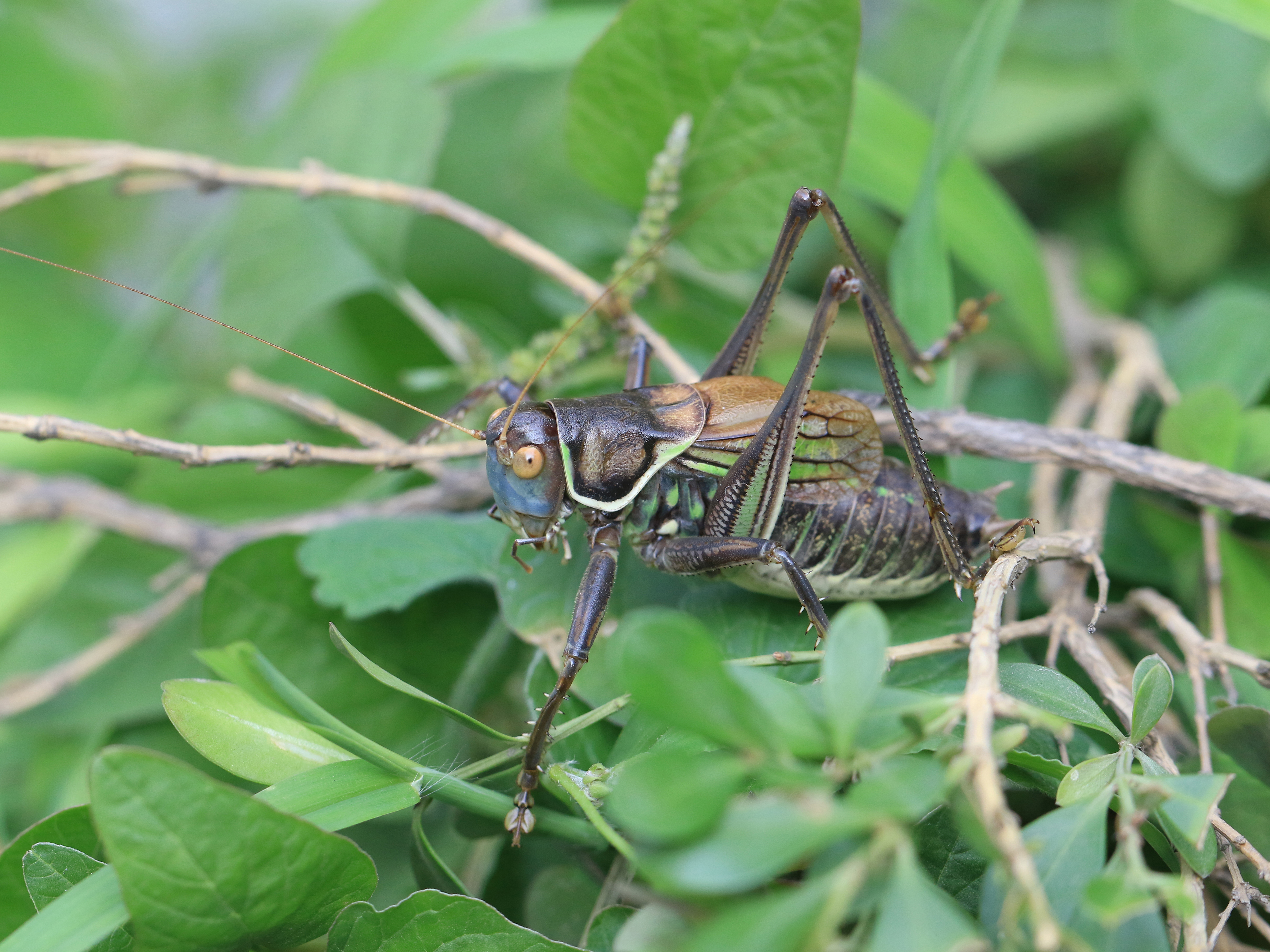
Bacterial Communities in the Feces of Laboratory Reared Gampsocleis gratiosa (Orthoptera: Tettigoniidae) across Different Developmental Stages and Sexes

1
Key Laboratory of Zoological Systematics and Application of Hebei Province, College of Life Sciences, Hebei University, Baoding 071002, China
2
Institute of Life Science and Green Development, Hebei University, Baoding 071002, China
*
Author to whom correspondence should be addressed.
Insects 2022, 13(4), 361; https://doi.org/10.3390/insects13040361
Submission received: 5 February 2022 / Revised: 5 April 2022 / Accepted: 6 April 2022 / Published: 7 April 2022
(This article belongs to the Section Insect Molecular Biology and Genomics)

Abstract

:

Simple Summary

Many insects host a diverse gut microbial community, ranging from pathogenic to obligate mutualistic organisms. Little is known about the bacteria associated with katydids. Gampsocleis gratiosa (Orthoptera, Tettigoniidae) is an economically important singing pet in China. In the present study, the bacterial communities of the laboratory-reared G. gratiosa feces were characterized using Illumina sequencing of the 16S rDNA V3-V4 region.

Abstract

We used Illumina sequencing of the 16S rDNA V3-V4 region to identify the bacterial community in laboratory-reared G. gratiosa feces across different developmental stages (1st–7th instar nymph day 0, and 0-, 7-, 14-, and 21-day adult) and sexes. In total, 14,480,559 high-quality reads were clustered into 2982 species-level operational taxonomic units (OTUs), with an average of 481.197 (±137.366) OTUs per sample. These OTUs were assigned into 25 phyla, 42 classes, 60 orders, 116 families, 241 genera, and some unclassified groups. Only 21 core OTUs were shared by all samples. The most representative phylum was Proteobacteria, followed by Firmicutes, Bacteroidetes, and Acidobacteria. At the genus level, Kluyvera (387 OTUs), Obesumbacterium (339 OTUs), Buttiauxella (296 OTUs), Lactobacillus (286 OTUs), and Hafnia (152 OTUs) were dominant bacteria. The early-instar nymphs harbored a similar bacterial community with other developmental stages, which contain higher species diversity. Both principal coordinate analysis (PCoA) and non-metric multidimensional scaling analysis (NMDS) failed to provide a clear clustering based on the developmental stages and sexes. Overall, we assume that G. gratiosa transmits bacteria vertically by eating contaminated eggshells, and both developmental stages and sexes had no significant effect on the fecal bacterial community.

Graphical Abstract

1. Introduction

Many insects harbor a diverse gut microbial community, including protists, fungi, archaea, and bacteria [1]. Numerous previous works have demonstrated that these symbiotic microbiotas potentially provide many beneficial services to the host’s overall ecological fitness: e.g., feeding, digestion, nutrient absorption, immunity, growing development, and even insecticide resistance [2,3,4,5,6,7]. For instance, the gut bacterial communities of camellia weevil Curculio chinensis are consistent with a potential microbial contribution to the detoxification of the genus Camellia tree defensive chemicals [8]. The gut microbial composition may vary widely from insect to insect because of their different feeding habits. On the whole, the gut microbial diversity of omnivorous insects was significantly higher than that of carnivorous and herbivorous insects [9]. For example, omnivorous cockroaches Shelfordella lateralis, Blattella germanica, and Periplaneta americana harbor relatively rich gut microbial species [10,11,12,13]. The majority of gut microbiota were not essential for host insect survival and obtained via the environment rather than via vertical transmission [10]. Large differences in annual temperature and humidity could alter the gut microbial composition and diversity, and assist host insects’ survival in these local environments [14].
The basic insect life cycle presents potential challenges for transmission of microorganisms between generations. The opportunity of direct transfer for gut symbionts between conspecifics are more limited in solitary insects with non-overlapping generations. Females sometimes display sophisticated mechanisms for inoculating eggs or progeny with microbial symbionts [15]. Adult females potentially transmit bacteria to progeny by defecating in the vicinity of eggs and having their gut bacteria ingested by their progeny [1]. Female red firebugs Pyrrhocoris apterus (Hemiptera: Pyrrhocoridae) and many lepidopterans transfer microbes vertically by smearing their microbiota on the eggs [16,17]. The stinkbug Riptortus clavatus postnatally acquires a beneficial gut symbiont from the environment every generation [18]. Caterpillar gut microbiomes are dominated by leaf-associated bacteria, further suggesting that resident, host-specific symbionts are sparse or absent [19]. Gut symbiotic communities can be dynamic, changing through time and developmental stage [20]. Several larvae–adult sample pairs were analyzed independently to further assess species-specific and diet-specific effects on conspecific individuals of varying life stages [21,22,23,24]. The shedding of foregut and hindgut exoskeletal lining during insect larval development severely disrupts or eliminates any attached bacterial populations [1]. The midgut bacteria of Drosophila have been associated with changes in host development [25,26].
Bacterial species comprise all or most organisms of most insect guts [1]. Bacterial diversity studies using culture-independent methods depended on the 16S rDNA gene. The bacterial 16S rDNA full-length [27] and hypervariable regions V1–V3 [12,28], V3–V4 [14,29,30], and V4 [31,32] have been widely used. High-throughput sequencing technologies can detect significantly higher diversity in microbial populations than traditional culture-based and conventional molecular methods [9,29]. Until recently, the Illumina platform had greater potential because it generated longer sequence reads and was more suited for smaller projects [33].
The diversity and composition of bacterial community vary substantially across different developmental stages of holometabolous insects, such as Dendroctonus rhizophagus [34], Monochamus alternatus and Psacothea hilaris [35], and Spodoptera littoralis [6]. Orthopterans are hemimetabolous insects with similar nutritional environments and requirements during their developmental stages. The egg hatches into a nymph, which feeds, ecdysis, and grows larger, then emerges as an adult that looks very similar to the late-instar nymph. Previous studies focused mainly on the orthopteran adult gut bacterial community [36,37,38]. Katydids and grasshoppers shared a characteristic bacterial community dominated by Proteobacteria, Firmicutes, and Actinobacteria [39]. The midgut bacteria of desert locust Schistocerca gregaria produce phenolics with antimicrobial properties that protect host insects from entomopathogens [39]. Both trophic behaviors and the evolution of the host may contribute to the shifts in prevalence among the core bacterial groups of six grasshopper species [36]. The gut bacterial communities of katydids having different feeding habits were obviously different, in which omnivorous katydids host the highest gut bacterial diversity [38].
Microbial symbiosis involves acquisition, colonization, and transmission. It is poorly understood whether the orthopteran bacterial community varies among the different developmental stages and between sexes. Omnivorous katydids Gampsocleis gratiosa (Orthoptera: Tettigoniidae) are well-known as singing pets in China. Hence, we used Illumina sequencing of 16S rRNA V3–V4 regions to assess the bacterial communities in laboratory-reared G. gratiosa feces across different developmental stages and sexes. We hypothesize that bacterial diversity increased as G. gratiosa progressed through first instar nymph to adult, and shared several core bacteria between different life stages and sexes. To the best of our knowledge, this is the first attempt to study the bacterial communities across the life history of a laboratory-reared katydid.

2. Materials and Methods

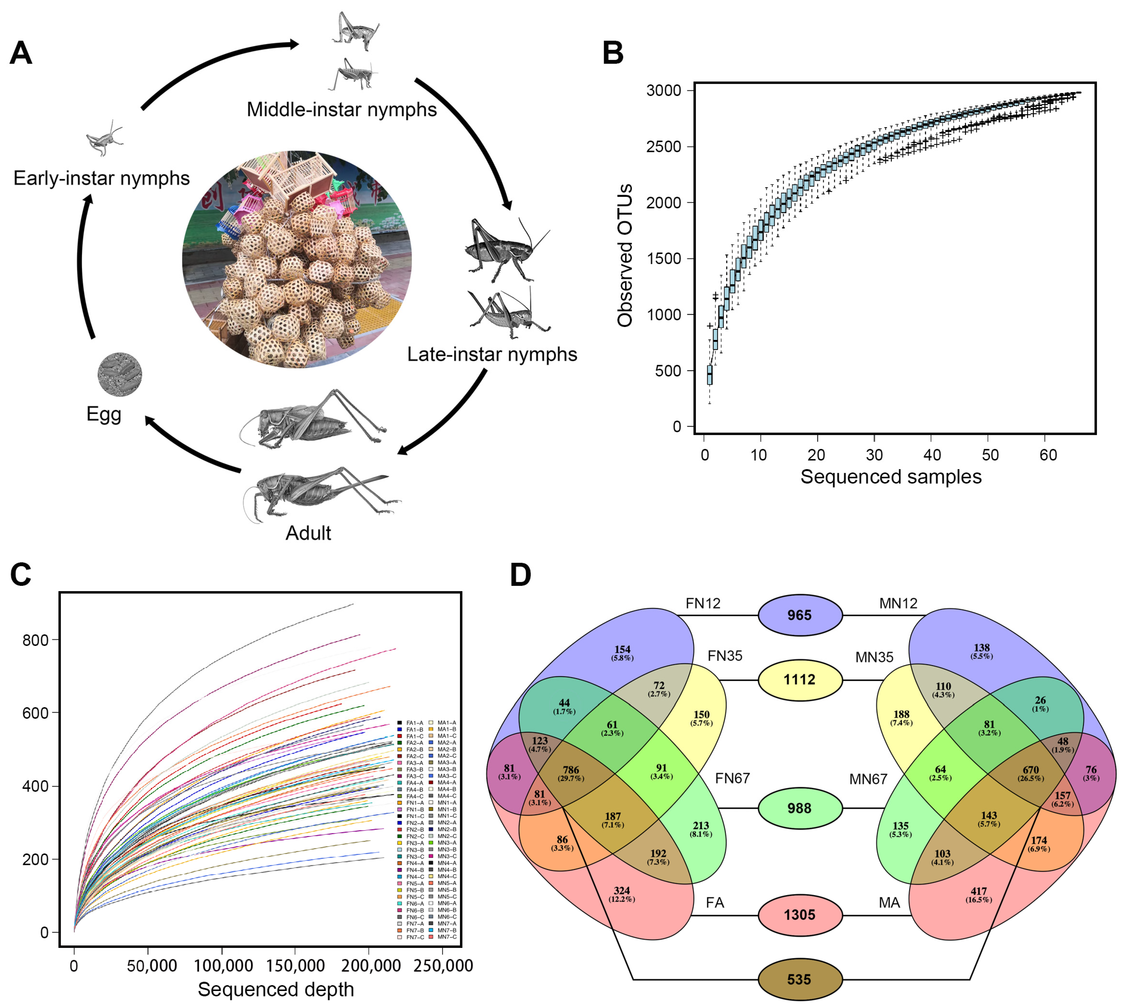
2.1. Insect Rearing and Fecal Sample Collection

Fifty-five newly hatched G. gratiosa nymph (N) were reared until reaching 21 d adult (A) after the final adult ecdysis. It was reared in separate cages under a regime of 12 h lightness at 32 °C, 12 h darkness at 18 °C, and 50 ± 5% relative humidity. Both nymphs and adults were fed with a 1:1:1 mixture of steamed carrot, chicken liver, and cooked soya. There are seven instars, and complete nymphal development in about 60 days. Fecal pellets were collected singly in aseptic microtube at 17:00 daily. Afterward, all fecal samples were stored at −80 °C until required. Avoided interrupting G. gratiosa molting when collecting. We recorded the molting date in detail.
The fecal samples of 36 G. gratiosa individuals (18 females and 18 males) were analyzed at 11 time points: 1st–7th instar nymph on molting day, and 0-, 7-, 14-, and 21-day adult. The feces of 6 G. gratiosa individuals were pooled as one sample and three biological replicates were established for each time point. The sample labels and grouping were shown in Table 1.

2.2. DNA Extraction, 16S rDNA V3–V4 Amplification and Illumina Sequencing

Bacterial DNA were extracted from each pool using the E.Z.N.A™ Mag-Bind Soil DNA Kit (Omega Bio-tek, Inc. Norcross, GA, USA). Bacterial 16S rDNA V3–V4 hypervariable region was utilized to assess gut bacterial diversity. Amplification was performed using the 314F (5′- CCT ACG GGN GGC WGC AG -3′) and 805R (5′- GAC TAC HVG GGT ATC TAA TCC -3′) primers [40]. Both primers contained Illumina adapters, and the reverse primer contained a 6 bp barcode sequence unique to each sample. Polymerase chain reaction (PCR) was performed using 2 × Taq Master Mix (Vazyme Biotech Co., Ltd. Nanjing, China) with the following conditions: 94 °C 3 min; 30 cycles of 94 °C 30 s, 50 °C 30 s, 72 °C 1 min; 72 °C 7 min. The PCR products were quantified with Qubit3.0 (Qubit® ssDNA Assay Kit, Thermo Fisher Scientific Inc., Shanghai, China) and pooled together in equimolar concentrations for sequencing. High throughput DNA sequencing was conducted using a paired-end, 2 × 300 bp cycle run on an Illumina MiseqTM platform at Shanghai Sangon Biotech Co., Ltd. (Shanghai, China). No technical replicates were performed on samples.

2.3. Bioinformatics and Statistical Analysis

Primers were trimmed from raw reads using Cutadapt 1.2.1 [41] and assigned to their respective samples according to the unique barcodes. The sequences were filtered by quality using Prinseq 0.20.4 [42], a Phred quality cutoff value of 20 (Q20) and a minimum cut length of 200 nucleotides were used for both strands. After the removal of barcodes and primers, forward and reverse reads were joined using PEAR v.0.9.10 [43].
Non-amplified region sequences were removed using Usearch 9.2 [44]. Chimeras were detected and removed using Uchime 4.2.40 [45]. Cleaned sequences were pooled and clustered into operational taxonomic units (OTUs) at a 97% similarity level using Usearch 9.2 [44]. OTUs with less than 0.005% of the total number of reads were further interpreted as likely contaminants and filtered from the OTU table [46]. Rarefaction curves of the observed OTUs were constructed in Usearch v10.0.240.
The most abundant sequence of each OTU was chosen as a representative sequence, and taxonomic assignment was made with the Bayesian RDP classifier 2.12 against the Ribosomal Project Database (Confidence level = 0.8) [47] and Silva databases (Similarly > 90% and Coverage > 90%) [48]. We removed the sequences from OTU tables, which were annotated as chloroplasts or mitochondria, and not assigned to any kingdom, to ensure only bacterial 16S rDNA sequences were included in downstream analyses. Relative abundance of each OTU was counted from phylum to genus level [49]. The core bacteria of G. gratiosa was defined as those OTUs present in all 66 fecal samples.
All statistical tests were performed in R v4.1.2 (http://cran.r-project.org accessed on 1 January 2022), mainly using vegan v2.5-7 package. The OTU table was subsampled to avoid sequencing depth effect according to the sample with the lowest number of reads (n = 168,005). Alpha and beta diversity analyses were calculated using the vegan package and plotted using the ggplot2 package. The differences among developmental stages and between sexes were analyzed by alpha diversity indices (ACE, Chao1, Shannon, and Simpson) using Kruskal-Wallis (KW) rank test [50] and paired t-test, respectively. Differences between compared groups were considered significant when p < 0.05. Non-metric multidimensional scaling (NMDS) was used to visualize the sample groupings based on the Bray-Curtis dissimilarity indices [51]. Analysis of similarity (ANOSIM) was performed to determine the statistical significance among groups with 9999 permutations. Principal coordinates analysis (PCoAs) was used to visualize the sample groupings based on the Bray-Curtis dissimilarity indices [51], and unweighted and weighted UniFrac distances [52]. Beta diversity was also performed through heatmap analysis for Top20 genus using a pheatmap (v1.0.12) package. Log-linear discriminant analysis effect size algorithm (LEfSe) [53] analysis was performed to identify potential taxonomic groups (LDA scores > 2.0, p < 0.05) that can serve as biomarkers for different groups.

3. Results

3.1. Sequencing Statistics

Like all orthopteran insects, G. gratiosa undergoes incomplete metamorphosis; nymph and adults had hardly any differentiations in form or function (Figure 1A). Illumina sequencing of 16S rDNA V3-V4 region for 66 fecal samples across the G. gratiosa life history yielded 14,907,483 raw reads. Following sequence trimming, quality filtering, and removal of contaminants (non-16S rDNA, organelle 16S rDNA and chimeras), remaining 14,480,559 high-quality clean reads were retained. A summary of the number of pre-filtering, post-filtering reads for each fecal sample is given in Table S1.

3.2. Bacterial Community Structures and OTUs

These clean reads fell to 80,492 OTUs at the 97% similarity interval. After removal of OTUs with less than 0.005% of the total number of reads, 2982 OTUs were counted across all samples. In general, these ultra-low abundance OTUs were interpreted as likely contaminants and discarded before the downstream analyses [46,54]. The number of observed OTUs varied from 204 to 899 per sample (Table 1), with an average of 481.197 (±137.366) OTUs per sample. The majority of OTUs (2507 of 2982 OTUs) were present in two or more samples. The species accumulation curves reached an asymptote after ~30 samples, indicating that the number of sequenced samples enough to observe most bacteria in G. gratiosa feces (Figure 1B). Rarefaction curves plotted from observed OTUs have reached plateau with a high Good’s coverage index (>99%) that indicated that the sequence depth were able to represent majority of the OTUs present in all the 66 fecal samples (Figure 1C). A higher OTUs count indicated a highly complex bacterial community in G. gratiosa feces.
The 2982 OTUs were assigned into 25 phyla, 42 classes, 60 orders, 116 families, 241 genera, and some unclassified groups. The number of unassigned OTUs across different taxonomic ranks was raised from 27 OTUs (representing 0.01% total reads) at phylum level to 860 OTUs (representing 2.07% total reads) at genus level.
The majority of OTUs fell into four major bacterial phyla: Proteobacteria, Firmicutes, Bacteroidetes, and Acidobacteria (Table 2 and Table S2), while the remaining 21 phyla represented less than 1% OTUs and total reads. Within the Proteobacteria phylum, Gammaproteobacteria, Alphaproteobacteria, and Betaproteobacteria were the most dominant. Within the Firmicutes phylum, Bacilli, and Clostridia were the most dominant. At the order and family level, the Enterobacteriales order all belonged to the Enterobacteriaceae family within class Gammaproteobacteria, while the Lactobacillales order was almost completely represented by the Lactobacillaceae family within class Bacilli. At the genus level, 72 of 241 (29.88%) genera occurred in all eight groups, and seven genera from Proteobacteria and Firmicutes were dominant (relative abundance >1%) (Table 2).
Nine of 25 phyla Acidobacteria, Actinobacteria, Bacteroidetes, Chloroflexi, Euryarchaeota, Firmicutes, Proteobacteria, Thermotogae, and Verrucomicrobia were detected in all eight groups. The majority of OTUs belonged to the most abundant phylum Proteobacteria (ranging from 71.76% in MA to 83.46% in the MN12 group), followed by Firmicutes (ranging from 10.80% in MN12 to 16.77% in the FA group). Bacteroidetes was the third most abundant phylum in all groups except for the FN67 group. Proteobacteria phylum had the highest relative abundances in all groups, except for the FN35 group (53.99% reads of Firmicutes higher than 45.84% reads of Proteobacteria). The remaining seven phyla had lower relative abundance (<1% reads of any sample). The relative abundance of the Kluyvera, Lactobacillus, and Hafnia genus were the top five genera in all groups. These three genera made up 2% or higher reads of any groups. The relative abundance of Kluyvera OTU3 in early-instar nymphs was apparently higher than other developmental stages (Table S2).

3.3. Dominant and Core Bacterial OTUs

Thirty-two dominant OTUs (representing >1% reads of any sample) represented 91.56% of total reads. Among these, 24 OTUs and 8 OTUs belonged to the Gammaproteobacteria class of Proteobacteria and the Bacilli class of Firmicutes, respectively. Within Gammaproteobacteria, 24 OTUs were assigned to the genus Aeromonas (OTU28), Buttiauxella (OTU263, 266, 386, 1000, 14,025), Citrobacter (OTU66), Erwinia (OTU204), Escherichia/Shigella (OTU6), Hafnia (OTU1, 8, 10), Kluyvera (OTU3, 7, 990, 14,028), Obesumbacterium (OTU221), Raoultella (OTU67), and Serratia (OTU5, 22), as well as four unidentified OTUs of Enterobacteriaceae (OTU27, 210, 294, 8791). Within Bacilli, eight OTUs were assigned to the genus Bacillus (OTU70), Enterococcus (OTU128), Lactobacillus (OTU2, 11, 122, 125, 205), and Pediococcus (OTU207).
Core OTUs were determined by the shared OTUs in all fecal samples. Twenty-one OTUs (representing 91.13% total reads) were classified into 3 phyla, 4 classes, 5 orders, 8 families, and 13 genera (Table 3).

3.4. Alpha and Beta Diversity

Alpha diversity of bacterial communities was assessed in terms of observed number of OTUs, richness estimate (Chao1 and ACE), and diversity index (Shannon and Simpson) (Figure 2, Table 1 and Table 2). In alpha diversity analysis based on the minimal read number (168,005 reads), the total OTU number was 2982 and ranged from 179 to 865 over all samples (Table 4). In general, the bacterial communities were slightly less abundant in early-instar nymph stages as compared to other developmental stages (Table 2). There were no significant differences among eight groups in all four diversity indices according KW rank tests: ACE (Chi-squared = 8.6308, Bonferroni-corrected p = 0.2803), Chao1 (Chi-squared = 10.51, Bonferroni-corrected p = 0.1615), Shannon (Chi-squared = 6.8006, Bonferroni-corrected p = 0.4499), and Simpson (Chi-squared = 7.1247, Bonferroni-corrected p = 0.416) (Table S3). Nonparametric paired t-test only revealed significant differences in the Shannon diversity indices between male vs. female (p < 2.2 × 10⁻¹⁶) and MN12 vs. FN12 (p = 0.028) (Table S4).
Analysis of similarity (ANOSIM) at the OTU level based on Bray-Curtis dissimilarity showed an overall significant difference among developmental stages and sexes (R = 0.148, p = 7 × 10⁻⁴). To better visualize the results, both PCoA and NMDS analyses based on the Bray-Curtis dissimilarity was plotted. The PCoA analyses showed significant differences at the OTU level among groups using two comparisons (ANOSIM: p = 7 × 10⁻⁴ using the Bray-Curtis dissimilarity indices, and p = 1 × 10⁻⁴ using unweighted UniFrac distances); the comparison using weighted UniFrac distances was not significant (p = 0.2341) (Figure 3A–C). The NMDS analysis (Stress value = 0.09) failed to provide a clear clustering based on the developmental stages and sexes (Figure 3D). LEfSe analysis identified two families, seven genera, and an unclassified Enterobacteriaceae family to be significantly associated with different development stages and sexes. Kluyvera and Mesorhizobium were more abundant in the MN12 group. Megamonas was more abundant in the MN35 group. The Enterococcaceae family and Enterococcus were more abundant in the MA group. The Brucellaceae family, Serratia, and Ochrobactrum were more abundant in the FN12 group. Hafnia was more abundant in the FN67 group. The unclassified Enterobacteriaceae family was more abundant in the FA group (Figure 3E). To demonstrate differences in genus abundance profiles between samples, a heat map was generated with R using the pheatmap package. The relative abundance and distribution of gut bacteria communities at genus level was shown by the heat map (Figure 3F). There were no significant differences in the abundance of fecal bacteria at the genus level among the eight groups. Compared to the G. gratiosa nymph group, adults contain more unique OTUs (together constituting 12.2% and 16.5% OTUs in the MA and FA groups, respectively). Beta diversity was further characterized by OTUs shared between groups, revealing that 17.94% OTUs were shared among all eight groups (Figure 1D).

4. Discussion

Bacteria can play beneficial, and often essential, roles for host insect survival. The most dominant role of the gut bacteria is essential nutrient provisioning, followed by digestion and detoxification [55]. While insects readily acquire several bacteria from their surroundings during their life cycle, others are vertically transmitted or inherited [56]. In the past decade, the bacterial communities of insects have been extensively studied between wild and laboratory-reared populations [57,58], and gut and fecal samples [59], as well as across different developmental stages [24,29,60,61]. It has been observed that an insect gut microbial community can be influenced by the host’s diet and surroundings. In this study, G. gratiosa were raised separately under the same conditions. Therefore, the bacterial communities were mainly affected by the developmental stages and sexes.
Omnivorous insects commonly have a higher gut bacterial diversity [38,62], which could partly be due to their highly diversified diets, as they eat whatever they find. We observed 2982 OTUs in omnivorous katydid G. gratiosa, which were assigned into 25 phyla, 42 classes, 60 orders, 116 families, 241 genera, and some unclassified groups. Approximately 30% of OTUs were not assigned to the genus level, suggesting the existence of previously uncharacterized taxa [63]. Similar higher gut bacterial diversity has been reported in the omnivorous cockroach Blattella germanica (2713 OTUs) [12]. It is worth noting that omnivorous cockroaches host a diverse gut microbiome that is not essential for host survival [10]. Most gut microbiota of omnivorous cockroaches come from their surroundings rather than vertical transmission [10].
An insect bacterial community is usually dominated by few phyla, such as Proteobacteria, Firmicutes, and Bacteroidetes [32,64,65]. Although the OTUs in G. gratiosa feces were assigned into 25 bacterial phyla, the majority of OTUs (91.18%) and reads (99.81%) are concentrated in Proteobacteria and Firmicutes. The phylum Proteobacteria and Firmicutes were also the most common found in other orthopteran species [36,38]. Proteobacteria were the mostly transient microbes of the variegated grasshopper Zonocerus variegatus [66]. Although male grasshoppers showed a significantly higher alpha diversity, the prevalence of the main phyla does not shift [36]. The Proteobacteria phylum is highly diverse and contains a large variety of species that are adapted to several environments [67]. Firmicutes and Proteobacteria play important roles in maintaining the growth and development of insects during the metabolism of secondary metabolites in host plants [39].
The families Clostridiales, Desulfovivrionales, and Bacteriodales are common to omnivorous cockroaches [11,68,69]. Enterobacteriaceae often represent one of the dominant bacterial families in insect guts [64,70]. The gut microbiota of G. gratiosa was dominated by Kluyvera, Obesumbacterium, Buttiauxella, Lactobacillus, and Hafnia genera. The gut microbiota of Apis cerana is dominated by Serratia (Proteobacteria: Enterobacteriaceae), Snodgrassella (Proteobacteria: Neisseriaceae), and Lactobacillus (Firmicutes: Lactobacillaceae) genera [71].
The gut core microbiota are quite stable over evolutionary time [72] and likely have an important role in host nutrition [73]. In contrast to higher bacterial richness (2982 OTUs), only 21 core OTUs were presented in all G. gratiosa fecal samples. Similar results (25 of 2713 bacterial OTUs) have been found in omnivorous cockroach B. germanica [12]. Only six core bacteria OTUs were shared across different developmental stages of Bactrocera minax (Diptera: Tephritidae) [70]. This suggests the possibility of vertical transmission of these core bacteria [70]. The variability of microbial composition within groups could be the random acquisition of microorganisms from surroundings [74]. Some of these core OTUs have a low abundance, which may become dominant in response to environmental changes and play a key role in the ecosystem [12,75].
The dietary change in different developmental stages played a substantial role in shaping bacterial communities of holometabolic insects [28,59,76,77,78,79]. For example, the bacterial diversity of Chironomus transvaalensis declines as it evolves from egg mass to adult, while the highest richness was observed in the pupal stage [60]. The proportion of each intestinal microbe is related to the host development process and gender, indicating that the physiological changes in the host affect the growth of microorganisms. Orthopterans are hemimetabolous insects with similar nutritional environments and requirements during their developmental stages. The bacterial richness of the G. gratiosa early-instar nymph is very similar to other developmental stages. The influence of sex is minimal on the insect bacterial composition [80,81]. Male grasshoppers have a significantly higher alpha diversity than the females, which could be attributed to rare taxa rather than main phyla [36]. It is therefore not surprising that minimal variation in G. gratiosa bacterial communities was observed among differential developmental stages and sexes.
The symbiotic association was due to the influence of diet, but also may have been related to vertical transmission [82]. Symbiotic bacteria of insects have also been reported to be transmitted horizontally and vertically [83,84]. The majority of symbionts are transmitted from the mother to the offspring vertically. Previous studies have revealed the host insects generally transmit their symbionts vertically by egg surface contamination, coprophagy, or symbiont capsule provisioning, etc. Transmission through eggs requires that larvae take up these bacteria when hatching. The fecal–oral route, early-instar nymph eating mother’s feces, may be key to a gut microbial community establishing in German cockroach B. germanica [85]. Newly hatched larvae take up symbionts maternally transferred via biting through and fully ingesting eggshells contaminated with bacteria, e.g., smeared over the egg surface following oviposition [6,17,86]. Some endophytic bacteria in plant-feeding insect gut were the same as plant tissues, suggesting that their microbial communities might be established by acquiring bacteria from plant tissues [17].
We assume that G. gratiosa acquired bacteria vertically by feeding on contaminated eggshells. Insect gut communities are dominated by widely distributed bacteria that appear to colonize hosts opportunistically [1]. The taxonomic composition of egg microbiota revealed a relatively high abundance of Proteobacteria, which represents the maternal and environmental sources of gut bacteria [6]. The horizontal transmission of microbial communities has been demonstrated in hemimetabolous Scaphoideus titanus [87]. The intestinal environment of G. gratiosa has strict limits on bacteria survival. Only a few bacteria can adapt to the intestinal environment, resulting in the diversity of intestinal microbe being much lower than the surroundings. Symbionts were readily acquired horizontally when Pyrrhocoris apterus nymphs were reared in the presence of symbionts on the surface of the eggs during oviposition, feces, or adult bugs [16].

5. Conclusions

Bacterial communities present in laboratory-reared G. gratiosa feces were studied using Illumina sequencing of the 16S rDNA V3-V4 region. G. gratiosa is an omnivorous and hemimetabolous insect, which passes through seven nymphal stages from egg to adult. Considering G. gratiosa were raised separately under the same conditions, the bacterial communities were not affected by diet and rearing conditions. The early-instar nymphs harbored a similar bacterial community with other developmental stages, which contain higher species diversity. Both PCoA and NMDS analysis at the OTU level failed to provide a clear clustering based on the developmental stages and sexes. One factor could be the extent to which the feeding ecology of hemimetabolous insects is very similar from early-instar nymphs up to adults. This proves that there is vertical transmission of microorganisms, early-instar nymphs eating eggshells to obtain the flora. These data will provide an overall view of the bacterial community in the feces across G. gratiosa life stages and sexes.

Supplementary Materials

The following supporting information can be downloaded at: https://www.mdpi.com/article/10.3390/insects13040361/s1, Table S1: Characteristics of 16S rDNA gene amplicon libraries of bacterial communities in each 66 Gampsocleis gratiosa fecal sample; Table S2: Relative abundance of different bacterial taxa and OTU in each sample; Table S3: Nonparametric Kruskal-Wallis tests; Table S4: Nonparametric paired t-test between groups.

Author Contributions

X.C. and Z.Z. designed the research; X.C. reared the katydids, collected the fecal samples; X.C. and H.H. conducted the research, analyzed the data; X.C. and Z.Z. wrote the manuscript. All authors approved the manuscript. All authors have read and agreed to the published version of the manuscript.

Funding

This research was funded by the Natural Science Foundation of Hebei Province, grant number C2021201002.

Institutional Review Board Statement

Not applicable.

Informed Consent Statement

Not applicable.

Data Availability Statement

The datasets generated during this study were deposited in NCBI under Bio-project PRJNA795812 (Bio-sample accessions SAMN24777588–SAMN24777653, SRA accession SRR17518483–SRR17518548).

Acknowledgments

Sangon Biotech (Shanghai) Co., Ltd., China, is acknowledged for its sequencing service.

Conflicts of Interest

The authors declare no conflict of interest.

References

  1. Engel, P.; Moran, N.A. The gut microbiota of insects—Diversity in structure and function. FEMS Microbiol. Rev. 2013, 37, 699–735. [Google Scholar] [CrossRef] [PubMed]
  2. Douglas, A.E. Nutritional interactions in insect-microbial symbioses: Aphids and their symbiotic bacteria Buchnera. Annu. Rev. Entomol. 1998, 43, 17–37. [Google Scholar] [CrossRef] [PubMed] [Green Version]
  3. Gibson, C.M.; Hunter, M.S. Extraordinarily widespread and fantastically complex: Comparative biology of endosymbiotic bacterial and fungal mutualists of insects. Ecol. Lett. 2010, 13, 223–234. [Google Scholar] [CrossRef] [PubMed]
  4. Tagliavia, M.; Messina, E.; Manachini, B.; Cappello, S.; Quatrini, P. The gut microbiota of larvae of Rhynchophorus ferrugineus Oliver (Coleoptera: Curculionidae). BMC Microbiol. 2014, 14, 136. [Google Scholar] [CrossRef] [PubMed] [Green Version]
  5. Berlanga, M.; Llorens, C.; Comas, J.; Guerrero, R. Gut bacterial community of the xylophagous cockroaches Cryptocercus punctulatus and Parasphaeria boleiriana. PLoS ONE 2016, 11, e0152400. [Google Scholar] [CrossRef] [Green Version]
  6. Chen, B.; Teh, B.S.; Sun, C.; Hu, S.; Lu, X.; Boland, W.; Shao, Y. Biodiversity and activity of the gut microbiota across the life history of the insect herbivore Spodoptera littoralis. Sci. Rep. 2016, 6, 29505. [Google Scholar] [CrossRef]
  7. Chen, B.; Du, K.; Sun, C.; Vimalanathan, A.; Liang, X.; Li, Y.; Wang, B.; Lu, X.; Li, L.; Shao, Y. Gut bacterial and fungal communities of the domesticated silkworm (Bombyx mori) and wild mulberry-feeding relatives. ISME J. 2018, 12, 2252–2262. [Google Scholar] [CrossRef] [Green Version]
  8. Zhang, S.; Shu, J.; Xue, H.; Zhang, W.; Zhang, Y.; Liu, Y.; Fang, L.; Wang, Y.; Wang, H. The gut microbiota in camellia weevils are influenced by plant secondary metabolites and contribute to saponin degradation. mSystems 2020, 5, e00692-e19. [Google Scholar] [CrossRef] [Green Version]
  9. Yun, J.H.; Roh, S.W.; Whon, T.W.; Jung, M.J.; Kim, M.S.; Park, D.S.; Yoon, C.; Nam, Y.D.; Kim, Y.J.; Choi, J.H.; et al. Insect gut bacterial diversity determined by environmental habitat, diet, developmental stage, and phylogeny of host. Appl. Environ. Microbiol. 2014, 80, 5254–5264. [Google Scholar] [CrossRef] [Green Version]
  10. Tinker, K.A.; Ottesen, E.A. Phylosymbiosis across deeply diverging lineages in omnivorous cockroaches. Appl. Environ. Microbiol. 2020, 86, e02513-19. [Google Scholar] [CrossRef]
  11. Schauer, C.; Thompson, C.L.; Brune, A. The bacterial community in the gut of the Cockroach Shelfordella lateralis reflects the close evolutionary relatedness of cockroaches and termites. Appl. Environ. Microbiol. 2012, 78, 2758–2767. [Google Scholar] [CrossRef] [Green Version]
  12. Perez-Cobas, A.E.; Maiques, E.; Angelova, A.; Carrasco, P.; Moya, A.; Latorre, A. Diet shapes the gut microbiota of the omnivorous cockroach Blattella germanica. FEMS Microbiol. Ecol. 2015, 91, fiv022. [Google Scholar] [CrossRef] [PubMed] [Green Version]
  13. Bertino-Grimaldi, D.; Medeiros, M.N.; Vieira, R.P.; Cardoso, A.M.; Turque, A.S.; Silveira, C.B.; Albano, R.M.; Bressan-Nascimento, S.; Garcia, E.S.; de Souza, W.; et al. Bacterial community composition shifts in the gut of Periplaneta americana fed on different lignocellulosic materials. Springerplus 2013, 2, 609. [Google Scholar] [CrossRef] [PubMed] [Green Version]
  14. Wang, H.; Wu, N.; Liu, Y.; Kundu, J.K.; Liu, W.; Wang, X. Higher bacterial diversity of gut microbiota in different natural populations of leafhopper vector does not influence WDV transmission. Front. Microbiol. 2019, 10, 1144. [Google Scholar] [CrossRef] [Green Version]
  15. Kuechler, S.M.; Renz, P.; Dettner, K.; Kehl, S. Diversity of symbiotic organs and bacterial endosymbionts of lygaeoid bugs of the families Blissidae and Lygaeidae (Hemiptera: Heteroptera: Lygaeoidea). Appl. Environ. Microbiol. 2012, 78, 2648–2659. [Google Scholar] [CrossRef] [PubMed] [Green Version]
  16. Kaltenpoth, M.; Winter, S.A.; Kleinhammer, A. Localization and transmission route of Coriobacterium glomerans, the endosymbiont of pyrrhocorid bugs. FEMS Microbiol. Ecol. 2009, 69, 373–383. [Google Scholar] [CrossRef] [Green Version]
  17. Paniagua Voirol, L.R.; Frago, E.; Kaltenpoth, M.; Hilker, M.; Fatouros, N.E. Bacterial symbionts in Lepidoptera: Their diversity, transmission, and impact on the host. Front. Microbiol. 2018, 9, 556. [Google Scholar] [CrossRef]
  18. Kikuchi, Y.; Hosokawa, T.; Fukatsu, T. Insect-microbe mutualism without vertical transmission: A stinkbug acquires a beneficial gut symbiont from the environment every generation. Appl. Environ. Microbiol. 2007, 73, 4308–4316. [Google Scholar] [CrossRef] [Green Version]
  19. Hammer, T.J.; Janzen, D.H.; Hallwachs, W.; Jaffe, S.P.; Fierer, N. Caterpillars lack a resident gut microbiome. Proc. Natl. Acad. Sci. USA 2017, 114, 9641–9646. [Google Scholar] [CrossRef] [Green Version]
  20. Augustinos, A.A.; Tsiamis, G.; Caceres, C.; Abd-Alla, A.M.M.; Bourtzis, K. Taxonomy, diet, and developmental stage contribute to the structuring of gut-associated bacterial communities in Tephritid pest species. Front. Microbiol. 2019, 10, 2004. [Google Scholar] [CrossRef] [Green Version]
  21. Andongma, A.A.; Wan, L.; Dong, Y.C.; Li, P.; Desneux, N.; White, J.A.; Niu, C.Y. Pyrosequencing reveals a shift in symbiotic bacteria populations across life stages of Bactrocera dorsalis. Sci. Rep. 2015, 5, 9470. [Google Scholar] [CrossRef] [PubMed]
  22. Shukla, S.P.; Sanders, J.G.; Byrne, M.J.; Pierce, N.E. Gut microbiota of dung beetles correspond to dietary specializations of adults and larvae. Mol. Ecol. 2016, 25, 6092–6106. [Google Scholar] [CrossRef] [PubMed]
  23. Ventura, C.; Briones-Roblero, C.I.; Hernandez, E.; Rivera-Orduna, F.N.; Zuniga, G. Comparative analysis of the gut bacterial community of four Anastrepha fruit flies (Diptera: Tephritidae) based on Pyrosequencing. Curr. Microbiol. 2018, 75, 966–976. [Google Scholar] [CrossRef] [PubMed]
  24. Wang, X.; Gao, Q.; Wang, W.; Wang, X.; Lei, C.; Zhu, F. The gut bacteria across life stages in the synanthropic fly Chrysomya megacephala. BMC Microbiol. 2018, 18, 131. [Google Scholar] [CrossRef] [PubMed]
  25. Shin, S.C.; Kim, S.H.; You, H.; Kim, B.; Kim, A.C.; Lee, K.A.; Yoon, J.H.; Ryu, J.H.; Lee, W.J. Drosophila microbiome modulates host developmental and metabolic homeostasis via insulin signaling. Science 2011, 334, 670–674. [Google Scholar] [CrossRef] [PubMed] [Green Version]
  26. Storelli, G.; Defaye, A.; Erkosar, B.; Hols, P.; Royet, J.; Leulier, F. Lactobacillus plantarum promotes Drosophila systemic growth by modulating hormonal signals through TOR-dependent nutrient sensing. Cell Metab. 2011, 14, 403–414. [Google Scholar] [CrossRef] [PubMed] [Green Version]
  27. Burke, C.M.; Darling, A.E. A method for high precision sequencing of near full-length 16S rRNA genes on an Illumina MiSeq. PeerJ 2016, 4, e2492. [Google Scholar] [CrossRef]
  28. Gonzalez-Escobedo, R.; Briones-Roblero, C.I.; Pineda-Mendoza, R.M.; Rivera-Orduna, F.N.; Zuniga, G. Bacteriome from Pinus arizonica and P. durangensis: Diversity, comparison of assemblages, and overlapping degree with the gut bacterial community of a bark beetle that kills pines. Front. Microbiol. 2018, 9, 77. [Google Scholar] [CrossRef]
  29. Zhang, Z.; Jiao, S.; Li, X.; Li, M. Bacterial and fungal gut communities of Agrilus mali at different developmental stages and fed different diets. Sci. Rep. 2018, 8, 15634. [Google Scholar] [CrossRef] [Green Version]
  30. Rimoldi, S.; Gini, E.; Iannini, F.; Gasco, L.; Terova, G. The effects of dietary insect meal from Hermetia illucens prepupae on autochthonous gut microbiota of rainbow trout (Oncorhynchus mykiss). Animals 2019, 9, 143. [Google Scholar] [CrossRef] [Green Version]
  31. Dubois, G.; Girard, C.; Lapointe, F.J.; Shapiro, B.J. The Inuit gut microbiome is dynamic over time and shaped by traditional foods. Microbiome 2017, 5, 151. [Google Scholar] [CrossRef] [PubMed] [Green Version]
  32. Zhao, Y.; Wang, W.; Zhu, F.; Wang, X.; Wang, X.; Lei, C. The gut microbiota in larvae of the housefly Musca domestica and their horizontal transfer through feeding. AMB Express 2017, 7, 147. [Google Scholar] [CrossRef] [PubMed] [Green Version]
  33. Caporaso, J.G.; Lauber, C.L.; Walters, W.A.; Berg-Lyons, D.; Huntley, J.; Fierer, N.; Owens, S.M.; Betley, J.; Fraser, L.; Bauer, M.; et al. Ultra-high-throughput microbial community analysis on the Illumina HiSeq and MiSeq platforms. ISME J. 2012, 6, 1621–1624. [Google Scholar] [CrossRef] [PubMed] [Green Version]
  34. Morales-Jimenez, J.; Zuniga, G.; Ramirez-Saad, H.C.; Hernandez-Rodriguez, C. Gut-associated bacteria throughout the life cycle of the bark beetle Dendroctonus rhizophagus Thomas and Bright (Curculionidae: Scolytinae) and their cellulolytic activities. Microb. Ecol. 2012, 64, 268–278. [Google Scholar] [CrossRef]
  35. Kim, J.M.; Choi, M.Y.; Kim, J.W.; Lee, S.A.; Ahn, J.H.; Song, J.; Kim, S.H.; Weon, H.Y. Effects of diet type, developmental stage, and gut compartment in the gut bacterial communities of two Cerambycidae species (Coleoptera). J. Microbiol. 2017, 55, 21–30. [Google Scholar] [CrossRef] [PubMed]
  36. Muratore, M.; Prather, C.; Sun, Y. The gut bacterial communities across six grasshopper species from a coastal tallgrass prairie. PLoS ONE 2020, 15, e0228406. [Google Scholar] [CrossRef] [Green Version]
  37. Muratore, M.; Sun, Y.; Prather, C. Environmental nutrients alter bacterial and fungal gut microbiomes in the common meadow katydid, Orchelimum vulgare. Front. Microbiol. 2020, 11, 557980. [Google Scholar] [CrossRef]
  38. Zheng, X.; Zhu, Q.; Zhou, Z.; Wu, F.; Chen, L.; Cao, Q.; Shi, F. Gut bacterial communities across 12 Ensifera (Orthoptera) at different feeding habits and its prediction for the insect with contrasting feeding habits. PLoS ONE 2021, 16, e0250675. [Google Scholar] [CrossRef]
  39. Dillon, R.; Charnley, K. Mutualism between the desert locust Schistocerca gregaria and its gut microbiota. Res. Microbiol. 2002, 153, 503–509. [Google Scholar] [CrossRef]
  40. Takahashi, S.; Tomita, J.; Nishioka, K.; Hisada, T.; Nishijima, M. Development of a prokaryotic universal primer for simultaneous analysis of Bacteria and Archaea using next-generation sequencing. PLoS ONE 2014, 9, e105592. [Google Scholar] [CrossRef] [Green Version]
  41. Martin, M. Cutadapt removes adapter sequences from high-throughput sequencing reads. Embnet. J. 2011, 17, 10–12. [Google Scholar] [CrossRef]
  42. Schmieder, R.; Edwards, R. Quality control and preprocessing of metagenomic datasets. Bioinformatics 2011, 27, 863–864. [Google Scholar] [CrossRef] [PubMed] [Green Version]
  43. Zhang, J.; Kobert, K.; Flouri, T.; Stamatakis, A. PEAR: A fast and accurate Illumina Paired-End reAd mergeR. Bioinformatics 2014, 30, 614–620. [Google Scholar] [CrossRef] [PubMed] [Green Version]
  44. Edgar, R.C. Search and clustering orders of magnitude faster than BLAST. Bioinformatics 2010, 26, 2460–2461. [Google Scholar] [CrossRef] [PubMed] [Green Version]
  45. Edgar, R.C.; Haas, B.J.; Clemente, J.C.; Quince, C.; Knight, R. UCHIME improves sensitivity and speed of chimera detection. Bioinformatics 2011, 27, 2194–2200. [Google Scholar] [CrossRef] [PubMed] [Green Version]
  46. Bokulich, N.A.; Subramanian, S.; Faith, J.J.; Gevers, D.; Gordon, J.I.; Knight, R.; Mills, D.A.; Caporaso, J.G. Quality-filtering vastly improves diversity estimates from Illumina amplicon sequencing. Nat. Methods 2013, 10, 57–59. [Google Scholar] [CrossRef] [PubMed]
  47. Wang, Q.; Garrity, G.M.; Tiedje, J.M.; Cole, J.R. Naive Bayesian classifier for rapid assignment of rRNA sequences into the new bacterial taxonomy. Appl. Environ. Microbiol. 2007, 73, 5261–5267. [Google Scholar] [CrossRef] [Green Version]
  48. Quast, C.; Pruesse, E.; Yilmaz, P.; Gerken, J.; Schweer, T.; Yarza, P.; Peplies, J.; Glockner, F.O. The SILVA ribosomal RNA gene database project: Improved data processing and web-based tools. Nucleic Acids Res. 2013, 41, D590–D596. [Google Scholar] [CrossRef]
  49. Cole, J.R.; Wang, Q.; Fish, J.A.; Chai, B.; McGarrell, D.M.; Sun, Y.; Brown, C.T.; Porras-Alfaro, A.; Kuske, C.R.; Tiedje, J.M. Ribosomal Database Project: Data and tools for high throughput rRNA analysis. Nucleic Acids Res. 2014, 42, D633–D642. [Google Scholar] [CrossRef] [Green Version]
  50. Kruskal, W.H.; Wallis, W.A. Use of ranks in one-criterion variance analysis. J. Am. Stat. Assoc. 1952, 47, 583–621. [Google Scholar] [CrossRef]
  51. Bray, J.R.; Curtis, J.T. An ordination of the upland forest communities of southern Wisconsin. Ecol. Monogr. 1957, 27, 325–349. [Google Scholar] [CrossRef]
  52. Lozupone, C.; Knight, R. UniFrac: A new phylogenetic method for comparing microbial communities. Appl. Environ. Microbiol. 2005, 71, 8228–8235. [Google Scholar] [CrossRef] [PubMed] [Green Version]
  53. Segata, N.; Izard, J.; Waldron, L.; Gevers, D.; Miropolsky, L.; Garrett, W.S.; Huttenhower, C. Metagenomic biomarker discovery and explanation. Genome Biol. 2011, 12, R60. [Google Scholar] [CrossRef] [PubMed] [Green Version]
  54. Navas-Molina, J.A.; Peralta-Sanchez, J.M.; Gonzalez, A.; McMurdie, P.J.; Vazquez-Baeza, Y.; Xu, Z.J.; Ursell, L.K.; Lauber, C.; Zhou, H.W.; Song, S.J.; et al. Advancing our understanding of the human microbiome using QIIME. Methods Enzymol. 2013, 531, 371–444. [Google Scholar]
  55. Jing, T.Z.; Qi, F.H.; Wang, Z.Y. Most dominant roles of insect gut bacteria: Digestion, detoxification, or essential nutrient provision? Microbiome 2020, 8, 38. [Google Scholar] [CrossRef] [PubMed] [Green Version]
  56. Gupta, A.; Nair, S. Dynamics of insect-microbiome interaction influence host and microbial symbiont. Front. Microbiol. 2020, 11, 1357. [Google Scholar] [CrossRef] [PubMed]
  57. Hegde, S.; Khanipov, K.; Albayrak, L.; Golovko, G.; Pimenova, M.; Saldana, M.A.; Rojas, M.M.; Hornett, E.A.; Motl, G.C.; Fredregill, C.L.; et al. Microbiome interaction networks and community structure from laboratory-reared and field-collected Aedes aegypti, Aedes albopictus, and Culex quinquefasciatus mosquito vectors. Front. Microbiol. 2018, 9, 2160. [Google Scholar] [CrossRef] [Green Version]
  58. Liu, G.; Zheng, X.; Long, H.; Rao, Z.; Cao, L.; Han, R. Gut bacterial and fungal communities of the wild and laboratory-reared Thitarodes larvae, host of the Chinese medicinal fungus Ophiocordyceps sinensis on Tibetan plateau. Insects 2021, 12, 327. [Google Scholar] [CrossRef]
  59. Oliveira, J.M.M.; Henriques, I.; Read, D.S.; Gweon, H.S.; Morgado, R.G.; Peixoto, S.; Correia, A.; Soares, A.M.V.M.; Loureiro, S. Gut and faecal bacterial community of the terrestrial isopod Porcellionides pruinosus: Potential use for monitoring exposure scenarios. Ecotoxicology 2021, 30, 2096–2108. [Google Scholar] [CrossRef]
  60. Sela, R.; Laviad-Shitrit, S.; Halpern, M. Changes in microbiota composition along the metamorphosis developmental stages of Chironomus transvaalensis. Front. Microbiol. 2020, 11, 586678. [Google Scholar] [CrossRef]
  61. Xue, H.; Zhu, X.; Wang, L.; Zhang, K.; Li, D.; Ji, J.; Niu, L.; Wu, C.; Gao, X.; Luo, J.; et al. Gut bacterial diversity in different life cycle stages of Adelphocoris suturalis (Hemiptera: Miridae). Front. Microbiol. 2021, 12, 670383. [Google Scholar] [CrossRef] [PubMed]
  62. Franzini, P.Z.; Ramond, J.B.; Scholtz, C.H.; Sole, C.L.; Ronca, S.; Cowan, D.A. The gut microbiomes of two Pachysoma macLeay desert dung beetle species (Coleoptera: Scarabaeidae: Scarabaeinae) feeding on different diets. PLoS ONE 2016, 11, e0161118. [Google Scholar]
  63. Wang, Y.; Gilbreath, T.M., 3rd; Kukutla, P.; Yan, G.; Xu, J. Dynamic gut microbiome across life history of the malaria mosquito Anopheles gambiae in Kenya. PLoS ONE 2011, 6, e24767. [Google Scholar] [CrossRef] [PubMed] [Green Version]
  64. Colman, D.R.; Toolson, E.C.; Takacs-Vesbach, C.D. Do diet and taxonomy influence insect gut bacterial communities? Mol. Ecol. 2012, 21, 5124–5137. [Google Scholar] [CrossRef] [PubMed]
  65. Bozorov, T.A.; Rasulov, B.A.; Zhang, D. Characterization of the gut microbiota of invasive Agrilus mali Matsumara (Coleoptera: Buprestidae) using high-throughput sequencing: Uncovering plant cell-wall degrading bacteria. Sci. Rep. 2019, 9, 4923. [Google Scholar] [CrossRef] [Green Version]
  66. Idowua, A.B.; Edemaa, M.O.; Oyedepoa, M.T. Extracellular enzyme production by microflora from the gut region of the variegated grasshopper Zonocerus variegatus (Orthoptera: Pyrgomorphidae). Int. J. Trop. Insect Sci. 2009, 29, 229–235. [Google Scholar] [CrossRef]
  67. Lima, M.S.; Laport, M.S.; Lorosa, E.S.; Jurberg, J.; Dos Santos, K.R.N.; da Silva Neto, M.A.C.; Rachid, C.; Atella, G.C. Bacterial community composition in the salivary glands of triatomines (Hemiptera: Reduviidae). PLoS Negl. Trop. Dis. 2018, 12, e0006739. [Google Scholar] [CrossRef] [Green Version]
  68. Sabree, Z.L.; Huang, C.Y.; Arakawa, G.; Tokuda, G.; Lo, N.; Watanabe, H.; Moran, N.A. Genome shrinkage and loss of nutrient-providing potential in the obligate symbiont of the primitive termite Mastotermes darwiniensis. Appl. Environ. Microbiol. 2012, 78, 204–210. [Google Scholar] [CrossRef] [Green Version]
  69. Schauer, C.; Thompson, C.; Brune, A. Pyrotag sequencing of the gut microbiota of the cockroach Shelfordella lateralis reveals a highly dynamic core but only limited effects of diet on community structure. PLoS ONE 2014, 9, e85861. [Google Scholar] [CrossRef]
  70. Andongma, A.A.; Wan, L.; Dong, Y.C.; Wang, Y.L.; He, J.; Niu, C.Y. Assessment of the bacteria community structure across life stages of the Chinese citrus fly, Bactrocera minax (Diptera: Tephritidae). BMC Microbiol. 2019, 19, 285. [Google Scholar] [CrossRef] [Green Version]
  71. Huang, S.K.; Ye, K.T.; Huang, W.F.; Ying, B.H.; Su, X.; Lin, L.H.; Li, J.H.; Chen, Y.P.; Li, J.L.; Bao, X.L.; et al. Influence of feeding type and Nosema ceranae infection on the gut microbiota of Apis cerana workers. mSystems 2018, 3, e00177-18. [Google Scholar] [CrossRef] [PubMed] [Green Version]
  72. Sanders, J.G.; Powell, S.; Kronauer, D.J.; Vasconcelos, H.L.; Frederickson, M.E.; Pierce, N.E. Stability and phylogenetic correlation in gut microbiota: Lessons from ants and apes. Mol. Ecol. 2014, 23, 1268–1283. [Google Scholar] [CrossRef] [PubMed]
  73. Anderson, K.E.; Russell, J.A.; Moreau, C.S.; Kautz, S.; Sullam, K.E.; Hu, Y.; Basinger, U.; Mott, B.M.; Buck, N.; Wheeler, D.E. Highly similar microbial communities are shared among related and trophically similar ant species. Mol. Ecol. 2012, 21, 2282–2296. [Google Scholar] [CrossRef]
  74. Curtis, T.P.; Sloan, W.T. Prokaryotic diversity and its limits: Microbial community structure in nature and implications for microbial ecology. Curr. Opin. Microbiol. 2004, 7, 221–226. [Google Scholar] [CrossRef]
  75. Sogin, M.L.; Morrison, H.G.; Huber, J.A.; Mark Welch, D.; Huse, S.M.; Neal, P.R.; Arrieta, J.M.; Herndl, G.J. Microbial diversity in the deep sea and the underexplored “rare biosphere”. Proc. Natl. Acad. Sci. USA 2006, 103, 12115–12120. [Google Scholar] [CrossRef] [PubMed] [Green Version]
  76. Gonzalez-Serrano, F.; Perez-Cobas, A.E.; Rosas, T.; Baixeras, J.; Latorre, A.; Moya, A. The gut microbiota composition of the moth Brithys crini reflects insect metamorphosis. Microb. Ecol. 2019, 79, 960–970. [Google Scholar] [CrossRef]
  77. Zhukova, M.; Sapountzis, P.; Schiott, M.; Boomsma, J.J. Diversity and transmission of gut bacteria in atta and acromyrmex leaf-cutting ants during development. Front. Microbiol. 2017, 8, 1942. [Google Scholar] [CrossRef]
  78. Zurek, K.; Nayduch, D. Bacterial associations across house fly life history: Evidence for transstadial carriage from managed manure. J. Insect Sci. 2016, 16, 2. [Google Scholar] [CrossRef]
  79. Mohlmann, T.W.R.; Ter Braak, C.J.F.; Te Beest, D.E.; Hendriks, M.; Nijhuis, E.H.; Warris, S.; Drolet, B.S.; van Overbeek, L.; Koenraadt, C.J.M. Species identity, life history, and geographic distance influence gut bacterial communities in lab-reared and European field-collected Culicoides biting midges. Microb. Ecol. 2021. Epub ahead of print. [Google Scholar] [CrossRef]
  80. Hadapad, A.B.; Shettigar, S.K.G.; Hire, R.S. Bacterial communities in the gut of wild and mass-reared Zeugodacus cucurbitae and Bactrocera dorsalis revealed by metagenomic sequencing. BMC Microbiol. 2019, 19, 282. [Google Scholar] [CrossRef]
  81. Wang, A.; Yao, Z.; Zheng, W.; Zhang, H. Bacterial communities in the gut and reproductive organs of Bactrocera minax (Diptera: Tephritidae) based on 454 pyrosequencing. PLoS ONE 2014, 9, e106988. [Google Scholar] [CrossRef] [PubMed]
  82. Ma, Q.Y.; Cui, Y.H.; Chu, X.; Li, G.Q.; Yang, M.J.; Wang, R.; Liang, G.H.; Wu, S.Q.; Tigabu, M.; Zhang, F.P.; et al. Gut bacterial communities of Lymantria xylina and their associations with host development and diet. Microorganisms 2021, 9, 1860. [Google Scholar] [CrossRef] [PubMed]
  83. Lauzon, C.R.; McCombs, S.D.; Potter, S.E.; Peabody, N.C. Establishment and vertical passage of Enterobacter (Pantoea) Agglomerans and Klebsiella pneumoniae through all life stages of the Mediterranean fruit fly (Diptera: Tephritidae). Ann. Entomol. Soc. Am. 2009, 102, 85–95. [Google Scholar] [CrossRef] [Green Version]
  84. Behar, A.; Jurkevitch, E.; Yuval, B. Bringing back the fruit into fruit fly-bacteria interactions. Mol. Ecol. 2008, 17, 1375–1386. [Google Scholar] [CrossRef] [PubMed]
  85. Rosas, T.; Garcia-Ferris, C.; Dominguez-Santos, R.; Llop, P.; Latorre, A.; Moya, A. Rifampicin treatment of Blattella germanica evidences a fecal transmission route of their gut microbiota. FEMS Microbiol. Ecol. 2018, 94, fiy002. [Google Scholar] [CrossRef] [PubMed] [Green Version]
  86. Salem, H.; Florez, L.; Gerardo, N.; Kaltenpoth, M. An out-of-body experience: The extracellular dimension for the transmission of mutualistic bacteria in insects. Proc. Biol. Sci. 2015, 282, 20142957. [Google Scholar] [CrossRef] [Green Version]
  87. Lopez-Fernandez, S.; Mazzoni, V.; Pedrazzoli, F.; Pertot, I.; Campisano, A. A phloem-feeding insect transfers bacterial endophytic communities between grapevine plants. Front. Microbiol. 2017, 8, 834. [Google Scholar] [CrossRef]
Figure 1. Changes in bacterial community diversity across life stages of Gampsocleis gratiosa. (A) Overview of development stages of Gampsocleis gratiosa, (B) species accumulation curves, (C) rarefaction analysis for each sample, (D) Venn diagram of OTU distribution.
Insects 13 00361 g001
Figure 2. The alpha diversity of bacterial composition in Gampsocleis gratiosa fecal samples. (A) ACE, (B) Chao1, (C) Shannon, (D) Simpson.
Insects 13 00361 g002
Figure 3. Bacterial beta diversity of Gampsocleis gratios. (AC) Principal coordinate analysis (PCoA) based on Bray-Curtis dissimilarity indices, unweighted UniFrac, and weighted UniFrac distance. (D) Non-metric multidimensional scaling analysis (NMDS) based on Bray-Curtis dissimilarity indices. (E) Differences in bacterial taxa among groups determined by linear discriminative analysis effect size (LEfSe), Abbreviation: g_, genus and f_, family. (F) Heat map of the relative abundance of bacterial communities at the genus level. The genus of which abundance is less than 0.2% in all samples were classified into “Other”. The OTUs was not classified into genus level were marked by “Unclassified”. Heatmap color scale represents the proportion of sequences assigned to the genus.
Insects 13 00361 g003
Table 1. The sample labels and group information of this study.
GroupSexDevelopmental StageTime Points
MN12MaleEarly nymphs1st instar male nymphs, MN1
2nd instar male nymphs, MN2
MN35MaleMiddle nymphs3rd instar male nymphs, MN3
4th instar male nymphs, MN4
5th instar male nymphs, MN5
MN67MaleLate nymphs6th instar male nymphs, MN6
7th instar male nymphs, MN7
MAMaleAdults0 day male adult, MA1
7 day male adult, MA2
14 day male adult, MA3
21 day male adult, MA4
FN12FemaleEarly nymphs1st instar female nymphs, FN1
2nd instar female nymphs, FN2
FN35FemaleMiddle nymphs3rd instar female nymphs, FN3
4th instar female nymphs, FN4
5th instar female nymphs, FN5
FN67FemaleLate nymphs6th instar female nymphs, FN6
7th instar female nymphs, FN7
FAFemaleAdults0 day female adult, FA1
7 day female adult, FA2
14 day female adult, FA3
21 day female adult, FA4
Table 2. No. of OTUs and relative abundance of dominant taxa at different taxonomic levels.
Taxonomic LevelsNo. OTUs (%)% Read Counts
Proteobacteria2208 (74.04%)57.22%
 Gammaproteobacteria2083 (69.85%)57.05%
  Enterobacteriales2000 (67.07%)56.39%
   Enterobacteriaceae2000 (67.07%)56.39%
     Kluyvera387 (12.98%)14.62%
     Obesumbacterium339 (11.37%)0.26%
     Buttiauxella296 (9.93%)1.01%
     Hafnia152 (5.10%)33.17%
     Serratia75 (2.52%)1.98%
     Raoultella44 (1.48%)0.16%
 Alphaproteobacteria57 (1.91%)0.08%
 Betaproteobacteria35 (1.17%)0.08%
Firmicutes511 (17.14%)42.59%
 Bacilli418 (14.02%)42.52%
  Lactobacillales367 (12.31%)41.83%
   Lactobacillaceae300 (10.06%)41.34%
     Lactobacillus286 (9.59%)40.71%
 Clostridia81 (2.72%)0.06%
Bacteroidetes58 (1.95%)0.04%
 Bacteroidia41 (1.37%)0.03%
Acidobacteria55 (1.84%)0.01%
Table 3. Abundance and classification of core OTUs.
OTU ID% Read CountsPhylumClassOrderFamilyGenus
Otu1280.14%FirmicutesBacilliLactobacillalesEnterococcaceaeEnterococcus
Otu238.95%FirmicutesBacilliLactobacillalesLactobacillaceaeLactobacillus
Otu4085090.04%FirmicutesBacilliLactobacillalesLactobacillaceaeLactobacillus
Otu954560.02%FirmicutesBacilliLactobacillalesLactobacillaceaeLactobacillus
Otu2110.09%FirmicutesBacilliLactobacillalesLeuconostocaceaeWeissella
Otu1250.18%FirmicutesBacilliLactobacillalesStreptococcaceaeLactococcus
Otu1070.11%FirmicutesBacilliLactobacillalesStreptococcaceaeLactococcus
Otu920.04%FirmicutesBacilliLactobacillalesStreptococcaceaeLactococcus
Otu2120.06%ProteobacteriaBetaproteobacteriaBurkholderialesBurkholderiaceaeBurkholderia
Otu140250.21%ProteobacteriaGammaproteobacteriaEnterobacterialesEnterobacteriaceaeButtiauxella
Otu661.48%ProteobacteriaGammaproteobacteriaEnterobacterialesEnterobacteriaceaeCitrobacter
Otu60.82%ProteobacteriaGammaproteobacteriaEnterobacterialesEnterobacteriaceaeEscherichia/Shigella
Otu113.90%ProteobacteriaGammaproteobacteriaEnterobacterialesEnterobacteriaceaeHafnia
Otu810.12%ProteobacteriaGammaproteobacteriaEnterobacterialesEnterobacteriaceaeHafnia
Otu107.70%ProteobacteriaGammaproteobacteriaEnterobacterialesEnterobacteriaceaeHafnia
Otu315.26%ProteobacteriaGammaproteobacteriaEnterobacterialesEnterobacteriaceaeKluyvera
Otu220.65%ProteobacteriaGammaproteobacteriaEnterobacterialesEnterobacteriaceaeSerratia
Otu270.87%ProteobacteriaGammaproteobacteriaEnterobacterialesEnterobacteriaceaeUnclassified
Otu2100.41%ProteobacteriaGammaproteobacteriaEnterobacterialesEnterobacteriaceaeUnclassified
Otu1230.07%ProteobacteriaGammaproteobacteriaPseudomonadalesPseudomonadaceaePseudomonas
Otu954570.02%ThermotogaeThermotogaePetrotogalesPetrotogaceaeDefluviitoga
Table 4. Alpha diversity indices (Mean ± SD) of bacterial community in feces across Gampsocleis gratiosa’s different developmental stages and sexes.
GroupSample SizeNumber of OTUsChao1ACEShannonSimpson
MN126475.667 ± 109.432710.841 ± 159.337722.539 ± 174.0951.568 ± 0.3830.667 ± 0.103
MN359459.333 ± 40.765682.463 ± 61.026705.487 ± 78.7751.602 ± 0.2620.659 ± 0.078
MN676396.667 ± 78.194659.140 ± 78.934664.879 ± 83.6891.567 ± 0.4030.644 ± 0.168
MA12355.500 ± 153.390565.359 ± 193.420582.558 ± 183.1281.620 ± 0.3180.683 ± 0.077
FN126512.000 ± 83.816720.064 ± 161.132736.850 ± 163.781.927 ± 0.2750.760 ± 0.084
FN359419.444 ± 61.561615.366 ± 110.787647.467 ± 119.321.577 ± 0.4180.632 ± 0.185
FN676592.000 ± 179.861803.358 ± 194.458821.251 ± 202.9581.707 ± 0.4300.663 ± 0.163
FA12446.833 ± 157.623663.298 ± 163.211667.523 ± 173.0581.812 ± 0.4080.714 ± 0.134
Publisher’s Note: MDPI stays neutral with regard to jurisdictional claims in published maps and institutional affiliations.

