3.2. Redescription of S. vannus
Family Tetranychidae Donnadieu
Subfamily Tetranychinae Berlese
Tribe Aponychini Rimando & Corpuz-Raros
Genus Stylophoronychus Prasad, 1975
Aponychus vannus Rimando, 1968
Aponychus (Stylophoronychus) vannus (Rimando); Prasad, 1975
Stylophoronychus vannus (Rimando); Meyer, 1987
Redescription
Figure 7,
Figure 8,
Figure 9,
Figure 10,
Figure 11,
Figure 12,
Figure 13,
Figure 14,
Figure 15,
Figure 16,
Figure 17,
Figure 18 and
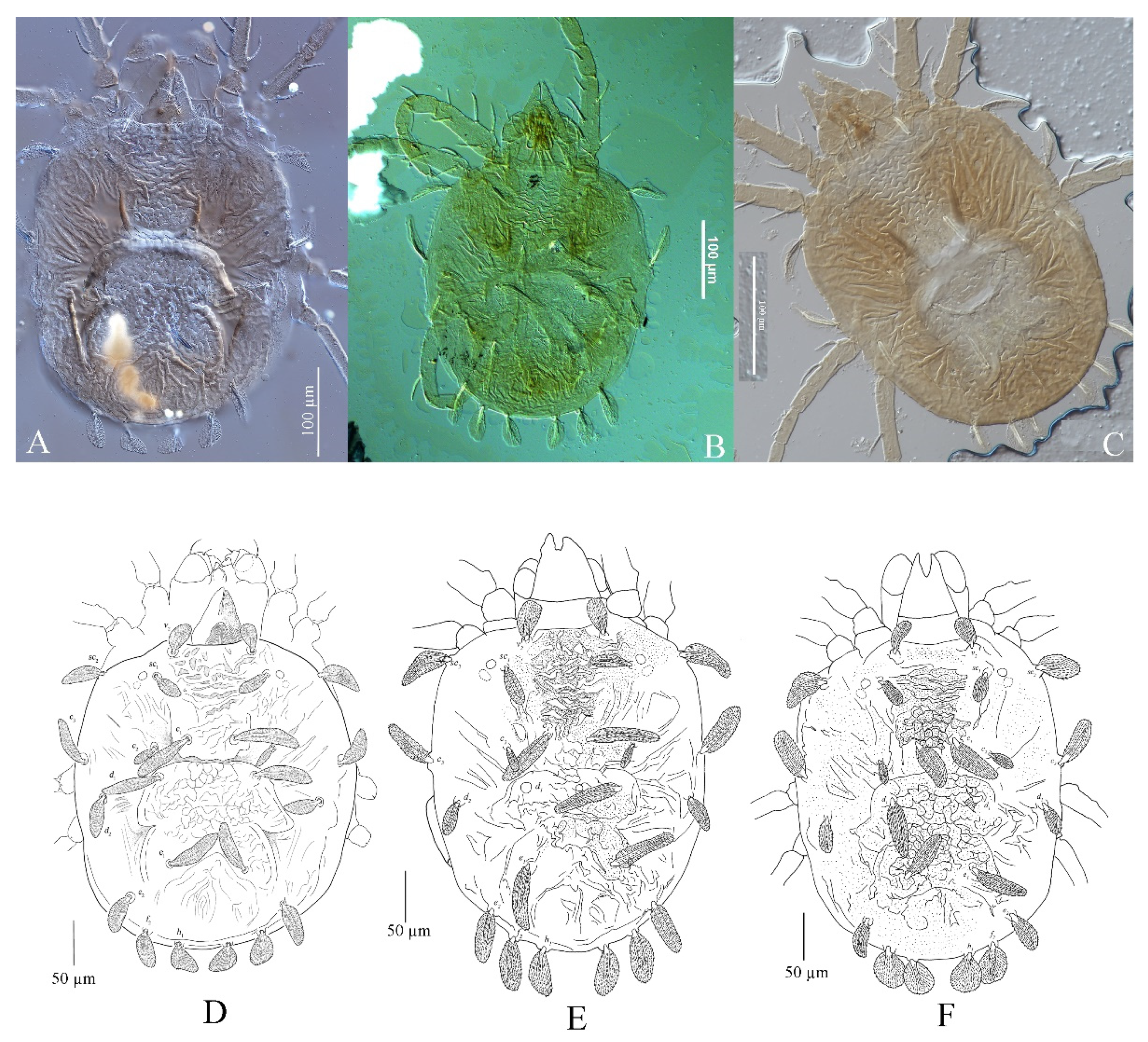
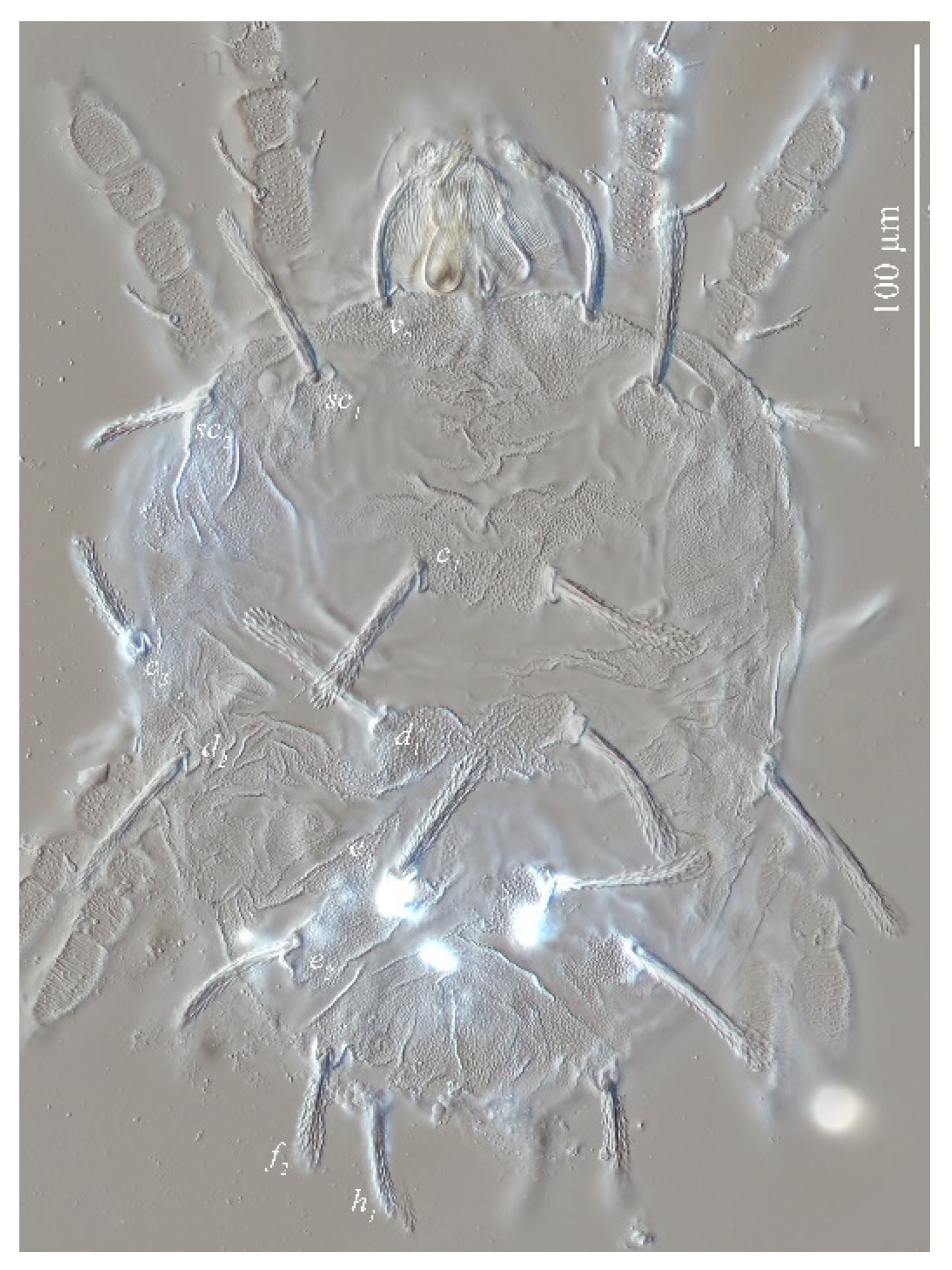
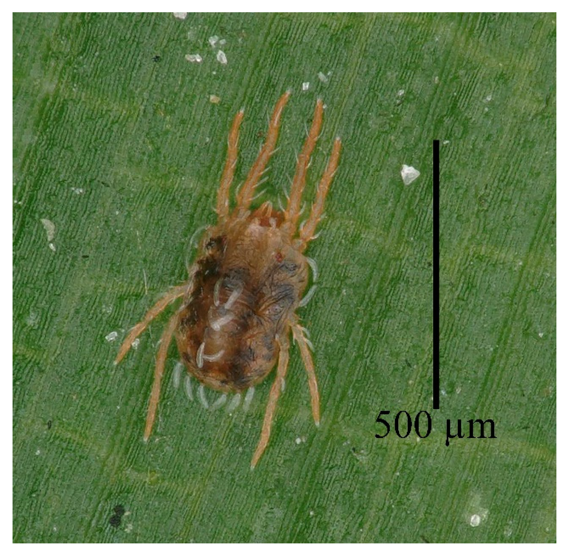
Figure 19 Adult female (n = 23)
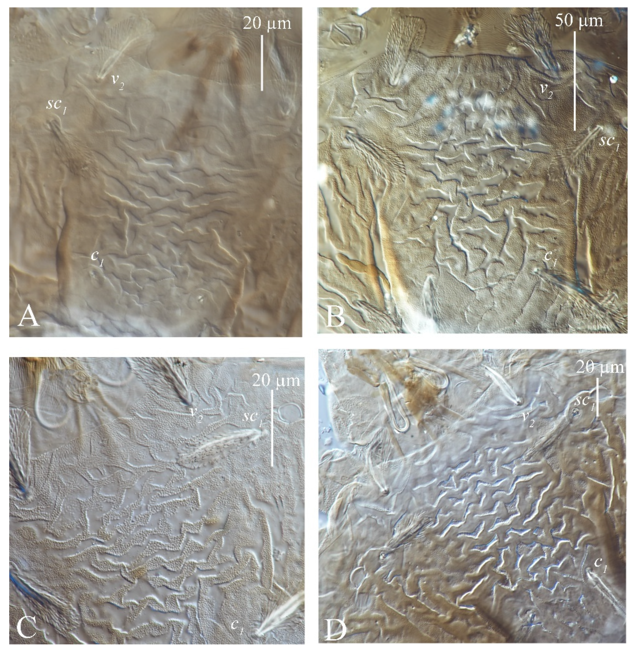
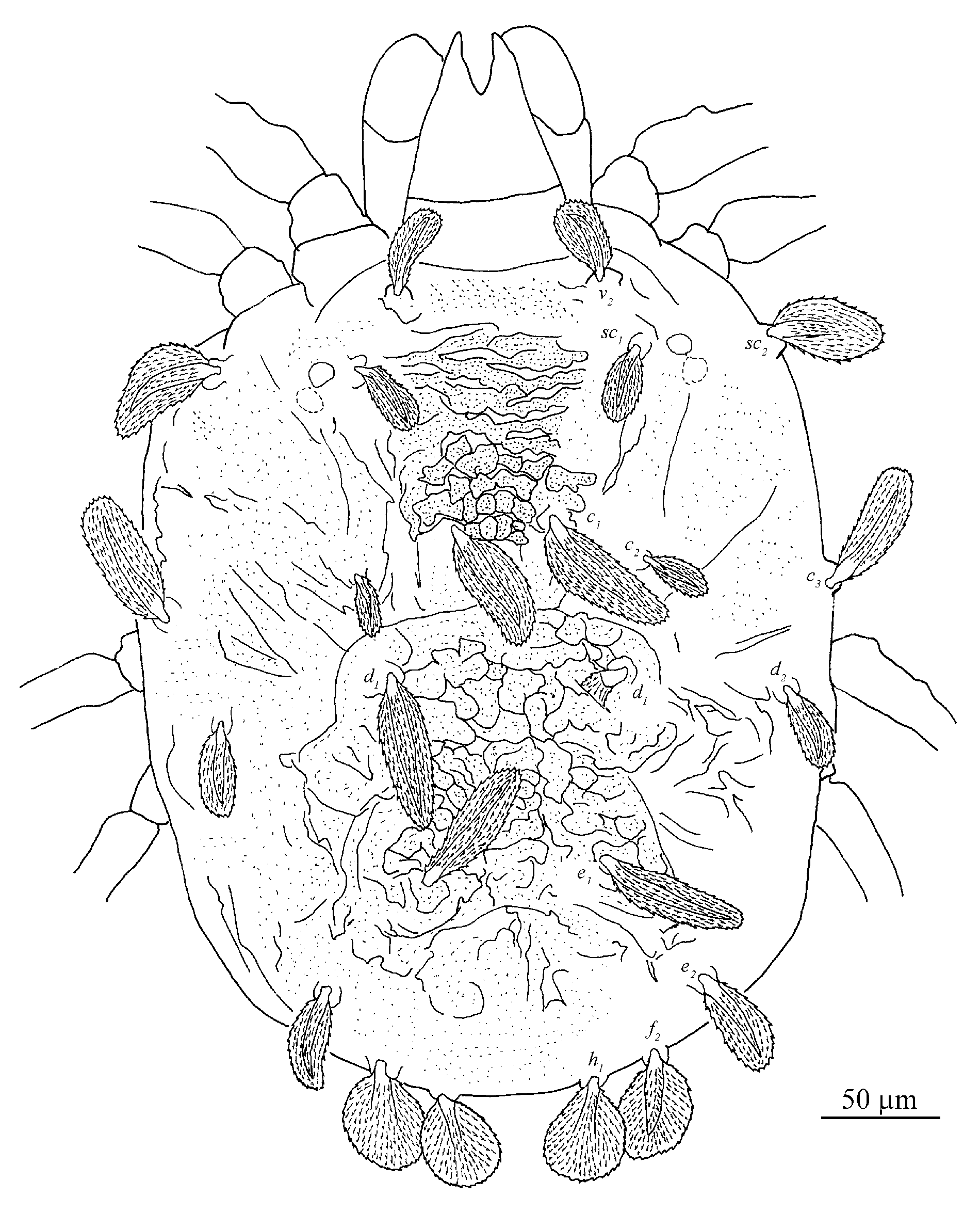
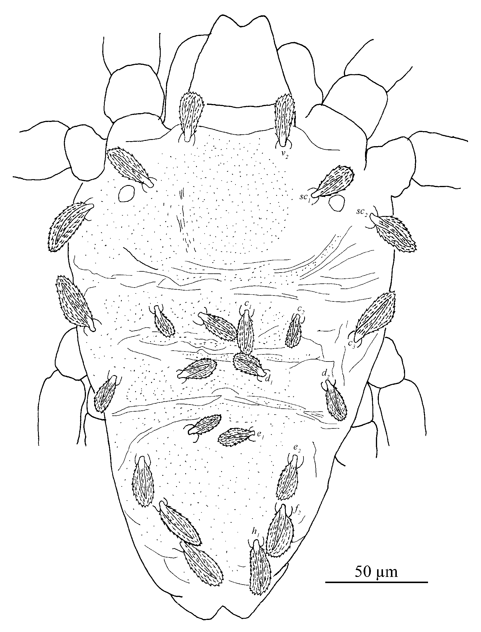
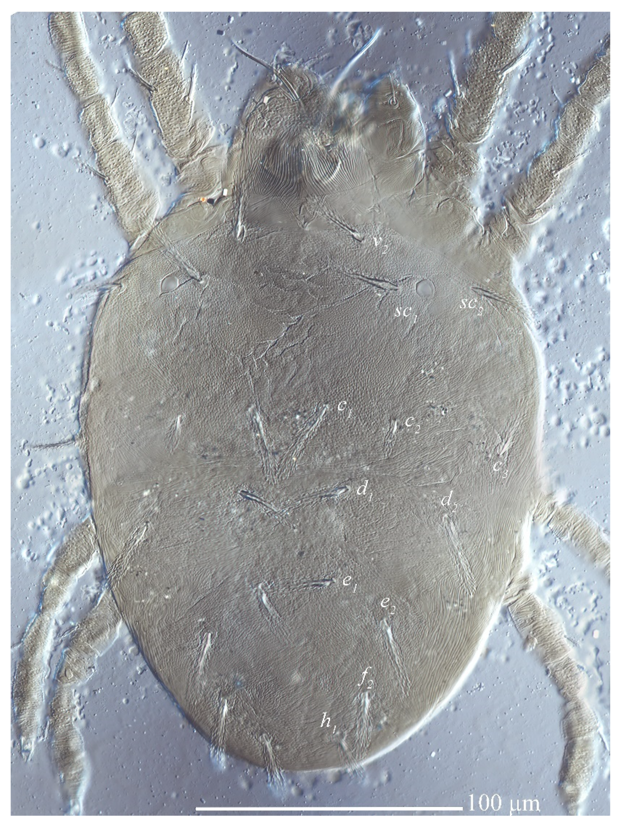
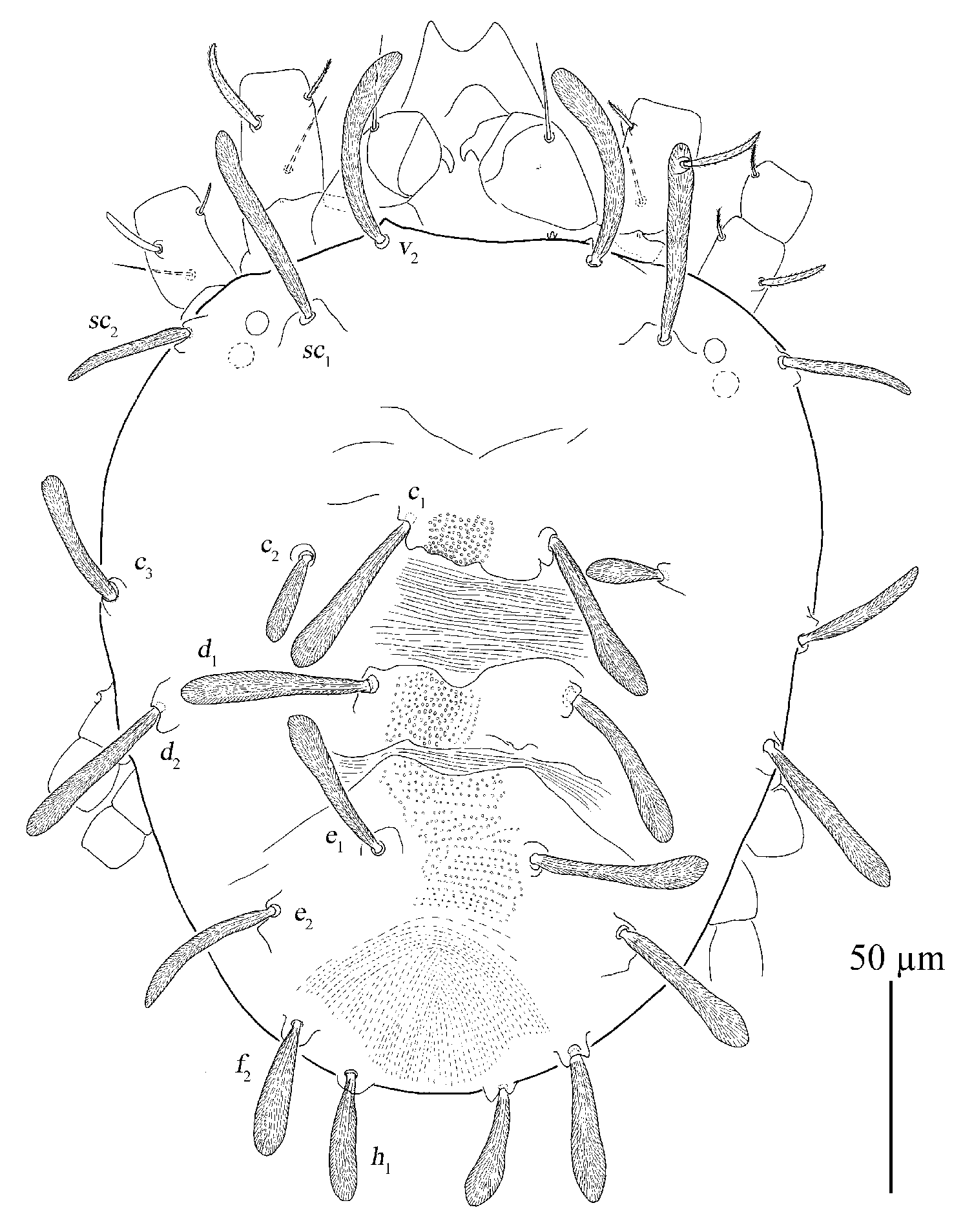
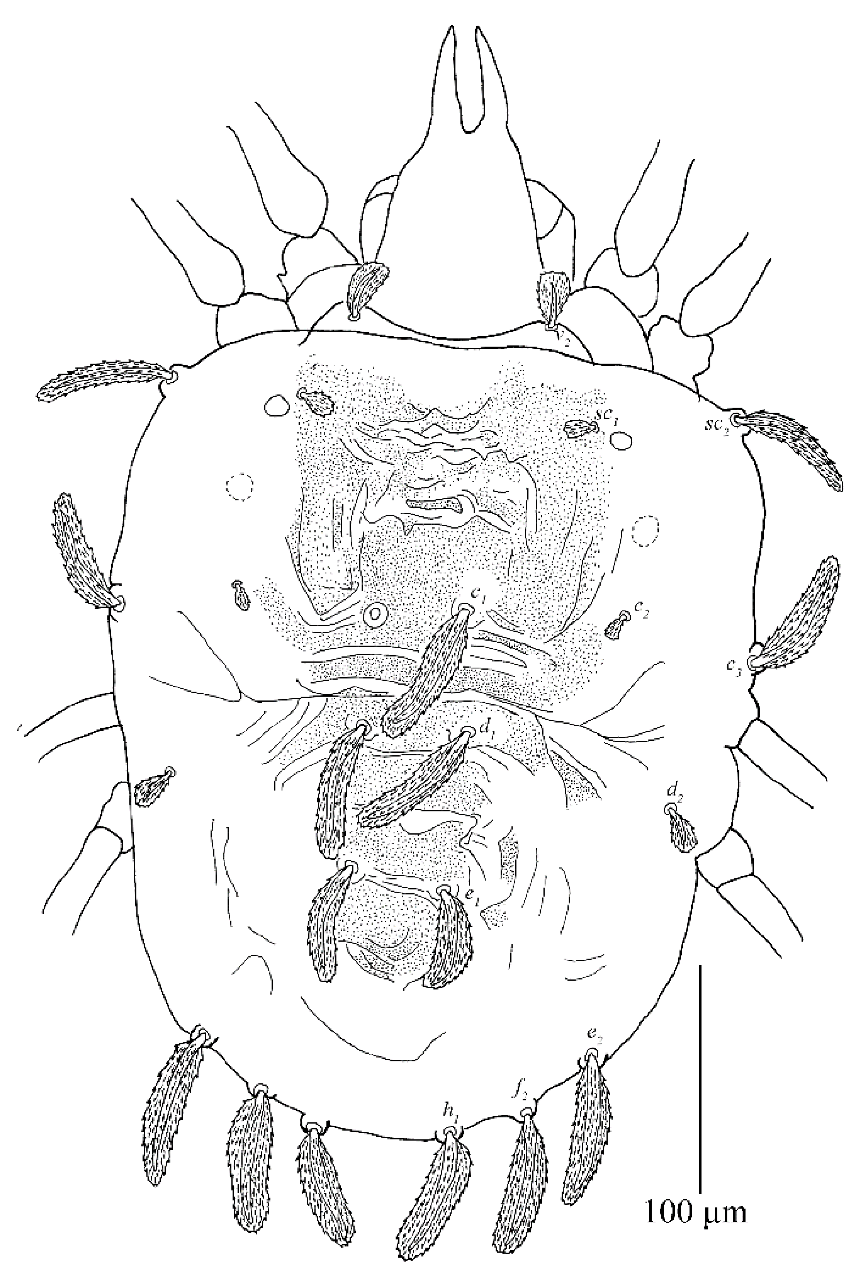
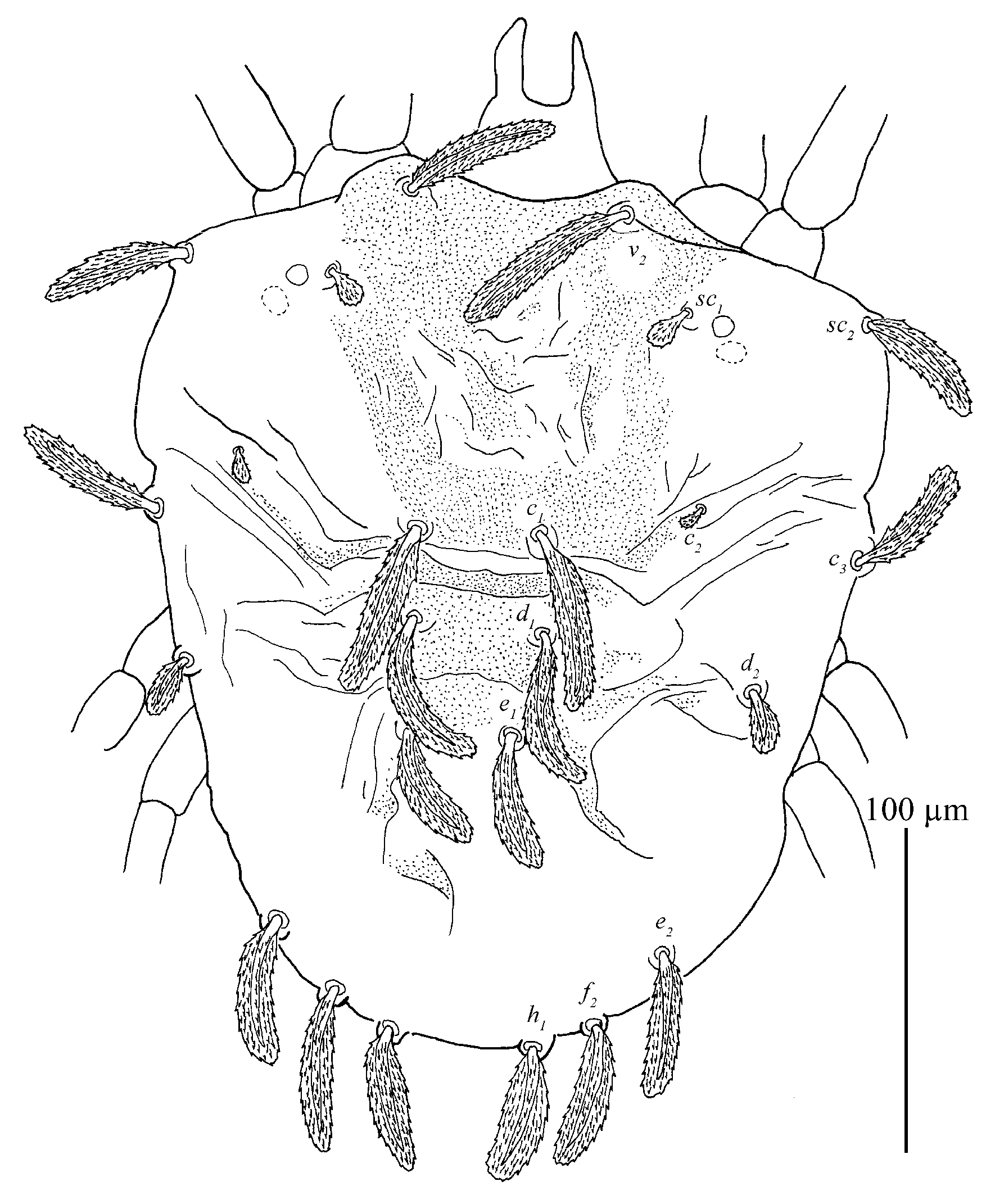
Dorsum (
Figure 7 and
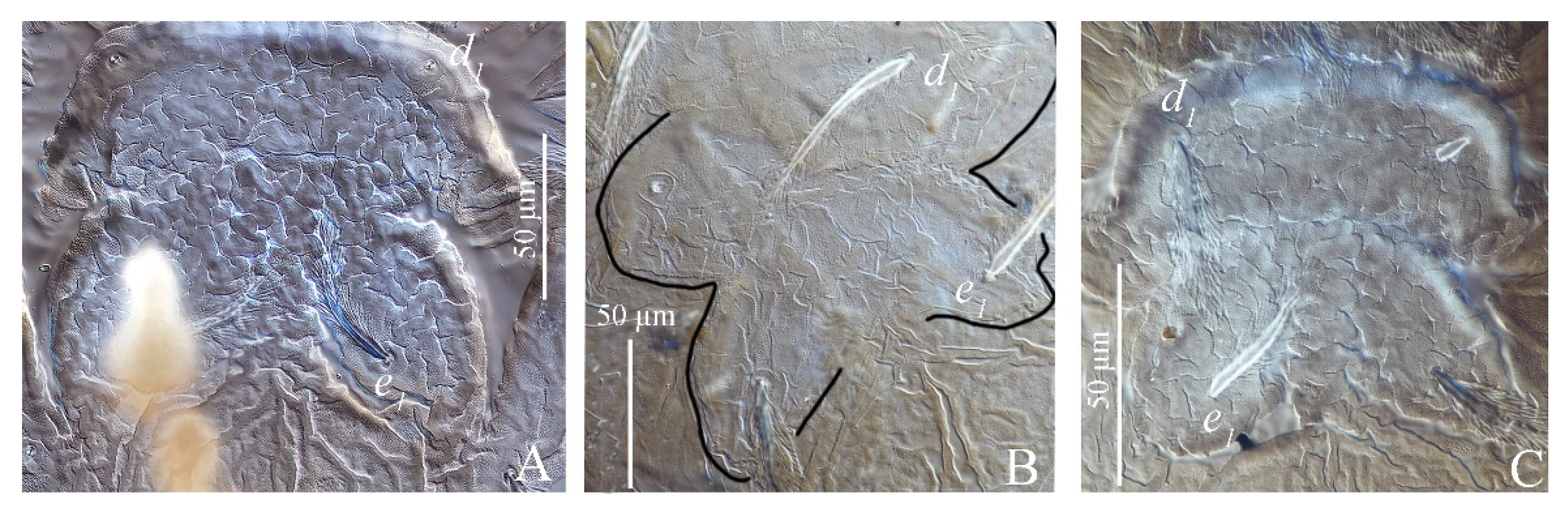
Figure 8). Idiosoma vermilion, 296–338 long, 264–297 wide, nearly oblong, slightly longer than wide and lateral margins approximately parallel. Dorsal setae white. Hysterosoma with two distinct protuberances anterior, one small and bears seta
c1, posterior, one large constricted laterally (between
d1 and
e1), and bears setae
d1 and
e1.
Prodorsum with three pairs of palmate to spatulate setae, covered with short longitudinally aligned spinules, v2 31–39, sc1 30–40 and sc2 44–56. Distances between setal bases: v2–v2 79–87, sc1–sc1 111–127, sc2–sc2 224–239. Prodorsum with a pattern resulting in highly wrinkled ornamentation on those transverse ridges.
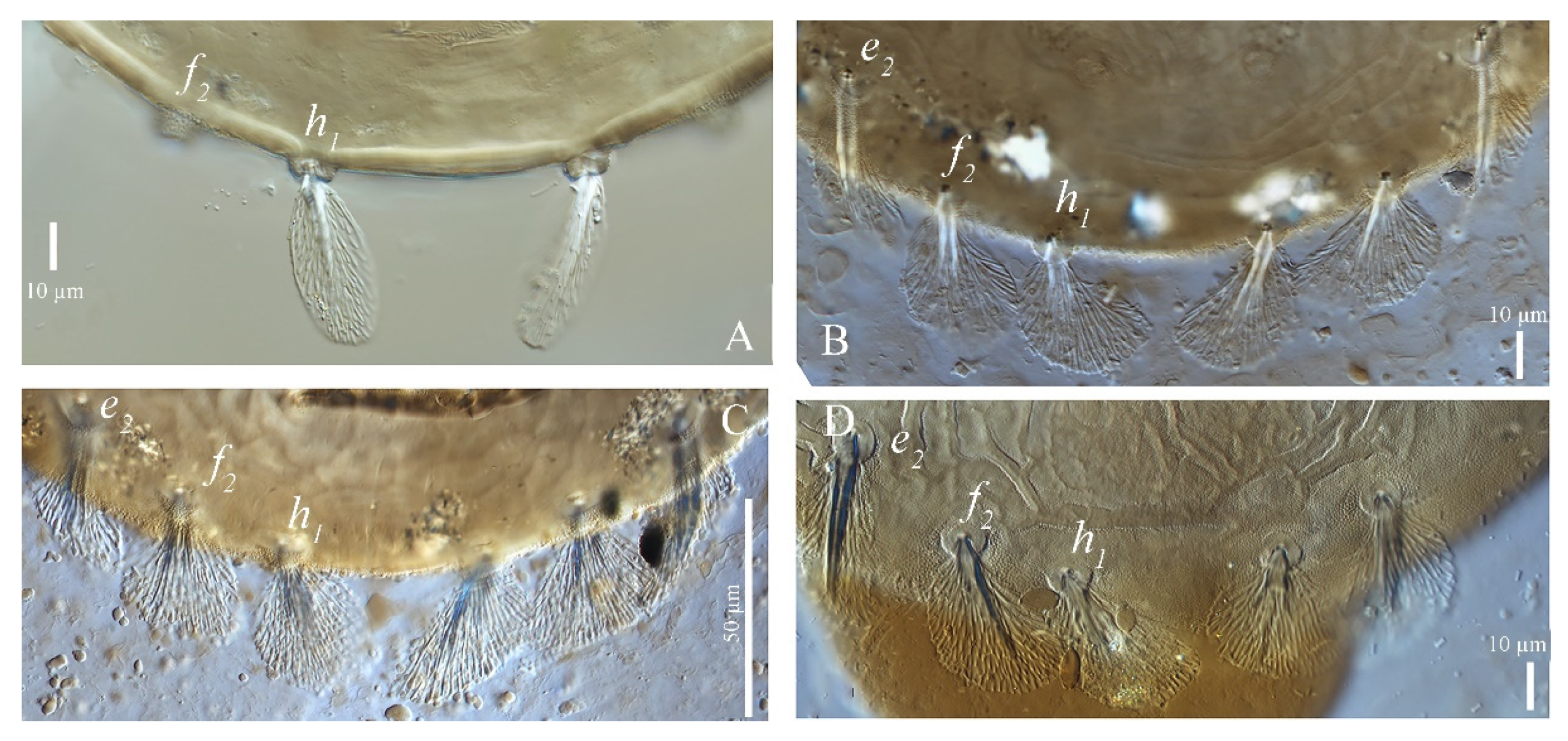
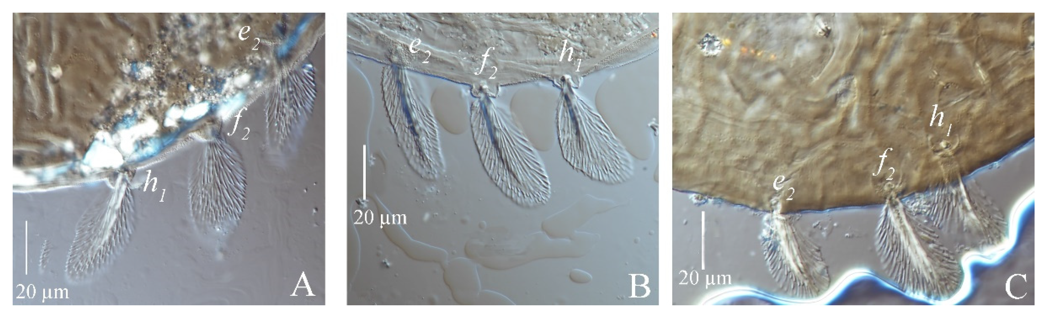
Hysterosoma with 11 pairs of setae (c1–3, d1–2, e1–2, f2, h1–3), f1 absent and f2 marginally positioned. Setae c1, c3, d1 and e1 elongate palmate to long linear, c2 much shorter than other dorsal setae, about as long as one third of seta c1; e2, f2 and h1 palmate or spatulate. Setae h2–3 of differing morphology, similar to other ventral setae and inserted posteroventrally. Seta c1 slightly longer than the distance to setae d1. Seta d1 shorter than the distance to seta e1. Length of setae: c1 58–78, c2 24–29, c3 50–65, d1 60–76, d2 34–51, e1 59–72, e2 43–48, f2 40–47, h1 37–42, h2 18–27, h3 15–25. Distances between setal bases: c1–c1 39–53, c2–c2 120–133, c3–c3 272–280, d1–d1 85–100, d2–d2 233–248, e1–e1 76–90, e2–e2 154–171, f2–f2 104–130, h1–h1 38–64, c1–d1 59–76, d1–e1 79–91, e1–f2 59–86, f2–h1 25–28. Hysterosoma dorsally with a pattern may be resulting in highly wrinkled ornamentation that somewhat round convex on protuberances, oblique wide ridges laterally, and opisthosoma dorsally with longitudinal ridges.
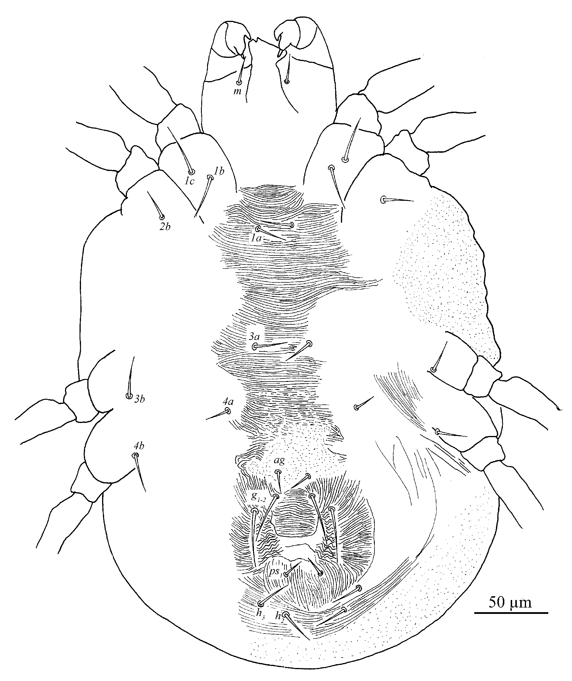
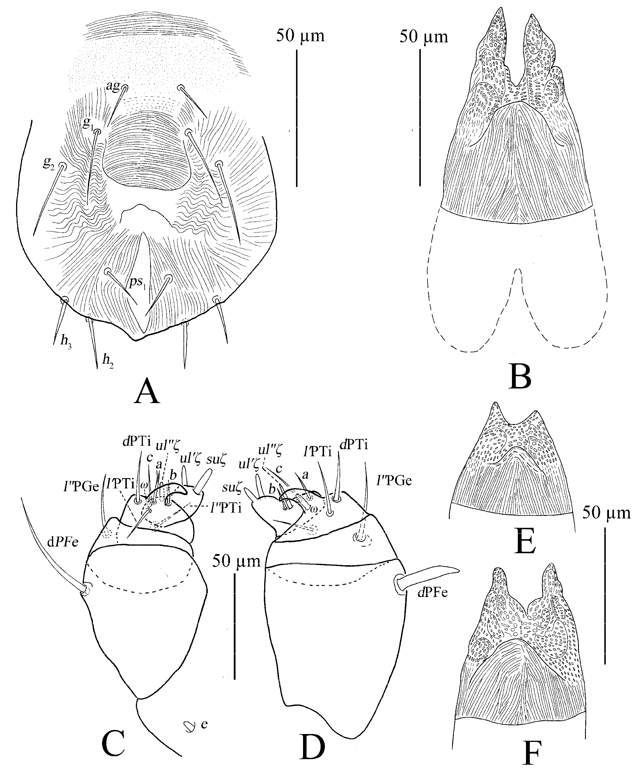
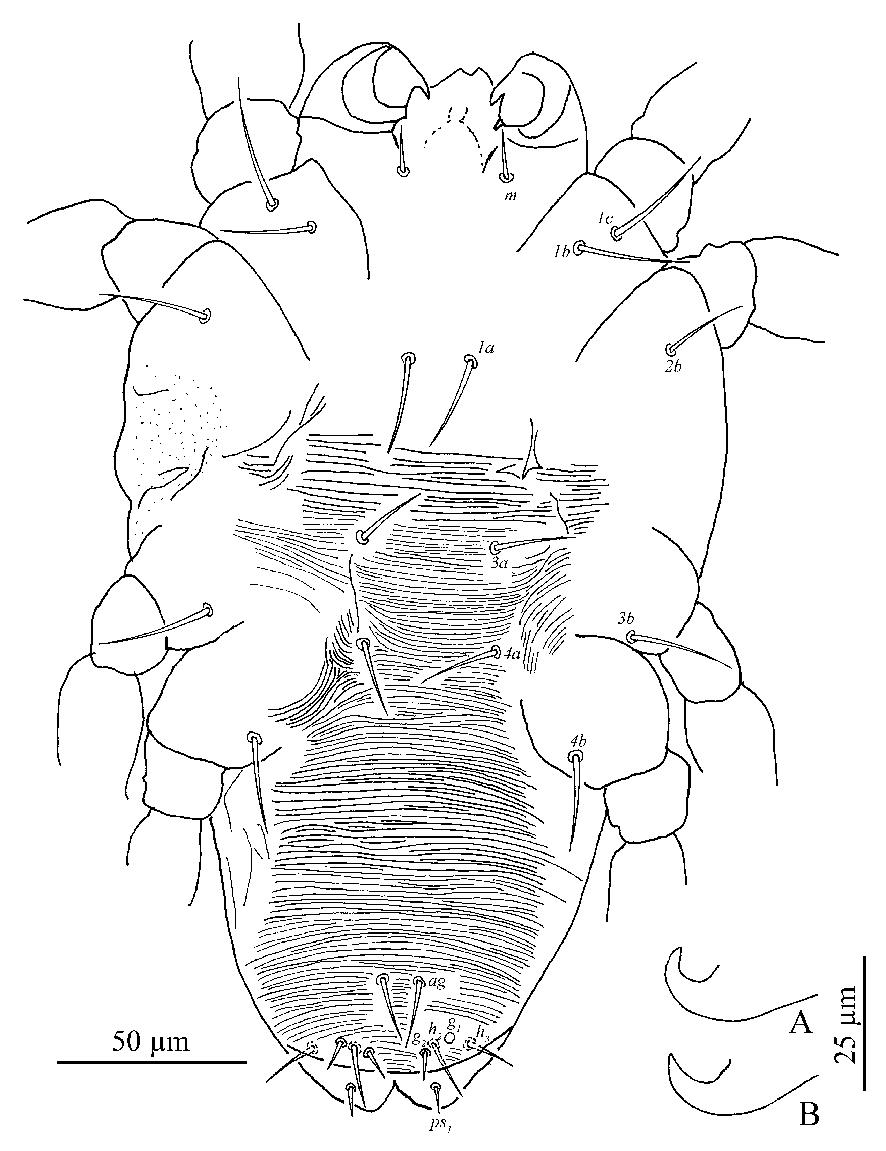
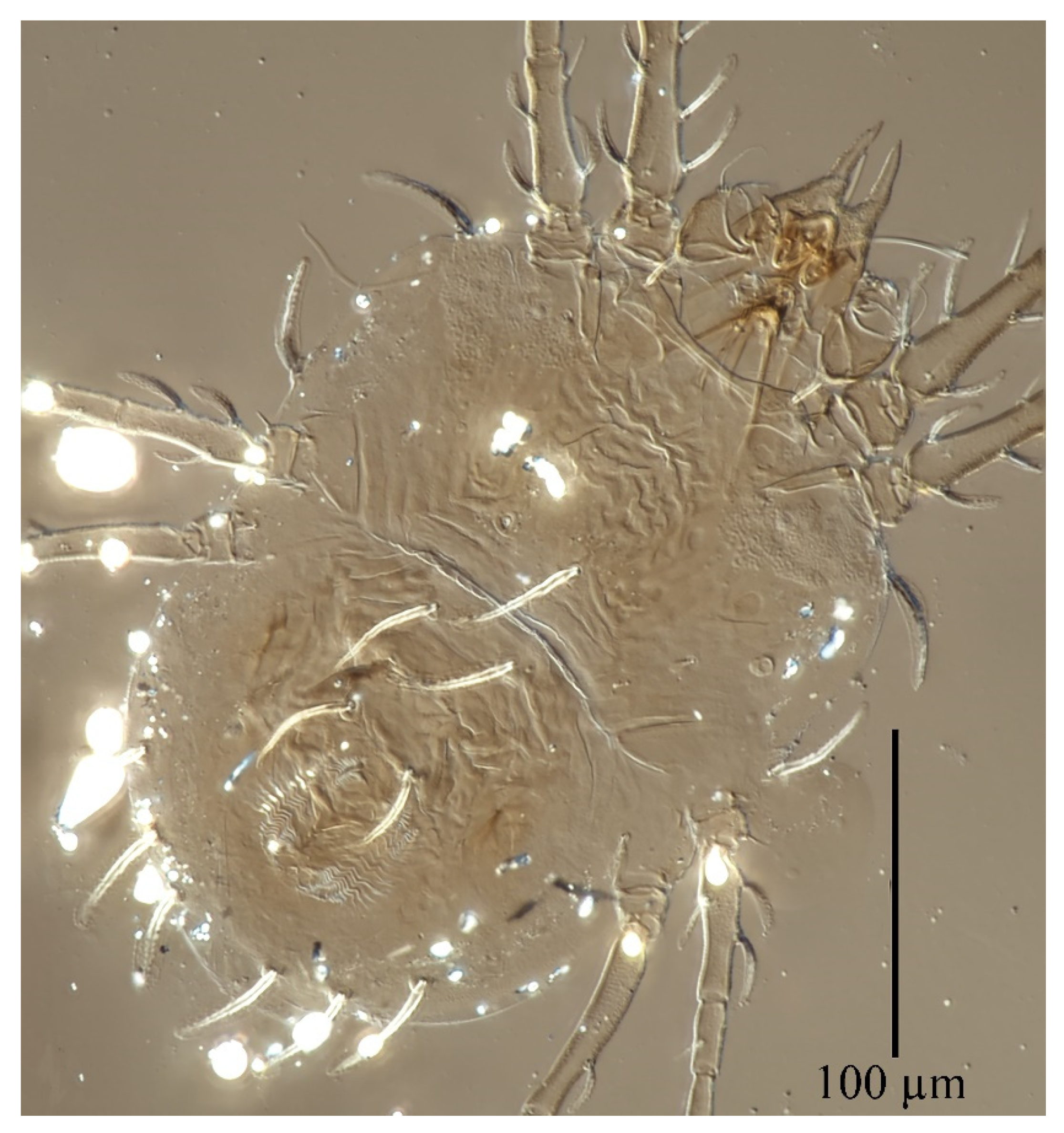
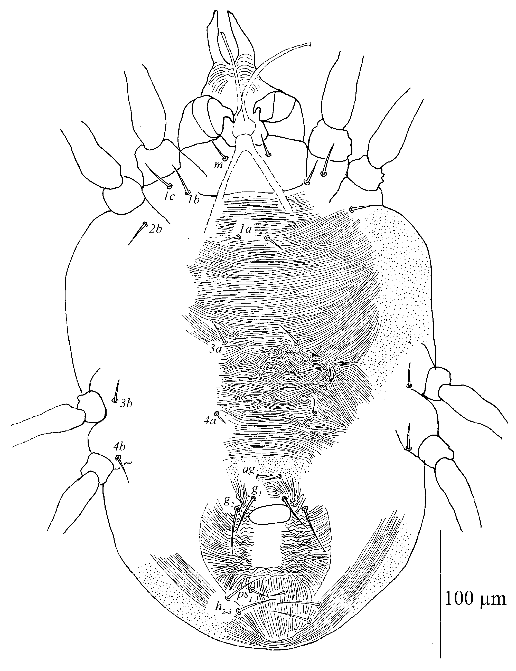
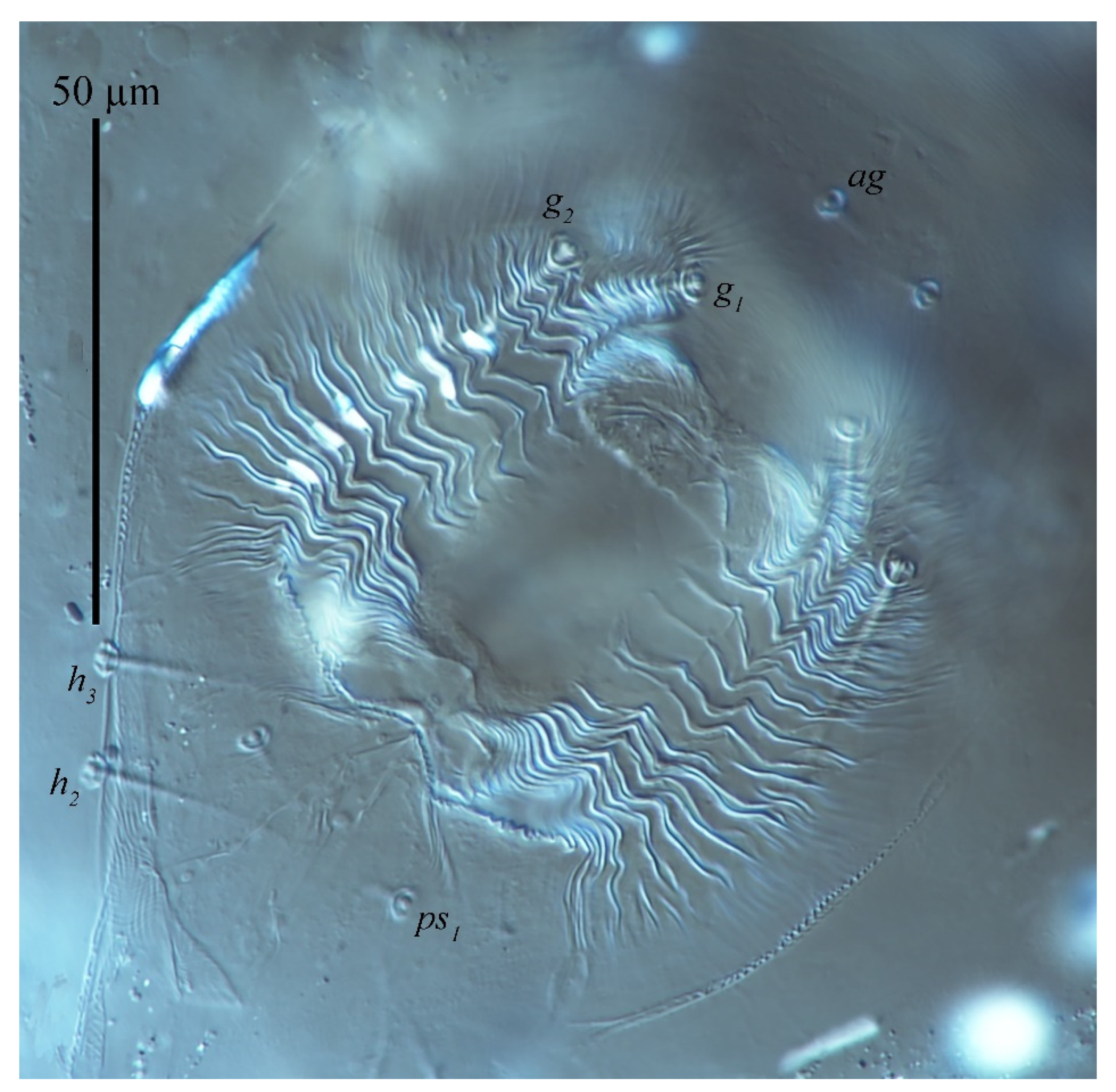
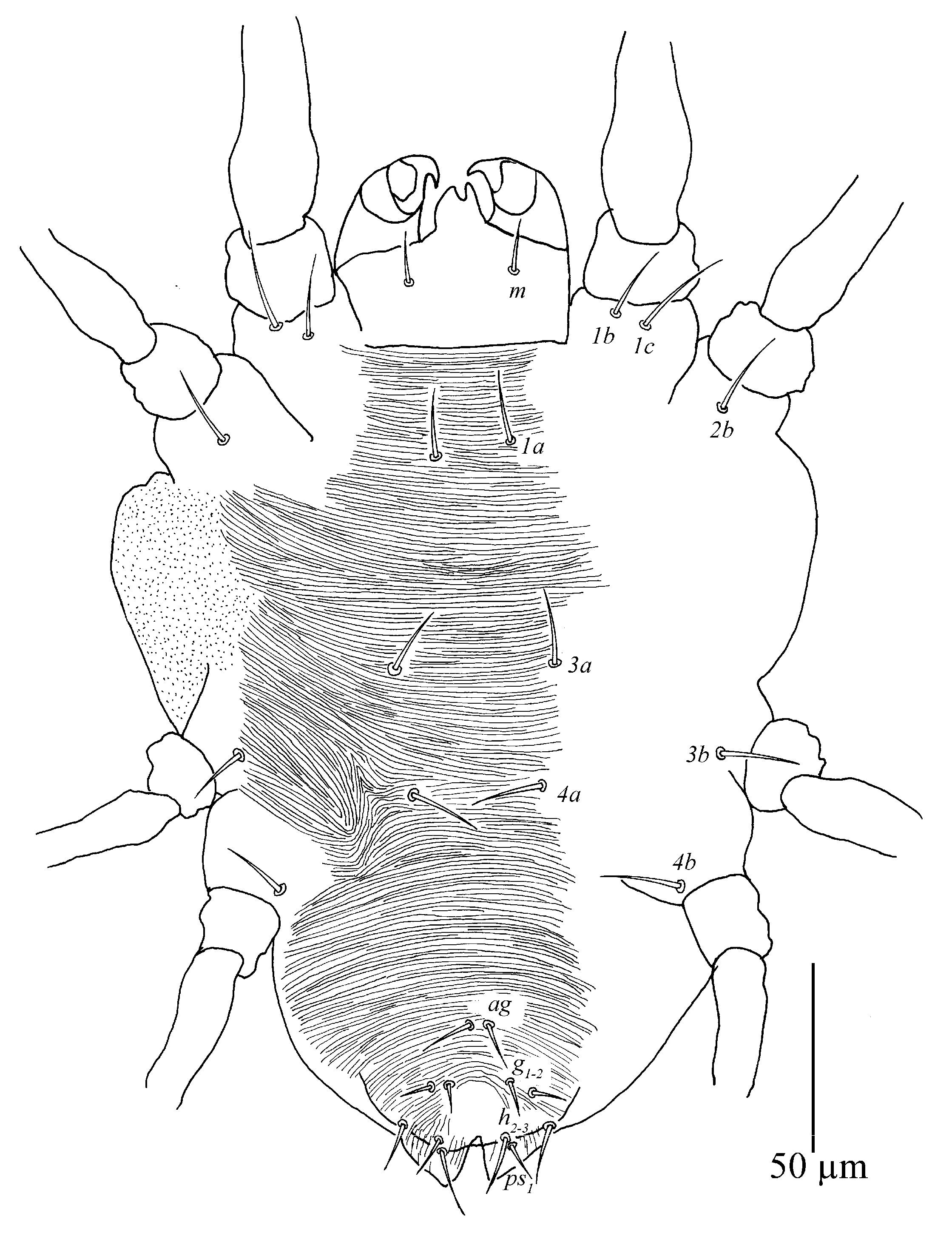
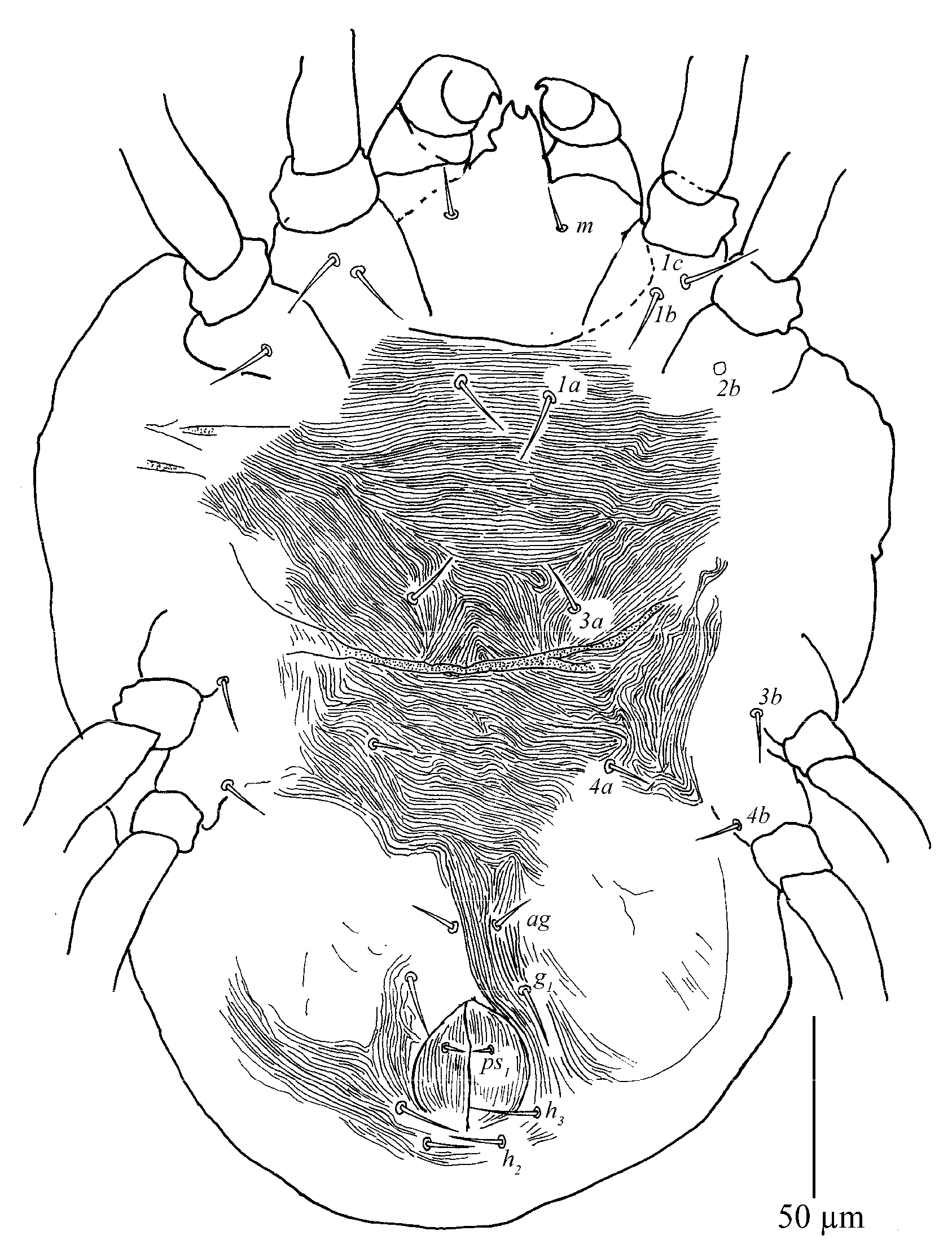
Venter (
Figure 9 and
Figure 10A). Striae mostly transverse, pregenital striae transverse and broken. Genital flap with transverse striae. All ventral setae thin and smooth. Setae
1a as long as distance between their bases; setae
3a and
4a shorter than distance between their bases. Coxal setae count 2-1-1-1, one pair of pseudanal setae (
ps1), two pairs of smooth genital setae (
g1–2). Length of setae:
1a 19–22,
3a 18–23,
4a 14–21,
ag 13–17,
g1 25–36,
g2 25–35,
ps1 11–15. Distances between setal bases:
1a–
1a 19–28,
3a–
3a 37–46,
4a–
4a 80–92,
ag–
ag 20–25,
g1–
g1 25–32,
g2–
g2 48–63,
ps1–
ps1 16–44.
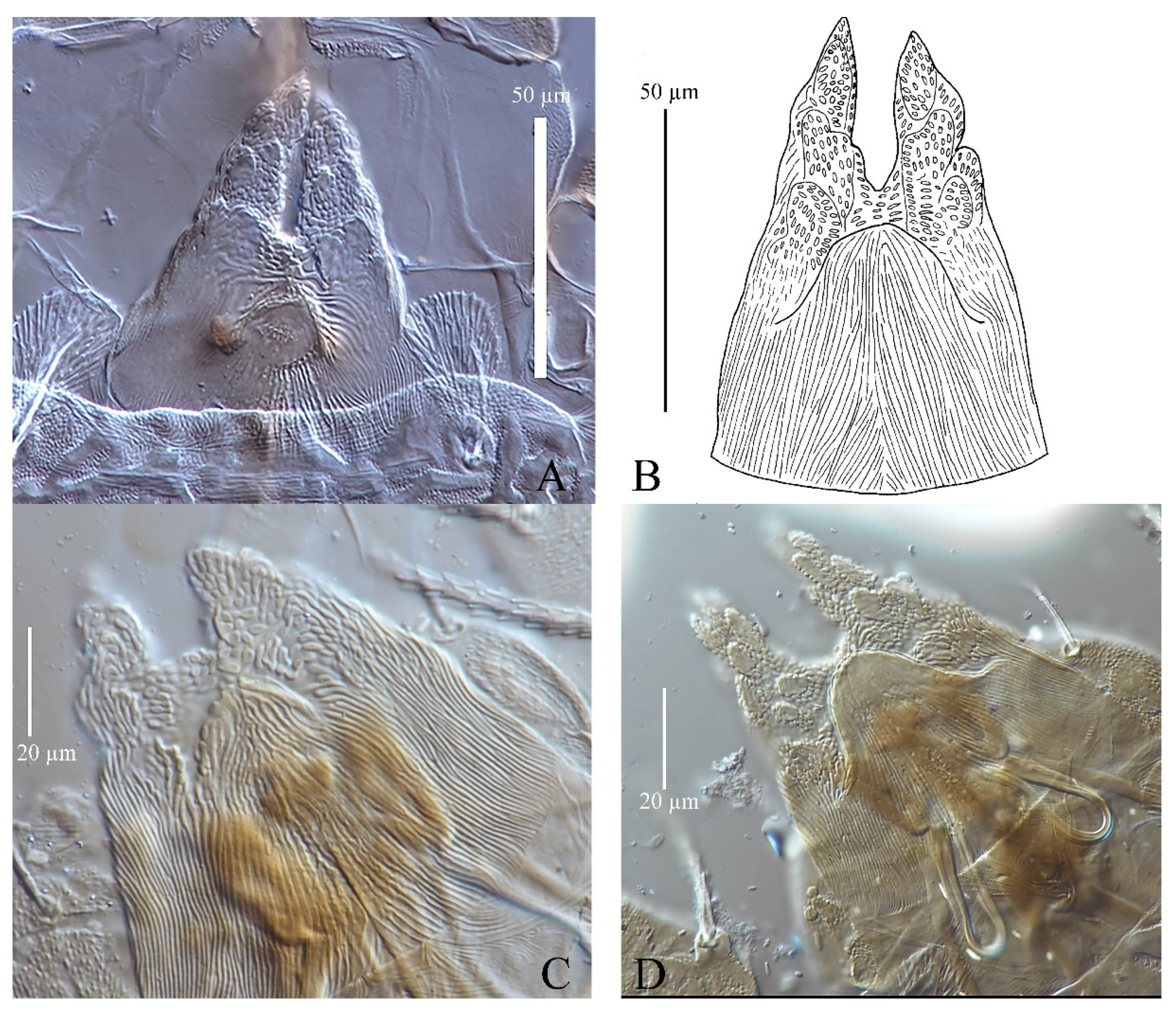
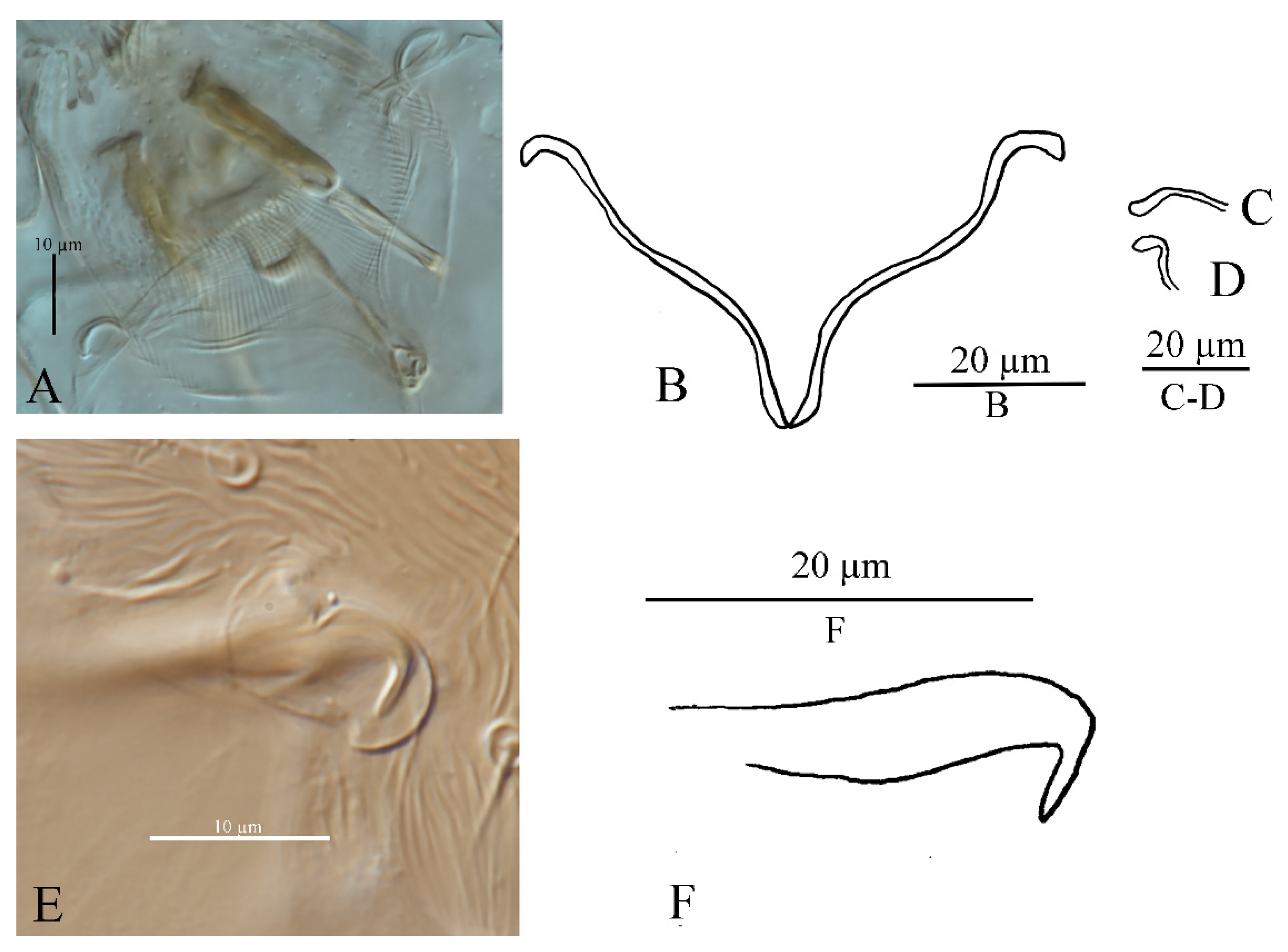
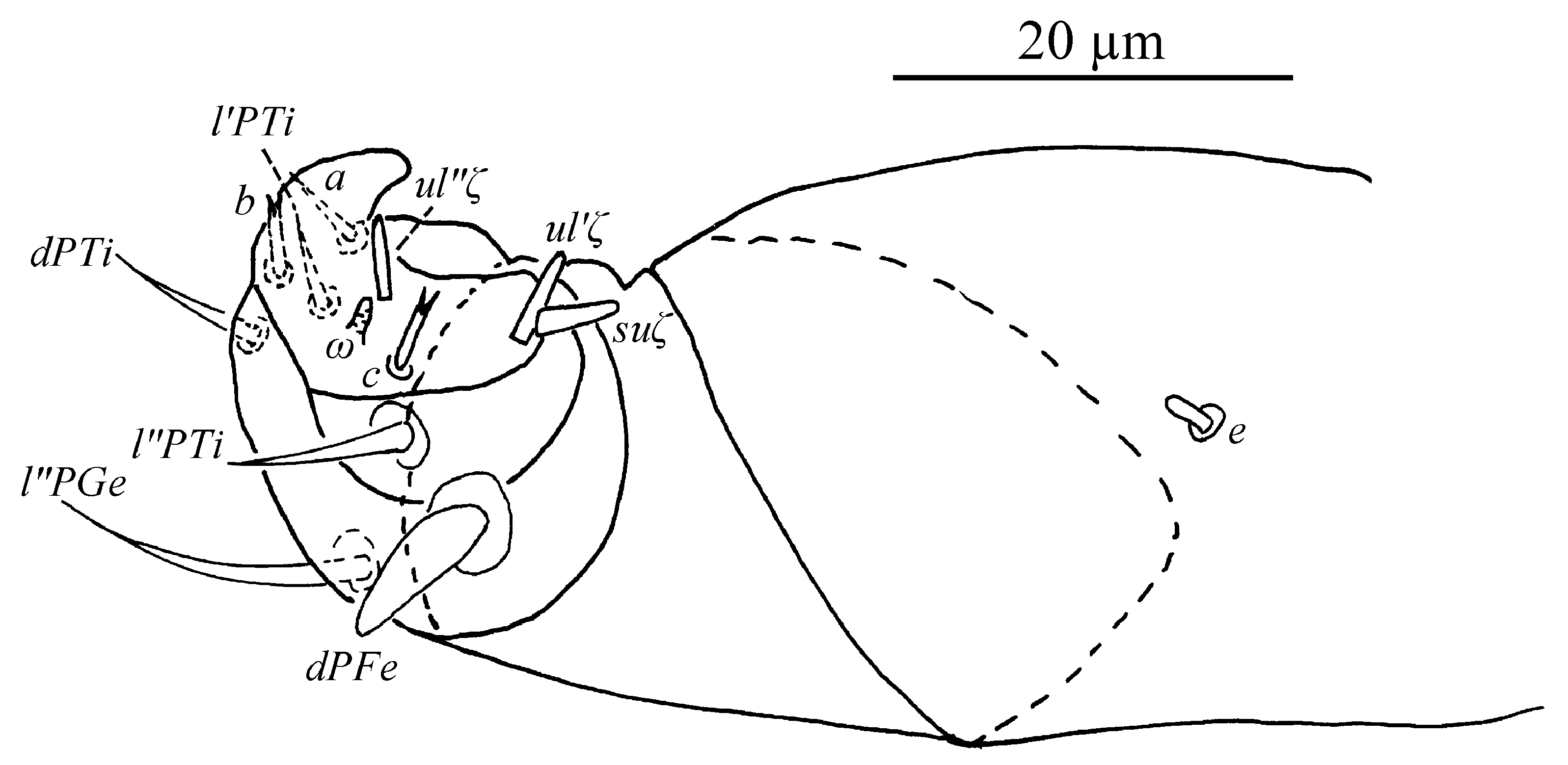
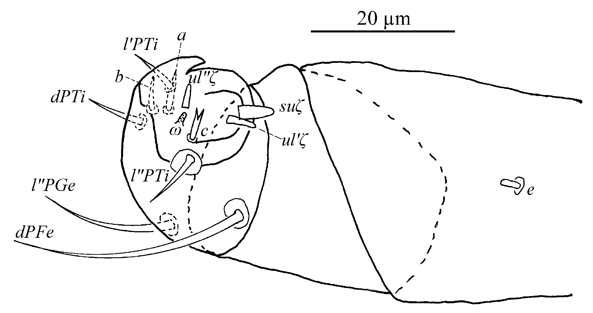
Gnathosoma (
Figure 10B,C). Stylophore with well-developed bilobed horn-like anterior projections. Integument of base of stylophore with longitudinal striae, projections with granulate pattern (
Figure 10B). Ventral infracapitular setae
m smooth, 18–19 in length. Palp setation and notation as shown in
Figure 10C. Dorsal surface of palp base with a pair of inconspicuous supracoxal setae (
e). Palptarsus: terminal eupathidium (
suζ) club-like with blunt tip end, 4.4–5.9 long, two lateral eupathidia,
ul′ζ 4.4–5.8 and
ul″ζ 4.3–5.3 long, one solenidion (
ω) 2.9–4 long; three short, smooth, tactile setae (
a,
b,
c).
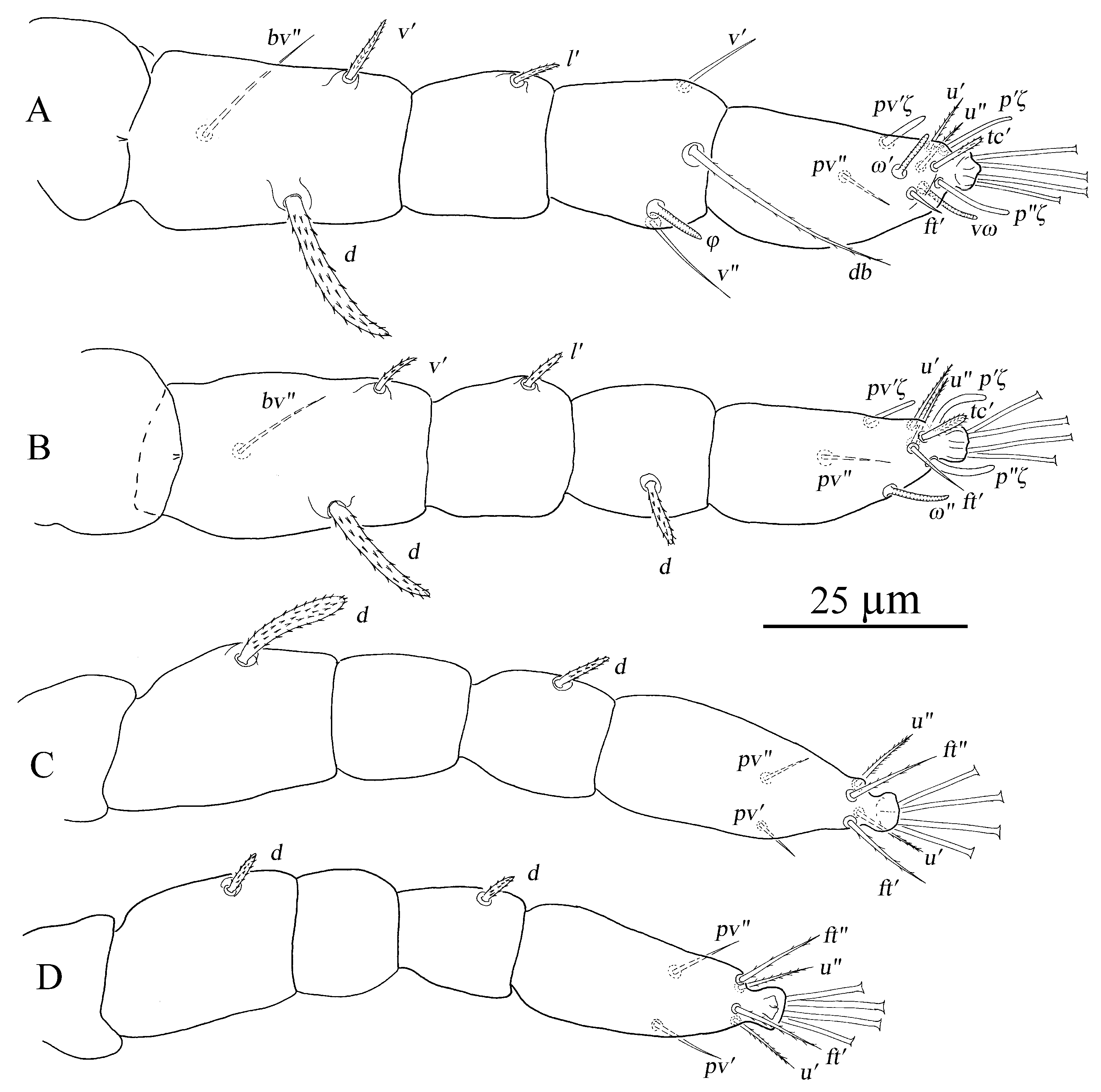
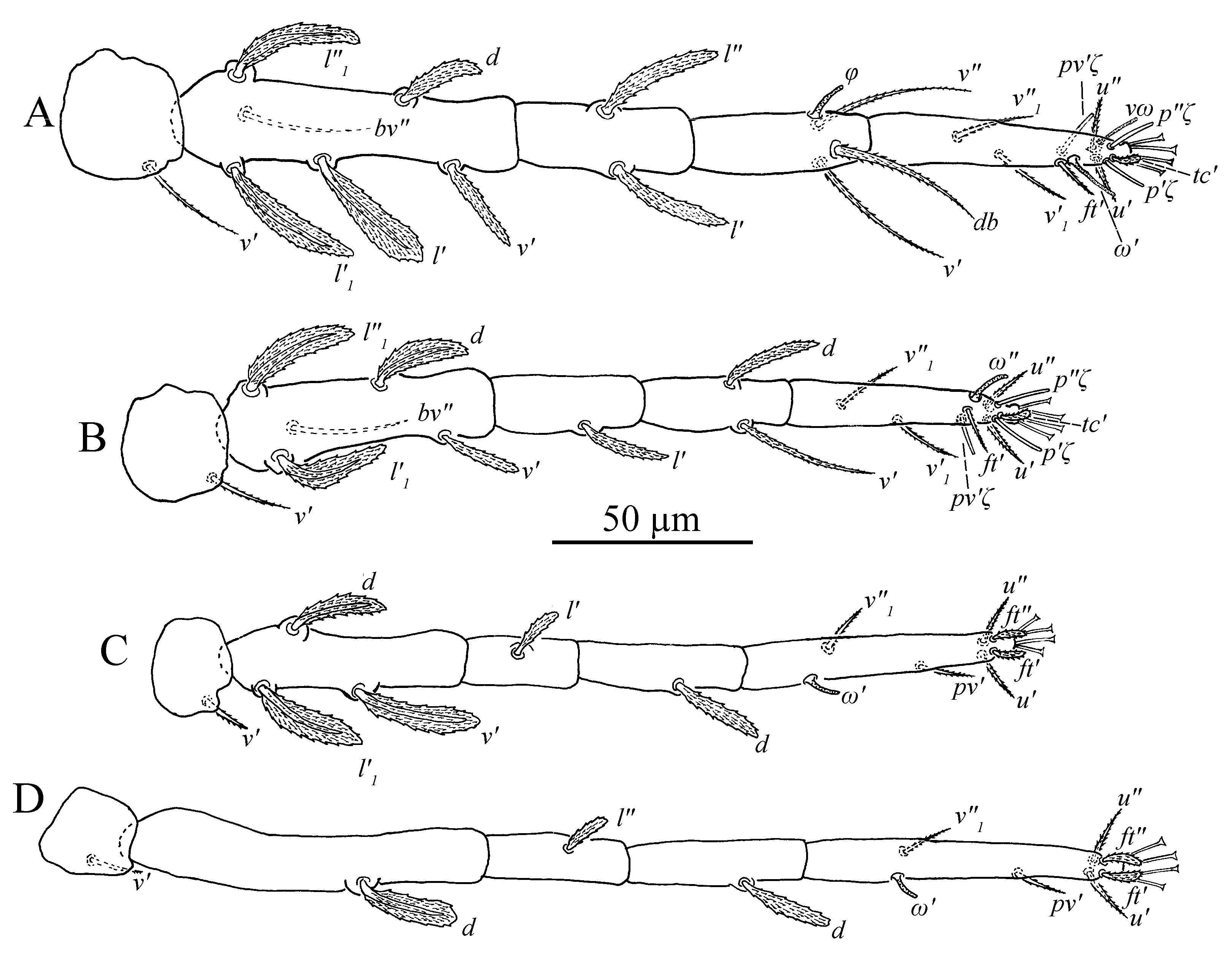
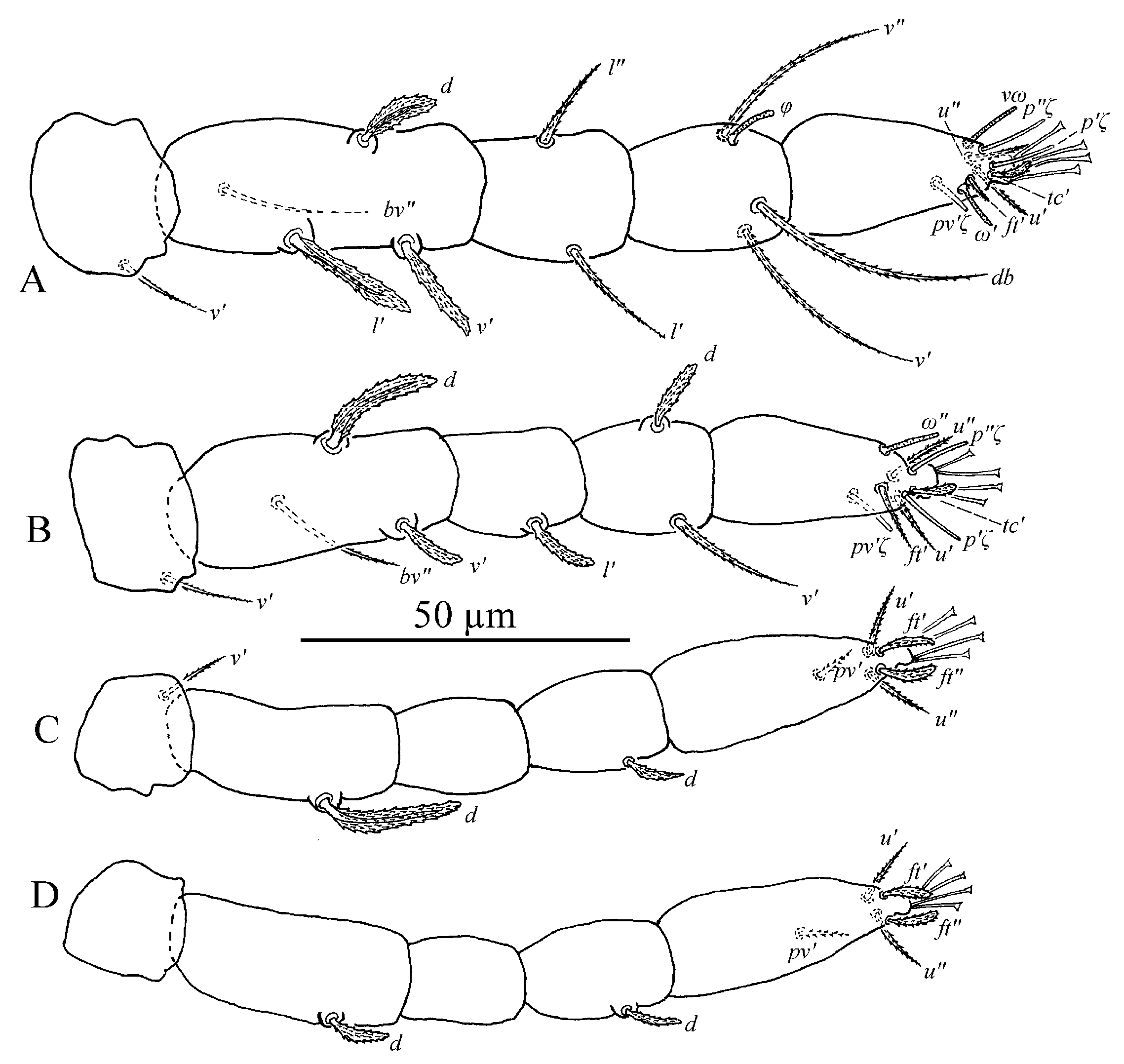
Legs (
Figure 11A–D). Empodial claws absent. One pair of duplex setae on tarsus I, solenidion
ω′ 8–9, one additional ventral solenidion (
vω) at the same transverse level with
u setae, 10–14 long, tectal seta (
tc′) unpaired, thicker than other tactile setae on tarsus I; tibia I with one solenidion
φ 10–12 long; tarsus II without duplex setae, solenidion
ω″ 10–13 long, tectal seta (
tc′) unpaired, thicker than other tactile setae; tarsus III without solenidion, tectal setae paired; tarsus IV with one proximal solenidion
ω′ 6–7 long, tectal setae paired. Number of tactile setae on leg (I–IV) segments: trochanters 1-1-1-1, femora 6-5-3-1, genua 1-1-1-1, tibiae 3-1-1-1, tarsus 7-7-6 (or 7) -6 (or 7). Number of eupathidia on tarsus I–IV: 3-3-0-0. Legs I–IV setation and notation as shown in
Figure 11A–D. Length of leg segments: femur I 99–110, genu I 41–46, tibia I 39–47, tarsus I 67–74; femur II 81–90, genu II 38–45, tibia II 31–38, tarsus II 62–71; femur III 67–78, genu III 33–36, tibia III 37–41, tarsus III 75–78; femur IV 96–102, genu IV 33–44, tibia IV 44–45, tarsus IV 79–85.
Number of tactile setae on Tarsus III–IV varies among specimens, and differs between right and left legs in the same specimen (
Table 2). Among 23 adult females, 12 (one from Thailand, one from India, six from Yunnan Province, China and four from Guangdong Province, China) with six tactile setae (
u′,
u″,
ft′,
ft″,
pv′,
pv″) on both right and left tarsus III; six (two from Thailand, four from Yunnan Province, China) with seven tactile setae (
u′,
u″,
ft′,
ft″,
pv′,
pv″,
v′1) on both right and left tarsus III; three (two from Yunnan Province, China and one from Guangdong Province, China) with seven tactile setae on right tarsus III and six tactile setae on left; one (from Guangdong Province China)with six tactile setae (
u′,
u″,
ft′,
ft″,
pv′,
pv″) and one solenidion (
ω′) on right tarsus III and six tactile setae on left; one (from Yunnan Province, China) with seven tactile setae on left tarsus III and unknown the right side due to the broken tarsus III. Among 23 adult females, 15 (one from Thailand, one from India, eight from Yunnan Province, China and five from Guangdong Province, China) with six tactile setae (
u′,
u″,
ft′,
ft″,
pv′,
pv″) and one solenidion (
ω′) on both right and left tarsus IV; four (two from Thailand, two from Yunnan Province, China) with seven tactile setae (
u′,
u″,
ft′,
ft″,
pv′,
pv″,
v′1) and one solenidion (
ω′) on both right and left tarsus IV; two (from Yunnan Province, China) with seven tactile setae and one solenidion on left tarsus IV and six tactile setae and one solenidion on right; one (from Guangdong Province, China) with seven tactile setae on left tarsus IV and six tactile setae and one solenidion on right; one (from Yunnan Province, China) with six tactile setae and one solenidion on left tarsus IV and unknown the right side due to the broken tarsus IV. The variations in the setal count of tarsus III and IV are here considered intraspecific in nature, and attributed to the geographical position of the samples and different host plant species. In order to express the ontogenetic development of leg chaetotaxy conveniently, tarsus III with six tactile setae and tarsus IV with six tactile setae and one solenidion are regarded as normal setal count.
Setal counts (solenidion in parentheses following tactile setae) on legs I–IV are: femora 6-5-3-1, genua 1-1-1-1, tibiae 3(1)-1-1-1, tarsus 7(2)-7(1)-6(0)-6(1). There is a significant amount of setal suppression on the legs in this species, with a total of 15 setae being added to the legs in the adult female stage of this species: pair
l1 on femur I, pair
v1 on tarsus I, pair
l1 on femur II, pair
v1 on tarsus II,
v′ on trochanter III,
v′ and
l′1 on femur III,
l′ on genua III,
v′ on trochanter IV,
l″ on genua IV and
ω′ on tarsus IV. According to the normal ontogenetic setal additions for the family [
10], seven of thirteen additional setae are delayed additions:
v′1 on tarsus I suppressed on protonymph stage,
v″1 on tarusus I,
v′1 on tarsus II,
v′ on trochanter III and IV,
l on genua III and IV are suppressed on deutonymph stage.
Male (n = 9)
Dorsum (
Figure 12). Idiosoma gradual narrowing caudally, 184–209 long, 123–147 wide, with length much longer than width. Dorsum without a protuberance.
Prodorsum with three pairs of palmate setae, covered with short longitudinally aligned spinules, v2 19–21, sc1 32–37 and sc2 19–21. Distances between setal bases: v2–v2 50–54, sc1–sc1 74–76, sc2–sc2 133–145. Integument with irregular fine granulate.
Hysterosoma with 11 pairs of setae (c1–3, d1–2, e1–2, f2, h1–3), similar in shape to prodorsal setae, except with setae h2–3 of differing morphology, similar to other ventral setae and inserted posterodorsally. Seta c2 slightly shorter than other dorsal setae. Dorsal central setae (c1, d1, e1) much shorter than the distance to setae in the next setal row. Length of setae: c1 17–19, c2 13–15, c3 24–26, d1 16–18, d2 19–20, e1 13–16, e2 20–22, f2 21–26, h1 17–23, h2 13–23, h3 13–16. Distances between setal bases: c1–c1 21–25, d1–d1 37–40, e1–e1 25–25, f2–f2 57–59, h1–h1 28–31. Hysterosoma dorsally with irregular fine granulate, except for band of transverse striae between paired c1 and d1.
Venter (
Figure 13). Striae mostly transverse. All ventral setae thin and smooth. Coxal setae count 2-1-1-1. Length of setae:
1a 20–21,
3a 16–21,
4a 13–15,
ag 14–17,
g1 6–8,
g2 5–9,
ps1 5–6. Distances between setal bases:
1a–
1a 22–24,
3a–
3a 31–38,
4a–
4a 34–38,
ag–
ag 7–8.
Gnathosoma (
Figure 10B–D). Stylophore with short bilobed horn-like anterior projections as shown in
Figure 10B. Ornamentation of integument similar to that of female. Ventral infracapitular setae
m smooth, 11–17 in length. Palp setation and notation as shown in
Figure 10D. Palptarsus: terminal eupathidium (
suζ) club-like with sharp tip end, 2.8–3 long, two lateral eupathidia,
ul′ζ 5–6 and
ul″ζ 5–7 long, one solenidion (
ω) 3–5 long; three short, smooth, tactile setae (
a,
b,
c).
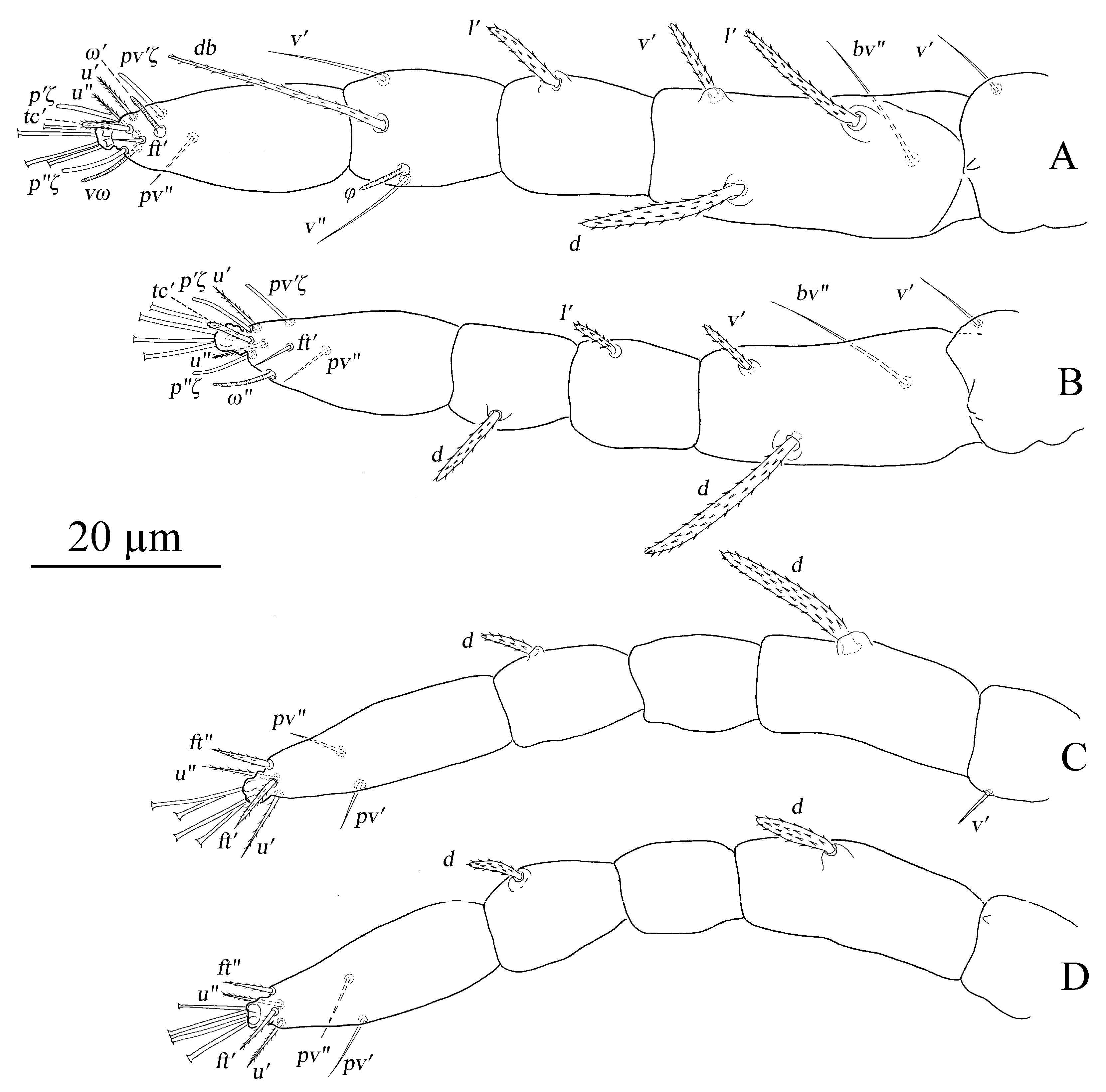
Legs (
Figure 14A–D). Empodial claws absent. One pair of duplex setae on tarsus I, solenidion
ω′ 12–14,
ω″1 8–11 long, one additional ventral solenidion (
vω) at the same level with
u setae, 13–16 long, tectal setae (
tc′) unpaired, thicker than other tactile setae on tarsus I; tibia I with four solenidion,
φ 12–13,
φ′ 8–9,
φ″ 6–8,
φ″1 8–10 long; tarsus II without duplex setae, solenidion
ω″ 11–14 long, tectal setae (
tc′) unpaired, thicker than other tactile setae; tibia II with two solenidion,
φ′ 8–9,
φ″ 8–9 long, tarsus III with one solenidion
ω′ 7–9,
tc paired; tarsus IV with one proximal solenidion
ω′ 7–9 long,
tc paired. Number of tactile setae on leg (I–IV) segments: trochanters 1-1-1-1, femora 6-5-3-1, genua 2-2-1-1, tibiae 3-1-1-1, tarsus 7-7-7-7. Number of eupathidia on tarsus I–V: 3-3-0-0. Legs I–IV setation and notation as shown in
Figure 14A–D. Length of leg segments: femur I 78–86, genu I 40–42, tibia I 46–47, tarsus I 67–68; femur II 74–76, genu II 39–42, tibia II 39–39, tarsus II 64–64; femur III 78–79, genu III 33–37, tibia III 32–40, tarsus III 69–73; femur IV 81–85, genu IV 35–38, tibia IV 44–44, tarsus IV 75–80.
The number of tactile setae on tarsus III and tarsus IV varies among specimens, and differs between right and left legs in the same specimen. Among nine adult males, seven (three from Thailand, four from Yunnan Province, China) with seven tactile setae (u′, u″, ft′, ft″, pv′, pv″, v′1) and one solenidion (ω′) on both right and left tarsus III; one (from Yunnan Province, China) with eight tactile setae (u′, u″, ft′, ft″, pv′, pv″, v′1, v″1) and one solenidion (ω′) on both right and left tarsus III; one (from Yunnan Province, China) with eight tactile setae and one solenidion on left and seven tactile and one solenidion on right. Among nine adult males, four (from Yunnan Province, China) with six tactile setae (u′, u″, ft′, ft″, pv′, pv″) and one solenidion (ω′) on both right and left tarsus IV; three (two from Thailand, one from Yunnan Province, China) with 7 tactile setae (u′, u″, ft′, ft″, pv′, pv″, v′1) and one solenidion (ω′) on both right and left tarsus IV; and two (one from Thailand, one from Yunnan Province, China) with eight tactile (u′, u″, ft′, ft″, pv′, pv″, v′1, v″1) and one solenidion (ω′) on right Tarsus IV and seven tactile setae and one solenidion on left. The variations in the setal count of tarsus III and IV are here considered intraspecific in nature, and attributed to the geographical position of the samples and different host plant species. In order to express the ontogenetic development of leg chaetotaxy conveniently, tarsus III–IV with seven tactile setae and one solenidion are regarded as normal setal counts.
The male has a slightly different chaetotactic formula to the female. Setal counts on legs I–IV: femur 6-5-3-1, genua 2-2-1-1, tibia 3(4)-1(2)-1-1, tarsus 7(3)-7(1)-7(1)-7(1). The male adds four more tactile setae to legs than the dose for the female, accurately, l’ on genua III and IV, v″1 on tarsus III and IV. The male also adds more solenidia to the legs than does the female: φ’, φ″, φ″1 on tibia I, ω″1 on tarsus I, φ’, φ″ on tibia II and ω′ on tarsus III.
A total of 26 setae were added to the legs in the adult male stage of this species, and 11 additional setae are delayed additions: v′ on Genua I and II suppressed in larva stage, v′1 on tarsus I suppressed on protonymph stage, and v″1, ω″1 on tarusus I, v′1 on tarsus II, v′ on trochanter III and IV, l′ on genua III and IV, and ω′ on tarsus III are suppressed on deutonymph stage. Tarsus III in male of S. vannus does not express standard adult seta v′1, replaced by v″1.
Aedeagus (
Figure 13A,B). Aedeagus dorsally curved, gradually narrowing and bent distally to form a somewhat right angle.
Deutonymph (n = 6)
Dorsum (
Figure 15). Idiosoma oval without protuberance on hysterosoma. 190–268 long, 168–230 wide.
Prodorsum with three pairs of club-like setae, covered with short longitudinally aligned spinules, v2 25–26, sc1 30–31 and sc2 26–30. Distances between setal bases: v2–v2 45–46, sc1–sc1 72–78 and sc2–sc2 108–173. Integument with irregular fine granulate medially and longitudinal stiae laterally.
Hysterosoma with 11 pairs of setae (c1–3, d1–2, e1–2, f2, h1–3), similar in shape to prodorsal setae, except with setae h2–3 of differing morphology, similar to other ventral setae and inserted posteroventrally. Seta c2 much shorter than other dorsal setae. Dorsal central setae (c1, d1, e1) shorter than the distance to setae in the next setal row. Length of setae: c1 27–30, c2 13–15, c3 24–26, d1 62–68, d2 25–25, e1 24–27, e2 26–29, f2 25–26, h1 22–25, h2 12–13, h3 14–15. Distances between setal bases: c1–c1 26–32, d1–d1 40–42, e1–e1 26–34, f2–f2 51–61, h1–h1 33–34. Hysterosoma dorsally with irregular fine granulate, except for band of transverse striae between paired c1 and d1, and oblique broken striae on opisthosoma.
Venter. Striae mostly transverse. All ventral setae thin and smooth. Setae 1a as long as distance between their bases; setae 3a and 4a shorter than distance between their bases. Coxal setae count 2-1-1-1, one pair of pseudanal setae (ps1) and one pair of smooth genital setae (g1). Length of setae: 1a 12–15, 3a 11–16, 4a 9–10, ag 9–11, g1 9–10, ps1 7–7. Distances between setal bases: 1a–1a 26–31, 3a–3a 40–47, 4a–4a 54–58, ag–ag 10–10.
Gnathosoma (
Figure 10F and
Figure 15). Stylophore with slightly or well-developed bilobed horn-like anterior projections. Ornamentation of integument similar to that of female. Ventral infracapitular setae
m smooth, 13–16 in length. Length of setae on palptarsus:
suζ 3–3.4,
ul′ζ 4.8–6,
ul″ζ 5.5–7,
ω 2–2.8.
Legs (
Figure 16A–D). Empodial claws absent. One pair of duplex setae on tarsus I, sometimes setal bases of
ft′ and
ω′ separated, solenidion
ω′ 5–6, one additional ventral solenidion (
vω) at the same level with
u setae, 7–10 long, tectal seta (
tc′) unpaired, thicker than other tactile setae on tarsus I; tibia I with one solenidion 5–8 long; tarsus II without duplex setae, solenidion
ω″ 8–10 long, tectal seta (
tc′) unpaired, thicker than other tactile setae; and tarsus III and tarsus IV without solenidion. Number of tactile setae on leg (I–IV) segments: trochanters 1-1-1-0, femora 4-3-1-1, genua 1-1-0-0, tibiae 3-1-1-1, tarsus 5-5-6-6. Number of eupathidia on tarsus I–V: 3-3-0-0. Legs I–IV setation and notation as shown in
Figure 16A–D. Length of leg segments: femur I 46–53, genu I 18–22, tibia I 21–26, tarsus I 36–39; femur II 40–44, genu II 18–19, tibia II 18–20, tarsus II 32–37; femur III 34–40, genu III 15–19, tibia III 17–22, tarsus III 35–38; femur IV 34–39, genu IV 15–18, tibia IV 18–22, tarsus IV 30–41.
Four setae are added to the legs of the deutonymph of this species during ontogeny: l′ is added to femur I. v′ on trochanter I–III, respectively. A total of 50 setae are suppressed on legs I–IV in the deutonymphal stage of this species: two on femur I, four on genua I, four on tibia I, eight on tarsus I, four on genua II, four on tibia II, three on tarsus II, one on femur III, three on genua III, four on tibia III, three on tarsus III, one on femur IV, three on genua IV, four on tibia IV, and two on tarsus IV.
Protonymph (n = 1)
Dorsum (
Figure 17 and
Figure 18). Idiosoma oval without protuberance on hysterosoma, 198 long, 148 wide.
Prodorsum with three pairs of club-like setae, covered with short longitudinally aligned spinules, v2 50, sc1 44 and sc2 32. Distances between setal bases: v2–v2 50, sc1–sc1 82, sc2–sc2 148. Integument with irregular fine granulate medially and broken longitudinal stiae laterally.
Hysterosoma with 11 pairs of setae (c1–3, d1–2, e1–2, f2, h1–3), similar in shape to prodorsal setae, except with setae h2–3 is of differing morphology, similar to other ventral setae and inserted posteroventrally. Seta c2 much shorter than other dorsal setae. Dorsal central setae (c1, d1, e1) slightly longer than the distance to setae in the next setal row. Length of setae: c1 43, c2 22, c3 34, d1 42, d2 44, e1 37, e2 38, f2 30, h1 29, h2 12, h3 10. Distances between setal bases: c1–c1 29, d1–d1 48, e1–e1 35, f2–f2 63, h1–h1 36. Hysterosoma dorsally with irregular fine granulate, except for band of transverse striae between paired c1 and d1, narrow band of transverse striae between d1 and e1, and oblique broken striae on opisthosoma.
Venter. Striae mostly transverse. All ventral setae thin and smooth. Setae 1a and 3a shorter distance between their bases. Coxal setae count 2-1-1-0, one pair of pseudanal setae (ps1). Length of setae: 1a 13, 3a 33, ag 11, ps1 7. Distances between setal bases: 1a–1a 24, 3a–3a 33, ag–ag 11.
Gnathosoma (
Figure 17). Stylophore with slightly bilobed horn-like anterior projections. Ornamentation of integument similar to that of female and deutonymph. Ventral infracapitular setae
m smooth, 12 in length. Length of setae on palptarsus:
suζ 3.6,
ul′ζ 5.6,
ul″ζ 6.7,
ω 2.
Legs (
Figure 19A–D). Empodial claws absent. One pair of duplex setae on tarsus I, sometimes setal bases of
ft′ and
ω′ separated, solenidion
ω′ 5, one additional ventral solenidion (
vω) at the same level with
u setae, 8 long, tectal seta (
tc′) unpaired, thicker than other tactile setae on tarsus I; tibia I with one solenidion
φ 7 long; tarsus II without duplex setae, solenidion
ω″ 6 long, tectal seta (
tc′), thicker than other tactile setae; tarsus III and tarsus IV without solenidion. Number of tactile setae on leg (I–IV) segments: trochanters 0-0-0-0, femora 3-3-1-1, genua 1-1-0-0, tibiae 3-1-1-1, tarsus 5-5-6-6. Number of eupathidia on tarsus I–V: 3-3-0-0. Legs I–IV setation and notation as shown in
Figure 19A–D. Length of leg segments: femur I 32, genu I 17, tibia I 16, tarsus I 28; femur II 26, genu II 15, tibia II 15, tarsus II 28; femur III 25, genu III 13, tibia III 15, tarsus III 32; femur IV 22, genu IV 11, tibia IV 12, tarsus IV 29.
As we do not have the larva to examine, we cannot determine which setae are added to the legs in the protonymph, although based on what is already known for the ontogenetic setal additions for the family, it would appear that the protonymph maintains the larval chaetotaxy on femora I–IV, genua I–IV, tibiae I–IV and tarsus III–IV, adding only the tectal (tc″) to tarsus I–II and ω′ to tarsus I, as is normal for the family. A total of 34 setae are suppressed on legs I–IV in the larval-protonymphal stage of this species: three on genua I, two on tibia I, four on tarsus I, three on genua II, four on tibia II, two on tarsus II, one on femur III, two on genua III, four on tibia III, two on tarsus III, one on femur IV, two on genua IV, and four on tibia IV.
3.3. New Species
Family Tetranychidae Donnadieu
Subfamily Tetranychinae Berlese
Tribe Aponychini Rimando & Corpuz-Raros
Genus Stylophoronychus Prasad
Stylophoronychus wangaePan, Jin & Yi sp. nov.
Material examined. Holotype, one female, ex. bamboo, from Majiang Country, Guizhou Province, China, on 3 August 2020, coll. Tian-Ci Yi. Paratype, 14 females, three males, one deutonymph, the same data as the holotype. All deposited at the Institute of Entomology, Guizhou University, Guiyang, P.R. China (GUGC).
Etymology. The name of the new species is named after the late Professor Huifu Wang in honor of her contributions to Acarology in China.
Description
Figure 20,
Figure 21,
Figure 22,
Figure 23,
Figure 24,
Figure 25,
Figure 26,
Figure 27,
Figure 28,
Figure 29,
Figure 30,
Figure 31,
Figure 32,
Figure 33,
Figure 34,
Figure 35 and
Figure 36 Female (n = 15)
Dorsum (
Figure 20,
Figure 21 and
Figure 22). Body oblong, 359 (311–366) long excluding gnathosoma, 495 (422–506) including gnathosoma, 277 (251–277) wide. Color: brownish yellow, with some black patches on the dorsum and two pairs of red eyes. Dorsocentral region idiosoma with a distinct convex protuberance. Integument finely granulate and with irregular striae. Prodorsum with three pairs of setae (
v2,
sc1,
sc2), seta
v2 spatulate, on two slightly developed anterior lobes, two times as long as wide, more than twice as long as
sc1, seta
sc1 smaller, fan-shaped; setae
sc2 set on strong tubercles, linear, all covered with short barbs;
v2 26 (25–30),
sc1 14 (12–15),
sc2 59 (50–62),
v2–
v2 87 (81–90),
sc1–
sc1 129 (120–129),
sc2–
sc2 244 (222–244).
Hysterosomal dorsum with convex bulge that bears setae c1, d1 and e1, oblique wide ridges laterally, full of irregular wrinkles and finely granulated; the dorsocentral setae (c1, d1, e1) long linear, similar in shape to sc2, the dorsolateral setae (c2, d2, e2) are greatly different in morphology and size, setae c2 and d2 spatulate but the former smaller, seta e2 long linear; setae e2, f2, h1 are nearly the same length and similar in shape to the dorsocentral setae. The length of dorsal central setae (c1, d1, e1) is equal to or longer than the distances between the seta and the next setal row (c1–d1, d1–e1). Length of dorsal setae: c1 68 (53–68), c2 13 (11–13), c3 62 (54–62), d1 66 (56–72), d2 19 (15–23), e1 56 (45–57), e2 61 (52–61), f2 57 (53–62), h1 58 (48–58); distance between dorsal setae: c1–c1 39 (39–47), c2–c2 168 (151–175), c3–c3 277 (251–277), d1–d1 54 (41–54), d2–d2 221 (206–230), e1–e1 42 (35–44), e2–e2 165 (150–165), f2–f2 115 (99–115), h1–h1 60 (53–60), c1–d1 53 (39–53), d1–e1 61 (41–67), e2–f2 118 (103–120), e1–h1 40 (33–40).
Venter (
Figure 23 and
Figure 24). Striae mostly transverse, pregenital striae with discontinuous slight fine lines. Genital flap with transverse striae, oblique striae anterior-laterally, longitudinal medially and transverse striae posteriorly. All ventral setae thin and smooth. Setae
1a,
3a and
4a shorter than distance between their bases, respectively. Coxal setal count 2-1-1-1, one pair of anal setae (
ps1), two pairs of genital setae (
g1–2). Length of ventral setae:
1a 18 (16–20),
3a 22 (12–23),
4a 15 (15–19),
1b 25 (23–28),
1c 28 (26–31),
2b 22 (17–28),
3b 17 (17–28),
4b 26 (22–26); distance between intercoxal and coxae setae:
1a–
1a 22 (21–27),
3a–
3a 59 (55–67),
4a–
4a 82 (53–82); aggenital setae:
ag 13 (9–15),
ag–
ag 17 (17–21); genital setae:
g1 31 (29–31),
g2 40 (34–40),
g1–
g1 25 (24–28),
g2–
g2 55 (49–55); anal setae one pair:
ps1 14 (10–14),
ps1–
ps1 28 (18–28); para-anal setae two pairs
h2 31 (25–32),
h3 32 (27–32),
h2–
h2 57 (28–57),
h3–
h3 77 (48–77).
Gnathosoma (
Figure 25 and
Figure 30C). Stylophore with longitudinal striae, having two strong lobes distally. Ventral infracapitular setae
m smooth, 19 (14–19) in length.
m–
m 34 (31–34). Palp setation and notation as shown in
Figure 25. Palptarsus: terminal eupathidium (
suζ) elongate, blunt tipped, 4.4 (3.5–5.8) in length, 2.4 (2.2–2.9) in width; two lateral eupathidia (
ul′ζ and
ul″ζ) subequal in length, 4.8 (4.2–5.8); one solenidion (
ω), 3.1 (2.2–3.1); three tactile setae:
a 4.5 (4.5–6.3),
b 5.3 (5.3–6.5),
c 7.5 (4.9–7.6). Measurements of setae on other palp segments:
dPFe 36 (32–38),
l″PGe 17 (17–19),
dPTi 11 (7–11),
l′PTi 12 (9–12),
l″PTi 14 (14–18). Peritreme slightly enlarged at distal end (
Figure 30C).
Legs (
Figure 26). Tarsus I with one pair of duplex setae and one additional ventral solenidion (
vω) at the same transverse level with
u. Two solenidia
vω 12 (11–14),
ω′ 13 (9–13), single
tc on tarsus I (
tc″ absent); tibia I with one solenidion
φ 13 (12–14) long; tarsus II with one solenidion
ω″ 12 (11–12) long; tarsi III and IV with one solenidion
ω′ 8 (7–10),
ω′ 6 (6–10), respectively. Segmental length of legs: leg I: trochanter 31 (27–32), femur 95 (93–102), genua 51 (45–51), tibia 58 (46–58), tarsus 80 (65–84); leg II: trochanter 26 (20–27), femur 79 (75–80), genu 44 (38–47), tibia 43 (35–43), tarsus 70 (57–76); leg III: trochanter 26 (20–26), femur 72 (62–72), genua 38 (33–38), tibia 50 (41–50), tarsus 80 (68–81); leg IV: trochanter 31 (24–31), femur 98 (84–98), genua 44 (38–44), tibia 56 (50–56), tarsus 92 (79–92); legs chaetotaxy I–IV (eupathidia and solenidia in parentheses): trochanters 1-1-1-1, femora 6-5-3-1, genua 2-1-1-1, tibiae 3(0)(1)-2-1-1, tarsi 6(3)(2)-6(3)(1)-6(0)(1)-6(0)(1).
Male (n = 3)
Dorsum (
Figure 27 and
Figure 28). Idiosoma subovate, narrowing posteriorly, brownish yellow, with some black pathes on the dorsum and two pairs of red eyes. Length of idiosoma 208 (208–217) long excluding gnathosoma, 271 (271–274) including gnathosoma, 183 (183–189) wide. Hysterosoma dorsally with irregular fine granulate, except for band of transverse striae between paired
c1,
d1 and
e1. The 13 pairs of dorsal setae shorter than those of female, mostly spatulate. Length of dorsal setae:
v2 17 (17–20),
sc1 10 (10–11),
sc2 18 (17–18),
c1 16 (16–20),
c2 7,
c3 27 (27–30),
d1 15 (15–17),
d2 13 (11–13),
e1 17 (14–17),
e2 20 (20–29),
f2 28 (28–29),
h1 26; distance between dorsal setae:
v2–
v2 60 (59–60),
sc1–
sc1 87,
sc2–
sc2 178 (177–178),
c1–
c1 21 (19–21),
c2–
c2 99 (99–102),
c3–
c3 173 (162–173),
d1–
d1 33 (29–33),
d2–
d2 117 (110–117),
e1–
e1 19 (18–19),
e2–
e2 83 (83–86),
f2–
f2 64,
h1–
h1 37 (37–38),
c1–
d1 26 (26–31),
d1–
e1 31 (29–31),
e1–
f2 59 (56–59),
f2–
h1 19 (17–19).
Venter (
Figure 29). Striae mostly transverse. All ventral setae thin and smooth. Setae
1a,
3a and
4a shorter than distance between their bases respectively. Coxal setal count 2-1-1-1, one pair of anal setae (
ps1), two pairs of genital setae (
g1–2). Length of ventral setae:
1a 20,
3a 20,
4a 14 (14–17),
1b 19 (19–23),
1c 17 (17–22),
2b 16 (16–23),
3b 20 (20–24),
4b 20 (18–20); distance between intercoxal and coxae setae:
1a–
1a 22 (19–22),
3a–
3a 43 (38–43),
4a–
4a 32 (32–37); aggenital setae:
ag 16 (15–16),
ag–
ag 5 (5–6); genital setae:
g1 5 (5–7),
g2 8,
g1–
g1 17 (13–17),
g2–
g2 29 (25–29); anal setae one pair:
ps1 8 (8–9),
ps1–
ps1 20; para-anal setae two pairs
h2 12 (6–12),
h3 12 (12–13),
h2–
h2 15 (15–18),
h3–
h3 35 (35–37).
Gnathosoma (
Figure 27,
Figure 30A,B and
Figure 31). Stylophore with short bilobed horn-like anterior projections as shown in
Figure 27. Subcapitular setae
m smooth, 16 (13–16) in length,
m–
m 28 (27–28). Palp setation and notation as shown in
Figure 31. Palptarsus: terminal eupathidium (
suζ) elongate, blunt tipped, 3.4 (3.4–3.8) in length, 1.6 (1.4–1.6) in width; two lateral eupathidia (
ul′ζ and
ul″ζ) subequal in length, 3.9 (3.8–3.9); one solenidion (
ω), 2.4 (2.4–2.5); three tactile setae:
a 3.8 (3.8–5.3),
b 3.8 (3.8–6.5),
c 3.4. Measurements of setae on other palp segments:
dPFe 17 (15–17),
l″PGe 9 (9–11),
dPTi 6.5 (6.5–10.1),
l′PTi 8.9 (8.9–9),
l″PTi 7.9 (6.9–10.8). Peritreme ending in small expansion (
Figure 30A,B).
Aedeagus (
Figure 30E,F). Aedeagus dorsally curved, gradually narrowing and distally dipping upturned forming an acute angle, blunt tipped.
Legs (
Figure 32). Tarsus I with one pair of duplex setae, one additional ventral solenidion (
vω) and one additional dorsal solenidion
ω″1, three solenidia,
ω′ 14 (13–14),
vω 11 (11–13) long,
ω″1 13 (13–14) long; tibia I with three solenidia,
φ 14 (9–14),
φ′ 11 (11–13),
φ″ 14 (14–16) long; tarsus II with two solenidia
ω″ 16 (13–16),
ω″1 12 long; tibia II with one solenidion,
φ 10 (10–12); tarsi III and IV with one solenidion
ω′ 12 (11–12),
ω′ 10 (9–10), respectively. Segmental length of legs: leg I: trochanter 27, femur 86 (86–94), genua 53 (53–54), tibia 58 (58–60), tarsus 67 (67–72); leg II: trochanter 22, femur 70 (70–77), genua 46 (46–47), tibia 48 (47–48), tarsus 59 (59–68); leg III: trochanter 20 (20–22), femur 63 (63–65), genua 33 (33–37), tibia 49 (49–50), tarsus 72 (72–74); leg IV: trochanter 26 (22–26), femur 81 (81–87), genua 41, tibia 55 (55–56), tarsus 72 (72–80); legs chaetotaxy I–IV (eupathidia and solenidia in parentheses): trochanters 1-1-1-1, femora 7-5-3-1, genua 3-3-1-1, tibiae 3(0)(3)-2(0)(1)-1-1, tarsi 6(3)(3)-6(3)(2)-6(0)(1)-6(0)(1).
Deutonymph (n = 1)
Dorsum (
Figure 33). Length of idiosoma 243 long excluding gnathosoma, 300 including gnathosoma, 211 wide. Integument finely granulated, having irregular wrinkles, slightly uplifted in the middle. The shape of dorsal setae similar to female and the length of dorsal central setae (
c1,
d1,
e1) is much longer than the distances between bases of setae and setae in next row (
c1–
d1,
d1–
e1). Length of dorsal setae:
v2 48,
sc1 13,
sc2 43,
c1 55,
c2 9,
c3 43,
d1 58,
d2 18,
e1 42,
e2 46,
f2 47,
h1 44; distance between dorsal setae:
v2–
v2 66,
sc1–
sc1 107,
sc2–
sc2 205,
c1–
c1 40,
c2–
c2 141,
c3–
c3 211,
d1–
d1 42,
d2–
d2 180,
e1–
e1 31,
e2–
e2 119,
f2–
f2 82,
h1–
h1 44,
c1–
d1 28,
d1–
e1 34,
e2–
f2 21,
e1–
h1 90.
Venter (
Figure 34). Ventral striae mostly transverse except for pregenital area with longitudinal striae, oblique striae anterior-laterally, longitudinal medially and transverse striae posteriorly. All ventral setae thin and smooth. Setae
1a,
3a and
4a shorter than distance between their bases, respectively. Coxal setal count 2-1-1-1, one pair of anal setae (
ps1), two pairs of genital setae (
g1–2). Length of ventral setae:
1a 11,
3a 14,
4a 13,
1b 20,
1c 18,
2b 17,
3b 13,
4b 12; distance between intercoxal and coxae setae:
1a–
1a 25,
3a–
3a 45,
4a–
4a 65; aggenital setae:
ag 10,
ag–
ag 13; genital setae:
g1 19,
g1–
g1 31; anal setae one pair:
ps1 7,
ps1–
ps1 11; para-anal setae two pairs
h2 14,
h3 19,
h2–
h2 19,
h3–
h3 34.
Gnathosoma (
Figure 30D,
Figure 33 and
Figure 35). Stylophore with two well-developed lobes distally as shown in
Figure 33. Subcapitular setae
m smooth, 13 in length,
m–
m 30. Palp setation and notation as shown in
Figure 35. Palptarsus: terminal eupathidium (
suζ) elongate, blunt tipped, 5.3 in length, 1.5 in width; two lateral eupathidia (
ul′ζ and
ul″ζ) subequal in length, 3.4; one solenidion (
ω), 2.3; three tactile setae:
a 4.3,
b 3.2,
c 3. Measurements of setae on other palp segments:
dPFe 32,
l″PGe 16,
dPTi 7,
l′PTi 7,
l″PTi 16. Peritreme ending in small expansion (
Figure 30D).
Legs (
Figure 36). Similar to female except for missing one or two ventral tactile setae and one solenidion, tarsus I with one pair of duplex setae
ω′ 7, and one additional ventral solenidion
vω 8 long, tibia I with one solenidion,
φ 9; tarsus II with one solenidion
ω″ 8 long. Segmental length of legs: leg I: trochanter 20, femur 53, genua 30, tibia 28, tarsus 49; leg II: trochanter 16, femur 44, genua 24, tibia 24, tarsus 41; leg III: trochanter 17, femur 33, genua 20, tibia23, tarsus 49; leg IV: trochanter 17, femur 36, genua 20, tibia 22, tarsus 47; legs chaetotaxy I−IV (eupathidia and solenidia in parentheses): trochanters 1-1-1-0; femora 4-3-1-1; genua 2-1-0-0; tibiae 3(0)(1)-2-1-1; tarsi 4(3)(2)-4(3)(1)-5-5.
Key to species of Stylophoronychus (females)
All dorsal setae club-like, coxal setal count 2-2-1-1...................S. insularis (Flechtmann)
- -
Most dorsal setae long linear or spatulate, coxal setal count 2-1-1-1.....................2
Hysterosoma with a central protuberance that arches upward, covered with an irregular pattern of circles or the fusion of many circles...........................S. vannus (Rimando)
- -
Hysterosoma arched upward or not and without protuberance..............................3
Length of c1 and d1 as long as, or longer than the distances between their respective setal bases and those of the setae in next row.................S. wangae Pan, Jin & Yi sp. nov.
- -
Length of c1 and d1 less than the distances between their respective setal bases and those of the setae in next row................................................................................4
Setae c1, d1 and e1 decreasing in size successively..................S. nakaoi (Ehara & Wongsiri)
- -
Setae c1, d1 and e1 subequal in length...............................................S. baghensis (Prasad)


