Article

European and Mediterranean Myzocallidini Aphid Species: DNA Barcoding and Remarks on Ecology with Taxonomic Modifications in An Integrated Framework

by
Giuseppe Eros Massimino Cocuzza
1,*,
Giulia Magoga
2,
Matteo Montagna
3,4,
Juan Manuel Nieto Nafría
5 and
Sebastiano Barbagallo
1
1
Dipartimento di Agricoltura, Alimentazione e Ambiente, University of Catania, 95123 Catania, Italy
2
Dipartimento di Scienze Agrarie ed Ambientali (DISAA), Università degli Studi di Milano, Via Celoria 2, 20133 Milano, Italy
3
Dipartimento di Agraria, Università degli Studi di Napoli Federico II, Via Università 100, 80055 Portici, Italy
4
BAT Center–Interuniversity Center for Studies on Bioinspired Agro-Environmental Technology, University of Napoli Federico II, Via Università 100, 80055 Portici, Italy
5
Departamento de Biodiversidad y Gestión Ambiental, University of León, 24071 León, Spain
*
Author to whom correspondence should be addressed.
Insects 2022, 13(11), 1006; https://doi.org/10.3390/insects13111006
Submission received: 23 September 2022 / Revised: 27 October 2022 / Accepted: 28 October 2022 / Published: 1 November 2022
(This article belongs to the Special Issue Comparative Cytogenetics and Molecular Systematics of Insects)

Abstract

:

Simple Summary

Myzocallis is a Holarctic genus of monoecious species of aphids mostly hosted by plants belonging to Fagales. To date, extensively morphological studies have been carried out on this group of aphids, but only sporadic molecular studies have been performed to understand the relationships among the different species. With the aim improve knowledges on these aspects, almost all species of the European and Mediterranean Myzocallidini species were investigated. As a consequence of the results obtained, Myzocallis (Agrioaphis) leclanti originally described as a subspecies of M. (A.) castanicola and M. (M.) schreiberi, considered a subspecies of M. (M.) boerneri, should be regarded at a rank of a full species. Moreover, the subgenus Agrioaphis, Lineomyzocallis, Neomyzocallis and Pasekia were elevated to the rank of genus, while Myzocallis remain as such.

Abstract

The genus Myzocallis Passerini (Hemiptera, Aphididae, Calaphidinae, Myzocallidini) is a rather primitive group of aphids currently comprising 45 species and 3 subspecies, subdivided into ten subgenera, three of them having a West Palaearctic distribution. The majority of the species inhabit Fagales plants and some of them are considered pests. Despite their ecological interest and the presence of some taxonomic controversies, there are only a few molecular studies on the group. Here, the main aims were to develop a DNA barcodes library for the molecular identification of West Palaearctic Myzocallis species, to evaluate the congruence among their morphological, ecological and DNA-based delimitation, and verify the congruence of the subgeneric subdivision presently adopted by comparing the results with those obtained for other Panaphidini species. These study findings indicate that Myzocallis (Agrioaphis) leclanti, originally described as a subspecies of M. (A.) castanicola and M. (M.) schreiberi, considered as a subspecies of M. (M.) boerneri, should be regarded at a rank of full species, and the subgenera Agrioaphis, Lineomyzocallis, Neomyzocallis, Pasekia were elevated to the rank of genus, while Myzocallis remain as such.

1. Introduction

The Holarctic genus Myzocallis Passerini (Hemiptera, Aphididae, Calaphidinae, Myzocallidini) currently lists 45 species, including a few subspecies [1,2]. It is subdivided into ten subgenera, three of them having a West Palaearctic native area of distribution as quoted in the revision of all species-group on a worldwide scale [3,4]. Except for the Nearctic M. asclepiadis (Monell) living on Asclepias (Apocynaceae), all other Myzocallis species inhabit plants belonging to Fagales. Most species live on host plant of the genera Quercus and Castanea (Fagales, Fagaceae), except for M. myricae (Kaltenbach) linked to Myrica (Myricales, Myricaceae), and M. carpini (Koch) and M. coryli (Goeze) living on the Betulaceae genera Carpinus and Corylus, respectively [1]. Compared to the more recent and numerous Aphidinae, Calaphidinae aphids are rather primitive, according to the hypothesis they had a parallel evolution with Fagales, an earlier differentiated group among Magnoliophyta or Angiospermae [4]. In a recent taxonomic revision integrating data from molecular and morphological analyses, the west Eurasian oaks have resulted inclusive of subgenus Quercus, with the sections ‘Quercus’ and ‘Ponticae’ and of subgenus Cerris, in turn subdivided into Sections ‘Cerris’ and ‘Ilex’ [5]. Noteworthy, each European oak taxa host at least one Calaphidinae aphid species.
All known species of Myzocallis were keyed and finely illustrated by [4]. Further papers on taxonomy, morphology, distribution and host plants of European Myzocallis species are those of [6,7,8,9]. Recently, some species of the genus Myzocallis have been also included in a molecular study on the subfamily Calaphidinae [10].
The three West Palaearctic subgenera of Myzocallis (i.e., Agrioaphis, Myzocallis and Pasekia) are represented so far by thirteen species and one subspecies [3]. One of these species, M. (M.) macrolepidis, was recently described from Italy [11]. Four of these taxa (viz. M. castanicola Baker s. str., M. carpini, M. coryli and M. boerneri) are found also outside the native area since they were introduced into other continents following the human activities [1,4]. In contrast, one species of the large Nearctic subgenus Lineomyzocallis, M. walshii (Monell) appeared in Europe around the end of the ‘80s and quickly became widespread on the introduced red oak, Q. rubra (subgenus Quercus, sect. Lobatae) [8,12,13]. The Mediterranean Myzocallis are monoecious and predominantly holocyclic, except for of M. schreiberi Hille Ris Lambers & Stroyan and M. cocciferina Quednau & Barbagallo, which develop anholocyclically on evergreen oaks. All the species live on the lower part of the leaves usually without causing appreciable damage and are not myrmecophilous. West Palaearctic Myzocallis species are characterized by small body size (1.3–2.6 mm) and body colour varying from pale straw yellow to ocherous. Some species dorsally show dark longitudinal strips on the head and thorax and dark spots on the abdomen, while in others those patches are barely visible. Moreover, the viviparous females of these species are characterized by knobbed cauda and bilobed anal plates. All the viviparous females of Myzocallis species are also alate, except for of M. glandulosa and occasionally M. coryli, for which apterous or apteroids forms are known. Nymphs, as well as apterous viviparous and oviparous females, have dorsal and sometimes basal antennal hairs rather long and capitate [1,7].
Within the Myzocallis subgenera, the species are sometimes rather difficult to be distinguished based on morphology, since only few diagnostic characters are present that are also frequently subject to biometric variations among the different populations due to the influence of various abiotic factors [4]. These species groups have been extensively studied from the morphological point of view, but only a few molecular analyses were performed on them. DNA barcoding method [14] represents a useful tool for insect taxonomy since, in most cases, it allows discrimination of species based on molecular information even when their morphological identification is difficult [15,16,17,18,19]. The method has been used in numerous researches carried out on various systematic groups of aphids for the identification of the species [20,21,22], to associate different morphs and hosts [23,24,25], to recognize crop pest species [26] and the invasion history of pest species [27]. Beyond the identification of species, DNA barcoding frequently allows highlighting inconsistencies between morphological and molecular species identification and DNA barcodes have been proved to be effective also in species delimitation [28,29].
The main aims of this research are (i) to develop a DNA barcodes library for the molecular identification of Mediterranean species of the genus Myzocallis; (ii) to evaluate the congruence among morphological, ecological and DNA-based delimitation of the taxa belonging to this genus, benefitting also of comparison with molecular data of other Panaphidini species; and (iii) verify the congruence of the subgeneric subdivision presently adopted.

2. Materials and Methods

2.1. Aphids Collection and Specimen Identification

The study was carried out on about 400 samples representatives of almost all Myzocallis species present in the Mediterranean area plus other closely related Panaphidini species (Apulicallis trojanae Barbagallo & Patti, Tuberculatus eggleri Börner, T. neglectus Krzywiec, T. remaudierei Nieto Nafría, Hoplocallis ruperti Pintera and H. picta Ferrari, Siculaphis vittoriensis Quednau & Barbagallo). The specimens were dropped on the surface of a wooden plate by beating the leaves and then placed in tubes containing 85% alcohol. The leaves and acorns of each plant from which the aphids were collected were taken and stored for subsequent recognition. The host plants on which the analysed individuals were collected were identified following most accredited European Flora handbooks [30,31]. Metadata on the analysed specimens, such as host plants, geographical coordinates, sampling date and GenBank accession, are reported in Table S1.
For each sample collected, a preliminary classification of individuals was carried out through observation under the microscope. Subsequently, to confirm the first visual morphological classification a number of specimens were mounted on slides, according to the current preparation method for aphids and particularly for the softest Calaphidinae species [32,33]. The identification was carried out using characters reported in the key [4] and the comparison to specimens in the collection of senior co-authors (S.B. and J.M.N.N.). One single alate viviparous has been seen for M. persica Quednau & Remaudière and also M. taurica Quednau & Remaudière; moreover, few specimens of M. glandulosa Hille Ris Lambers (either as alate and apterous viviparous females) have been seen, thanks to the courtesy of Mr. Paul Brown—The Natural History Museum of London. Slides are available in the co-authors’ collections (Dept. of Agri-Food and Environmental Systems Management, University of Catania, Italy and the Dept. of Biodiversity and Environmental Management, Univ. of León, Spain). The largest part of the collected specimens was then stored in 95% (−20 °C) ethanol for the subsequent molecular analyses.

2.2. DNA Extraction, Amplification and Sequencing

After the morphological examination under the stereo-microscope, 3–4 samples, representative of each sample, were randomly selected for the molecular analysis. Total DNA was extracted from single individuals using the DNeasy Blood & Tissue kit (Qiagen, Hilden, Germany) following the instruction suggested by the manufacturing company. The non-destructive method [10] was used, so to not preclude subsequent morphological analyses of the specimen if molecular analysis makes it necessary. A fragment of mitochondrial COI was amplified using the universal primers LCO1490 and HCO2198 [34]. All PCRs were performed in 10 µL, with 4.25 µL buffer premix 2 × F (FailSafe tm PCR Premix Selection Kit, Epicentre Technologies, Thane, India), 0.5 µL of each primer (10 pmol, 0.25 µL Taq polymerase (Life Technologies, Carlsbad, CA, USA) and 2 µL DNA template. Thermal PCR cycle and electrophoresis conditions see [25]. PCR products were sent for sequencing to BMR Genomics (Padua, Italy) using ABI PRISM 3730XL DNA sequencer. All chromatograms were evaluated using 4Peaks [35], low-quality sequences were excluded from the following analyses and doubtful initial or final regions were pruned. The presence of open reading frame was assessed in order exclude nuclear mitochondrial pseudogenes. The developed sequences were deposited in Genbank (accessions list in Table S1).

2.3. Nucleotide Distance Analyses and Taxa Molecular Delimitation

All COI sequences of the Panaphidini subfamily publicly available on BOLD [36] were retrieved, and then aligned together with those developed in this study. Alignment was performed using MUSCLE algorithm [37] implemented in MEGA X [38]. Alignment was trimmed to retain the region shared among the majority of the sequences (616 bp segment within Folmer region [34] and then all the sequences with length <390 bp were excluded. R library Haplotypes (https://biolsystematics.wordpress.com/r/ accessed on 18 March 2022) was used for reducing haplotypes within each species. Finally, some of the sequences retrieved from BOLD were removed from the dataset or their identifier modified for the following reasons: incompatibility between the individual morphological identification and the collection host plant; sequences related to misidentifications (previously signalled by other scientific works) (Table S2). This dataset was then split into two sub-datasets, i.e., Myzocallis genus sub-dataset and a dataset including all Panaphidini sequences except for Myzocallis ones, in order to perform the analyses described hereafter.
From the Panaphidini dataset, the sequences belonging to the Myzocallis genus were extracted using R software and a Kimura-two parameter (K2P) [39] pairwise nucleotide distance matrix was estimated starting from them using ape R library [40]. The obtained nucleotide distance matrix was analysed for extrapolating summary statistics on intraspecific and interspecific distances, intrasubgeneric and intersubgeneric distances, and intrageneric distances using the R library spider [41].
Species delimitation analyses were performed on the same Myzocallis nucleotide sequences dataset using two species delimitation methods (i) 2% nucleotide distance threshold as species clustering threshold, a value corresponding to the maximum intraspecific distance estimated by [23] on aphids COI sequences. This analysis was carried out using the R package spider [41]. (ii) Assemble Species by Automatic Partitioning Estimation (ASAP) [42]. The delimitation was performed on the server (https://bioinfo.mnhn.fr/abi/public/asap) using the K2P model [39] and the remaining parameters were set as default. ASAP delimitation was defined by evaluating both the partitions with the first and the second best asap-score.
The previously aligned COI gene sequences of the genus Myzocallis were used to infer a single-gene phylogenetic tree using both Bayesian and Maximum Likelihood approaches. The best nucleotide substitution model was estimated using PartitionFinder2 [43] and selected according to the Bayesian information criterion (BIC) [44]. According to BIC, the best model of nucleotide substitution resulted the HKY model [45] with gamma distribution ( Γ ) and proportion of invariable sites (I). Bayesian inference was performed using MrBayes 3.2.2 [46] with two independent runs of 3 × 10⁷ generations (sample frequencies: every 100 generations; stationarity reached when the average standard deviation of split frequencies <0.01) and the nucleotide substitution model settled according to the results of the model selection analysis. The convergence of the runs was visually inspected using TRACER [47] and an appropriate number of sampled trees were discarded as burn-in. The Maximum Likelihood inference was performed using PhyML 3.0 [48] implementing: the selected model of nucleotide substitutions; tree searching operations accounting for the best between the nearest neighbour interchange and the subtree pruning and regrafting; approximate Likelihood-Ratio Test (aLRT) [49] as branch support.
The sub-dataset of Panaphidini sequences not including Myzocallis genus was created excluding the sequences of the Myzocallis genus from the full Panaphidini dataset using R software. This dataset was used for estimating a K2P pairwise nucleotide distance matrix from which intergeneric distances were derived. From the same dataset, the sequences of genera represented from at least two species were extracted and used for the estimation of K2P intrageneric distances.

3. Results

In this study, a DNA barcode library including sequences from 63 Panaphidini individuals was developed. Processed individuals belonged to 19 species, 13 of them of the genus Myzocallis, while the remaining of the genera Tuberculatus (three species), Hoplocallis (one species), Apulicallis (one species) and Siculaphis (one species) (Table S1). The mean length of the obtained barcode sequences was of 616 bp [range: 600–616 bp] with the following average base composition: A = 34.6%, C = 15.1%, T = 40.5%, G = 9.8%.

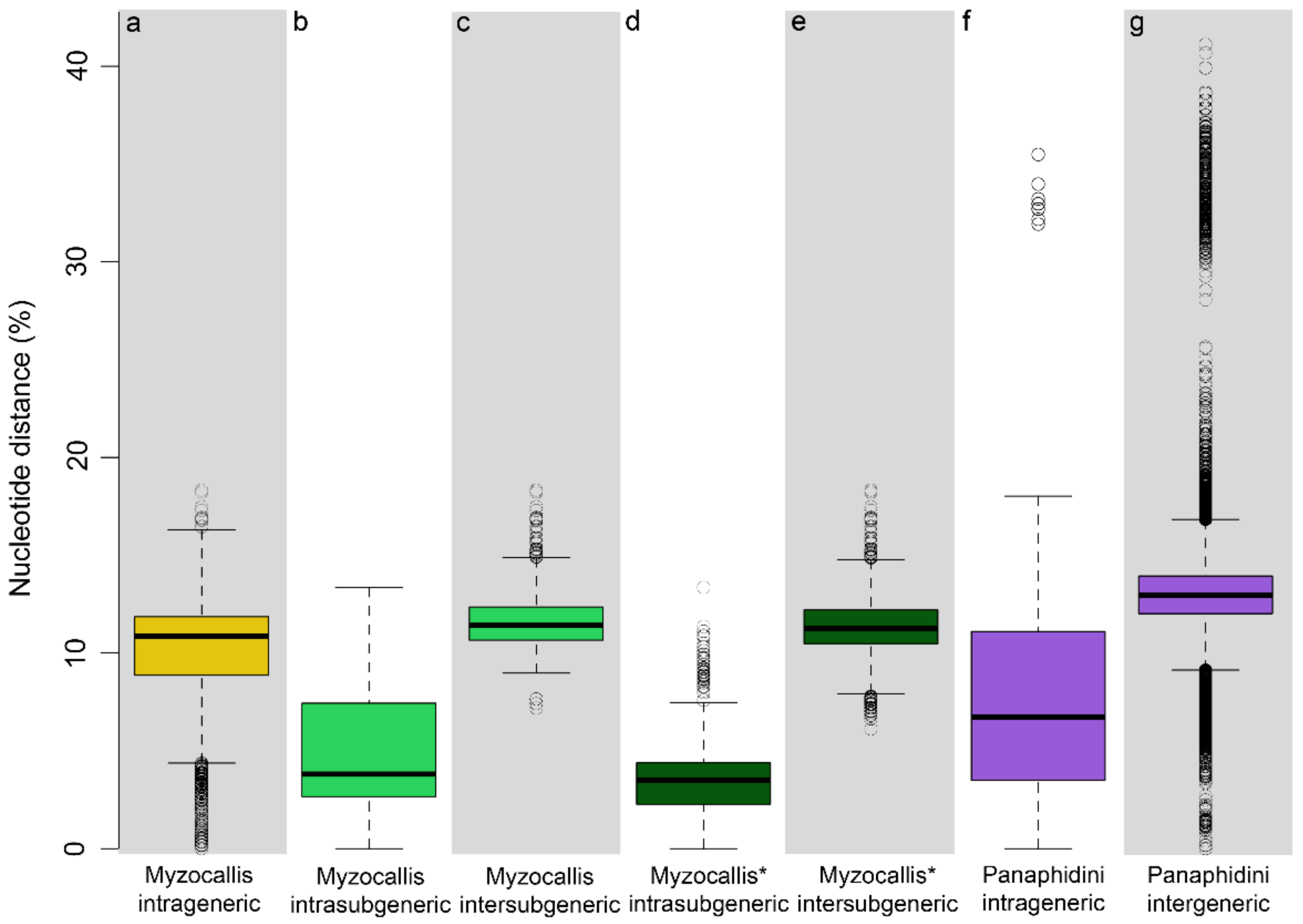
3.1. Nucleotide Distance Analyses

The Myzocallis COI sequences developed in this study plus all the sequences available for this genus in BOLD, for a total of 90 sequences belonging to 17 species (representative of five different Myzocallis subgenera, i.e., M. Agrioaphis, M. Lineomyzocallis, M. Myzocallis, M. Neomyzocallis, M. Pasekia) were assembled in a dataset that includes ~43% of the species currently described for this genus.
Nucleotide divergence within and among Myzocallis species resulted in a mean value of 2.1% (range: 0.2–11.4%) and 10.4% (range: 0–18.3%) respectively, while intrageneric divergence was estimated to be in mean of 9.6% (Figure 1a). Myzocallis subgenera intra- and inter-subgeneric nucleotide distances resulted to be in mean of 4.5% and 11.6%, respectively (Figure 1b,c). The highest inter-subgeneric nucleotide distances have emerged between subgenera Myzocallis and M. Lineomyzocallis (mean value 13.4%) and between the latter and Pasekia (mean value 11.9%). A notable situation emerged within the subgenus Myzocallis, where two groups showing a considerable nucleotide distance (in mean 8.1%, range: 6.1–10.7%) were recognised, one including M. coryli, M. carpini, M. boerneri and M. occidentalis (M. Myzocallis group A) and the other including M. schreiberi, M. glandulosa, M. macrolepidis (M. Myzocallis group B). When intra-/inter-subgeneric divergences were estimated considering these groups as two different subgenera, a mean value of 3.7% and 11.4%, respectively, were found (Figure 1d,e).
The dataset assembled in this study including all sequences of Panaphidini except for Myzocallis (de novo developed plus BOLD database publicly available barcodes) resulted to be composed of 580 COI sequences. Specifically, it included 118 species belonging to 41 genera with a mean intergeneric divergence between genera of 13.1% (Figure 1g) and mean intrageneric divergence, estimated only on genera represented by at least two species (520 sequences of 20 genera), of 6.9% (Figure 1f).

3.2. Species Delimitation of the Genus Myzocallis

The species delimitation analyses performed on the genus Myzocallis dataset using two molecular delimitation methods (i.e., the 2% clustering threshold and ASAP) produced comparable quite results and partially reflect the classical subdivision based on morphology. Specifically, 2% threshold delimitated 21 evolutionary units within the dataset, 11 of them exactly matched the morphological species. The species M. walshii and M. castanicola were both split in three evolutionary units, while all M. carpini and M. coryli were merged in the same one (except for two samples). Finally, also M. asclepiadis and M. punctata were merged in a single unit (Figure 2). Species delimitation adopting ASAP method led to almost identical results with the exceptions of M. carpini and M. coryli whose sequences were split into different evolutionary units but never merged together, and of M. castanicola, whose sequences were split in four units, one of them including only M. leclanti sequences (Figure 2).
In the Myzocallis genus dendrogram, inferred using the same COI sequences dataset, not all the species resulted monophyletic, with incongruences between specimens’ morphological identification and monophyly in tree involving some of the species already highlighted from species delimitation analyses (six species, Figure 2). Whereas, all Myzocallis subgenera, except for Agrioaphis, formed monophyletic clusters (nodes support aLRT ≥ 0.85, except for Pasekia; Figure 2). Noteworthy, the two groups identified within the subgenus Myzocallis from nucleotide distance analyses (i.e., subgen Myzocallis group A and Myzocallis group B) resulted in two well supported monophyletic groups (aLRT = 0.95 node support) (Figure 2).

4. Discussion

The analyses performed in this study shed light on some aspects of the taxonomy of the genus Myzocallis. The main taxonomic inferences derived from this study can be summarized as follow: (i) two different groups are present within the subgenus Myzocallis whose taxonomic rank should be better investigated; (ii) the nominal taxon M. castanicola leclanti Quednau & Remaudière possibly should be elevated to the level of full species; (iii) the status of valid species of M. schreiberi Hille Ris Lambers & Stroyan, which in the past was considered as a synonym of M. boerneri Stroyan, has been confirmed also based on molecular analyses; (iv) the taxonomic hierarchy of Myzocallis subgenera have to be revised, together with other related genera of Myzocallidini.

4.1. Myzocallis Subgenus Myzocallis

The analyses revealed that the subgenus Myzocallis is divided into two well separated clusters (in terms of nucleotide distance) based on COI gene sequences. The first cluster joints together M. coryli, M. carpini, M. boerneri and M. occidentalis (Myzocallis group A), while the second includes M. schreiberi, M. glandulosa, and M. macrolepis (Myzocallis group B). All these species show common morphological characters that allow to cluster them as a single homogeneous group based on morphology. The observed genetic differences could be the result of an ecological divergence of the two species groups. The host plants of the species of Myzocallis group A are all deciduous, on the contrary, those of Myzocallis group B are all semi-evergreen. Aphids’ cycles have to fit the biology of the host plants, and in fact the aphids of the M. Myzocallis group B often develop anholocyclic on them. Except for M. coryli, all other species of M. Myzocallis group A showed a very low intraspecific nucleotide divergence based on COI, independently from their geographical origin (except for one sequence of M. carpini that will be further discussed). M. coryli was morphologically well-studied by several authors, but the presence of morphological variation among the populations of this species was never reported. However, for this species an unusually high intraspecific genetic variability was already observed (on the COI gene), especially between individuals from different geographical areas [10,50]. In previous researches, the authors hypothesized that M. coryli is a cryptic species complex. The nucleotide sequences of M. coryli analysed in this study (18 sequences in total, 14 of which were mined from BOLD) resulted subdivided into three clusters plus one independent sequence in the COI dendrogram and were differentially delimited by the two species delimitation methods adopted, i.e., two (clustering threshold) and three (ASAP) different evolutionary units (Figure 2). Intraspecific nucleotide distance of such a species ranged from 0.02% to 5%. Based on this evidence, M. coryli may represent a complex of cryptic species. Anyway, since M. coryli is a holocyclic and monoecious species on Corylus genus, the nucleotide distances and clusters observed could be explained by the wide geographic distribution of its host plant. Although the latter has a European-Caucasian origin, currently it is widespread throughout the world for agricultural production with M. coryli that followed its distribution. This may have determined a progressive adaptation of the aphid populations to the different environmental conditions and led to some genetic divergence between them, without evolving morphological differences and maybe nether incurring in speciation. This situation might have some analogy B. helichrysi, where a COI nucleotide distance of 2.7% was detected between two different populations and for which the existence of two cryptic species has been hypothesized [51,52]. However, further investigation within this group should be undertaken even with additional mitochondrial and nuclear markers.
The analyses performed in this study clearly distinguished the holm oak aphid M. (M.) schreiberi from the Turkey oak aphid M. (M.) boerneri Stroyan. Both species delimitation methods recognised them as separated species, but also, they have fallen into separate clusters in COI tree (Myzocallis group A, Myzocallis group B) (Figure 2). The two species are morphologically very similar, so in the past, they have often been confused, doubting whether they were distinct species or assuming that M. schreiberi was an anholocyclic form of M. boerneri [3,4,53]. This misunderstanding is probably at the origin of the incorrect classification of the COI sequences ACEA810-14 and GBMHH5717-14 available on BOLD (renamed as M. schreiberi for these study analyses; see Materials and Methods). While describing M. (M.) schreiberi from specimens collected on Q. ilex in Italy and England, Hille Ris Lambers & Stroyan [54] listed the morphological characters distinguishing this species from other European congeneric taxa known at the time. While the identification keys to distinguish M. schreiberi and M. boerneri are provided by Barbagallo & Massimino Cocuzza [11]. These two aphids have a rather different host plant preference. M. schreiberi usually lives on Q. ilex and on other evergreen—leaved oak species, such as Q. suber and Q. x crenata. All these oaks have coriaceous leaves with a grey-tomentose texture beneath, to which this aphid species is likely adapted to feed on through its rather acute shaped last rostral joint. In the present study, no genetic difference was found among the populations of M. schreiberi living on Q. ilex, Q. suber and Q. x crenata. M. boerneri is widely distributed in the West Palaearctic region (Europe, Middle East) and its main host plant is the deciduous Q. cerris.
The M. (M.) glandulosa Hille Ris Lambers [55] and M. (M.) occidentalis Remaudière and Nieto Nafria [56] are both characterized by a rather long last rostral joint. The former was described from Q. ithaburensis in the Middle East [48]. Several years later, the alate viviparous female of M. (M.) glandulosa was figured and Q. persica was added as a further host plant for this species [4]. The individuals whose COI sequences were analysed in the present study were collected in Israel from the type locality. M. (M.) occidentalis has been described from specimens collected in South-Western Europe (France, Spain); the only host plant known is Q. pyrenaica, on which the aphid performs a holocyclic life cycle [8,55]. Also included in this group is M. macrolepidis, a new species of recent description [11]. Moreover, the sequence labelled as M. occidentalis presents on NCBI (accession number GBMIN66582) obtained from a specimen collected in California on Quercus sp. [10] should be regarded as belonging to a different taxon and would deserve in-depth analysis. Finally, also the classification as M. occidentalis of the samples caught with suction or yellow water traps in Serbia and Greece could be incorrect [57,58].

4.2. Myzocallis Subgenus Agrioaphis

The subgenus Agrioaphis Walker is represented so far by two species Myzocallis myricae and M. castanicola (the latter including two subspecies, M. castanicola castanicola and M. castanicola leclanti) [3]. M. castanicola mainly lives on Quercus, but it was recorded as well from Castanea by several authors. The very common populations living on C. sativa in southern Europe and the Middle East, usually belong to the subspecies M. castanicola leclanti. On chestnut it performs a monophagous and holocyclic life cycle and not rarely is considered a noxious aphid species. In Italy, M. castanicola castanicola is quite common in northern areas of the peninsula, mainly on Q. petraea and its hybrids x Q. pubescens (group) and unlikely it can be found there on different Quercus-species (including Q. robur) or on C. sativa, unless perhaps as occasional vagrant alates. In Spain, the aphid commonly lives on Q. pyrenaica and it is occasionally detected on additional oak species [8].
Based on the results obtained in the present study, the bog myrtle aphid M. myricae (Kaltenbach) is clearly distinguishable from M. castanicola. Interestingly, M. castanicola was split into three or four evolutionary units based on the delimitation methods adopted (Figure 2). These units appear to be related with the host plant on which individuals were collected. In the case of ASAP, the evolutionary units were composed as follow (i) individuals collected from Q. pyrenaica and Q. cerris (plus two sequences mined from BOLD for which the host plants are not specified), (ii) individuals collected from Q. petraea in North Est Italy, (iii) individuals collected on Castanea sativa and currently classified as M. castanicola subsp. leclanti, (vi) a sole individual whose sequence was mined from BOLD (GBMHH16898-19). A 2% delimitation threshold partially confirmed these results, but units (ii) and (iii) were merged together since the nucleotide distance between individuals is ~2%. Further analyses are needed to shed light on the taxonomic status of the individuals following in these clusters, but we hypothesis M. castanicola GBMHH16898-19 is a misidentification (actually this sequence may belong to a species not present in the dataset of this study). On the other hand, the analyses here performed clearly discriminate M. castanicola castanicola and M. castanicola leclanti, as belonging to two separate evolutionary units. M. castanicola castanicola and M. castanicola leclanti also clearly differ from the morphological point of view. Specifically, M. castanicola leclanti has (i) paler yellow colour of nymphs, alate viviparous females and sexuales; (ii) paler and less extensive dorsal sclerified areas, particularly those on abdomen; (iii) more haired last rostral joint. Quednau & Remaudière [3] and Quednau, [4] provided the morphological description and illustration of M. castanicola leclanti, adding the comparative range of their variation within the different seasonal morphs of M. castanicola castanicola.

4.3. Myzocallis Subgenus Pasekia

The subgenus Pasekia Aizenberg is represented so far by five species, i.e., Myzocallis persica, Myzocallis taurica, Myzocallis komareki, Myzocallis mediterranea and Myzocallis cocciferina. All of them are well studied from the morphological point of view and a key for their identification (as viviparous alates and nymphs, including males) has been reported [3,4].
M. (Pasekia) persica Quednau & Remaudière and M. (P.) taurica Quednau & Remaudière are apparently confined to eastern Mediterranean. Both species are holocyclic, the former on Q. persica, and the latter on Q. coccifera (maybe also other oak species). Unluckily, these species were not included in the present study since no specimen was available for the molecular analyses.
M. (P.) komareki Pašek has a wider distribution, it was recorded from Central Europe southward to the Mediterranean and the Middle East on several oak species, as well as on Castanea sativa [1,3,8,59]. The nucleotide distance analysis of this study showed low intraspecific variability (<2.1%) for M. komareki, despite the analysed specimens being collected from different oak species and various geographical localities. It can be argued that, due to its polyphagy, this species can easily adapt to the different oak species, but without occurring in isolation of its populations.
M. (P.) mediterranea Quednau & Remaudière is recorded so far from France, Spain and Italy, and probably it is widespread also in other Mediterranean countries. The species inhabits several oaks such as Q. pubescens and strictly allied taxa (Q. congesta, Q. virgiliana and Q. dalechampii) [60] and only occasionally it has been collected on oaks of different groups (i.e., Q. ilex). In the past, this aphid was frequently confused with M. (P.) komareki, because of their morphological similarity [61,62]. Usually, M. (P.) mediterranea develops through a regular holocycle. Nevertheless, in more warm habitats (such as in southern Italy) the amphigonic morphs appear very late in the season (from the end of December to the first half of February), inhabiting the host plant leaves still green in Q. pubescens-group species (semi-evergreen oaks). In such a case, part of the aphid population can overwinter anholocyclically on these oaks and the survived specimens move to new oak blossoms by the end of March or early in April [63] (as M. komareki). Also, in the case of M. mediterranea, a low intraspecific variability (<1% nucleotide distance) was found between specimens considered in this study, however all of them were collected from a limited geographic area (Sicily) on Q. pubescens.
Finally, M. (P.) cocciferina Quednau & Barbagallo is present in southern Europe and Mediterranean countries, from Spain eastward to Lebanon on Q. coccifera s. lat. and occasionally on Q. ilex or their hybrids (ilex x coccifera). In Spain, the aphid is recorded on Q. ilex and reported as well for Portugal and North Africa [8]. This species was originally attributed [64] to subgenus Agrioaphis sensu Richards (1968) and later transferred to Pasekia [3,64]. In the present study, molecular and morphological species delimitation were consistent for M. cocciferina. A nucleotide distance of ~1.5% was estimated between specimens collected from Q. coccifera in Sicily and Apulia and those collected from Q. ilex or its hybrid x Q. coccifera (postulated as Q. soluntina Tineo ex Lojacono by Giardina et al. [65]).

4.4. Nearctic Myzocallis Subgenera Lineomyzocallis and Neomyzocallis

In this study, a few specimens belonging to the Nearctic subgenera Lineomyzocallis (M. walshii, M. bellus and M. ephemerata) and Neomyzocallis (M. asclepiadis and M. punctata) were included. Here, two relevant situations were observed: (i) a high intraspecific variability of M. walshii (range intraspecific nucleotide distance: 0.2–8%); (ii) a very low nucleotide distance between M. asclepiadis and M. punctata (<0.2%). All the species delimitation methods adopted split M. walshii into three evolutionary units, two of them including one individual each (Figure 2). The black-bordered oak aphid, M. walshii lives on Quercus of the group of red oaks (Quercus section Lobatae) and is native to North America, where it is widely distributed. In 1989 the species was found for the first time in France and in about twenty years it spread throughout the European continent [1]. None of the numerous studies performed in the last decades on M. walshii reported the presence of intraspecific morphological or biological variability. Possibly, the high genetic divergence observed in this study could be related to specimen misidentifications, rather than to the existence of cryptic diversity. Both the molecular species delimitation methods used in this study delimited M. asclepiadis and M. punctata in a single species, in accordance with what was already observed [10], i.e., some sequences present on BOLD may have been mistakenly attributed to M. asclepiadis.

5. Conclusions

Systematics is a hierarchic science fundamentally built on the relationships of affinity between species [66]. COI is a DNA marker known to be effective in discriminating insect taxa at the lowest taxonomic levels. Combining molecular (COI gene), morphological and ecological information in an integrative framework makes possible to resolve numerous taxonomic issues concerning insect species [67,68,69,70,71]. In particular, the most accurate results are obtained when expert morphologists on the target group are involved in the evaluation of the signal resulting from molecular analyses, in this case is also easier to distinguish between extrinsic and intrinsic errors related to the species molecular identification and delimitation [71,72,73]). For some insect groups, as in the case of aphids, the evaluation of the ecological information is important in the process of species identification and delimitation as well. Hence the relevance of coupling DNA sequences with specimen’s collection locality and date, and further ecological information if present (e.g., collection habitat or host plant), even in the phase of molecular data publication.
However, also in this context, the use of integrative taxonomy (and COI as DNA marker) to define other taxonomic categories than species could sound as improper. Whereas, in previous studies it was done and valuable results were obtained. For example, the COI nucleotide distances have been examined for evaluating the range of variation between genera and subfamilies of Aphididae [10,23,27]. Lee et al. [10] developed the barcode sequences for 154 Aphididae species (72 genera, 11 subfamilies), and reported a mean pairwise divergence between specimens of different genera of 8.9% (range from 1.6 to 19%). Other studies, on Adelgidae and Eulachnini aphids [23], found a mean intergeneric distance of 9.6% and 11.7%, respectively. In a further investigation, a mean intergeneric distance of 7.7% (from 5 to 9.7%) in the tribe of Macrosiphini and 10.4 (range 8.9% to 12.4%) in Aphidini were found [27].
The Myzocallis subgenera analysed in this work are morphologically distinguishable through characters of well-verified validity [4], recognized as adequate by aphid-taxonomists. In the present study, a substantial nucleotide distance was found between the Myzocallis subgenera (Figure 1), reinforcing the validity of these taxa, regardless of their hierarchical position. The nucleotide distance values are sufficient to justify the elevation of the subgenera to the rank of genus. In particular, considering that the inter-subgeneric distances estimated in this study for Myzocallis subgenera are comparable to those observed between genera of the Panaphidini tribe (Figure 1), that certainly include species phylogenetically more distant than those of the Myzocallis genus.
Several authors stated that different genera have to be monophyletic [66,74], and the conclusion drawn by the present work meets this condition. Furthermore, this evidence could be supported also from the morphological point of view. Some of the most important morphological characters [3,4] (i.e., spinal hairs on abdominal tergites, chaetotaxy of immature morphs and oviparae) to distinguish Myzocallis subgenera from each other, are the same that were used to distinguish the genera Hoplocallis (until a few years ago considered as a subgenus of Myzocallis), Apulicallis and Siculaphis.
Consequently, we elevated these subgenera to the rank of genus: Agrioaphis Walker, stat. n. [described as genus, type species Aphis myricae Kaltenbach], Lineomyzocallis Richards, stat. n. [described as subgenus of Myzocallis, type species Aphis bella Walsh], Neomyzocallis Richards stat. n. [described as subgenus of Myzocallis, type species Callipterus punctatus Monell], and Pasekia Aizenberg stat. n. [described as subgenus of Myzocallis, type species Hoplocallis komareki Pašek, 1953]. The genus Myzocallis remains as such, including the nominotypical genus and the subgenera whose species have not been considered in this work: Californicallis [3], Castaneomyzocallis [3] Quednau & Remaudière, Globulicaudaphis Hille Ris Lambers, Neodryomyzus Quednau & Remaudière, Neodryomyzus Quednau & Remaudière, and Paramyzocallis Quednau & Remaudière, for which further studies are needed.
Finally, Myzocallis (Agrioaphis) leclanti Quednau & Remaudière, n. stat., originally described as a subspecies of M. (A.) castanicola (Quednau & Remaudière, 1994) and M. (M.) schreiberi, considered as a subspecies of M. (M.) boerneri, should be regarded at a rank of full species.

Supplementary Materials

The following supporting information can be downloaded at: https://www.mdpi.com/article/10.3390/insects13111006/s1, Table S1: Host plants, sampling date, geographical coordinates, voucher numbers, BIN and GenBank accession number of Myzocallis spp. analysed. Table S2. List of sequences used for analysis.

Author Contributions

Conceptualization, G.E.M.C.; methodology, G.E.M.C., G.M. and M.M.; validation, S.B. and J.M.N.N.; formal analysis, G.M.; data curation, G.E.M.C. and G.M.; writing—original draft preparation, G.E.M.C. and G.M.; writing—review and editing, G.E.M.C., G.M., M.M., S.B. and J.M.N.N. All authors have read and agreed to the published version of the manuscript.

Funding

This research was funded by University of Catania, within the project “Strategie di controllo eco-sostenibile di fitofagi d’interesse economico” (COSOFI) Piano Incentivi per la Ricerca di Ateneo 2020–22.

Data Availability Statement

Data presented in this research are available in the article.

Acknowledgments

Authors are much grateful to Zvi Mendel (The Hebrew University, Tel Aviv, Israel) for freshly collected samples of Myzocallis glandulosa from the type locality, Stefano Convertini to help provided in the collection of M. macrolepidis samples and to the anonymous reviewer for useful suggestions.

Conflicts of Interest

The authors declare no conflict of interest.

References

  1. Blackman, R.L.; Eastop, V.F. Aphids on the World’s Trees: An Identification and Information Guide; CAB International: Wallingford, UK, 1994; p. 987. Available online: www.aphidsonworldsplants.info (accessed on 23 September 2022).
  2. Favret, C. Aphid Speciesfile. Version 5.0/5.0. 2022. Available online: https://Aphid.SpeciesFile.org (accessed on 23 September 2022).
  3. Quednau, F.W.; Remaudière, G. Le genre Myzocallis Passerini, 1860: Classification mondiale des sous-genres et nouvelle espèces paléarctiques (Homoptera: Aphididae). Can. Entomol. 1994, 126, 303–326. [Google Scholar] [CrossRef]
  4. Quednau, W. Atlas of the Drepanosiphine aphids of the world. Part I: Panaphidini—Myzocallidina. Contrib. Am. Entomol. Inst. 1999, 31, 1–281. [Google Scholar]
  5. Denk, T.; Grimm, G.W.; Manos, P.S.; Deng, M.; Hipp, A.L. An updates infrageneric classification of the oaks: Review of previous taxonomic schemes and synthesis of evolutionary patterns. In Oaks Physiological Ecology. Exploring the Functional Diversity of Genus Quercus, L.; Gil-Pelegrín, E., Peguero-Pina, J., Sancho-Knapik, D., Eds.; Springer: Cham, Switzerland, 2017. [Google Scholar] [CrossRef]
  6. Heie, O.E. The Aphidoidea (Hemiptera) of Fennoscandia and Denmark. II. The family Drepanosiphidae; Scandinavian Science Press: Klampenborg, Denamrk, 1982; p. 176. [Google Scholar]
  7. Stroyan, H.L.G. Homoptera Aphidoidea, Chaitophoridae & Callaphidinae. In Handbook Identification British Insects vol. II; Royal Entomological Society: London, UK, 1977; p. 130. [Google Scholar]
  8. Nieto Nafría, J.M.; Mier Durante, M.P. Hemiptera, Aphididae I., Fauna Iberica; CSIC: Madrid, Spain, 1998. [Google Scholar]
  9. Osiadacz, B.; Wieczorek, K.M. Myzocallis (Lineomyzocallis) walshii Monell, 1879 (Hemiptera, Aphidoidea), an aphid species new for Poland. Pol. J. Entom. 2006, 75, 233–338. [Google Scholar]
  10. Lee, Y.; Lee, W.; Kanturski, M.; Foottit, R.G.; Akimoto, S.I.; Lee, S. Cryptic diversity of the subfamily Calaphidinae (Hemiptera: Aphididae) revealed by comprehensive DNA barcoding. PLoS ONE 2016, 12, e0176582. [Google Scholar] [CrossRef] [PubMed] [Green Version]
  11. Barbagallo, S.; Massimino Cocuzza, G.E. Description of a new Myzocallis (Hemiptera Aphididae) living on Valonia oak in Southern Italy with DNA barcoding accounts on allied species-group. Zootaxa 2022, 5183, 187–202. [Google Scholar] [CrossRef] [PubMed]
  12. Remaudière, G. Découverte en France de l’espèce américaine Myzocallis (Lineomyzocallis) walshii (Monell) (Hom. Aphididae). Ann. Société Entomol. De Fr. 1989, 25, 117. [Google Scholar]
  13. Patti, I.; Lozzia, G.C. Presenza in Italia dell’afide neartico della quercia rossa, Myzocallis (Lineomyzocallis) walshii (Mon.). Boll. Zool. Agric. E Bachic. 1994, 26, 141–145. [Google Scholar]
  14. Hebert, P.D.N.; Cywinska, A.; Ball, S.L.; de Waard, J.R. Biological identification through DNA barcodes. Proc. R. Soc. Lond. B 2003, 270, 313–321. [Google Scholar] [CrossRef] [Green Version]
  15. Schindel, D.E.; Miller, S.E. DNA barcoding a useful tool for taxonomists. Nature 2005, 435, 17. [Google Scholar] [CrossRef] [Green Version]
  16. Virgilio, M.; Backeljan, I.; Nevado, B.; De Meyer, M. Comparative performances of DNA barcoding across insect orders. BMC Bioinform. 2010, 11, 206. [Google Scholar] [CrossRef] [Green Version]
  17. Jimbo, U.; Kato, T.; Ito, M. Current progress in DNA barcoding and future implications for entomology. Entomol. Sci. 2011, 14, 107–124. [Google Scholar] [CrossRef]
  18. Magoga, G.; Sassi, D.; Daccordi, M.; Leonardi, C.; Mirzaei, M.; Regalin, R.; Lozzia, G.; Montagna, M. Barcoding Chrysomelidae: A resource for taxonomy and biodiversity conservation in the Mediterranean Region. Zookeys 2016, 597, 27–38. [Google Scholar] [CrossRef]
  19. Magoga, G.; Sahin, D.C.; Fontaneto, D.; Montagna, M. Barcoding of Chrysomelidae of Euro-Mediterranean area: Efficiency and problematic species. Sci. Rep. 2018, 8, 13398. [Google Scholar] [CrossRef]
  20. Massimino Cocuzza, G.E.; Cavalieri, V. Identification of aphids of Aphis frangulae-group living in Lamiaceae species through DNA barcode. Mol. Ecol. Resour. 2014, 14, 447–457. [Google Scholar] [CrossRef] [PubMed]
  21. Kanturski, M.; Lee, Y.; Choi, J.; Lee, S. DNA barcoding and a precise morphological comparison revealed a cryptic species in the Nippolachnus piri complex (Hemiptera: Aphididae: Lachninae). Sci. Rep. 2018, 8, 8998. [Google Scholar] [CrossRef] [PubMed]
  22. Xi-Cha, Z.; Jing, C.; Rui, C.; Li-Yun, J.; Ge-Xia, Q. DNA barcoding and species delimitation of Chaitophorinae (Hemiptera, Aphididae). Zootaxa 2017, 656, 25–50. [Google Scholar]
  23. Foottit, R.G.; Maw, H.E.L.; Pike, K.S. DNA barcodes to explore diversity in aphids (Hemiptera Aphididae and Adelgidae). Redia 2009, 92, 87–91. [Google Scholar]
  24. Kim, H.; Lee, S.; Jang, Y. Macroevolutionary Patterns in the Aphidini Aphids (Hemiptera: Aphididae): Diversification, Host Association, and Biogeographic Origins. PLoS ONE 2011, 6, e24749. [Google Scholar] [CrossRef]
  25. Massimino Cocuzza, G.E.; Di Silvestro, S.; Giordano, R.; Rapisarda, C. Congruence between cytochrome oxidase I (COI) and morphological data in Anuraphis spp. (Hemiptera, Aphididae) with a comparison between the utility of the 5′ barcode and 3′ COI regions. Zookeys 2015, 529, 123–144. [Google Scholar] [CrossRef] [Green Version]
  26. Kinyanjui, G.; Khamis, F.M.; Mohamed, S.; Ombura, L.O.; Warigia, M.; Ekesi, S. Identification of aphid (Hemiptera: Aphididae) species of economic importance in Kenya using DNA barcodes and PCR-RFLP based approach. Bull. Entomol. Res. 2016, 106, 63–72. [Google Scholar] [CrossRef]
  27. Rebijith, K.B.; Asokan, R.; Krishna Kumar, N.K.; Krishna, V.; Chaitanya, B.N.; Ramamurthy, V.V. DNA barcoding and elucidation of cryptic aphid species (Hemiptera: Aphididae) in India. Bull. Entomol. Res. 2013, 103, 601–610. [Google Scholar] [CrossRef] [PubMed]
  28. Montagna, M.; Sassi, D.; Giorgi, A. Pachybrachis holerorum (Coleoptera: Chrysomelidae: Cryptocephalinae), a new species from the Apennines, Italy, identified by integration of morphological and molecular data. Zootaxa 2013, 3741, 243–253. [Google Scholar] [CrossRef] [PubMed]
  29. Magoga, G.; Fontaneto, D.; Montagna, M. Factors affecting the efficiency of molecular species delimitation in a species-rich insect family. Mol. Ecol. Res. 2021, 21, 1475–1489. [Google Scholar] [CrossRef] [PubMed]
  30. Tutin, T.G.; Heywood, V.H.; Burges, N.A.; Valentine, D.H.; Walters, S.M.; Webb, D.A. Flora Europea; Cambridge University Press: Cambridge, UK, 2001; Volume V, p. 2392. [Google Scholar]
  31. Pignatti, S. Flora d’Italia; Edagrcole-New Business Media: Bologna, Italy, 2017; Volume I, p. 1064. [Google Scholar]
  32. Quednau, F.W. Atlas of the drepanosiphine aphids of the world. Part II: Panaphidini Oestlund, 1923—Panaphidina Oestlund, 1923 (Hemiptera: Aphididae: Callaphidinae). Mem. Am. Entomol. Inst. 2003, 72, 301. [Google Scholar]
  33. Quednau, F.W. Atlas of the Drepanosiphine aphids of the world. Part III: Mindarinae Tullgren, 1909; Neophyllaphidinae Takahashi, 1921; Lizeriinae, E.E. Blanchard, 1923; Pterastheniinae Remaudière & Quednau, 1988; Macropodaphidinae Zachvatkin & Aizenberg, 1960; Taiwanaphidinae Quednau & Remaudière, 1994; Spicaphidinae Essig, 1953; Phyllaphidinae Herrich-Schaeffer in Koch, 1957; Israelaphidinae Ilharco, 1961; Saltusaphidinae Baker, 1920 (Hemiptera: Sternorrhycha, Aphididae). Mem. Am. Entomol. Inst. 2010, 83, 361. [Google Scholar]
  34. Folmer, O.; Black, M.; Hoeh, W.; Lutz, R.; Vrijenhoek, R. DNA primers for amplification of mitochondrial cytochrome c oxidase subunit I from diverse metazoan invertebrates. Mol. Mar. Biol. Biotech. 1994, 3, 294–299. [Google Scholar]
  35. Griekspoor, A.; Groothuis, T. 4Peaks V 1.7. 2019. Available online: https://nucleobytes.com (accessed on 20 September 2022).
  36. Ratnasingham, S.; Hebert, P.D.N. BOLD: The Barcode of Life Data System (www.barcodinglife.org). Mol. Ecol. Notes 2007, 7, 355–364. [Google Scholar] [CrossRef] [Green Version]
  37. Edgar, R.C. MUSCLE: A multiple sequence alignment method with reduced time and space complexity. BMC Bioinform. 2004, 5, 113. [Google Scholar] [CrossRef]
  38. Kumar, S.; Stecher, G.; Li, M.; Knyaz, C.; Taura, K. MEGA X: Molecular Evolutionary Genetics Analysis across computing platforms. Mol. Biol. Evol. 2018, 35, 1547–1549. [Google Scholar] [CrossRef]
  39. Kimura, M. A simple method for estimating evolutionary rates of base substitutions through comparative studies of nucleotide sequences. J. Mol. Evol. 1980, 16, 111–120. [Google Scholar] [CrossRef]
  40. Popescu, A.A.; Huber, K.T.; Paradis, E. Ape 3.0: New tools for distance based phylogenetics and evolutionary analysis in R. Bioinformatics 2012, 28, 1536–1537. [Google Scholar] [CrossRef] [PubMed] [Green Version]
  41. Brown, S.D.J.; Collins, R.A.; Boyer, S.; Lefort, M.-C.; Malumbres-Olarte, J.; Vink, C.J.; Cruickshank, R.H. SPIDER: An R package for the analysis of species identity and evolution, with particular reference to DNA barcoding. Mol. Ecol. Res. 2012, 12, 562–565. [Google Scholar] [CrossRef] [PubMed]
  42. Puillandre, N.; Lambert, A.; Brouillet, S.; Achaz, G. ABGD, Automatic Barcode Gap Discovery for primary species delimitation. Mol. Ecol. 2012, 21, 1864–1877. [Google Scholar] [CrossRef]
  43. Lanfaer, R.; Frandsen, P.B.; Wright, A.M.; Senfeld, T.; Calcott, B. PartitionFinder 2: New methods for selecting partitioned models of evolution for molecular and morphological phylogenetic analyses. Mol. Biol. Evol. 2017, 34, 772–773. [Google Scholar] [CrossRef] [Green Version]
  44. Schwarz, G. Estimating the dimension of a model. Ann. Stat. 1974, 6, 461–464. [Google Scholar] [CrossRef]
  45. Hasegawa, M.; Kishino, H.; Yano, T. Dating of the human-ape splitting by a molecular clock of mitochondrial DNA. J. Mol. Evol. 1985, 22, 160–174. [Google Scholar] [CrossRef] [PubMed]
  46. Ronquist, F.; Teslenko, M.; van der Mark, P.; Ayres, D.L.; Darling, A.; Höhna, S.; Larget, B.; Liu, L.; Suchard, M.A.; Huelsenbeck, J.P. MrBayes 3.2: Efficient Bayesian phylogenetic inference and model choice across a large model space. Syst. Biol. 2012, 61, 539–542. [Google Scholar] [CrossRef] [Green Version]
  47. Drummond, A.J.; Suchard, M.A.; Xie, D.; Rambaut, A. Bayesian phylogenetics with BEAUti and the BEAST 1.7. Mol. Biol. Evol. 2012, 29, 1969–1973. [Google Scholar] [CrossRef]
  48. Guindon, S.; Dufayard, J.F.; Lefort, V.; Anisimova, M.; Hordijk, W.; Gascuel, O. New Algorithms and Methods to Estimate Maximum-Likelihood Phylogenies: Assessing the Performance of PhyML 3.0. Syst. Biol. 2010, 59, 307–321. [Google Scholar] [CrossRef] [Green Version]
  49. Anisimova, M.; Gascuel, O. Approximate likelihood ratio test for branches: A fast, accurate, and powerful alternative. Syst. Biol. 2006, 55, 539–552. [Google Scholar] [CrossRef]
  50. Coeur D’acier, A.; Cruaud, A.; Artige, E.; Genson, G.; Clamens, A.L.; Pierce, E.; Hudaverdian, S.; Simon, J.-C.; Jousselin, E.; Rasplus, J.Y. DNA barcoding and the associated PhylAphidB@se Web site for the identification of European aphids (Insecta: Hemiptera: Aphididae). PLoS ONE 2014, 9, e97620. [Google Scholar] [CrossRef] [PubMed]
  51. Piffaretti, J.; Vanlenberghe-Masutti, F.; Tayeh, A.; Clamens, A.L.; Coeur D’acier, A.; Jousselin, E. Molecular phylogeny reveals the existence of two sibling species in the aphid pest Brachycaudus helichrysi (Hemiptera: Aphididae). Zool. Scr. 2012, 41, 266–280. [Google Scholar] [CrossRef]
  52. Popkin, M.; Piffaretti, J.; Clamens, A.L.; Qiao, G.X.; Chen, J.; Vitalis, K.; Vanlerberghe-Masutti, F.; Gupta, R.K.; Mamaari, M.; Langella, O.; et al. Large-scale phylogeographic study on the cosmopolitan aphid pest Brachycaudus helichrysi reveals host plant associated lineages that evolved in allopatry. Biol. J. Linn. Soc. 2017, 120, 102–114. [Google Scholar]
  53. Richards, W.R. A synopsis of the world fauna of Myzocallis. Mem. Entom. Soc. Can. 1968, 57, 1–76. [Google Scholar]
  54. Hille Ris Lambers, D.; Stroyan, H.L.G. Two new species and a new subspecies of aphid from Italy. Mem. Soc. Entomol. Ital. 1959, 38, 84–94. [Google Scholar]
  55. Hille Ris Lambers, D. On Palestine aphids, with description of new subgenera and new species (Homoptera, Aphididae). Trans. R. Entom. Soc. Lond. 1948, 99, 269–289. [Google Scholar] [CrossRef]
  56. Remaudière, G.; Nieto Nafría, J.M. Un Myzocallis nouveau du Sud-Ouest de l’Europe (Hom. Aphididae). Ann. Soc. Entom. Fr. 1974, 10, 751–756. [Google Scholar]
  57. Tsitsipis, J.A.; Katis, N.I.; Margaritopoulos, J.T.; Lykourresis, D.P.; Avgelis, A.D.; Gargalianou, I.; Zarpas, K.D.; Perdikis, D.C.; Papapanayotoua, A. A contribution to the aphid fauna of Greece. Bull. Insectology 2007, 60, 31–38. [Google Scholar]
  58. Vučetic, A.; Jovičić, I.; Petrović-Obradović, O. Several new and one nvasive aphid species (Aphididae, Hemiptera) caught by yellow water trap in Serbia. Phytoparasitica 2014, 42, 247–257. [Google Scholar] [CrossRef]
  59. Holman, J. Host Plant Catalogue of Aphids: Palaearctic Region; Springer: Berlin/Heidelberg, Germany, 2009; p. 1216. [Google Scholar]
  60. Patti, I.; Barbagallo, S. Annotazioni integrative sugli afidi della Sicilia. Redia 1999, 82, 49–82. [Google Scholar]
  61. Barbagallo, S.; Stroyan, H.L.G. Osservazioni biologiche, ecologiche e tassinomiche dell’afidofauna della Sicilia. Frustula Entomol. 1982, 3, 1–182. [Google Scholar]
  62. Barbagallo, S.; Binazzi, A. Gli Afidi delle Querce in Italia. In Atti Convegno “Problematiche fitopatologiche del genere Quercus in Italia”; Stamperia Granducale: Firenze, Italia, 1990; pp. 142–160. [Google Scholar]
  63. Barbagallo, S.; Stroyan, H.L.G. Rilievi preliminari sulla composizione dell’afidofauna siciliana. In Proceedings of the Atti XI Congresso Nazionale Italiano di Entomologia, Portici-Sorrento, Italy, 10–15 May 1976; pp. 241–251. [Google Scholar]
  64. Quednau, F.W.; Barbagallo, S. A new genus and three new species of aphids from Quercus calliprinos Webb in the mediterranean countries (Homoptera: Aphididae). Can. Ent. 1991, 123, 581–593. [Google Scholar] [CrossRef]
  65. Giardina, G.; Raimondo, F.M.; Spadaro, V. A catalogue of plants growing in Sicily. Bocconea 2007, 20, 5–582. [Google Scholar]
  66. La Greca, M. L’uso delle categorie sistematiche sottogenere e sottospecie in tassonomia, alla luce della ricerca biogeografica. Boll. Dell'istituto Di Entomol. Dell'universita Degli Studi Di Bologna 1987, 41, 159–171. [Google Scholar]
  67. Gebiola, M.; Bernardo, U.; Ribes, A.; Gibson, G.A.P. An integrative study of Necremnus Thomson (Hymenoptera: Eulophidae) associated with invasive pests in Europe and North America: Taxonomic and ecological implications. Zool J. Linn. Soc. 2015, 173, 352–423. [Google Scholar] [CrossRef] [Green Version]
  68. Montagna, M.; Kubisz, D.; Mazur, M.A.; Magoga, G.; Ścibior, R.; Kajtoch, Ł. Exploring species-level taxonomy in the Cryptocephalus flavipes species complex (Coleoptera: Chrysomelidae). Zool. J. Linn. Soc. 2017, 179, 92–109. [Google Scholar] [CrossRef] [Green Version]
  69. Montagna, M.; Gonella, E.; Mereghetti, V.; Magoga, G.; Ferrari, E.; Pontini, M.; Ferracini, C.; Alma, A. Molecular species delimitation of the Asian chestnut gall wasp biocontrol agent released in Italy. Insect Syst. Evol. 2019, 50, 327–345. [Google Scholar] [CrossRef]
  70. Masonick, P.; Weirauch, C. Integrative species delimitation in Nearctic ambush bugs (Heteroptera: Reduviidae: Phymatinae): Insights from molecules, geometric morphometrics and ecological associations. Syst. Entomol. 2020, 45, 205–223. [Google Scholar] [CrossRef]
  71. Gadawski, P.; Montagna, M.; Rossaro, B.; Gilka, W.; Pesic, V.; Grabowski, M.; Magoga, G. DNA barcoding of Chironomidae from the Lake Skadar region: Reference library and a comparative analysis of the European fauna. Divers. Distrib. 2022, 28, 1–20. [Google Scholar] [CrossRef]
  72. Salvi, D.; Berrilli, E.; D’Alessandro, P.; Biondi, M. Sharpening the DNA barcoding tool through a posteriori taxonomic validation: The case of Longitarsus flea beetles (Coleoptera: Chrysomelidae). PLoS ONE 2020, 15, e0233573. [Google Scholar] [CrossRef]
  73. Marconi, M.; Modesti, A.; Alvarez, L.P.; Ogoña, P.V.; Mendoza, A.C.; Vecco-Giove, C.D.; Luna, J.O.; Di Giulio, A.; Mancini, E. DNA Barcoding of Stingless Bees (Hymenoptera: Meliponini) in Northern Peruvian Forests: A Plea for Integrative Taxonomy. Diversity 2022, 14, 632. [Google Scholar] [CrossRef]
  74. Mayr, E. Principles of Systematic Zoology; Mc Grew-Hill: New York, NY, USA, 1969; p. 428. [Google Scholar]
Figure 1. Boxplot of K2P pairwise nucleotide distances (a) within the genus Myzocallis; (b) within Myzocallis subgenera (c) between Myzocallis subgenera (d) within Myzocallis subgenera considering M. Myzocallis A and M. Myzocallis B as two separated subgenera (e) between Myzocallis subgenera considering M. Myzocallis A and M. Myzocallis B as two separated subgenera (f) within Panaphidini genera, excluding Myzocallis (g) between Panaphidini genera, excluding Myzocallis.
Insects 13 01006 g001
Figure 2. Myzocallis genus COI dendrogram and molecular species delimitation results. Results of molecular species delimitation analyses (i.e., 2% distance threshold and ASAP) and morphological delimitation are reported through vertical bars on the right side of the tree. On the same side, Myzocallis species host plants are reported (deciduous are written in red, semi evergreen in green), information on the host plants of species of the subgenera M. Lineomyzocallis and M. Neomyzocallis was taken from the scientific literature. BOLD id and identifiers of sequences developed in this study are indicated on the tips. On nodes aLRT values are reported, * represents aLRT values < 0.70. The tree scale bar indicates the distance in substitutions per site.
Insects 13 01006 g002
Publisher’s Note: MDPI stays neutral with regard to jurisdictional claims in published maps and institutional affiliations.

