Article

The Intracellular Symbiont Wolbachia pipientis Enhances Recombination in a Dose-Dependent Manner

by
Kaeli N. Bryant
and
Irene L. G. Newton
*
Department of Biology, Indiana University, 1001 E 3rd Street, Bloomington, IN 47405, USA
*
Author to whom correspondence should be addressed.
Insects 2020, 11(5), 284; https://doi.org/10.3390/insects11050284
Submission received: 14 April 2020 / Revised: 24 April 2020 / Accepted: 27 April 2020 / Published: 6 May 2020
(This article belongs to the Collection Insect Symbionts: Evolution and Application)

Abstract

:
Wolbachia pipientis is an intracellular alphaproteobacterium that infects 40%–60% of insect species and is well known for host reproductive manipulations. Although Wolbachia are primarily maternally transmitted, evidence of horizontal transmission can be found in incongruent host–symbiont phylogenies and recent acquisitions of the same Wolbachia strain by distantly related species. Parasitoids and predator–prey interactions may indeed facilitate the transfer of Wolbachia between insect lineages, but it is likely that Wolbachia are acquired via introgression in many cases. Many hypotheses exist to explain Wolbachia prevalence and penetrance, such as nutritional supplementation, protection from parasites, protection from viruses, or direct reproductive parasitism. Using classical genetics, we show that Wolbachia increase recombination in infected lineages across two genomic intervals. This increase in recombination is titer-dependent as the wMelPop variant, which infects at higher load in Drosophila melanogaster, increases recombination 5% more than the wMel variant. In addition, we also show that Spiroplasma poulsonii, another bacterial intracellular symbiont of D. melanogaster, does not induce an increase in recombination. Our results suggest that Wolbachia infection specifically alters its host’s recombination landscape in a dose-dependent manner.

1. Introduction

Recombination, the exchange of genetic material during meiosis, is thought to be largely beneficial, as it increases the efficacy of natural selection [1,2]. Because of chromosome architecture, loci that are physically linked to each other can interfere with selection such that selection at one locus reduces the effective population size, and therefore the efficacy of selection, at linked loci. This phenomenon, termed “Hill–Robertson interference,” means that positive or negative selection at one site can interfere with selection at another site. By allowing loci to shuffle between chromosomes, recombination mitigates Hill–Robertson interference [3]. As a result of this re-shuffling, areas of the genome subject to high recombination rates show higher nucleotide diversity, either because of the inherent mutagenic effect of recombination, or by the indirect influence of recombination on natural selection in a population. Overall, a large body of literature supports the assertion that recombination increases efficacy of selection and enhances adaptation in animals, as reviewed and studied in various Drosophila species [1,2,3].
One factor that may influence recombination is bacterial infection. For example, injection of Drosophila melanogaster with the bacterial pathogen Serratia increases recombination post infection [4]. Many Drosophila species are colonized persistently by Wolbachia pipientis, an alpha-proteobacterium within the Rickettsiales and a highly prevalent infection, found in 40%–60% of all insects [5,6]. Wolbachia’s prevalence in populations is likely modulated by reproductive manipulations, induced to benefit infected females [7]. However, this reproductive parasitism alone is not sufficient to explain the prevalence of Wolbachia infection. Indeed, there are many recently discovered, insect-infecting strains that do not seem to induce any reproductive phenotype at all, suggesting other potential benefits provided by the symbiont [8,9,10]. One known benefit is pathogen blocking, where Wolbachia repress virus replication within the insect host [11,12,13]. This phenomenon has important implications for the use of Wolbachia in vector control [14]. In addition to protecting their hosts from pathogens, Wolbachia also improve the fitness and fecundity of some hosts (by protecting against virus infection or enhancing survival and fecundity), and removal of the endosymbiont can cause a decrease in host fitness [15,16]. Finally, Wolbachia can rescue oogenesis defects in mutant Drosophila strains [17] and have also made themselves a necessary component of oogenesis in some wasp species, thereby making the infection indispensable [18].
One recently discovered phenotype of Wolbachia pipientis is that it may increase the frequency of recombination events on the X chromosome, but not on the third, in D. melanogaster [19,20]. This phenotype contrasts with that observed for Serratia infection, where elevated recombination was observed on the third chromosome [4]. Does Wolbachia infection actually lead to increased recombination? If so, would any infection of the reproductive tract result in increased recombination? Here, we answer these questions using classical genetics in D. melanogaster with different Wolbachia variants and using another maternally transmitted bacterial infection, Spiroplasma poulsonii. We confirm that Wolbachia significantly increase recombination across two intervals, one on the X and one on the second chromosome, but we could not detect any effect on the third chromosome interval queried. In addition, there is a clear correlation between Wolbachia load and recombination events, suggesting Wolbachia infection itself is the cause of the elevated recombination; clearing the host of Wolbachia restores recombination rate to a basal level while infection with a high-titer variant, in the same genetic background, increases recombination. Another intracellular symbiont, Spiroplasma, does not increase recombination rate, suggesting this phenomenon is not simply due to the presence of a bacterial infection in the gonads, but is induced by Wolbachia. These results, in sum, confirm that Wolbachia infection elevates host recombination.

2. Materials and Methods

2.1. Fly Rearing

Flies were ordered from the Bloomington Drosophila Stock Center. Three marker stocks were selected as recombination trackers for the X, 2nd, and 3rd chromosomes in D. melanogaster: #1509, which is marked with yellow (y) and vermillion (v) on the X chromosome (33.0 cM); #433, which is marked with vestigial wings (vg) and brown (bw) on the 2nd chromosome (37.5 cM); and #496, which is marked with ebony (e) and rough (ro) on the 3rd chromosome (20.4 cM). Two stocks, one Wolbachia infected and one uninfected, were selected at random from the Drosophila Genetic Reference Panel (DGRP): DGRP-320 and DGRP-83, respectively. To modulate infection status, we introduced high-titer Wolbachia infections into our stock DGRP-320 using the Wolbachia variant wMelPop from stock BDSC #65284. To clear DGRP-320 of its infection, flies were raised on fly food containing 50 ug/mL tetracycline for 3 generations and then allowed to be recolonized by their extracellular microbiome, and recover from tetracycline, for 1 generation. All crosses were conducted at room temperature.

2.2. Spiroplasma Methods

An original Spiroplasma-infected, wild-caught D. melanogaster line was a generous gift from John Jaenike. Hemolymph from 1-month-old Spiroplasma-infected female flies, which are heavily colonized by the symbiont [21], was suspended in sterile phosphate buffered saline (PBS), then injected intrathoracically into virgin uninfected OreR-modENCODE (Wolbachia-negative) females with a microinjector (n = 67). Flies were allowed to recover in individual vials for 3 days prior to adding 3—5 uninfected OreR-modENCODE males. A successful infection was identified first by observing sex ratios of progeny, then verified by PCR using the PCR protocol below. Spiroplasma-infected lines are maintained on standard Bloomington food at 25 °C on a 12 h light/dark cycle with an addition of OreR-modENCODE uninfected males each generation.

2.3. DNA Extraction and Polymerase Chain Reaction

Wolbachia and Spiroplasma infection across all flies was confirmed by PCR, in triplicate reactions with no template controls. DNA was extracted using a single-fly extraction method. Whole flies were ground with a pipette tip containing 50 μL lysis buffer (10 mM Tris-HCl pH 8.2, 1 mM EDTA, and 25 mM NaCl) and 5 μL Proteinase K. They were incubated at room temperature for 20 min then heated to 95 °C for 2 min to deactivate the enzyme. After a quick spin, this lysate was directly used as a template for PCR. For Wolbachia, primers used for this are as follows: wsp F1 5′-GTC CAA TAR STG ATG ARG AAA C-3′ and wsp R1 5′-CYG CAC CAA YAG YRC TRT AAA-3′. The cycling conditions were: 98 °C for 2 min, followed by 30 cycles of 98 °C for 30 s, 59 °C for 45 s, and 72 °C for 1 min 30 s, then finished with 72 °C for 10 min. For Spiroplasma, primers used were: SpiroDnaA_F 5′–TTAAGAGCAGTTTCAAAATCGGG–3′ and SpiroDnaA_R 5′–TGAAAAAAACAAACAACAAATTGTTATTACTTC—3’ [22,23]. The cycling conditions were: 98 °C for 2 min, followed by 35 cycles of 98 °C for 30 s, 56.7 °C for 1 min, and 72 °C for 30 s, then finished with 72 °C for 5 min. Amplified DNA was visualized using agarose gel electrophoresis. Quantitative PCR was performed to confirm the titer difference in Wolbachia infection between wMel and wMelPop. Data were collected using an Applied Biosystems StepOne Real-time PCR system and iTaq universal SYBR Green supermix. The Wolbachia primers used are as follows: wspF 5′-CATTGGTGTTGGTGTTGGTG-3′ and wspR 5′-ACCGAAATAACGAGCTCCAG-3′. The host primers used are as follows: Rpl32F 5′-CCGCTTCAAGGGACAGTATC-3′ and Rpl32R 5′-CAATCTCCTTGCGCTTCTTG-3′. The cycling conditions are as follows: 50 °C for 2 min, 95 °C for 10 min, followed by 40 cycles of 95 °C for 30 s and 59 °C for 1 min. The reaction was carried out in a 96-well plate. Gene expression was determined by the Livak and Pfaffl methods.

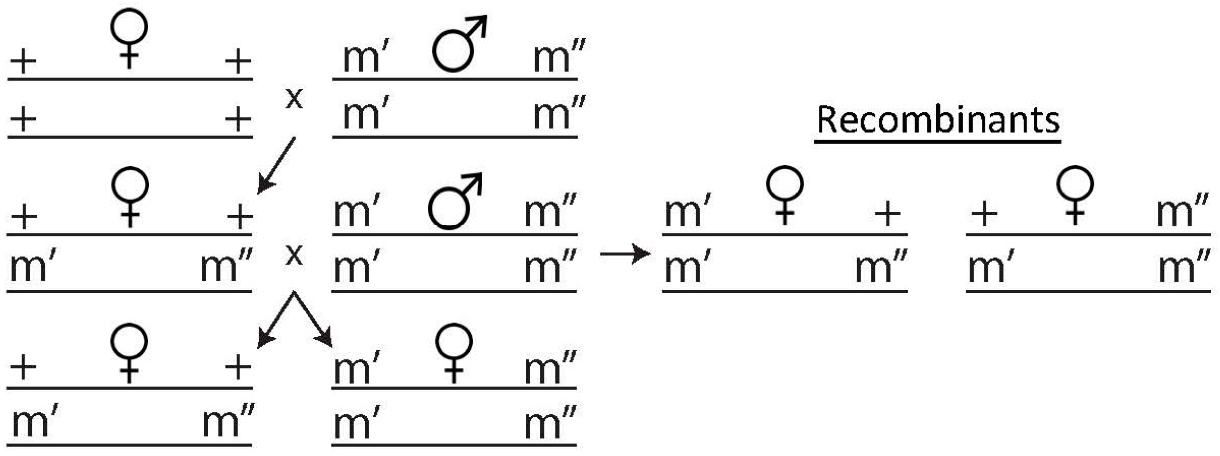
2.4. Recombination Assay

To determine if recombination events had occurred, a two-step crossing method was devised, shown in Figure 1. Ten virgin DGRP females aged 3–5 days were housed with ten phenotypically marked males and were allowed to mate until the appearance of pupae, after which the parents were cleared from the bottle. Virgin female F1 progeny was collected and crossed to the male parental line in the same ratio as before and allowed to mate for 10 days before being cleared from the bottle. All F2 progeny from this cross was collected and frozen within 10 days of clearing the parental F1s. The flies were scored according to their visible phenotypes and sorted into two groups. The proportion of recombinants in each cross was calculated by adding the total exhibiting visible phenotypes and dividing by the total number of progeny. Differences in these proportions between Wolbachia infected and uninfected flies were tested using a logistic regression and Mann–Whitney U tests in SPSS.

3. Results

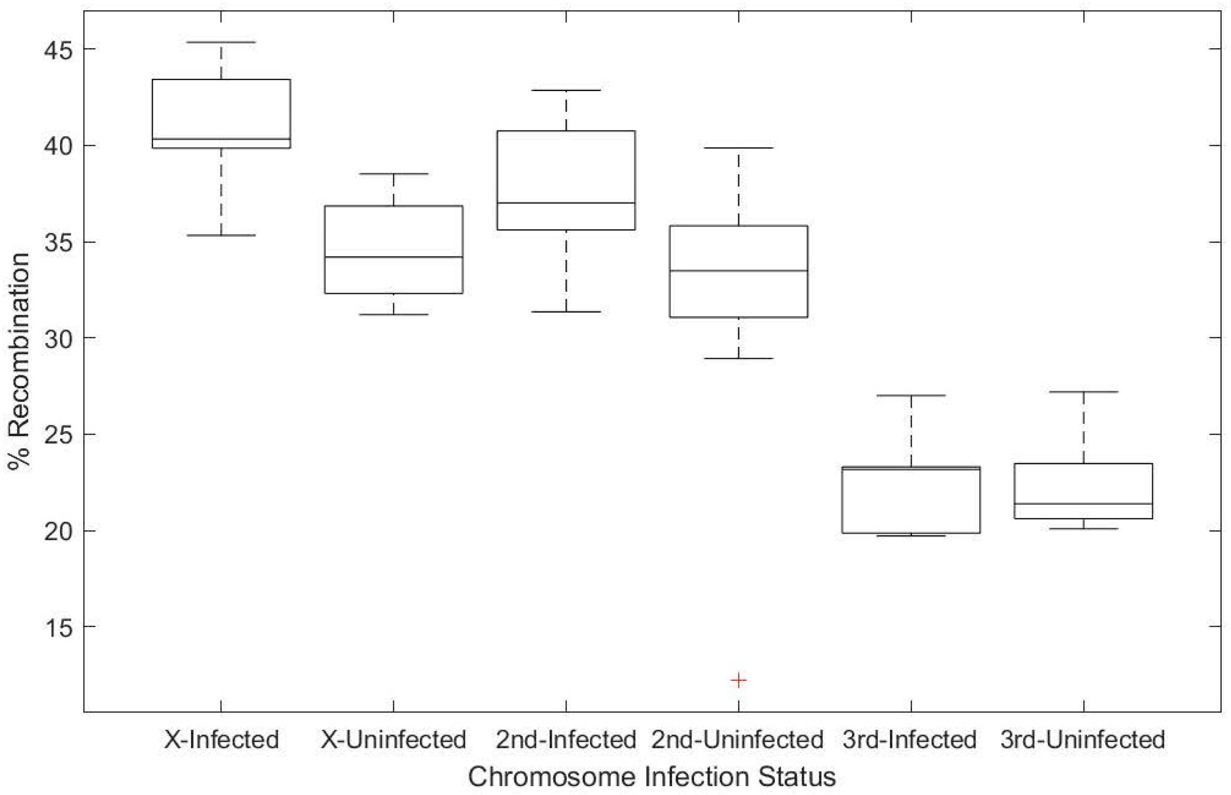
3.1. Wolbachia Infection Increases Host Recombination Rate

Prior work has shown that Wolbachia infection is correlated with an increased frequency of recombination in natural populations [19,20]. We used the same set of isogenized flies, sampled from a wild-caught population in North Carolina, the Drosophila Genetic Reference Panel [24], to confirm prior observations of increased recombination in a Wolbachia-infected fly. Virgin females from two backgrounds (DGRP-320, infected with Wolbachia, and DGRP-83, Wolbachia-free) were crossed independently to three different lines carrying chromosomal markers, allowing us to distinguish recombinants along certain genomic intervals based on the presence of dominant markers (Figure 1). For the X and second chromosomes, we observed an increase in recombination rate of 6.4% and 6.1%, respectively, based on Wolbachia infection status (df = 1, χ2 = 14.316, p < 0.001 and df = 1, χ2 = 5.597, p = 0.018, for the X and second chromosome data) (Figure 2). This observation is well within the range of what had previously been observed for these same genotypes [19]. No statistically significant effect of Wolbachia infection was observed on the third chromosome (df = 1, χ2 = 2.156, p = 0.142). It may be that this difference in recombination rate across chromosomes reflects the natural variation in recombination observed across genomic intervals for D. melanogaster, or it may be an artifact of the genomic interval sampled and not an influence of Wolbachia on specific chromosomes [19].
Although this first experiment established we could replicate prior results, it did not control for host genetic background. To control for host genotype, we cleared the Wolbachia infection from two fly stocks by rearing the flies on tetracycline for three generations and then repopulating the extracellular microbiome for one generation (Table S1). We performed genetic crosses using these cleared backgrounds, comparing them to the uncleared progenitor lines in each case, focusing on the X chromosome interval as we had already established that Wolbachia significantly increased recombination across that genomic interval. For both of these fly stocks, we observed an increase in recombination on the X chromosome when flies were infected with Wolbachia (Figure 3A; df = 1, χ2 = 16.363, p < 0.001 for the OreR-modENCODE background and df = 1, χ2 = 4.461, p = 0.035 for OreR-C). Specifically, we observed an increase in recombination of 6.4%; this increase was well within the range we had observed in our first experiments (Figure 2).

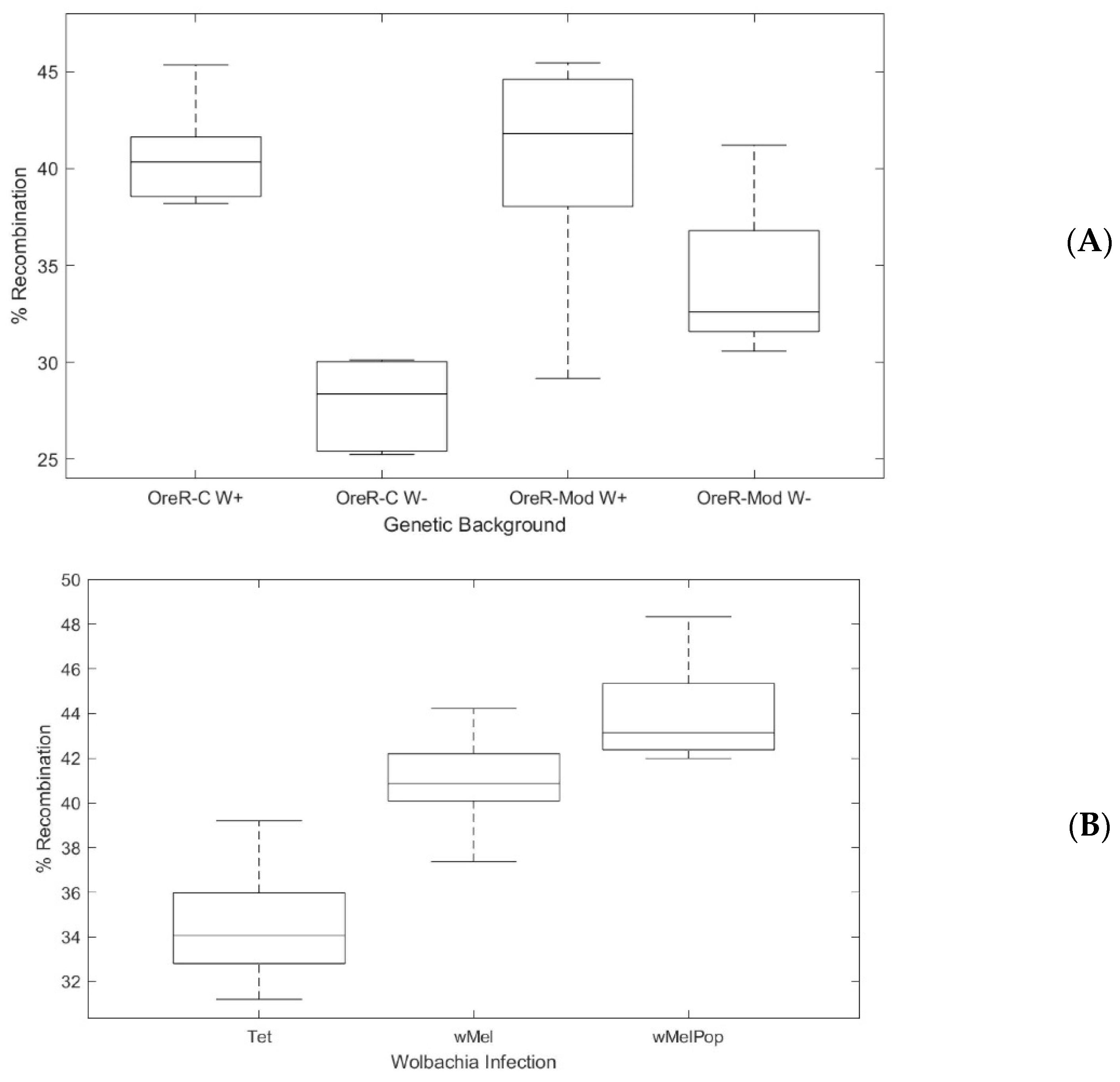
3.2. Dose-Dependent Effect of Wolbachia on Host Recombination Rate

Because we observed an influence of Wolbachia infection status on the host recombination rate, we sought to modulate infection status by using high-titer Wolbachia infections in our experiment. Wolbachia colonize D. melanogaster at different titers depending on the amplification of a specific genomic interval in the Wolbachia genome termed “octomom” [25]. We crossed females carrying the highest-titer, pathogenic Wolbachia variant, wMelPop, (w[1118]/Dp(1;Y)y[+] |Wolbachia-wMelpop; BDSC stock #65284), with males of stock DGRP-320. The lines were introgressed for three generations within the #DGRP-320 genetic background before use in experiments (Figure S2). We also cleared the Wolbachia infection from the DGRP-320 line, using the same tetracycline method as noted above. We looked specifically at the X chromosome interval. Again, we observed a significant effect of Wolbachia infection on recombination rate in this experiment (df = 3, χ2 = 33.4, p < 0.001) (Figure 3). Interestingly, we observed a significant effect of Wolbachia titer on recombination rate—the high-titer wMelPop variant increased recombination on the X chromosome in F2 progeny by 9.5% compared to the 6.3% observed for wMel (df = 3, χ2 = 4.866, p = 0.027) (Figure 3).

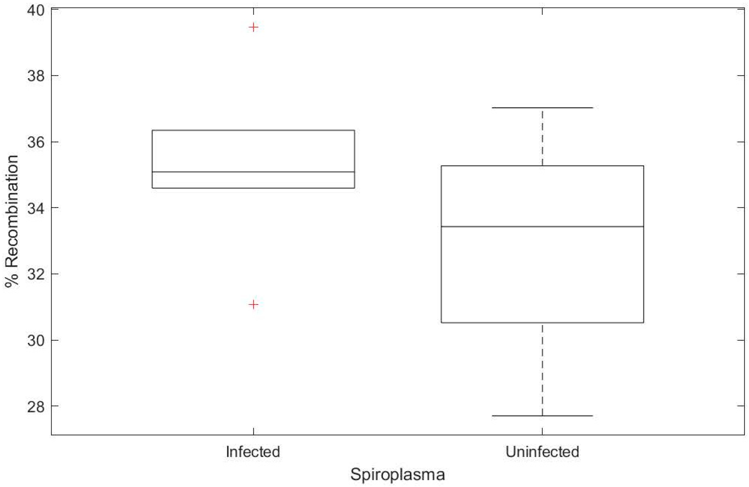
3.3. Spiroplasma Does Not Increase Host Recombination Rate

We hypothesized that Wolbachia may be a stress on the host cell, increasing recombination rate as a result of increased reactive oxygen species or other immune activation pathways. We therefore reasoned that any bacterial infection may increase recombination rate. To test this hypothesis, we procured Spiroplasma poulsonii MSRO (a gift from John Jaenike), which we used to infect a Wolbachia-free OreR lab stock (Oregon-R-modENCODE, BDSC #25211) (Figure S2). We used the same crossing scheme as above to introduce Spiroplasma into the y[1] v[1] background (Figure 1), carrying phenotypic markers on the X chromosome. As a genetic control, we used stock #25211. Counter to our hypothesis, we observed no significant increase in recombination based on Spiroplasma infection (Figure 4 df = 1, χ2 = 0.057, p = 0.812).

4. Discussion

For sexually reproducing organisms, recombination is both a source of genetic diversity within a population and a mechanism by which to decouple differential selection on sites across the chromosome. Therefore, recombination is thought to be beneficial. Here, we observed that Wolbachia infection significantly increased the recombination rate observed across two genomic intervals (for both the X and the second chromosomes). Our goal was to identify whether the Wolbachia infection itself was responsible for the increase in recombination rate that was previously observed. We showed that it is because an increase in Wolbachia titer increases the recombination rate proportionally and because another endosymbiont, Spiroplasma, does not elevate recombination rate. Recombination rates vary dramatically across animals, even within a genus, as best illustrated within the Drosophila clade [26,27]. The mechanism behind this difference is not well understood, but our data suggest that the symbiont Wolbachia may influence the recombination rate of infected D. melanogaster.
The mechanism by which Wolbachia infection elevates recombination is not currently known. Wolbachia have an active type IV secretion system that they use to secrete proteins into the host and modulate host cell biology. It is possible that some of these proteins may influence the recombination rate directly or indirectly, although no effectors have yet been identified that bind to host DNA. Importantly, some of the recently identified and characterized cytoplasmic incompatibility (CI) factors contain nuclease domains [28], and for the cin loci, nuclease activity against DNA in vitro has been directly shown [29]. Wolbachia also encode many other secreted effectors that might directly interact with host proteins involved in recombination [30]. It is not clear how an increase in double stranded breaks would increase the recombination rate, nor what host loci might modulate this increase in recombination. That said, variation in the recombination rate across Drosophila melanogaster genotypes has been clearly observed [19], and a similar approach, modulating Wolbachia infection status in each of these backgrounds, could identify host loci that modulate the recombination phenotype we observe.
Here, we used two different Wolbachia variants and multiple host genotypes to support the hypothesis that Wolbachia increases host recombination rate. However, it is possible that strains outside of the wMel clade do not increase the host recombination and a comparative genomic framework could be used to identify loci in Wolbachia that confer the phenotype. Wolbachia pipientis is certainly not monolithic, and genome content differs significantly across the genus [31]. The clearest comparison would come from using closely related strains, in the same host D. melanogaster background, identifying loss of recombination rate increase. As titer is a confounding variable, this must be controlled in the experiment. Finally, a recent publication suggested Wolbachia wMel-infected Drosophila prefer cooler temperatures [32]. Increases in temperature modulate recombination in D. melanogaster [33], and it is possible that Wolbachia infection elevates host temperature enough to generate an increase in the number of detected recombinants, in a laboratory setting where flies are kept at a constant temperature.

5. Conclusions

In this study, we used genetic crosses and varying titers of Wolbachia pipientis to support the hypothesis that Wolbachia infection increases recombination rate in Drosophila melanogaster. Because Spiroplasma infection does not induce a similar phenotype, we hypothesize that Wolbachia pipientis specifically upregulates recombination, in a dose-dependent manner and that all bacterial infections do not increase recombination rate per se. The mechanism by which this increase in recombination is achieved is not known but we hypothesize that some manipulation of recombination machinery (expression, localization, etc) coupled to DNA damage might induce the phenotype observed.

Supplementary Materials

The following are available online at https://www.mdpi.com/2075-4450/11/5/284/s1, Figure S1: Schematic of backcrossing to introduce wMelPop into the DGRP-320 background, Figure S2A: Wolbachia titer quantification for infection in background DGRP-320 when infected with strain wMelPop compared to strain wMel, Figure S2B: Verification of Spiroplasma infection by PCR, Table S1: List of stocks, their genotypes, and infection status for flies used in this study.

Author Contributions

I.L.G.N. and K.N.B. conceived of the project, and I.L.G.N. and K.N.B. implemented the experiments and wrote the manuscript. All authors have read and agreed to the published version of the manuscript.

Funding

This work was funded in part by the National Science Foundation (NSF 1456545 to ILGN).

Acknowledgments

We thank Lindsay B.M. Nevalainen for contributing key reagents and confirming Spiroplasma infected stocks. We would like to thank two anonymous reviewers for their feedback. We also thank MaryAnn Martin for their assistance and guidance throughout the course of this project and Nadia Singh for discussions at earlier stages of data analysis.

Conflicts of Interest

The authors note no known conflicts of interest.

Data Deposition and Reagent Availability

D. melanogaster strains used in this work are publicly available through the Bloomington Drosophila Stock Center. Spiroplasma-infected OreR is available from the Newton laboratory—please contact irnewton@indiana.edu. Recombination raw count data are available on Dryad (doi:10.5061/dryad.4b8gtht8d).

References

  1. Haddrill, P.R.; Halligan, D.L.; Tomaras, D.; Charlesworth, B. Reduced efficacy of selection in regions of the Drosophila genome that lack crossing over. Genome Biol. 2007, 8, R18. [Google Scholar] [CrossRef] [PubMed] [Green Version]
  2. McGaugh, S.E.; Heil, C.S.; Manzano-Winkler, B.; Loewe, L.; Goldstein, S.; Himmel, T.L.; Noor, M.A. Recombination modulates how selection affects linked sites in Drosophila. PLoS Biol. 2012, 10, e1001422. [Google Scholar] [CrossRef] [PubMed] [Green Version]
  3. Presgraves, D.C. Recombination enhances protein adaptation in Drosophila melanogaster. Curr Biol. 2005, 15, 1651–1656. [Google Scholar] [CrossRef] [PubMed] [Green Version]
  4. Singh, N.D.; Criscoe, D.R.; Skolfield, S.; Kohl, K.P.; Keebaugh, E.S.; Schlenke, T. Fruit flies diversify their offspring in response to parasite infection. Science 2015, 349, 747–750. [Google Scholar] [CrossRef] [PubMed] [Green Version]
  5. Zug, R.; Hammerstein, P. Still a host of hosts for Wolbachia. analysis of recent data suggests that 40% of terrestrial arthropod species are infected. PLoS ONE 2012, 7, e38544. [Google Scholar] [CrossRef] [PubMed] [Green Version]
  6. Werren, J.H.; Windsor, D.; Guo, L.R. Distribution of Wolbachia among neotropical arthropods. Proc. R. Soc. Lond. Ser. B 1995, 262, 197–204. [Google Scholar]
  7. Werren, J.H.; Baldo, L.; Clark, M.E. Wolbachia: Master manipulators of invertebrate biology. Nat. Rev. Microbiol. 2008, 6, 741–751. [Google Scholar] [CrossRef]
  8. Hamm, C.A.; Begun, D.J.; Vo, A.; Smith, C.C.; Saelao, P.; Shaver, A.O.; Jaenike, J.; Turelli, M. Wolbachia do not live by reproductive manipulation alone: Infection polymorphism in Drosophila suzukii and D. subpulchrella. Mol. Ecol. 2014, 23, 4871–4885. [Google Scholar] [CrossRef] [Green Version]
  9. Zug, R.; Hammerstein, P. Bad guys turned nice? A critical assessment of Wolbachia mutualisms in arthropod hosts. Biol. Rev. Camb. Philos Soc. 2015, 90, 89–111. [Google Scholar] [CrossRef]
  10. Hosokawa, T.; Koga, R.; Kikuchi, Y.; Meng, X.Y.; Fukatsu, T. Wolbachia as a bacteriocyte-associated nutritional mutualist. Proc. Natl. Acad. Sci. USA 2010, 107, 769–774. [Google Scholar] [CrossRef] [Green Version]
  11. Chrostek, E.; Marialva, M.S.; Esteves, S.S.; Weinert, L.A.; Martinez, J.; Jiggins, F.M.; Teixeira, L. Wolbachia variants induce differential protection to viruses in Drosophila melanogaster: A phenotypic and phylogenomic analysis. PLoS Genet. 2013, 9, e1003896. [Google Scholar] [CrossRef] [Green Version]
  12. Martinez, J.; Longdon, B.; Bauer, S.; Chan, Y.S.; Miller, W.J.; Bourtzis, K.; Teixeira, L.; Jiggins, F.M. Symbionts commonly provide broad spectrum resistance to viruses in insects: A comparative analysis of Wolbachia strains. PLoS Pathog. 2014, 10, e1004369. [Google Scholar] [CrossRef] [PubMed] [Green Version]
  13. Teixeira, L.; Ferreira, A.; Ashburner, M. The bacterial symbiont Wolbachia induces resistance to RNA viral infections in Drosophila melanogaster. PLoS Biol. 2008, 6, e2. [Google Scholar] [CrossRef] [PubMed] [Green Version]
  14. O’Neill, S.L. The Use of Wolbachia by the World Mosquito Program to Interrupt Transmission of Aedes aegypti Transmitted Viruses. Adv. Exp. Med. Biol. 2018, 1062, 355–360. [Google Scholar] [PubMed]
  15. Fry, A.J.; Rand, D.M. Wolbachia Interactions That Determine Drosophila melanogaster Survival. Evolution 2002, 56, 1976–1981. [Google Scholar] [CrossRef]
  16. Fry, A.J.; Palmer, M.R.; Rand, D.M. Variable fitness effects of Wolbachia infection in Drosophila melanogaster. Heredity 2004, 93, 379–389. [Google Scholar] [CrossRef] [Green Version]
  17. Starr, D.J.; Cline, T.W. A host parasite interaction rescues Drosophila oogenesis defects. Nature 2002, 418, 76–79. [Google Scholar] [CrossRef]
  18. Dedeine, F.; Vavre, F.; Shoemaker, D.D.; Boulétreau, M. Intra-individual coexistence of a wolbachia strain required for host oogenesis with two strains inducing cytoplasmic incompatibility in the wasp asobara tabida. Evolution 2004, 58, 2167–2174. [Google Scholar] [CrossRef]
  19. Hunter, C.M.; Huang, W.; Mackay, T.F.; Singh, N.D. The Genetic Architecture of Natural Variation in Recombination Rate in Drosophila melanogaster. PLoS Genet. 2016, 12, e1005951. [Google Scholar] [CrossRef] [Green Version]
  20. Singh, N.D. Wolbachia Infection Associated with Increased Recombination in Drosophila. G3 (Bethesda) 2019, 9, 229–237. [Google Scholar] [CrossRef] [Green Version]
  21. Herren, J.K.; Paredes, J.C.; Schupfer, F.; Arafah, K.; Bulet, P.; Lemaitre, B. Insect endosymbiont proliferation is limited by lipid availability. Elife 2014, 3, e02964. [Google Scholar] [CrossRef] [PubMed]
  22. Herren, J.K.; Lemaitre, B. Spiroplasma and host immunity: Activation of humoral immune responses increases endosymbiont load and susceptibility to certain Gram-negative bacterial pathogens in Drosophila melanogaster. Cell Microbiol. 2011, 13, 1385–1396. [Google Scholar] [CrossRef] [PubMed] [Green Version]
  23. Anbutsu, H.; Fukatsu, T. Evasion, suppression and tolerance of Drosophila innate immunity by a male-killing Spiroplasma endosymbiont. Insect. Mol. Biol. 2010, 19, 481–488. [Google Scholar] [CrossRef] [PubMed]
  24. Mackay, T.F.; Richards, S.; Stone, E.A.; Barbadilla, A.; Ayroles, J.F.; Zhu, D.; Casillas, S.; Han, Y.; Magwire, M.M.; Cridland, J.M.; et al. The Drosophila melanogaster Genetic Reference Panel. Nature 2012, 482, 173–178. [Google Scholar] [CrossRef] [PubMed] [Green Version]
  25. Chrostek, E.; Teixeira, L. Mutualism breakdown by amplification of Wolbachia genes. PLoS Biol. 2015, 13, e1002065. [Google Scholar] [CrossRef] [Green Version]
  26. Hamblin, M.T.; Aquadro, C.F. High nucleotide sequence variation in a region of low recombination in Drosophila simulans is consistent with the background selection model. Mol. Biol. Evol. 1996, 13, 1133–1140. [Google Scholar] [CrossRef] [Green Version]
  27. Aquadro, C.F.; Begun, D.J.; Kindahl, E.C. Selection, recombination, and DNA polymorphism in Drosophila; Springer: Boston, MA, USA, 1994; pp. 46–56. [Google Scholar]
  28. Lindsey, A.R.I.; Rice, D.W.; Bordenstein, S.R.; Brooks, A.W.; Bordenstein, S.R.; Newton, I.L.G. Evolutionary Genetics of Cytoplasmic Incompatibility Genes cifA and cifB in Prophage WO of Wolbachia. Genome Biol. Evol. 2018, 10, 434–451. [Google Scholar] [CrossRef]
  29. Chen, H.; Ronau, J.A.; Beckmann, J.F.; Hochstrasser, M. A Wolbachia nuclease and its binding partner provide a distinct mechanism for cytoplasmic incompatibility. Proc. Natl. Acad. Sci. USA 2019, 116, 22314–22321. [Google Scholar] [CrossRef]
  30. Rice, D.W.; Sheehan, K.B.; Newton, I.L.G. Large-Scale Identification of Wolbachia pipientis Effectors. Genome Biol. Evol. 2017, 9, 1925–1937. [Google Scholar] [CrossRef] [Green Version]
  31. Lindsey, A.R.; Werren, J.H.; Richards, S.; Stouthamer, R. Comparative Genomics of a Parthenogenesis-Inducing Wolbachia Symbiont. G3 (Bethesda) 2016, 6, 2113–2123. [Google Scholar] [CrossRef]
  32. Truitt, A.M.; Kapun, M.; Kaur, R.; Miller, W.J. Wolbachia modifies thermal preference in Drosophila melanogaster. Environ. Microbiol. 2019, 21, 3259–3268. [Google Scholar] [CrossRef] [PubMed] [Green Version]
  33. Parsons, P.A. Evolutionary Rates-Effects of Stress Upon Recombination. Biol. J. Linn. Soc. 1988, 35, 49–68. [Google Scholar] [CrossRef]
Figure 1. Schematic of fly crossing used to track recombination events. ++ refers to wild type and m’ m” refers to genetic markers on each chromosome (y v on the X; vg bw on the 2nd; e ro on the 3rd). Recombination is tracked by looking at the ratio of recombinants to the total number of progeny produced.
Insects 11 00284 g001
Figure 2. Box and whisker plots showing percentage of recombinants observed, based on genetic markers on the X, 2nd, and 3rd chromosome of Drosophila. Wolbachia infection significantly increased the recombination rate observed on both the X and 2nd chromosome by 6.4 and 6.1%, respectively. A significant increase was observed for the X (df = 1, χ2 = 14.316, p < 0.001) and the 2nd chromosome (df = 1, χ2 = 5.597, p = 0.018), but not on the 3rd chromosome (df = 1, χ2 = 2.156, p = 0.142). Uninfected = DGRP-83; Infected =DGRP-320.
Insects 11 00284 g002
Figure 3. Wolbachia increase in recombination is titer depedent and independent of fly genetic background. (A). Percentage of recombinants observed based on genetic markers on the X chromosome of Drosophila in two different genetic backgrounds, OreR-modENCODE (BDSC stock #25211)and OreR-C (BDSC stock #5), shown as box and whisker plots. Wolbachia infection significantly increased the recombination rate observed in both genetic backgrounds. Significance was determined by logistic regression OreR-modENCODE (df = 1, χ2 = 16.363, p < 0.001) and OreR-C (df = 1, χ2 = 4.461, p = 0.035). (B). Box and whisker plots showing percentage of recombinants observed, based on genetic markers on the X chromosome of Drosophila, when Wolbachia titer is varied. A uniform genetic background was used for comparisons across Wolbachia-uninfected (Tet) flies, Wolbachia infected (wMel) flies, and flies infected with a high-titer variant (wMelPop) (see Figure S1). Increased Wolbachia increased recombination in a load-dependent manner by 6.3% for wMel and 9.5% for wMelPop (df = 3, χ2 = 33.4, p < 0.001). Significance between Tet and wMel (U = 1.0, Z = −3.064, p = 0.002), wMel and wMelPop (U = 8, Z = −2.870, p = 0.004), and Tet and wMelPop (U = 0, Z = −3.182, p = 0.001) was determined with Mann-Whitney U test implemented in SPSS.
Insects 11 00284 g003
Figure 4. Spiroplasma infection does not increase the percentage of recombinants observed based on genetic markers on the X chromosome of Drosophila. Box and whisker plots showing that Spiroplasma infection did not significantly increase the observed recombination rate (df = 1, χ2 = 0.057, p = 0.812). Genetic background for all flies was OreR-modENCODE.
Insects 11 00284 g004

