Review

Efficacy of Two Entomopathogenic Fungi, Metarhizium brunneum, Strain F52 Alone and Combined with Paranosema locustae against the Migratory Grasshopper, Melanoplus sanguinipes, under Laboratory and Greenhouse Conditions

by
Wahid H. Dakhel
1,*,
Alexandre V. Latchininsky
2 and
Stefan T. Jaronski
3
1
Department of Veterinary Pathobiology, College of Veterinary Medicine, University of Missouri, Columbia, MO 65211, USA
2
Food and Agriculture Organization of the United Nations (FAO UN), 00153 Rome, Italy
3
Department of Agriculture, USDA, Agriculture Research Service (ARS), Sidney, MT 59270, USA
*
Author to whom correspondence should be addressed.
Insects 2019, 10(4), 94; https://doi.org/10.3390/insects10040094
Submission received: 8 March 2019 / Revised: 25 March 2019 / Accepted: 29 March 2019 / Published: 30 March 2019
(This article belongs to the Special Issue Biological Control of Plant Pests in Protected Culture)

Abstract

:
Grasshopper outbreaks cause significant damage to crops and grasslands in US. Chemical control is widely used to suppress these pests but it reduces environmental quality. Biological control of insect pests is an alternative way to reduce the use of chemical insecticides. In this context, two entomopathogenic fungi, Metarhizium brunneum strain F52 and Paranosema locustae were evaluated as control agents for the pest migratory grasshopper Melanoplus sanguinipes under laboratory and greenhouse conditions. Third-instar grasshoppers, reared in the laboratory, were exposed up to fourteen days to wheat bran treated with different concentrations of each of the fungi alone or the two pathogens combined. In the greenhouse, nymphs were placed individually in cages where they were able to increase their body temperatures by basking in the sun in an attempt to inhibit the fungal infection, and treated with each pathogen alone or in combination. Mortality was recorded daily and presence of fungal outgrowth in cadavers was confirmed by recording fungal mycosis for two weeks’ post-treatment (PT). For combination treatment, the nature of the pathogen interaction (synergistic, additive, or antagonistic effects) was also determined. In laboratory conditions, all treatments except P. locustae alone resulted in grasshopper mortality. The application of the pathogen combinations caused 75% and 77%, mortality for lower and higher concentrations, respectively than each of the pathogens alone. We infer a synergistic effect occurred between the two agents. In greenhouse conditions, the highest mortalities were recorded in combination fungal treatments with a M. brunneum dose (60% mortality) and with a combination of the two pathogens in which M. brunneum was applied at high rate (50%) two weeks after application. This latter combination also exhibited a synergistic effect. Exposure to the P. locustae treatment did not lead to mortality until day 14 PT. We infer that these pathogens are promising for developing a biopesticide formulation for rangeland pest grasshopper management.

1. Introduction

Grasshoppers (Orthoptera: Acrididae) are important rangeland herbivores that compete with other wildlife and livestock for forage [1,2]. Each year in western United States (US) grasshoppers consume more than 20% of rangeland forages, causing damage estimated at $400 million annually in 1983, close to $1 billion in today’s dollars [3]. There are nearly 500 species of grasshoppers in the 17 US western states and over 100 grasshopper species in Wyoming [4,5]. Of those, about a dozen species are recurrent economic pests including five species in the genus Melanoplus, particularly, the migratory grasshopper M. sanguinipes (Fabricius) [4,6].
During grasshopper outbreaks, a significant effort is needed to prevent damage to rangelands and crops, which is done by large-scale insecticide applications. For instance, more than 8 million hectares of rangelands and crops were blanketed by five million liters of broad-spectrum chemical insecticides at a cost of $75 million in the mid-1980s [7]. Insecticides are still broadcast in rangelands, with almost 2.5 million hectares of Wyoming rangelands treated with insecticides to manage a severe grasshopper infestation [8].
Currently, chemical pesticides, particularly carbaryl, diflubenzuron and malathion, are used to suppress grasshopper outbreaks in the US [9]. The risks of using chemical pesticides to control grasshopper outbreaks include potential deleterious effects on non-target organisms [10] and the recognized environmental side effects. These risks have encouraged interest in developing fungal pathogens because of their environmentally compatible qualities and reduced impacts on non-target beneficial arthropods including pollinators [11]. Possible uses of biological control agents to reduce use of chemical insecticides are under study in federal grasshopper control programs [12].
In 2005, the insect pathogenic fungus Metarhizium anisopliae (Metchnikoff) Sorokin, strain F52, more recently classified as M. brunneum F52 (Hypocreales: Clavicipitaceae), was commercialized as a microbial pesticide in the US to control Coleoptera in horticulture and turf management and soft-bodied ticks [12]. M. brunneum F52 is now recognized as a pathogen of more than 100 insect species from the Hemiptera, Lepidoptera, Thysanoptera, and Diptera orders [13]. It has led to high mortality against the Mormon cricket, Anabrus simplex (Orthoptera: Tettigoniidae), especially under laboratory conditions [12]. The mode of infection can be summarized in the following steps: adhesion, germination, differentiation and penetration. Adhesion involves spore attaching to the cuticle wall, where it germinates to produce an initial hyphal tube (appressorium). This hyphal tube penetrates directly through the exoskeleton and epidermis to reach the hemocoel, in which it develops as yeast-like blastospores [14]. The infection process occurs with the aid of both mechanical and enzymatic degradation of the cuticle. The host will be killed as a result of starvation, nutrient depletion or body obstruction by proliferation of the hyphal bodies [15].
Paranosema (Nosema) locustae (Canning), Sokolova (Microsporidia: Nosematidae) was isolated from the African migratory locust, Locusta migratoria migratorioides in 1953. Presently, this pathogen is the only microsporidium commercially produced and registered in the US for control of rangeland insects [16,17]. P. locustae has a host range of more than 100 orthopteran species and is found across North and South America, Africa, Australia, China, and India [16,18]. The infection of P. locustae spores begins in the grasshopper midgut, and then spreads to the fat body. Once these spores are ingested, the infection occurs through the sporoplasm, which is inoculated by the spores through a polar filament into the midgut epithelial cell. Spores will then germinate in the body to affect tissues and hemocytes [19].Compared to chemical insecticides, the Paranosema and Metarhizium fungal pathogens are slower in killing infected insects [20,21]. To address this shortcoming, the efficacy and/or speed of acridid control may be improved by combining two pathogens [22,23].
Many insect species are able to thermoregulate across changing ambient temperatures [24]. Aside from maintaining optimal thoracic temperatures for locomotion, grasshoppers infected with fungal conidia in the field develop “behavioral fevers”. Many insect orders can generate behavioral fevers, including house flies and stable flies. They assume basking postures or locations that elevate their body temperature above optimal temperatures for microbial growth [25,26]. The mortality rates in field experiments are often lower compared to controlled conditions in a laboratory [11,12]. Performing the experiment in a greenhouse, where the grasshoppers are exposed to sunlight and could thermoregulate, is an intermediate step between laboratory and the field settings. Typically, formulations of the Metarhizium-based biopesticides used in acridid control consist of suspensions of fungal spores in oil [20]. P. locustae is formulated as solid bait on wheat bran carrier [27]. In the present study, the wheat bran based solid bait formulation was used for both fungal pathogens, M. brunneum F52 and P. locustae. We tested the hypothesis that despite the potential thermoregulation, the pathogen treatments will yield high mortality levels to consider them as effective grasshopper biocontrol agents. In this paper we report on the outcomes of experiments designed to test our hypothesis.

2. Materials and Methods

2.1. Experimental Setting

The study was conducted in the Department of Ecosystem Science and Management at the University of Wyoming’s College of Agriculture and Natural Resources insect rearing laboratory and in the greenhouse located at the University of Wyoming Laramie Research and Extension Center, Laramie (41°14′ N, 105°5′ W; elevation 2184 m), Wyoming, USA. In the laboratory, M. sanguinipes were reared using a modified protocol of Hinks and Erlandson [28] in which grasshoppers were maintained in cages (Figure 1a) under laboratory conditions at 27 ± 1 °C, 16D:8L photoperiod, and 31 ± 1% relative humidity (RH). M. sanguinipes nymphs of a non-diapausing strain from the USDA-ARS-NPARL colony were reared in insect cages (BugDorm-1®, MegaView Science Co., Ltd., Taiwan) and provided daily with organic lettuce (Pure Pacific®; Romaine) plus wheat bran (ConAgra Mills) as a dietary supplement until they reached the third instar.
For the greenhouse experiment, the nymphs were transferred to a greenhouse where the temperature and relative humidity were kept at 27 ± 1 °C and 33 ± 1% RH, with a natural photoperiod of approximately 16D:8L. One hundred and twenty cylinder-shaped cages (30 cm H × 8 cm D) were constructed of metal cloth (Figure 1b). This size was chosen to fit over sprouted spring wheat (Triticum aestivum, Prairie Gold® brand from Wheat Montana Farms) grown in round plastic plant pots (12.5 cm H × 8 cm D). All pots with cages were kept on trays placed on a 1m high growing bench.

2.2. Fungal and Baits Preparations

Prior to bait formulation, viability of M. brunneum and P. locustae was determined. Dry spores were suspended in sterile water and diluted in 0.1% surfactant (Silwet® L-77, Helena Agri Enterprises LLC, Collierville, TN, USA). Conidial viability was determined by placing a dilute suspension of conidia onto potato dextrose agar, incubating at 27 °C for 18 h and then examining the conidia for germination using 650× phase contrast magnification. A minimum of 400 conidia were examined for germination and a conidium was considered viable (germinated) if it had formed a visible germination peg throughout the specified incubation time.
M. brunneum F52 conidia from the USDA-ARS-NPARL (Sidney, MT) were applied to wheat bran carrier (ConAgra Mills) or to commercial P. locustae bait (NoLo®, M&R Durango, Ignacio, CO, USA). The baits were prepared with 100 g of wheat bran spread on a large baking sheet and sprayed using a fine spray (DeVilbiss®, Carlisle Fluid Technologies, Scottsdale, AZ, USA) hand atomizer connected to a pressurized air supply, with culinary canola oil at the rate of 3% volume/weight with constant mixing; the canola oil acted as a binder for fungal conidia. The P. locustae commercial formulation consisted of wheat bran carrier at 1 × 10⁹ spores per 454 g (EPA registration# 46149-2). The baits were stored for five days at 4 °C prior to use in the assay.
In the laboratory experiment, treatments of M. brunneum alone came in two concentrations, low and high, with six replicates each. Therefore, Metarhizium was added in quantities sufficient to achieve 0.15 × 10⁹ viable conidia g−1 wheat bran (low dose) and 0.49 × 10⁹ viable conidia g−1 (high dose). These rates are equivalent to 3.3 × 10¹¹ conidia/hectare and 10.78 × 10¹¹ conidia/hectare when the baits are delivered at 2.2 kg ha−1, a typical dose rate used in grasshopper control [9]. Treated bran was then transferred to a 1L container and the requisite amount of conidial powder added. The container was then sealed and agitated for 10 min to thoroughly coat the bran. Treatment with P. locustae alone was another treatment (2.2 × 10⁶ P. locustae spores/g) with six replicates.

2.3. Treatments Application to the Hosts

In laboratory, ten third instar unsexed M. sanguinipes were randomly placed into a 22.86 × 12.7 × 13.97 cm plastic cage® covered with window screen mesh (Figure 1a). The eight experimental treatments were applied to a total of 48 cages (480 grasshoppers). Each treatment was replicated 6 times with biologically independent replicates. The caged nymphs were exposed to continuous, long-term exposure (LTE) with treated bait for 14 day (d), or short-term exposure (STE) for 3 d.
Treatments were applied as two formulations: single pathogen (individual treatment) or mixed pathogens (combination). Applications of M. brunneum alone were delivered to caged grasshoppers for 3 d at low concentration (0.15 × 10⁹ conidia/g; 3DM1) and high concentration (0.49 × 10⁹ conidia/g; 3DM2), and then substituted with untreated wheat bran for remaining 11 d of the experiment (STE). Besides STE, the LTE (14 d) was applied as low 0.15 × 10⁹ conidia/g (EDM1) and high 0.49 × 10⁹ conidia/g (EDM2) concentrations. Application of P. locustae bait was fed to grasshoppers for 3 d a single dose rate of 2.2 × 10⁶ spores/g (N), and then substituted with untreated wheat bran for 11 d.
Combined M. brunneum and P. locustae treatments were set in in two concentrations, low 0.15 × 10⁹ conidia/g (CMN1) and high 0.49 × 10⁹ conidia/g (CMN2), with six replicates each. In these combinations, the dose rate of P. locustae remained the same as in the single pathogen treatment, equaling 2.2 × 10⁶ spores/g. The combination treatments were provided to the grasshoppers for STE (3 d), and then substituted with untreated wheat bran for the remaining 11 d. Untreated wheat bran was provided continuously as the control (C) to groups of nymphs. The details of all eight treatments and dose rates are presented in Table 1. All 48 cages were placed randomly in environmental chambers. All treatments, including untreated wheat bran, were delivered to grasshopper populations in plastic square weighing dishes (89 mm × 89 mm × 25 mm) as 31 flakes of formulation bait per cage (Figure 1a). This amount was based on a standard bait application rate of 2.2 kg ha−1 extrapolated to the area under each cage.
In the greenhouse, one unsexed grasshopper was caged and received six flakes of each of the six different formulations of pathogen-treated wheat bran bait, and those flakes were applied directly on the bottom of each pot once at the start of experiment. Each treatment was replicated 20 times. Over a two-week period, nymphs had continuous access to the bran bait and wheat tillers. The rate of bait (0.0011 g, which is equivalent to six flakes/cage), was calculated taking into account the area of the cage (50.3 cm2) and the standard rate for P. locustae grasshopper bait recommended by the Nolo® bait company, equivalent to 2.2 kg of bait ha−1. In each of the 120 cages, a single unsexed third-instar M. sanguinipes nymph was placed for continuous exposure to different mixture rates of M. brunneum F52 and P. locustae similar to the laboratory experiment. The treatments included EDM1, EDM2, CMN1, CMN2, N, and Control (C).

2.4. Data Collection

Grasshopper mortality was registered daily for 16 d. Dead grasshoppers as well as grasshopper frass, were removed from the cages daily to avoid scavenging by grasshoppers that were still alive. Collected cadavers were washed in 0.5% NaOCl for 1 min and rinsed in distilled water and placed in Petri dishes containing a cotton ball moistened with distilled water to provide high humidity and promote fungal outgrowth in the cadaver. The Petri plates were then incubated at 27 °C in order to observe fungal mycosis on dead grasshoppers [29] (Figure 2).

2.5. Statistical Analysis
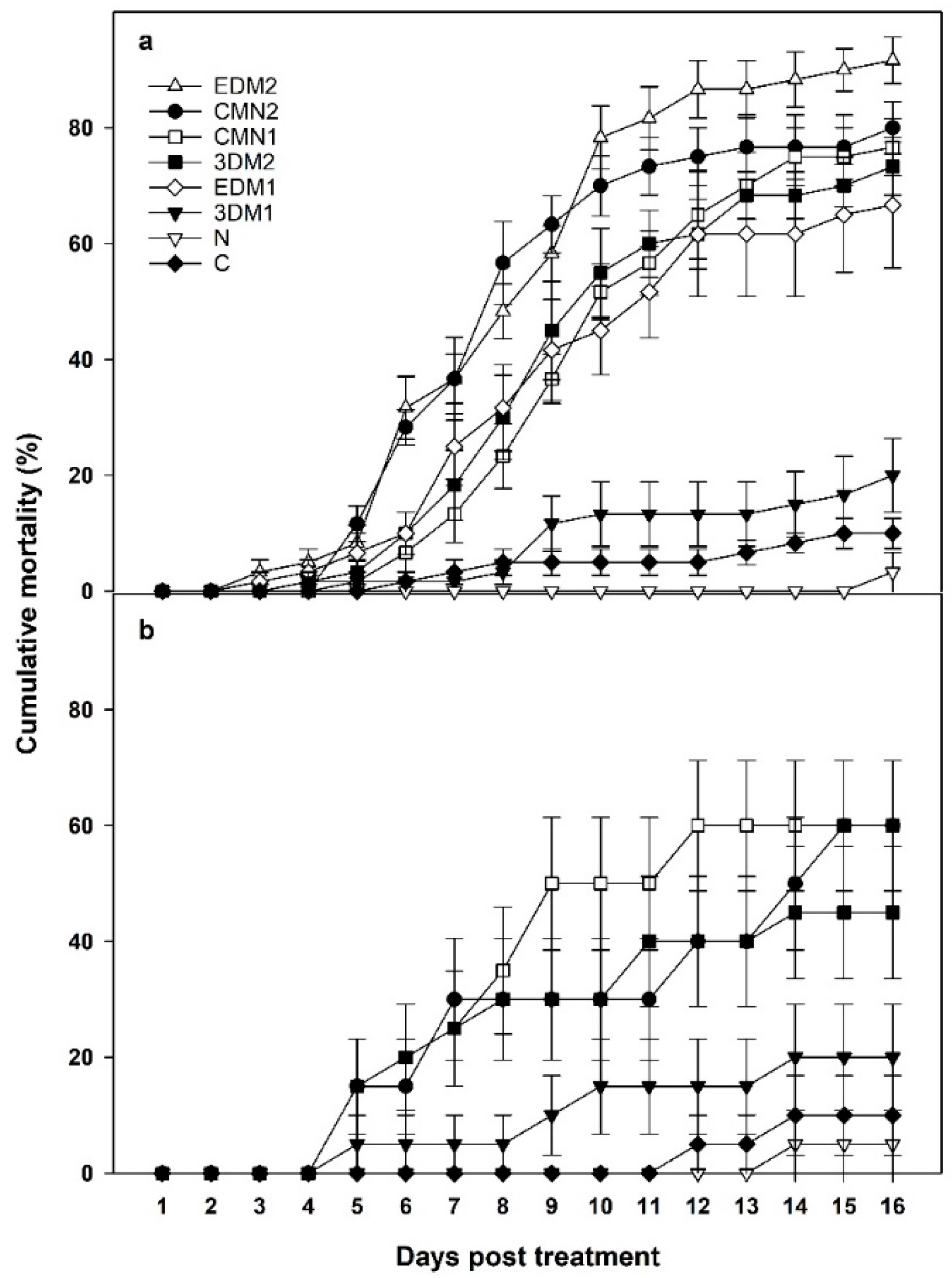
Both experiments were laid out as a randomized complete block design with six replicates under laboratory and twenty replicates under greenhouse conditions. Data of dead grasshoppers were collected at 7, 10 and 14 d after treatment using a one factor factorial analysis of variance (ANOVA) [30]. Post-hoc tests were performed using Fisher’s protected LSD (α = 0.05) procedure, and calculated following Milliken and Johnson method [31]. Homoscedasticity of variances was tested using Hartley’s ( s m a x 2 / s m i n 2 ) statistic and, if the variances were found to be heteroscedastic, the analysis was performed using ( 1 / s i 2 ) as weights, where s i 2 was the variances of the i t h treatment. Normality of residuals was assessed by the Shapiro-Wilk test and normal probability plots. Statistical computations for normality were facilitated using the UNIVARIATE procedure of SAS [32]. Virulence of the pathogen treatments was evaluated using the median survival times (MST) calculated from Kaplan and Meier survivorship analysis 16 d post-treatment (PT) [33]. Combination treatments (M. brunneum + P. locustae) were analyzed at 14 d PT for additive, antagonistic or synergistic interactions using bioassay method one of Nishimatsu and Jackson [34]. First, the expected percentage mortality was calculated using the formula Pe = P0 + (1 − P0) (P1) + (1 − P0) (1 − P1) (P2) where, Pe = expected percentage mortality from combined treatments divided by 100, P0 = percentage control mortality divided by 100, P1 = percentage mortality from agent 1 divided by 100; and P2 = percentage mortality from agent 2 divided by 100. Second, a Chi-square statistic was calculated using the formula χ2 = (L0Le)2/Le + (D0De)2/De where, L0 = Observed # living larvae, Le = Expected # living larvae, D0 = Expected # dead larvae, De = Observed # dead larvae. Finally, the determination of the effect (antagonism, additivity or synergism) was done based on the hypothesis with 1 df and α = 0.05. If χ2 < 3.84 additivity is indicated. If χ2 > 3.84, and Pc < Pe (Pc = observed mortality of combination, and Pe = expected mortality from combined treatments) antagonism is indicated. If χ2 > 3.84 and Pc > Pe, synergism is indicated. If p ≤ 0.05 the observed mortality differs from the expected in at least two treatments. However, if p ≥ 0.05 the observed mortality is the same as the expected over all treatments.
Further analysis was performed on greenhouse experiment. Data were subjected to a two dimensional χ2 test. As the number of grasshoppers per treatment was fixed, the appropriate sampling model for these analyses was that of a product-multinomial [35], with the null hypotheses being the ratio of dead: alive grasshoppers is the same among the treatments versus the alternate that at least two were different (α = 0.05). If the null hypothesis was rejected and the alternate was accepted, then all possible pair-wise 2 × 2 χ2 tests were conducted to determine which treatments were similar and which were different. Finally, as the number of grasshoppers that were dead represented a response variable (and not an explanatory variable) [35], results of the pair-wise tests were summarized as % mortality, along with their observed dead: alive ratios, for each of the three times. Statistical computations were facilitated using the FREQ procedure of SAS [32]. Over a period of 16 days, analysis of speed of lethal action was conducted to compare the virulence between the combination treatments and the single fungus treatments. Survival analysis was conducted using MST, and all treatments that caused more than 50% of grasshopper mortality were analyzed using Kaplan–Meier method and log-rank tests [33].

3. Results

Cumulative mortality of M. sanguinipes nymphs subject to various treatments at the laboratory and greenhouse is shown in Figure 3.

3.1. Treatments Exposure in the Laboratory

Analysis of variance indicated a significant differences occurred between at least two treatments at d 14 (df = (7, 40), F = 120.99, p < 0.0001; Table 2). All experimental treatments except 3DM1 were significantly different from the control. Additionally, a significant effect of grasshopper mortality was observed in treatment of EDM2, which differed significantly when compared to 3DM1, EDM1 and 3DM2.

3.1.1. Single Pathogen Treatments

In the laboratory, at short-term (3 d) exposure to M. brunneum (STE), mortality from the low-dose (low conidial concentration) treatment (3DM1) was consistently lower than mortality from the high dose (high conidial concentration, 3DM2) at 7, 10 and 14 d PT (Table 2). Similarly, high-dose of M. brunneum treatments at continuous exposure (LTE) showed high mortality, with significant difference between high-dose (EDM2) and low-doses (EDM1) of M. brunneum at 10 and 14 d PT (Table 2). The treatment with a high-dose, continuous exposure to M. brunneum (EDM2) resulted in the highest numerical grasshopper mortality (87.3%) in the entire experiment 14 d after treatment.
Mortality of M. sanguinipes nymphs exposed to M. brunneum treatments for only 3 d (STE; 3DM1) was significantly lower than the mortality from LTE-low M. brunneum concentration (EDM1) at 7, 10 and 14 d PT. Similarly, mortality after STE-high M. brunneum concentration (3DM2) was significantly lower than the mortality after LTE-high M. brunneum concentration (EDM2) at 7, 10 and 14 d PT. The other M. brunneum treatments (3DM2, EDM1 and EDM2) produced mortalities statistically different compared to that observed in the control (C) (Table 2).
During most of the observation period, P. locustae did not cause mortality of M. sanguinipes nymphs and only on the 16th d death was first observed on grasshoppers treated with this pathogen (Table 2).

3.1.2. Combination of Treatments

At 10 and 14 d PT, but not 7 d, the grasshopper mortality in the combination treatment with low dose of M. brunneum CMN1 resulted in mortalities statistically different from untreated control (Table 2). At two earlier dates (7 and 10 d after application in the laboratory) the combination treatment with high dose of M. brunneum (CMN2) showed high significant mortality compared to the combination treatment with low (CMN1) dose of M. brunneum (Table 2). Overall, the combination treatments resulted in cumulative mortalities over 70% two weeks after application in the laboratory (Table 2).

3.2. Treatments Exposure in the Greenhouse

A χ2 analyses at 7, 10, and 14 days’ post initial exposure indicated there were differences in dead: alive ratios among the six treatments at each assessment date (7 days: χ 5 2 = 15.968, p = 0.0069; 10 days: χ 5 2 = 23.2926, p = 0.0003; 14 days: χ 5 2 = 24.3389, p = 0.0002). Subsequent comparisons among treatments indicated that for 7-day continuous exposure time, mortality from the M. brunneum at high dosage + P. locustae treatment was significantly greater than mortality from the M. brunneum alone treatment at low dosage, as well as the mortality that occurred from both the untreated control and the P. locustae alone treatment (Table 2). In addition, the mortality that occurred from the M. brunneum + P. locustae treatment at low dosage and the M. brunneum alone treatment at high dosage was significantly greater than the mortality that occurred from both the untreated control and the P. locustae alone treatments.
Results from comparisons at 10-day continuous exposure were identical to those observed at the 7-day continuous exposure time with the exception that the mortality occurring from the M. brunneum at low dosage + P. locustae treatment was significantly greater than the mortality that occurred from the M. brunneum alone treatment at low dosage, the untreated control treatment, and the P. locustae alone treatment (Table 2).
Results from comparisons made at the 14-day continuous exposure had mortality from the M. brunneum + P. locustae treatment at both dosages being significantly greater than the mortalities that occurred from the M. brunneum treatment alone at low dosage, the untreated control, and the P. locustae alone treatment (Table 2). In addition, mortality occurring from the M. brunneum alone treatment at high dosage was significantly greater than the mortality that occurred at both the untreated control and the P. locustae alone treatment (Table 2).
Finally, it bears mentioning that mortalities that occurred from the M. brunneum + P. locustae treatments at both dosages plus the mortalities that occurred from the M. brunneum alone treatment at high dosage were not significantly different, regardless of the duration of exposure time (up to and including 14 days). Likewise, mortalities that occurred from the M. brunneum alone treatment at low dosage, the untreated control, and the P. locustae alone treatments, were not significantly different. Lastly, mortalities from the M. brunneum alone treatment at high dosage were not significantly different from the M. brunneum alone treatment at low dosage (Table 2).

3.3. Virulence

The laboratory results of the virulence bioassay, in which the median survival time (MST) for third-instar nymphs of M. sanguinipes was assessed at 16 d are presented in Table 3. Numerically, median survival time of grasshoppers treated with LTE-high concentration of fungus EDM2 was 8.2 d. Similar to EDM2 treatment, the MST for the combination treatment at high concentration CMN2 had the same length of 8.2 d. Because mortalities of grasshoppers over 16 d were less than 50% (Table 2), the MST for P. locustae alone, untreated control, and STE-low M. brunneum concentration (3DM1) treatments could not be determined. Cox Proportional Hazards analysis [36] with daily observation data showed that fungal treatments with a high concentration of EDM2 and CMN2 were statistically similar in their virulence (Z = −0.84, p = 0.4013, Relative Risk of death (Rel. Risk) = 0.92 for α = 0.05). At the same time, the two treatments were significantly more virulent than control (Table 4).
In the greenhouse, survival analysis showed that the effect of combined treatments with a mixture of two fungi resulted in the highest mortality, thus their median survival time (MST) was determined at 16 days PT in Table 3. Numerically, killing speed of grasshopper nymphs with the combination at low concentration (CMN1) was 10.1 days. In addition, fungus combinations at high concentration (CMN2) had MST of 13.5 days. Thus, based on log rank test (α = 0.05), the virulence of CMN1 and CMN2 were not significantly different (Z = −0.30, p = 0.7654, Relative Risk of death (Rel. Risk) = 0.88) (Table 4). However, virulence in both of combination fungal treatments were statistically different when compared to untreated control (Table 4). MST for other fungal treatments was not calculated for survival analysis because nymphal mortality was below 50% (Table 4).

3.4. Interactions between the Pathogens

Results of the analysis of the interactions between the pathogens at the laboratory and greenhouse are presented in Table 5. In the laboratory, combination treatment of M. brunneum and P. locustae together at high concentration versus LTE-high concentration of M. brunneum and P. locustae alone (CMN2 vs. EDM2, N), the observed mortality in the pathogen combination treatment was lower than the expected based on the separate performance of each pathogen. This means that in this combination, the pathogens exhibited antagonism. In three other cases, the observed mortality from the combination treatments exceeded the expected mortality. Out of those, in one case, comparing combination treatment of M. brunneum and P. locustae together at low concentration versus STE-low of M. brunneum and P. locustae alone (CMN1 vs. 3DM1, N), the pathogen combination produced a synergistic effect while in two others (CMN1 vs. EDM1, N and CMN2 vs. 3DM2, N), the combination resulted in additivity (Table 5).
For the greenhouse, at two weeks post treatment (PT), the combination of the two entomopathogenic fungi M. brunneum F52 and P. locustae at low rate (CMN1) resulted in a synergistic effect (i.e., Supplemental effect that is greater than the sum of each effects alone) when compared to M. brunneum F52 at low rate (EDM1) and P. locustae alone (N) treatments (Pc = 60%, Pe = 31.6%, χ2 = 7.463, df = 1; Table 5). Additive effect (an effect that occurs when the two agents of insect control act independently of each other) was observed with CMN2 treatment when compared to EDM2 and N alone (Pc = 50%, Pe = 53.0%, χ2 = 7.463, df = 1; Table 5). No antagonistic effects between the two fungal treatments were detected.

4. Discussion

In this study, two fungal pathogens, M. brunneum F52 and P. locustae were tested either singly or together under co-infection, delivered on wheat bran carrier, to control 3rd instar nymphs of the grasshopper M. sanguinipes. When applied alone, the M. brunneum caused high mortality of tested insects, especially at high dose 3-d exposure and continuous exposure (3DM2; EDM2). Such treatments resulted in 68 to 88% mortality 14 d after application, respectively. This effect was likely due to production of insecticidal toxins (destruxins) that impair host metabolism and the development of the fungal mycelia, which consume the host nutrients in the hemolymph [37]. Our findings are consistent with the results of Foster et al. [12] who also found high effects of M. brunneum F52 against Mormon crickets, A. simplex, under laboratory conditions.
In contrast, grasshoppers subject to a 3-d exposure to P. locustae did not exhibit mortality until the 16th day PT, which is consistent with previous studies. In Africa, P. locustae was evaluated in the laboratory against first, second, and third-instar nymphs of Senegalese grasshopper, Oedaleus senegalensis [38]. There was higher mortality of early instars that eventually died as fourth-instar nymphs or became moribund as fifth instars at 30 d of observation. There are several potential explanations why the single microsporidian pathogen did not lead to the disease. In terms of pathology, microsporidiosis is categorized as a chronic disease, meaning the disease is slow-acting instead of producing an acute, or fast-acting, infection [39]. Previously, a study concluded that P. locustae needs ample time to invade insect tissues and proliferate [40]. This is similar to the findings of the present study, which indicate that mortality of immature grasshoppers caused by feeding on P. locustae alone occurred only at d 16. Additionally, low mortality rate of grasshoppers being treated with bait consisting of just P. locustae might be explained by grasshoppers either not consuming the microsporidia-treated bait or avoiding it altogether, a behavior which has been sometimes observed in laboratory and field studies [12]. P. locustae alone acts very slowly, produces low and inconsistent mortality among host grasshoppers and therefore should not be considered as a viable microbial control agent.
However, when P. locustae and M. brunneum were applied together, the combination two-pathogen led to additive or even synergistic effects two weeks after treatment. The synergistic reaction occurred, somewhat unexpectedly, when low (and not high) doses of M. brunneum were used (CMN1). Such treatment resulted in ten-fold increase in mortality compared to that produced by M. brunneum by itself. At high dose rates (CMN2), the combination effect resulted only in one- fold increase in grasshopper mortality, compared to that produced by M. brunneum by itself [41]. A plausible explanation is that P. locustae infection develops primarily in midgut and then migrates to fat body. This may impair host metabolism and energy storage, resulting in sluggish behavior [39]. This could also cause the infected insects to become stressed, which favors the second infection by M. brunneum F52.
A study investigated the effects of simultaneous and individual administration of P. locustae and M. anisopliae var. acridum (known as M. acridum) to fifth-instar nymphs of desert locust, Schistocerca gregaria, under laboratory conditions [22]. As a carrier, they used solid wheat bran bait for 21 d. They recorded synergistic effects in treatments of the highest concentration of M. acridum, thus confirming our result that combined pathogens killed grasshoppers faster and more efficiently than either pathogen on its own. However, we found a synergistic interaction when the two pathogens were applied at lower rates, which is more advantageous economically and environmentally. Our findings support the results of Ericsson et al. [42] who demonstrated that dual infections consisting of the bioinsecticide spinosad and biological agent M. anisopliae caused more in reductions in wireworm populations, Agriotes spp. (Coleoptera: Elateridae) than either treatment by itself. Our results indicate that combining even a low dose rate of M. brunneum with P. locustae on wheat bran bait increases the pathogenicity and rate of mortality in host grasshoppers.
Although the fungal pathogen such as M. brunneum does not need to be ingested to cause a mycosis, it was nevertheless quite efficacious against grasshoppers that consumed the wheat bran bait with fungal conidia. We infer the infection occurs via conidia penetration through the insect cuticle on mouthparts as suggested by Hajek and St. Leger [37].
Our study was performed under controlled conditions of a laboratory. Numerous environmental factors may influence the performance of the microbiological entomopathogens under the field conditions [12]. One of the most important is solar UV radiation, which may kill the pathogen conidia directly or provoke a behavioral response in grasshoppers allowing them to increase their body temperature and overcome the infection [43]. Therefore, the results of the greenhouse experiment showed that, despite the assumed host thermoregulation, certain pathogen treatments caused grasshopper mortality under greenhouse conditions. For example, a high rate of M. brunneum application resulted in 45% mortality 14-day PT. Conversely, the application of the low M. brunneum rate yielded only 20% mortality after the same length of incubation (Table 2). Grasshoppers treated with P. locustae did not die until 14-day PT, understanding their mortality was lower than in untreated control. These results are consistent with data obtained in the laboratory experiments as well as in previous studies [44,45].
Combinations of the two pathogens resulted in mortality rates of 50% or higher at 14-day PT. As expected, these mortalities were lower than those obtained under the controlled laboratory conditions (close to 80%, Table 2). Also, the pathogens acted slower: the first signs of mortality appeared 5 and 6-day PT versus 3-day PT in the lab. Yet the fact that at 14-day PT the two combination treatments yielded more than 50% mortality allows consideration of the combination of M. brunneum and P. locustae as promising for developing an operational biological control formulation against pest grasshoppers, most of whom died of mycosis, despite behavioral fevers.
The simulated field study demonstrated that, applied alone, M. brunneum was quite efficacious at a high rate despite the fact that the tested grasshoppers were presumably able to thermoregulate. This suggests that possibly, M. brunneum exhibits tolerance to high body temperatures of grasshoppers, is an important trait of a potential microbiological control agent. Compared to another entomopathogenic fungus Beauveria bassiana, fungi from the genus Metarhizium appear more virulent against grasshoppers Schistocerca americana and Melanoplus sanguinipes even after 21 d of thermoregulation regime [46]. Previous research findings suggest that Mormon cricket Anabrus simplex Haldeman (Orthoptera: Tettigoniidae) also can be affected by M. brunneum. Even though Mormon cricket produces body heat of 39–40 °C during thermoregulation, M. brunneum achieved consistent and satisfactory efficacy, especially under laboratory conditions [12].

5. Conclusions

The application of two fungal pathogens, M. brunneum F52 and P. locustae formulated on wheat bran bait proved effective against migratory grasshoppers M. sanguinipes nymphs under both laboratory and greenhouse conditions. This fungal pathogen combination showed promise in developing a biopesticide against pest grasshoppers. Further research is required to evaluate the efficacy, virulence and persistence of this perspective pathogen combination in the field.

Author Contributions

W.H.D. designed the experiment, collected and analyzed data and wrote the manuscript; A.V.L. administred and supervised the project, provided funds, and reviewed the manuscript; S.T.J. helped in designing the study, provided the necessary lab materials and reviewed the manuscript.

Funding

This research received no external funding.

Acknowledgments

We gratefully acknowledge Scott Schell for laboratory assistance throughout this study and Jaronski for generously providing the entomopathogenic fungi strains.

Conflicts of Interest

The authors declare no conflict of interest.

References

  1. Sergeev, M.G. Ecogeographical distribution of Orthoptera. In The Bionomics of Grasshoppers, Katydids, and their Kin; Gangwere, S.K., Muralirangan, M.C., Muralirangan, M., Eds.; CAB International: Wallingford, UK, 1997; pp. 129–146. [Google Scholar]
  2. Branson, D.H.; Joern, A.; Sword, G.A. Sustainable management of insect herbivores in grassland ecosystems: New perspectives in grasshopper control. Bioscience 2006, 56, 743–755. [Google Scholar] [CrossRef]
  3. Pfadt, R.E. Field Guide to Common Western Grasshoppers; Wyoming Agricultural Experiment Station Bulletin 912: Laramie, WY, USA, 1988. [Google Scholar]
  4. Pfadt, R.E. A Field Guide to Common Western Grasshoppers, 3rd ed.; Wyoming Agricultural Experiment Station Bulletin 912: Laramie, WY, USA, 2002; p. 228. [Google Scholar]
  5. Lockwood, J.A.; McNary, T.J.; Larsen, J.C.; Zimmerman, K.; Shambaugh, B.A.; Latchininsky, A.V.; Herring, B.; Legg, C. Distribution Atlas for Grasshoppers and Mormon Crickets in Wyoming, 1988-92. Available online: http://www.uwyo.edu/entomology/grasshoppers/ghatlas.htm (accessed on 8 March 2019).
  6. Fielding, D.J.; Brusven, M.A.; Shafii, B.; Price, W.J. Spatial heterogeneity of low-density populations of Melanoplus sanguinipes (Orthoptera: Acrididae) associated with grazing and vegetation treatments. J. Can. Entomol. 2001, 133, 843–855. [Google Scholar] [CrossRef]
  7. Lockwood, J.A.; Anderson-Sprecher, R.; Schell, S.P. When less is more: Optimization of reduced agent-area treatments (RAATs) for management of rangeland grasshoppers. Crop Prot. 2002, 21, 551–562. [Google Scholar] [CrossRef]
  8. McNary, T.J.; Shambaugh, B.A.; Elliston, R.J.; Brown, C.L. Cooperative rangeland grasshopper suppression in Wyoming (USA) in 2010. Metaleptea 2011, 31, 10–12. [Google Scholar]
  9. USDA APHIS. Rangeland Grasshopper and Mormon Cricket Suppression Program. Final Environmental Impact Statement; United States Department of Agriculture, Marketing and Regulatory Programs: Riverdale, MD, USA, 2002; p. 336.
  10. Smith, D.I.; Lockwood, J.A.; Latchininsky, A.V.; Legg, D.E. Changes in non-target arthropod populations following application of liquid bait formulations of insecticides for control of rangeland grasshoppers. J. Int. Pest Manag. 2006, 52, 125–139. [Google Scholar] [CrossRef]
  11. Hajek, A.E. Natural Enemies: An Introduction to Biological Control; Cambridge University Press: Cambridge, UK, 2004; p. 378. [Google Scholar]
  12. Foster, R.N.; Jaronski, S.; Reuter, K.C.; Black, L.R.; Schlothauer, R.; Harper, J.; Jech, L.E. Simulated aerial sprays for field cage evaluation of Beauveria bassiana and Metarhizium brunneum (Ascomycetes: Hypocreales) against Anabrus simplex (Orthoptera: Tettigoniidae) in Montana. Biocontrol Sci. Technol. 2011, 21, 1331–1350. [Google Scholar] [CrossRef]
  13. Jaronski, S.T.; Jackson, M.A. Efficacy of Metarhizium anisopliae microsclerotial granules. Biocontrol Sci. Technol. 2008, 18, 849–863. [Google Scholar] [CrossRef]
  14. Furlong, M.J.; Pell, J.K.; Reddy, G.V.P. Premortality effects of Zoophthora radicans infection in Plutella xylostella. J. Invertebr. Pathol. 1997, 70, 214–220. [Google Scholar] [CrossRef] [PubMed]
  15. Reddy, G.V.P.; Furlong, M.J.; Pell, J.K.; Poppy, G.M. Zoophthora radicans infection inhibits the response to and production of sex pheromone in the diamondback moth (Note). J. Invertebr. Pathol. 1998, 72, 167–169. [Google Scholar] [CrossRef] [PubMed]
  16. USEPA. Reregistration Eligibility Document (RED) Facts: Nosema locustae; US Environmental Protection Agency, EPA: Washington, DC, USA, 1992; 738-F-92-011.
  17. Lockwood, J.A.; Bomar, C.R.; Ewen, A.B. The history of biological control with Nosema locustae: Lessons for locust management. J. Int. Tro. Insect Sci. 1999, 19, 333–350. [Google Scholar] [CrossRef]
  18. Lange, C.E. The host and geographical range of the grasshopper pathogen Paranosema (Nosema) locustae revisited. J. Orthoptera Res. 2005, 14, 137–141. [Google Scholar] [CrossRef]
  19. Solter, L.F.; Maddox, J.V. Timing of an early sporulation sequence of microsporidia in the Genus Vairimorpha (Microsporidia: Burenellidae). J. Invertebr. Pathol. 1998, 72, 323–329. [Google Scholar] [CrossRef]
  20. Milner, R.J.; Hunter, D.M. Recent developments in the use of fungi as biopesticides against locusts and grasshoppers in Australia. J. Orthoptera Ress. 2001, 10, 271–276. [Google Scholar] [CrossRef]
  21. Milner, R.J.; Lozano, L.B.; Driver, F.; Hunter, D. A comparative study of two Mexican isolates with an Australian isolate of Metarhizium anisopliae var. acridum–strain characterisation, temperature profile and virulence for wingless grasshopper, Phaulacridium vittatum. Biocontrol 2003, 48, 335–348. [Google Scholar] [CrossRef]
  22. Tounou, A.K.; Kooyman, C.; Douro-Kpindou, O.K.; Poehling, H.M. Interaction between Paranosema locustae and Metarhizium anisopliae var. acridum, two pathogens of the desert locust, Schistocerca gregaria under laboratory conditions. J. Invertebr. Pathol. 2008, 97, 203–210. [Google Scholar] [CrossRef]
  23. Tounou, A.K.; Kooyman, C.; Douro-Kpindou, O.K.; Poehling, H.M. Combined field efficacy of Paranosema locustae and Metarhizium anisopliae var. acridum for the control of sahelian grasshoppers. Biocontrol 2008, 53, 813–828. [Google Scholar] [CrossRef]
  24. Roxburgh, L.; Pinshow, B.; Prange, H.D. Temperature regulation by evaporative cooling in a desert grasshopper, Calliptamus barbarous. J. Therm. Biol. 1996, 21, 331–337. [Google Scholar] [CrossRef]
  25. Ouedraogo, R.M.; Cusson, M.; Goettel, M.S.; Brodeur, J. Inhibition of fungal growth in thermoregulating locusts, Locusta migratoria, infected by the fungus Metarhizium anisopliae var acridum. J. Invertebr. Pathol. 2003, 82, 103–109. [Google Scholar] [CrossRef]
  26. Ouedraogo, R.M.; Goettel, M.S.; Brodeur, J. Behavioral thermoregulation in the migratory locust: A therapy to overcome fungal infection. Oecologia 2004, 138, 312–319. [Google Scholar] [CrossRef]
  27. USEPA (United States Environmental Protection Administration). Nosema locustae (117001) Fact Sheet. 2000. Available online: https://www3.epa.gov/pesticides/chem_search/reg_actions/registration/fs_PC-117001_01-Oct-00.pdf (accessed on 13 February 2019).
  28. Hinks, C.F.; Erlandson, M.A. Rearing grasshoppers and locusts: Review, rationale and update. J. Orthoptera Res. 1994, 1, 1–10. [Google Scholar] [CrossRef]
  29. Elliot, S.L.; Blanford, S.; Thomas, M.B. Host–pathogen interactions in a varying environment: Temperature, behavioral fever and fitness. Proc. R. Soc. London B. 2002, 269, 1599–1607. [Google Scholar] [CrossRef] [PubMed]
  30. Steel, R.G.D.; Torrie, J.H. Principles and Procedures of Statistics: A Biometrical Approach; McGraw-Hill Book Company: New York, NY, USA, 1980. [Google Scholar]
  31. Milliken, G.A.; Johnson, D.E. Analysis of Messy Data; Chapman and Hall: New York, NY, USA, 1992. [Google Scholar]
  32. SAS Institute. SAS/STAT User’s Guide, 2nd ed.; Version 13.2 for Windows; SAS Institute: Cary, NC, USA, 2013. [Google Scholar]
  33. Kaplan, E.L.; Meier, P. Nonparametric estimation from incomplete observations. J. Am. Stat. Assoc. 1958, 53, 457–481. [Google Scholar] [CrossRef]
  34. Nishimatsu, T.; Jackson, J.J. Interaction of insecticides, entomopathogenic nematodes, and larvae of the western corn root worm (Coleoptera: Chrysomelidae). J. Econ. Entomol. 1998, 91, 410–418. [Google Scholar] [CrossRef]
  35. Feinberg, S.E. The Analysis of Cross-Classified Categorical Data, 2nd ed.; The MIT Press: Cambridge, MA, USA, 1980; p. 198. [Google Scholar]
  36. Cox, F.E.G. Concomitant infections, parasites and immune responses. Parasitology 2001, 122, 23–38. [Google Scholar] [CrossRef]
  37. Hajek, A.E.; St. Leger, R.J. Interactions between fungal pathogens and insect hosts. Annu. Rev. Entomol. 1994, 39, 293–322. [Google Scholar] [CrossRef]
  38. Tounou, A.K.; Kooyman, C.; Douro-Kpindou, O.K.; Gumedzoe, Y.M.; Poehlingn, H.M. Laboratory assessment of the potential of Paranosema locustae to control immature stages of Schistocerca gregaria and Oedaleus senegalensis and vertical transmission of the pathogen in host populations. Biocontrol Sci. Technol. 2011, 21, 605–617. [Google Scholar] [CrossRef]
  39. Solter, L.F.; Becnel, J.J.; Oi, D.H. Microsporidian entomopathogens. In Insect Pathology, 2nd ed.; Vega, F.E., Kaya, H.K., Eds.; Elsevier: San Diego, CA, USA, 2012; pp. 221–263. [Google Scholar]
  40. Henry, J.E. Epizootiology of infections by Nosema locustae Canning (Microsporida: Nosematidae) in grasshoppers. Acrida 1972, 1, 111–120. [Google Scholar]
  41. Tounou, A.K. The potential of Paranosema (Nosema) locustae (Microsporidia: Nosematidae) and its combination with Metarhizium anisopliae var. acridum (Deuteromycotina: Hyphomycetes) for the control of locusts and grasshoppers in West Africa. Ph.D. Thesis, Leibniz University of Hanover, Hanover, Germany, September 2007; p. 126. [Google Scholar]
  42. Ericsson, J.D.; Kabaluk, J.T.; Goettel, M.S.; Myers, J.H. Spinosad interacts synergistically with the insect pathogen Metarhizium anisopliae against two exotic wireworms, Agriotes lineatus and Agriotes obscurus (Coleoptera: Elateridae). J. Econ. Entomol. 2007, 100, 31–38. [Google Scholar] [CrossRef]
  43. Inglis, G.D.; Johnson, D.L.; Goettel, M.S. Effects of temperature and thermoregulation on Mycosis by Beauveria bassiana in Grasshoppers. Biol. Control 1996, 7, 131–139. [Google Scholar] [CrossRef]
  44. Ewen, A.B.; Mukerji, M.K. Evaluation of Nosema locustae (Microsporida) as a control agent of grasshopper populations in Saskatchewan. J. Invertebr. Pathol. 1980, 35, 295–303. [Google Scholar] [CrossRef]
  45. Johnson, D.L.; Henry, J.E. Low rates of insecticides and Nosema locustae (Microsporidia: Nosematidae) on baits applied to roadsides for grasshopper (Orthoptera: Acrididae) control. J. Econ. Entomol. 1987, 80, 685–689. [Google Scholar] [CrossRef]
  46. Sieglaff, D.H.; Pereira, R.M.; Capinera, J.L. Pathogenicity of Beauveria bassiana and Metarhizium flavoviride (Deuteromycotina) to Schistocerca americana (Orthoptera: Acrididae). J. Econ. Entomol. 1997, 90, 1539–1545. [Google Scholar] [CrossRef]
Figure 1. Plastic cage® covered with window screens mesh in the laboratory (a); cylindrical metal hardware cloth cages over the sprouted wheat in the greenhouse (b).
Insects 10 00094 g001aInsects 10 00094 g001b
Figure 2. M. brunneum F52 sporulation on the body of grasshopper M. sanguinipes.
Insects 10 00094 g002
Figure 3. Cumulative mortality (%SE) of laboratory (a) and greenhouse (b) Melanoplus sanguinipes nymphs treated with Metarhizium brunneum F52 and Paranosema locustae. 3DM1 = 3 days’ exposure (STE) to M. brunneum at low concentration. 3DM2 = 3 days’ exposure (STE) to M. brunneum at high concentration. EDM1 = Continuous exposure (LTE) to M. brunneum at low concentration. EDM2 = Continuous exposure (LTE) to M. brunneum at high concentration. CMN1 = 3 days’ exposure (STE) to M. brunneum and P. locustae at low concentration. CMN2 = 3 days’ exposure (STE) to M. brunneum and P. locustae at high concentration. N = 3 days’ exposure to P. locustae. C = Untreated control.
Insects 10 00094 g003
Table 1. Materials and rates of baits for treatment applied in study of 3rd instar grasshopper control. These rates were made according to 2.2 kg ha−1.
Treatment MaterialRate
3DM1Feeding low concentration of M. brunneum F52 for only 3 days0.15 × 109 conidia g−1
3DM2Feeding high concentration of M. brunneum F52 for only 3 days0.49 × 109 conidia g−1
EDM1Feeding low concentration of M. brunneum F52 continuously for 14 days0.15 × 109 conidia g−1
EDM2Feeding high concentration of M. brunneum F52 continuously for 14 days0.49 × 109 conidia g−1
CMN1Feeding low concentration of both M. brunneum F52 and P. locustae for only 3 days0.15 × 109 spores g−1
CMN2Feeding high concentration of both M. brunneum F52 and P. locustae for only 3 days0.49 × 109 spores g−1
NExposure grasshoppers for P. locustae by itself2.2 × 106 spores g−1
CUntreated controlUntreated control
3DM1 = 3 days’ exposure (STE) to M. brunneum at low concentration. 3DM2 = 3 days’ exposure (STE) to M. brunneum at high concentration. EDM1 = Continuous exposure (LTE) to M. brunneum at low concentration. EDM2 = Continuous exposure (LTE) to M. brunneum at high concentration. CMN1 = 3 days’ exposure (STE) to M. brunneum and P. locustae at low concentration of M. brunneum. CMN2 = 3 days’ exposure (STE) to M. brunneum and P. locustae at high concentration of M. brunneum. N = 3 days’ exposure to P. locustae. C = Untreated control.
Table 2. Mean percentage of observed mortality of third-instar nymphs of Melanoplus sanguinipes after exposure to bait treated with wheat bran of Metarhizium brunneum F52 and Paranosema locustae for 7, 10, 14 days in laboratory and greenhouse conditions.
Treatment 7 Days10 Days14 Days
LaboratoryGreenhouseLaboratoryGreenhouseLaboratoryGreenhouse
EDM237 a25 ab78 a30 ab88 a45 ab
CMN237 a30 a70 ab30 ab77 ab50 a
CMN113 bc25 ab52 c50 a75 ab60 a
3DM218 b--55 bc--68 b--
EDM125 ab5 b,c45 c15 bc62 b20 bc
3DM12 d--13 d--15 c--
C3 cd0 c5 de0 c8 c10 c
N0 d0 c0 e0 c0 d5 c
3DM1 = 3 days’ exposure (STE) to M. brunneum at low concentration (0.15 × 10⁹ conidia g−1). 3DM2 = 3 days’ exposure (STE) to M. brunneum at high concentration (0.49 × 10⁹ conidia g−1). EDM1 = Continuous exposure (LTE) to M. brunneum at low concentration (0.15 × 10⁹ conidia g−1). EDM2 = Continuous exposure (LTE) to M. brunneum at high concentration (0.49 × 10⁹ conidia g−1). CMN1 = 3 days’ exposure (STE) to M. brunneum and P. locustae at low concentration of M. brunneum (0.15 × 10⁹ spore g−1). CMN2 = 3 days’ exposure (STE) to M. brunneum and P. locustae at high concentration of M. brunneum (0.49 × 10⁹ spore g−1). N = 3 days’ exposure to P. locustae (2.2 × 10⁶ spores g−1). C = Untreated control. Within column, means followed by the same letter are not statistically different at p ≤ 0.05.
Table 3. Median survival time (MST) in days of third-instar nymphs of the migratory grasshoppers, Melanoplus sanguinipes, treated with different concentrations of Paranosema locustae and Metarhizium brunneum F52 for 16 days in laboratory and greenhouse conditions.
Treatment MST(days) §Lower 95%Upper 95%
LaboratoryGreenhouseLaboratoryGreenhouseLaboratoryGreenhouse
EDM28.2--7.2--10.3--
CMN28.213.57.18.49.218.4
CMN110.510.19.17.311.912.8
3DM210.3--9.1--11.5--
EDM110.7--9.6--11.9--
§ MST = Median Survival Time. 3DM1 = 3 days’ exposure (STE) to M. brunneum at low concentration. EDM2 = Continuous exposure (LTE) to M. brunneum at high concentration. CMN2 = 3 days’ exposure (STE) to M. brunneum and P. locustae at high concentration of M. brunneum. CMN1 = 3 days’ exposure (STE) to M. brunneum and P. locustae at low concentration of M. brunneum. 3DM2 = 3 days’ exposure (STE) to M. brunneum at high concentration. EDM1 = Continuous exposure (LTE) to M. brunneum at low concentration.
Table 4. Comparing different treatments in laboratory and greenhouse using Cox’s Proportional Hazards model based on the survivorship of third-instar grasshopper nymphs (α = 0.05).
Variables CoefficientStandard ErrorZ £pR.R ¥
Laboratory
 EDM2 vs. CMN2−0.084490.10067−0.840.40130.92
 EDM2 vs. untreated (C)1.042200.158646.570.00002.84
 CMN2 vs. untreated (C)0.548480.094945.780.00001.73
Greenhouse
 CMN1 vs. CMN2−0.122170.40944−0.300.76540.88
 CMN1 vs. untreated (C)2.189750.767312.850.00438.93
 CMN2 vs. untreated (C)1.049860.382772.740.00612.86
EDM2 = Continuous exposure (LTE) to M. brunneum at high concentration. CMN2 = 3 days’ exposure (STE) to M. brunneum and P. locustae at high concentration. C = Untreated control. CMN1 = exposure to M. brunneum and P. locustae at low concentration. Z£ = Covariate (Coefficient/Standard error). ¥ R.R = Relative Risk of death.
Table 5. Interactions between treatments applied to third-instar nymphs of Melanoplus sanguinipes for 14 days in laboratory and greenhouse conditions.
Treatment Pc% ζPe% §χ2Interaction
Laboratory
 CMN1 vs. 3DM1, and N752297.838Synergism
 CMN2 vs. 3DM2, and N77710.965Additive
 CMN1 vs. EDM1, and N75652.691Additive
 CMN2 vs. EDM2, and N77899.909Antagonism
Greenhouse
 CMN1 vs. EDM1, and N60327.463Synergism
 CMN2 vs. EDM2, and N50530.071Additive
3DM1 = 3 days’ exposure (STE) to M. brunneum at low concentration. 3DM2 = 3 days’ exposure (STE) to M. brunneum at high concentration. EDM1 = Continuous exposure (LTE) to M. brunneum at low concentration. EDM2 = Continuous exposure (LTE) to M. brunneum at high concentration. CMN1 = 3 days’ exposure (STE) to M. brunneum and P. locustae at low concentration of M. brunneum. CMN2 = 3 days’ exposure (STE) to M. brunneum and P. locustae at high concentration of M. brunneum. N = 3 days’ exposure to P. locustae. C = Untreated control. χ2 = Chi square. ζ Pc = observed percentage mortality from CMN1/CMN2 vs. § Pe = expected percentage mortality from 3DM1/3DM2/EDM1/EDM2, N, and Control treatments.

