Article

Knockdown of β-N-acetylglucosaminidase 2 Impairs Molting and Wing Development in Lasioderma serricorne (Fabricius)

Guizhou Provincial Key Laboratory for Rare Animal and Economic Insect of the Mountainous Region, College of Biology and Environmental Engineering, Guiyang University, Guiyang 550005, China
*
Author to whom correspondence should be addressed.
Insects 2019, 10(11), 396; https://doi.org/10.3390/insects10110396
Submission received: 8 October 2019 / Revised: 31 October 2019 / Accepted: 6 November 2019 / Published: 8 November 2019
(This article belongs to the Special Issue RNAi in Insect Pest Control)

Abstract

:
β-N-acetylglucosaminidases (NAGs) are carbohydrate enzymes that degrade chitin oligosaccharides into N-acetylglucosamine monomers. This process is important for chitin degradation during insect development and metamorphosis. We identified and evaluated a β-N-acetylglucosaminidase 2 gene (LsNAG2) from the cigarette beetle, Lasioderma serricorne (Fabricius). The full-length open reading frame of LsNAG2 was 1776 bp and encoded a 591 amino acid protein. The glycoside hydrolase family 20 (GH20) catalytic domain and an additional GH20b domain of the LsNAG2 protein were highly conserved. Phylogenetic analysis revealed that LsNAG2 clustered with the group II NAGs. Quantitative real-time PCR analyses showed that LsNAG2 was expressed in all developmental stages and was most highly expressed in the late larval and late pupal stages. In the larval stage, LsNAG2 was predominantly expressed in the integument. Knockdown of LsNAG2 in fifth instar larvae disrupted larval–pupal molting and reduced the expression of four chitin synthesis genes (trehalase 1 (LsTRE1), UDP-N-acetylglucosamine pyrophosphorylase 1 and 2 (LsUAP1 and LsUAP2), and chitin synthase 1 (LsCHS1)). In late pupae, LsNAG2 depletion resulted in abnormal adult eclosion and wing deformities. The expression of five wing development-related genes (teashirt (LsTSH), vestigial (LsVG), wingless (LsWG), ventral veins lacking (LsVVL), and distal-less (LsDLL)) significantly declined in the LsNAG2-depleted beetles. These findings suggest that LsNAG2 is important for successful molting and wing development of L. serricorne.

1. Introduction

Chitin is a polymer of β-1,4-linked N-acetylglucosamine residues and an essential component of the integument, trachea, salivary gland, foregut, hindgut, and intestinal peritrophic matrix in insects [1,2]. Chitin plays major roles in maintenance of the shape, size, and protection from external forces such as mechanical injuries and infection by bacteria, fungi, and viruses [3,4]. The regulation of chitin metabolism is crucial for insect growth and development [5,6]. Among the enzymes involved in chitin metabolism, β-N-acetylglucosaminidases (NAGs, EC 3.2.1.30) are chitin-degrading enzymes that hydrolyze the chitin oligosaccharides into N-acetylglucosamine monomers [7]. NAGs (also known as β-N-acetylhexosaminidases) are exosplitting enzymes that belong to the glycoside hydrolase family 20 (GH20) [8]. Insect NAGs are divided into four subgroups: chitinolytic group I NAGs, chitinolytic group II NAGs, glycan processing group III NAGs, and hexosaminidases group IV, on the basis of their sequence similarity and functions [9,10,11].
Insect NAGs are widely distributed and occur in many insect species, including Coleoptera, Diptera, Hemiptera, Lepidoptera, and Orthoptera [10,11,12,13,14,15]. NAGs mainly function in chitin degradation, and their roles have been studied in numerous insect species. In Aedes aegypti, the activity of NAG enzyme increased after feeding on blood or artificial diet [16]. RNA interference (RNAi)-mediated knockdown of NAGs causes molting failure in Tribolium castaneum [10], Locusta migratoria [13], Nilaparvata lugens [11], Mamestra brassicae [14], and Heortia vitessoides [17]. In Ostrinia furnacalis [17] and Lasioderma serricorne [18], NAGs are essential for the proper formation of adult wings. In addition to their central role in chitin degradation, NAGs participate in diverse physiological processes, such as post-translational modification of N–glycan proteins [19], degradation of glycoconjugates [20,21], and fertilization [22,23].
The cigarette beetle, Lasioderma serricorne (Fabricius) (Coleoptera: Anobiidae), is a serious pest of stored products worldwide [24]. The beetle causes economic losses to stored products such as tobacco, grains, traditional Chinese medicine materials, cereals, and dry foods [25,26]. Control of L. serricorne is primarily dependent on the use of insecticides, including phosphine, essential oils, pyrethrin, and organophosphates [27,28]. Excessive and frequent insecticide applications have resulted in insecticide resistance within many L. serricorne populations. In addition, insecticide residues in stored products are harmful to human health and the environment [29,30]. Therefore, new strategies are needed to effectively manage L. serricorne infestations. RNAi-based technologies have been used for the management of several insect pests [31,32,33]. Screening for an ideal silencing target is critical for the development of an RNAi-based control method [34,35]. In terms of practice, transgenic plants in expressing sequence-specific double-stranded RNA (dsRNA) [36] and spray-based dsRNA reagent application [37] have potential in implementation as future pest control, and a trial of Diabrotica virgifera virgifera via targeting DvSnf7 was firstly tested in filed as RNAi-based pest control incoperated with Bacillus thuringiensis Cry3Bb1 protein [38]. Chitin degrading enzymes are potential targets for novel insecticides [17,39,40,41]. In this study, we identified and cloned the full-length open reading frame (ORF) sequence of β-N-acetylglucosaminidase 2 gene (LsNAG2) in L. serricorne and analyzed its expression patterns in different developmental stages and tissues. We functionally characterized the knockdown effect of LsNAG2 on L. serricorne molting and the responses of genes related to wing development. The data provide significant insight into the functions of LsNAG2 in L. serricorne development.

2. Materials and Methods

2.1. Insect Culture

Lasioderna serricorne were collected from a tobacco warehouse in Guiyang City, Guizhou Province, China, in 2014. A colony was established in the laboratory and maintained at 28 °C with 40% relative humidity under a scotoperiod of 24 h. Larvae were fed on dried roots of Angelica sinensis, as described previously [42].

2.2. RNA Extraction and Cloning of LsNAG2 Gene

Total RNA was extracted from entire bodies of 30 L. serricorne larvae with TRIzol reagent according to manufacturer protocol (Invitrogen, Carlsbad, CA, USA), and treated with DNase I (Promega, Madison, WI, USA). The concentration and quantity of total RNA were measured by a NanoDrop 2000C spectrophotometer (Thermo Scientific, Waltham, MA, USA). RNA integrity was evaluated by agarose gel electrophoresis. First-strand complementary DNA was synthesized by the TransScript Synthesis Supermix (TransGen, Beijing, China) with oligo (dT)18 primers.
A candidate cDNA sequence of β-N-acetylglucosaminidase 2 gene was identified by Basic Local Alignment Search Tool Nucleotide (BLASTN) and Translated Basic Local Alignment Search Tool Nucleotide (TBLASTN) analyses from the transcriptome database of L. serricorne (unpublished data). The full-length ORF of the sequence was found using ORF Finder at the National Center for Biotechnology Information (NCBI) website (http://www.ncbi.nlm.nih.gov/gorf/gorf.html). Two pairs of gene-specific primers (Table S1) were designed to amplify the full-length cDNA of LsNAG2 by reverse transcription PCR (RT-PCR). The PCR amplification was carried out with an initial denaturation at 95 °C for 4 min, 34 cycles of 95 °C for 30 s, 56 °C for 30 s, and 72 °C for 2 min, and a final elongation at 72 °C for 10 min. The PCR products were purified using a Wizard SV Gel and PCR Clean-Up System (Promega, Madison, WI, USA) and sequenced in both directions.

2.3. Sequence Analysis

Sequence similarities and domain predictions were produced using the Basic Local Alignment Search Tool program at the NCBI website (http://blast.ncbi.nlm.nih.gov/). The nucleotide and deduced amino acid sequences were analyzed using DNAMAN7 (Lynnon Biosoft, Vaudreuil, Quebec, Canada). The molecular weight and isoelectric point were calculated using ExPASy tools (http://cn.expasy.org/tools/pi_tool.html). The N-glycosylation sites were predicted using the NetNGlyc1.0 Server (http://www.cbs.dtu.dk/services/NetNGlyc/), and the signal peptide was predicted using the SignalP4.1 program (http://www.cbs.dtu.dk/services/SignalP4.1/). The phylogenetic tree was constructed by MEGA7 (MEGA, PA, USA) [43] using the neighbor-joining method with bootstrap values using 1000 replicates at the cut off of 50% similarity.

2.4. Quantitative Real-Time PCR

For the stage-specific expression analyses, samples at different developmental stages of L. serricorne were prepared as described previously [42]. To analyze tissue-specific expression, eight tissues (integument, brain, foregut, midgut, hindgut, fat body, and Malpighian tubules) were dissected from the late larvae. RNA extraction and cDNA synthesis were performed as described above. The relative expression levels of LsNAG2 were detected by quantitative real-time PCR (qPCR). The qPCR assay was performed with GoTaq qPCR Master Mix (Promega, Madison, WI, USA) using the CFX-96 real-time PCR system (Bio-Rad, Hercules, CA, USA). The reaction was run at 95 °C for 5 min, followed by 40 cycles of 95 °C for 15 s and 60 °C for 1 min. A melt curve analysis was performed to assess the specificity of the qPCR amplifications. On the basis of the evaluation, the L. serricorne elongation factor 1-alpha (EF1α, GenBank: KY549658) was used for normalization (unpublished data). All of the experiments were performed in triplicate, and the results were calculated by the 2−ΔΔCt method [44].

2.5. RNA Interference

RNAi was performed to study the effects of LsNAG2 on L. serricorne development. The primers (Table S1) used for dsRNA synthesis were designed to add the T7 polymerase promoter sequence at the 5′-ends. The dsRNAs were synthesized using a TranscriptAid T7 High Yield Transcription Kit (Thermo Scientific, Wilmington, DE, USA). The quality and concentration of dsRNAs were measured with a NanoDrop 2000C spectrophotometer (Thermo Scientific), and their sizes were verified by agarose gel electrophoresis. The dsRNA of green fluorescent protein (GFP) served as the negative control. The day 2 fifth-instar larvae and day 5 pupae were used for microinjection; 50 individual insects were used for each group, and the experiment was performed in triplicate. About 300 ng dsRNA of GFP or LsNAG2 were slowly injected into the hemocoel between the second and third abdominal segments of each larva and pupa using a Nanoliter 2010 injector (World Precision Instruments, Sarasota, FL, USA). All of the injected insects were kept under the conditions mentioned above. To determine silencing efficiency, relative expression levels of LsNAG2 in both dsLsNAG2 and dsGFP group at 3 and 5 days after injection were measured by qPCR as described above. Insect survival rate and phenotype changes were observed and recorded. Photos were taken using a Keyence VHX-6000 stereomicroscope (Keyence Corporation, Osaka, Japan).
To determine the expression of chitin synthesis genes, samples were collected from the larvae treated with dsLsNAG2 and dsGFP for 5 days. The qPCR was used to analyze the expression of trehalase (LsTRE1 and LsTRE2), UDP-N-acetylglucosamine pyrophosphorylase (LsUAP1 and LsUAP2), and chitin synthase 1 (LsCHS1) as described above. We also evaluated the expressions of wing development-related genes after gene silencing in late pupae. The insect samples were collected 5 days after dsRNA injection in late pupae for expression analysis. The relative expression levels of nine wing development network genes, including miniature (LsMT), singed (LsSG), engrailed (LsEN), teashirt (LsTSH), wingless (LsWG), distal-less (LsDLL), vestigial (LsVG), ventral veins lacking (LsVVL), and apterous (LsAP), were detected by qPCR.

2.6. Statistical Analysis

All the expression data are presented as mean ± standard error (SE) and evaluated using SPSS 20.0 software (IBM Corp, Chicago, IL, USA). One-way analysis of variance (ANOVA) followed by a least significant difference test was used to analyze differences among different developmental stages and tissues. Student’s t-test was used to analyze data from the RNAi-treatments after dsRNA injection. The survival rates were analyzed using the Kaplan–Meier method.

3. Results

3.1. Identification and Characterization of LsNAG2

The full-length ORF of LsNAG2 (GenBank accession number MN310607) was obtained and confirmed by RT-PCR. The LsNAG2 cDNA sequence contained an ORF of 1776 bp that encoded a protein of 591 amino acids. The predicted protein of LsNAG2 had a molecular weight of 66.6 kDa and an isoelectric point of 5.92. Signal peptides with 16 amino acids were found at the N-terminal ends of LsNAG2, which suggested it may be secreted. Two potential N-glycosylation sites at positions 150 and 549 were predicted in the N-terminal extracellular domain.
The predicted amino acid sequences contained a conserved GH20 catalytic domain (residues 213–572), and an additional GH20b domain (residues 136–186). The conserved H×GGDEV×××CW motif was considered to be the catalytic site. The conserved active site residues R224, D377, E378, W435, W459, Y486, D488, W522, and E524 were found in the LsNAG2 sequence (Figure 1). Sequence alignment showed that LsNAG2 exhibited 60%, 54%, 53%, and 52% identity with NAG2 (XP_018336054) from Agrilus planipennis, NAG2 (XP_015833058) from T. castaneum, NAG (XP_023711735) from Cryptotermes secundus, and NAG (XP_022185539) from Nilaparvata lugens, respectively. A phylogenetic tree was generated using MEGA7 on the basis of the amino acid sequences of insect NAGs and related hexosaminidases. The tree showed that these proteins were classified into four major groups: NAG group I, NAG group II, N-glycan processing NAGs (group III), and hexosaminidase (group IV). The LsNAG2 protein was located on a branch with the group II NAGs of other insects (Figure 2).

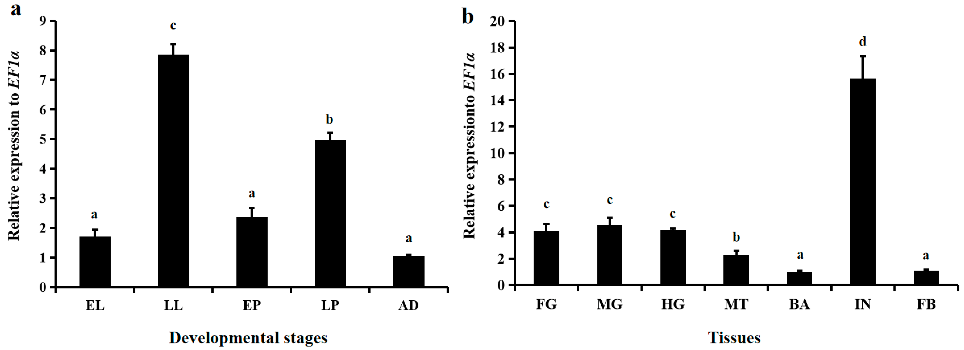
3.2. Spatiotemporal Expression Patterns of LsNAG2

Among the five tested stages, that is, early larvae (<24 h post-hatching), late larvae (older than fourth instar but before prepupae), early pupae (>72 h post-pupation), late pupae (>144 h post-pupation), and adults (>72 h post-eclosion), LsNAG2 was continuously expressed but displayed different expression patterns. LsNAG2 was more highly expressed in the late larval and late pupal stages than in the other stages (Figure 3a). The late larval stage had the highest expression of LsNAG2 and was investigated for spatial expression patterns. The highest expression level of LsNAG2 was observed in the integument and was approximately 15.6-fold greater than in the brain (Figure 3b).

3.3. Effects of LsNAG2 RNAi on the Expressions of Chitin Synthesis Genes

To study the functions of LsNAG2 in the molting process, specific dsRNA for LsNAG2 and GFP were synthesized in vitro and injected into fifth instar larvae and late pupae. Compared to the control, the expression of LsNAG2 was significantly reduced by 78% and 87% at 3 and 5 days after injection with dsLsNAG2 in the fifth instar larvae, respectively (Figure 4a). After inhibition of LsNAG2, the survival rate of L. serricorne was reduced to 57% at 15 days in the dsLsNAG2 group (Figure 4b).
After RNAi with dsLsNAG2 or dsGFP at larvae, qPCR was used to study the expression of chitin synthesis genes. Expression levels of LsTRE1, LsUAP1, LsUAP2 and LsCHS1 were significantly decreased in larvae treated with dsLsNAG2 compared to levels in control insects (Figure 4c). This suggested that LsNAG2 is involved in the regulation of genes involved in chitin synthesis during the larval–pupal transition. The lethal phenotypes included the following: (1) 28% of the individuals were unable to molt, retained the larval form, and ultimately died; and (2) 15% of the individuals eventually shed their old cuticles and completed the larval–pupal transformation but died after molting (Figure 4d).

3.4. Effects of LsNAG2 RNAi on the Expressions of Wing Development-Related Genes

Injection of late pupae with dsLsNAG2 significantly decreased the expression of LsNAG2 by 85% and 91% at 3 and 5 days, respectively, compared to the control (Figure 5a). In the control group, 96% of pupae molted normally to adults during the pupa to adult transition. Pupae injected with dsLsNAG2 exhibited lethal phenotypes. The accumulative survival rate of pupae was reduced to 45% at 15 days after treatment compared with the controls (Figure 5b). Approximately 11% of the individuals had difficulty shedding the old cuticle, failed to develop into adults, and eventually died. Interestingly, 44% of the individuals successfully molted but they did not complete wing development and died with an asymmetric wing pattern (Figure 5c).
To detect effects of dsLsNAG2 injection on wing development, we examined the expression of nine genes related to wing development after RNAi with dsLsNAG2 or dsGFP at late pupae. The qPCR results showed that the mRNA levels of five transcripts (LsTSH, LsVG, LsWG, LsVVL, and LsDLL) were significantly reduced in the LsNAG2-depleted pupae, compared to levels in the controls (Figure 5d). In contrast, the expression levels of LsAP and LsEN were significantly higher in LsNAG2 RNAi pupae.

4. Discussion

Chitin synthesis and chitin degradation are both important in insect development [5]. Chitinases and NAGs are critical enzymes for insect chitin metabolism. Chitinases hydrolyze chitin into oligosaccharides, whereas NAGs further degrade the small oligomers into N-acetylglucosamine monomers [7]. Several NAG genes have been characterized in many insect species. There are 4 in T. castaneum [10], 6 in Bombyx mori [45], 5 in Drosophila melanogaster [22], and 11 in N. lugens [11]. In L. serricorne, one NAG gene (LsNAG1) has been previously characterized [18]. In this study, we describe a second NAG, LsNAG2, in the cigarette beetle L. serricorne. The GH20 and GH20b domains were predicted in LsNAG1 and LsNAG2 proteins, which indicated that these two genes belong to the glycoside hydrolase family 20. Both domains were also predicted in the NAG of Exopalaemon carinicauda [46]. The phylogenetic relationships indicate that LsNAG2 belongs to the chitinolytic group II NAG [10]. All insect NAGs are related to chitin degradation, but group I and II are well characterized in chitin decomposition. The sequence characterization and phylogenies showed that TcNAG2 in T. castaneum and DmNAG2 in D. melanogaster are closely related and LsNAG2 possibly has a chitin degradation function [10,20].
NAGs are widely expressed in all developmental stages of insects because of their multiple physiological functions. For example, four NAGs are expressed in all stages of N. lugens [11]. In this study, LsNAG2 was expressed in all tested stages of L. serricorne but most highly expressed in late instar larvae and late pupae. This indicates its vital role in the degradation of old cuticle during molting. Similar high expression in late larvae and late pupae also occur in LsNAG1 [18]. High expression of NAG was observed at late fourth and fifth instar larvae of H. vitessoides [16] and O. furnacalis [47]. High expression was also seen in the last instar larvae of M. brassicae [14]. Increased expression of TcNAG1 and TcNAG3 was investigated in late instar larvae and pupae of T. castaneum, respectively [10]. Several NAGs were also highly expressed in the early pupal stage in other insects [10,14].
Insect NAGs are widely expressed in multiple tissues [11,45]. In N. lugens, a group II NAG (also defined as β-N-acetylhexosaminidases) gene, NlHex3, was stably expressed in all tissues, whereas the others showed a tissue-biased pattern, such as the gut, Malpighian tubules, salivary gland, and reproductive organ [11]. The spatial expression indicated tissue-specific functions of NAGs in insects. In B. mori, ovary- and testis-specific expressions were observed [45]. A fertilization-related function was demonstrated in D. melanogaster, in which three NAG genes were expressed in the male germ line [22]. In this study, LsNAG2 was prominently expressed in integument. A similar expression pattern was also demonstrated in NlHex4 of N. lugens [11]. The high expression of NAGs in integument may be associated with chitin metabolism during the molting process. In Choristoneura fumiferana, an immunohistochemical staining assay revealed that CfNAG occurred in the degrading exocuticle and molting fluid during late ecdysis [48]. Ecdysone is a hormone involved in the insect molting process, and it helps to regulate NAG activity during development. In H. vitessoides and M. brassicae, the NAG gene was upregulated by exogenous 20-hydroxyecdysone [14,16]. This induced expression can be suppressed by juvenile hormone [7]. The 20E-induced expression was studied in LsNAG1 and shown to be involved in the larval–pupal transition [18].
The suppressed LsNAG1 disrupted the metamorphic transition from larva to pupa and pupa to adult [18]. The expression profiling also showed a similar role in the transition. When the LsNAG2 was successfully suppressed by exogenous dsRNA micro-injection, almost 43% of the larvae exhibited a lethal phenotype (failure to successfully molt). A similar disruption was observed in N. lugens when NlHex4 was suppressed, but it did not reveal any abnormal phenotypes in the remaining 10 NAG homologs [11]. In L. serricorne, suppression of LsNAG2 led to molting failure of LsNAG1 and chitin deacetylase 1 gene [18,49]. The function of NAGs in molting was also studied in L. migratoria [13], M. brassicae [14], and O. furnacalis [18,47]. NAGs were present in the molting fluid and showed a positive synergistic effect on chitin degradation together with chitinases [50,51]. Gene expression of the chitin synthesis pathway was studied by qPCR, and four out of the selected five genes were downregulated by silencing LsNAG2. This indicated that the disrupted chitin degradation also inhibited new chitin biosynthesis when the chitin metabolism-related LsNAG2 was suppressed at the transcriptional level. However, the relative expression of the chitin synthesis-related genes was not influenced when NAG was knockdown by dsRNA in H. vitessoides [16]. In addition, the genes involved with chitin degradation showed no change when HvNAG1 was silenced. Group I NAGs are all secreted proteins that have been isolated from molting fluid, integument, and gut. They are functionally responsible for chitin degradation in the cuticle [51]. In this study, LsNAG2 had a role similar to other group I NAGs and was involved in chitin degradation within the cuticle. The phenotypes produced in T. castaneum by knocking down the other NAGs was studied, and the efficiency was not as strong as that produced by the group I NAG (TcNAG1) [10].
In M. brassica, suppression of NAG expression in larvae also increased mortality at the pupal-adult transition [14]. The continuous suppression was also reflected in the following stages [13,16]. We studied the role of LsNAG2 in the pupa-adult transition and observed high mortality when LsNAG2 was silenced. The higher mortality of LsNAG1 indicated a stringent efficiency during pupal–adult molting, which can also be concluded by the comparison of accumulative mortality of NAG1 and NAG2 knocked phenotype in L. serricorne [10,18]. Similar to the LsNAG1 RNAi bioassay, asymmetric wing development was also observed in the LsNAG2 RNAi, indicating a critical role in the wing development. Suppressed HvNAG1 in H. vitessoides also led to an asymmetric wing pattern in adults [16]. In O. furnacalis, suppressed OfHex2 resulted in 23.3% of the adults having abnormal wings [17]. The relationship of chitin-degrading enzymes and the upstream chitinase 7 with wing development was also documented in T. castaneum [52]. However, the mechanism of this abnormality in wing development is unclear. Therefore, we studied the gene expression of several wing development-related genes after RNAi of LsNAG2. Five out of the nine candidate genes showed downregulated expression. These genes are involved in wing development. In N. lugens, downregulated expression of apterous and teashirt genes induced by the suppressed trehalase gene resulted in wing deformity [53]. The vital roles of wingless and vestigial involved in wing development have been studied in Drosophila [54]. In T. castaneum, RNAi analysis showed that the vestigial gene is essential for normal wing formation [55]. Downregulation of such genes indicates that chitin metabolism may be functionally related with wing development and formation. Given that NAG is crucial for insect molting and wing development, it may be possible to serve as a candidate insecticide target. Further studies are needed for developing transgenic plants expressing specific NAG dsRNA or spray-based dsRNA reagents to control insect pests.

5. Conclusions

We identified and cloned a NAG gene (LsNAG2) in L. serricorne, which belongs to the group II NAG. Quantitative expression analysis showed that LsNAG2 was predominantly expressed during the larval–pupal and pupal–adult molting transitions, especially in the integument tissue. An RNAi bioassay showed that LsNAG2 is functionally involved in metamorphic transitions by regulating the chitin degradation of the old cuticle. LsNAG2 was also involved in the wing development of L. serricorne. These results demonstrated that LsNAG2 is a promising RNAi target for L. serricorne control, and this gene might be useful for developing novel pest control strategies.

Supplementary Materials

The following are available online at https://www.mdpi.com/2075-4450/10/11/396/s1: Table S1: Primers used in this study. Table S2: Details of β-N-acetylglucosaminidase protein sequences used for phylogenetic analysis.

Author Contributions

W.-J.Y. and C.L. conceived and designed the experiments; K.-K.X. and X.Y. contributed materials and performed the experiments; W.-J.Y. analyzed the data; W.-J.Y. and C.L. wrote the paper.

Funding

This work was supported in part by the National Natural Science Foundation of China (31501649), the Program for Science & Technology Outstanding talents in Higher Learning Institution of Guizhou Province (2017083), the Program for Science and Technology Youth Talents in Department of Education in Guizhou Province (2018298), the Discipline and Master’s Site Construction Project of Guiyang University by Guiyang City Financial Support Guiyang University (SH-2019), and the Program for First-class Discipline Construction in Guizhou Province (201785).

Conflicts of Interest

The authors declare no conflict of interest. The funders had no role in the design of the study; in the collection, analyses, or interpretation of data; in the writing of the manuscript, or in the decision to publish the results.

References

  1. Kramer, K.J.; Koga, D. Insect chitin: Physical state, synthesis, degradation and metabolic regulation. Insect Biochem. 1986, 16, 851–877. [Google Scholar] [CrossRef]
  2. Moussian, B. The apical plasma membrane of chitin-synthesizing epithelia. Insect Sci. 2013, 20, 139–146. [Google Scholar] [CrossRef] [PubMed]
  3. Merzendorfer, H.; Zimoch, L. Chitin metabolism in insects: Structure, function and regulation of chitin synthases and chitinases. J. Exp. Biol. 2003, 206, 4393–4412. [Google Scholar] [CrossRef] [PubMed]
  4. Moussian, B.; Schwarz, H.; Bartoszewski, S.; Nusslein-Volhard, C. Involvement of chitin in exoskeleton morphogenesis in Drosophila melanogaster. J. Morphol. 2005, 264, 117–130. [Google Scholar] [CrossRef] [PubMed]
  5. Kramer, K.; Muthukrishnan, S. Chitin metabolism in insects. Compr. Mol. Insect Sci. 2005, 4, 111–144. [Google Scholar]
  6. Nation, J.L. Insect Physiology and Biochemistry, 2nd ed.; CRC Press: Boca Raton, FL, USA, 2008. [Google Scholar]
  7. Zen, K.C.; Choi, H.K.; Krishnamachary, N.; Muthukrishnan, S.; Kramer, K.J. Cloning, expression, and hormonal regulation of an insect β-N-acetylhexosaminidase gene. Insect Biochem. Mol. Biol. 1996, 26, 435–444. [Google Scholar] [CrossRef]
  8. Slámová, K.; Bojarová, P.; Petrásková, L.; Kren, V. beta-N-acetylhexosaminidase: What’s in a name? Biotechnol. Adv. 2010, 28, 682–693. [Google Scholar] [CrossRef]
  9. Qu, M.B.; Liu, T.; Chen, L.; Chen, Q.; Yang, Q. Research progress in insect glycosyl hydrolyase family 20 β-N-acetylhexosaminidase. Sci. Agric. Sin. 2014, 47, 1303–1312. [Google Scholar]
  10. Hogenkamp, D.G.; Arakane, Y.; Kramer, K.J.; Muthukrishnan, S.; Beeman, R.W. Characterization and expression of the beta-N-acetylhexosaminidase gene family of Tribolium castaneum. Insect Biochem. Mol. Biol. 2008, 38, 478–489. [Google Scholar] [CrossRef]
  11. Xi, Y.; Pan, P.L.; Zhang, C.X. The β-N-acetylhexosaminidase gene family in the brown planthopper, Nilaparvata lugens. Insect Mol. Biol. 2015, 24, 601–610. [Google Scholar] [CrossRef]
  12. Cattaneo, F.; Ogiso, M.; Hoshi, M.; Perotti, M.E.; Pasini, M.E. Purification and characterization of the plasma membrane glycosidases of Drosophila melanogaster spermatozoa. Insect Biochem. Mol. Biol. 2002, 32, 929–941. [Google Scholar] [CrossRef]
  13. Rong, S.; Li, D.Q.; Zhang, X.Y.; Li, S.; Zhu, K.Y.; Guo, Y.P.; Ma, E.B.; Zhang, J.Z. RNA interference to reveal roles of β-N-acetylglucosaminidase gene during molting process in Locusta migratoria. Insect Sci. 2013, 20, 109–119. [Google Scholar] [CrossRef] [PubMed]
  14. Zhang, H.; Zhao, K.J.; Fan, D. Molecular cloning and RNA interference analysis of β-N-acetylglucosaminidase in Mamestra brassicae L. J. Asia Pac. Entomol. 2016, 19, 721–728. [Google Scholar] [CrossRef]
  15. Filho, B.P.; Lemos, F.J.; Secundino, N.F.; Pascoa, V.; Pereira, S.T.; Pimenta, P.F. Presence of chitinase and beta-N-acetylglucosaminidase in the Aedes aegypti: A chitinolytic system involving peritrophic matrix formation and degradation. Insect Biochem. Mol. Biol. 2002, 32, 1723–1729. [Google Scholar] [CrossRef]
  16. Lyu, Z.H.; Chen, J.X.; Li, Z.X.; Cheng, J.; Wang, C.Y.; Lin, T. Knockdown of β-N-acetylglucosaminidase gene disrupts molting process in Heortia vitessoides Moore. Arch. Insect Biochem. Physiol. 2019, 101, e21561. [Google Scholar] [CrossRef]
  17. Liu, F.Y.; Yang, Q. Investigation of as a potential target with RNA interference in Ostrinia furnacalis. Chin. J. Pestic. Sci. 2013, 15, 145–152. [Google Scholar]
  18. Chen, X.Y.L.; Xu, K.K.; Yan, X.; Chen, C.X.; Cao, Y.; Wang, Y.W.; Li, C.; Yang, W.J. Characterization of a β-N-acetylglucosaminidase gene and its involvement in the development of Lasioderma serricorne (Fabricius). J. Stored Prod. Res. 2018, 77, 156–165. [Google Scholar] [CrossRef]
  19. Liu, F.Y.; Liu, T.; Qu, M.B.; Yang, Q. Molecular and biochemical characterization of a novel beta-N-acetyl-D-hexosaminidase with broad substrate-spectrum from the Asian corn borer, Ostrinia furnacalis. Int. J. Biol. Sci. 2012, 8, 1085–1096. [Google Scholar] [CrossRef]
  20. Leonard, R.; Rendic, D.; Rabouille, C.; Wilson, I.B.H.; Preat, T.; Altmann, F. The Drosophila fused lobes gene encodes an N-acetylglucosaminidase involved in N-glycan processing. J. Biol. Chem. 2006, 281, 4867–4875. [Google Scholar] [CrossRef]
  21. Huo, Y.M.; Chen, L.; Qu, M.B.; Chen, Q.; Yang, Q. Biochemical characterization of a novel β-N-acetyl-D-hexosaminidase from the insect Ostrinia furnacalis. Arch. Insect Biochem. Physiol. 2013, 83, 115–126. [Google Scholar] [CrossRef]
  22. Cattaneo, F.; Pasini, M.E.; Intra, J.; Matsumoto, M.; Briani, F.; Hoshi, M.; Perotti, M.E. Identification and expression analysis of Drosophila melanogaster genes encoding beta-hexosaminidases of the sperm plasma membrane. Glycobiology 2006, 16, 786–800. [Google Scholar] [CrossRef] [PubMed]
  23. Pasini, M.E.; Intra, J.; Gomulski, L.M.; Calvenzani, V.; Petroni, K.; Briani, F.; Perotti, M.E. Identification and expression profiling of Ceratitis capitata genes coding for β-hexosaminidases. Gene 2011, 473, 44–56. [Google Scholar] [CrossRef] [PubMed]
  24. Ashworth, J.R. The biology of Lasioderma serricorne. J. Stored Prod. Res. 1993, 29, 291–303. [Google Scholar] [CrossRef]
  25. Mahroof, R.M.; Phillips, T.W. Life history parameters of Lasioderma serricorne (F.) as influenced by food sources. J. Stored Prod. Res. 2008, 44, 219–226. [Google Scholar] [CrossRef]
  26. Li, C.; Li, Z.Z.; Cao, Y.; Zhou, B.; Zheng, X.W. Partial characterization of stress-induced carboxylesterase from adults of Stegobium paniceum and Lasioderma serricorne (Coleoptera: Anobiidae) subjected to CO2-enriched atmosphere. J. Pest Sci. 2009, 82, 7–11. [Google Scholar] [CrossRef]
  27. Kim, S.; Park, C.; Ohh, M.H.; Cho, H.C.; Ahn, Y.J. Contact and fumigant activity of aromatic plant extracts and essential oils against Lasioderma serricorne (Coleoptera: Anobiidae). J. Stored Prod. Res. 2003, 39, 11–19. [Google Scholar] [CrossRef]
  28. Allahvaisi, S. Controlling Lasioderma serricorne F. (Col.: Anobiidae) by fumigation and packaging. World Appl. Sci. J. 2013, 28, 1983–1988. [Google Scholar]
  29. Rajendran, S.; Narasimhan, K.S. Phosphine resistance in the cigarette beetle Lasioderma serricorne (Coleoptera: Anobiidae) and overcoming control failures during fumigation of stored tobacco. Int. J. Pest Manag. 1994, 40, 207–210. [Google Scholar] [CrossRef]
  30. Sağlam, Ö.; Edde, P.A.; Phillips, T.W. Resistance of Lasioderma serricorne (Coleoptera: Anobiidae) to fumigation with phosphine. J. Econ. Entomol. 2015, 108, 2489–2495. [Google Scholar] [CrossRef]
  31. Huvenne, H.; Smagghe, G. Mechanisms of dsRNA uptake in insects and potential of RNAi for pest control: A review. J. Insect Physiol. 2010, 56, 227–235. [Google Scholar] [CrossRef]
  32. Khan, A.M.; Ashfaq, M.; Khan, A.A.; Naseem, M.T.; Mansoor, S. Evaluation of potential RNA-interference-target genes to control cotton mealybug, Phenacoccus solenopsis (Hemiptera: Pseudococcuidae). Insect Sci. 2018, 25, 778–786. [Google Scholar] [CrossRef] [PubMed]
  33. Knorr, E.; Fishilevich, E.; Tenbusch, L.; Frey, M.L.F.; Rangasamy, M.; Billion, A.; Worden, S.E.; Gandra, P.; Arora, K.; Lo, W.; et al. Gene silencing in Tribolium castaneum as a tool for the targeted identification of candidate RNAi targets in crop pests. Sci. Rep. 2018, 8, 2061. [Google Scholar] [CrossRef] [PubMed]
  34. Kola, V.S.R.; Renuka, P.; Madhav, M.S.; Mangrauthia, S.K. Key enzymes and proteins of crop insects as candidate for RNAi based gene silencing. Front. Physiol. 2015, 6, 119. [Google Scholar] [CrossRef] [PubMed]
  35. Li, H.; Jiang, W.H.; Zhang, Z.; Xing, Y.R.; Li, F. Transcriptome analysis and screening for potential target genes for RNAi-mediated pest control of the beet armyworm, Spodoptera exigua. PLoS ONE 2013, 8, e65931. [Google Scholar] [CrossRef] [PubMed]
  36. Zhang, J.; Khan, S.A.; Hasse, C.; Ruf, S.; Heckel, D.G.; Bock, R. Full crop protection from an insect pest by expression of long double-stranded rnas in plastids. Science 2015, 347, 991–994. [Google Scholar] [CrossRef] [PubMed]
  37. Yan, S.; Qian, J.; Cai, C.; Ma, Z.Z.; Li, H.J.; Yin, M.Z.; Ren, B.Y.; Shen, J. Spray method application of transdermal dsRNA delivery system for efficient gene silencing and pest control on soybean aphid Aphis glycines. J. Pest Sci. 2019, 1–11. [Google Scholar] [CrossRef]
  38. Khajuria, C.; Ivashuta, S.; Wiggins, E.; Flagel, L.; Moar, W.; Pleau, M.; Miller, K.; Zhang, Y.J.; Ramaseshadri, P.; Jiang, C.J.; et al. Development and characterization of the first dsRNA-resistant insect population from western corn rootworm, Diabrotica virgifera virgifera LeConte. PLoS ONE 2018, 13, e0197059. [Google Scholar] [CrossRef]
  39. Cohen, E. Chitin synthesis and degradation as targets for pesticide action. Arch. Insect Biochem. Physiol. 1993, 22, 245–261. [Google Scholar] [CrossRef]
  40. Kramer, K.J.; Muthukrishnan, S. Insect chitinases: Molecular biology and potential use as biopesticides. Insect Biochem. Mol. Biol. 1997, 27, 887–900. [Google Scholar] [CrossRef]
  41. Zhao, X.X.; Situ, G.M.; He, K.; Xiao, H.M.; Su, C.C.; Li, F. Function analysis of eight chitinase genes in rice stem borer and their potential application in pest control. Insect Mol. Biol. 2018, 27, 835–846. [Google Scholar] [CrossRef]
  42. Yang, W.J.; Xu, K.K.; Cao, Y.; Meng, Y.L.; Liu, Y.; Li, C. Identification and expression analysis of four small heat shock protein genes in cigarette beetle, Lasioderma serricorne (Fabricius). Insects 2019, 10, 139. [Google Scholar] [CrossRef] [PubMed]
  43. Kumar, S.; Stecher, G.; Tamura, K. MEGA7: Molecular evolutionary genetics analysis version 7.0 for bigger datasets. Mol. Biol. Evol. 2016, 33, 1870–1874. [Google Scholar] [CrossRef] [PubMed]
  44. Livak, K.J.; Schmittgen, T.D. Analysis of relative gene expression data using real-time quantitative PCR and the 2−ΔΔCT method. Methods 2001, 25, 402–408. [Google Scholar] [CrossRef]
  45. Zhai, Y.F.; Huang, M.X.; Wu, Y.; Zhao, G.D.; Du, J.; Li, B.; Shen, W.D.; Wei, Z.G. The expression profile and promoter analysis of β-N-acetylglucosaminidases in the silkworm Bombyx mori. Mol. Biol. Rep. 2014, 41, 6667–6678. [Google Scholar] [CrossRef] [PubMed]
  46. Sun, Y.; Zhang, J.; Xiang, J. Molecular characterization and function of β-N-acetylglucosaminidase from ridgetail white prawn Exopalaemon carinicauda. Gene 2018, 648, 12–20. [Google Scholar] [CrossRef] [PubMed]
  47. Liu, T.; Zhang, H.T.; Liu, F.Y.; Wu, Q.Y.; Shen, X.; Yang, Q. Structural determinants of an insect β-N-acetyl-D-hexosaminidase specialized as a chitinolytic enzyme. J. Biol. Chem. 2011, 286, 4049–4058. [Google Scholar] [CrossRef]
  48. Zheng, Y.P.; Krell, P.J.; Doucet, D.; Arif, B.M.; Feng, Q.L. Cloning, expression, and localization of a molt-related β-N-acetylglucosaminidase in the spruce budworm, Choristoneura fumiferana. Arch. Insect Biochem. Physiol. 2008, 68, 49–59. [Google Scholar] [CrossRef]
  49. Yang, W.J.; Xu, K.K.; Yan, X.; Chen, C.X.; Cao, Y.; Meng, Y.L.; Li, C. Functional characterization of chitin deacetylase 1 gene disrupting larval–pupal transition in the drugstore beetle using RNA interference. Comp. Biochem. Physiol. Part B 2018, 219, 10–16. [Google Scholar] [CrossRef]
  50. Wu, Q.; Liu, T.; Yang, Q. Cloning, expression and biocharacterization of OfCht5, the chitinase from the insect Ostrinia furnacalis. Insect Sci. 2013, 20, 147–157. [Google Scholar] [CrossRef]
  51. Zhu, K.Y.; Merzendorfer, H.; Zhang, W.; Zhang, J.Z.; Muthukrishnan, S. Biosynthesis, turnover, and functions of chitin in insects. Annu. Rev. Entomol. 2016, 61, 177–196. [Google Scholar] [CrossRef]
  52. Zhu, Q.; Arakane, Y.; Beeman, R.W.; Kramer, K.J.; Muthukrishnan, S. Functional specialization among insect chitinase family genes revealed by RNA interference. Proc. Natl. Acad. Sci. USA 2008, 105, 6650–6655. [Google Scholar] [CrossRef] [PubMed] [Green Version]
  53. Zhang, L.; Qiu, L.Y.; Yang, H.L.; Wang, H.J.; Zhou, M.; Wang, S.G.; Tang, B. Study on the effect of wing bud chitin metabolism and its developmental network genes in the brown planthopper, Nilaparvata lugens, by knockdown of TRE gene. Front. Physiol. 2017, 8, 750. [Google Scholar] [CrossRef] [PubMed]
  54. Brook, W.J.; Sm, D.B.F. Organizing spatial pattern in limb development. Annu. Rev. Cell Dev. Biol. 1996, 12, 161–180. [Google Scholar] [CrossRef] [PubMed]
  55. Clark-Hachtel, C.M.; Linz, D.M.; Tomoyasu, Y. Insights into insect wing origin provided by functional analysis of vestigial in the red flour beetle, Tribolium castaneum. Proc. Natl. Acad. Sci. USA 2013, 110, 16951–16956. [Google Scholar] [CrossRef] [PubMed]
Figure 1. Nucleotide and deduced amino acid sequences of LsNAG2 complementary DNA. The start codon is indicated in bold and the stop codon in bold with an asterisk. The predicted signal peptide, glycoside hydrolase family 20 (GH20) catalytic domain, and an additional GH20b domain are underlined. The conserved catalytic sites are shaded. The potential glycosylation sites are in white with a blank background, and the catalytically active sites are boxed.
Insects 10 00396 g001
Figure 2. Phylogenetic analysis of insect β-N-acetylglucosaminidases. The phylogenetic tree was constructed using neighbor-joining method and bootstrap support values on 1000 replicates by MEGA7. LsNAG2 is marked with black dots. Table S2 shows the protein sequences of the β-N-acetylglucosaminidases used in the phylogenetic tree.
Insects 10 00396 g002
Figure 3. The spatiotemporal expression patterns of LsNAG2 in Lasioderma serricorne. (a) The relative expression levels of LsNAG2 in different developmental stages. EL, early larave; LL, late larvae; EP, early pupae; LP, late pupae; AD, adults. (b) The relative expression levels of LsNAG2 in different tissues including integument (IN), brain (BA), fat body (FB), foregut (FG), midgut (MG), hindgut (HG), and Malpighian tubules (MT). Elongation factor 1-alpha (EF1α) was used as the reference control. Lowercase letters above bars indicate statistical difference based on one-way ANOVA followed by a least significant difference test (p < 0.05).
Insects 10 00396 g003
Figure 4. Effect of LsNAG2 RNA interference (RNAi) on the larval–pupal transition in L. serricorne. (a) Relative expression levels of LsNAG2 at 3 and 5 days after the LsNAG2 or green fluorescent protein (GFP) double-stranded RNA injection at day 2 of fifth-instar larvae. (b) Changes in survival of L. serricorne larvae after LsNAG2 or GFP dsRNA injection. (c) Effects of LsNAG2 knockdown on the expression of chitin synthesis pathway genes. The expression level of chitin synthesis pathway genes in the control is marked with a dashed line. (d) Representative phenotypes of the larvae after LsNAG2 or GFP dsRNA injection. Injection of dsLsNAG2 resulted in two lethal phenotypes. P1: Larvae were unable to molt, retained the larval form, and ultimately died; P2: insects shed their old cuticles and completed the larval–pupal transformation but died after molting. Larvae were alive and successfully molted to pupae at day 5 after injection with dsGFP. Significant differences between treatment and control with Student’s t-test are indicted by * p < 0.05, ** p < 0.01.
Insects 10 00396 g004
Figure 5. Effect of LsNAG2 RNAi on the pupal–adult transition in L. serricorne. (a) Relative expression levels of LsNAG2 at 3 and 5 days after the LsNAG2 or GFP dsRNA injection at day 5 of pupae. (b) Changes in survival of L. serricorne pupae after LsNAG2 or GFP dsRNA injection. (c) Representative phenotypes of the pupae after LsNAG2 or GFP dsRNA injection. Injection of dsLsNAG2 resulted in two lethal phenotypes. P1: pupae were unable to shed off their old cuticles and died without completing adult eclosion; P2: pupae molted to adults but died with an asymmetric wing pattern. Pupae were alive and successfully molted to adults at day 3 after injection with dsGFP. (d) Effects of LsNAG2 knockdown on the expression of genes involved in wing development. The expression levels of wing development-related genes in the control is marked with a dashed line. Significant differences between treatment and control with Student’s t-test are indicted by * p < 0.05, ** p < 0.01.
Insects 10 00396 g005

