Article

The Comparison of Predicting Factors and Outcomes of MINOCA and STEMI Patients in the 5-Year Follow-Up

1
Department of Cardiology, Provincial Integrated Hospital, 09-400 Plock, Poland
2
Department of Cardiology and Internal Medicine, University of Warmia and Mazury, 10-082 Olsztyn, Poland
3
Department of Invasive Cardiology, Centre of Postgraduate Medical Education, 02-508 Warsaw, Poland
4
Department of Internal Medicine, Independent Public Complex of Healthcare Institutions, 09-300 Zuromin, Poland
5
State Medical Institute of the Ministry of Interior and Administration, 02-508 Warsaw, Poland
*
Author to whom correspondence should be addressed.
J. Pers. Med. 2023, 13(5), 856; https://doi.org/10.3390/jpm13050856
Submission received: 1 April 2023 / Revised: 18 May 2023 / Accepted: 18 May 2023 / Published: 19 May 2023

Abstract

:
The long-term outcomes of patients with myocardial infarction with non-obstructive coronary arteries (MINOCA) are still not well known. This study aimed to compare the characteristics and outcomes between MINOCA and STEMI patients in a 5-year follow-up. Between 2010 and 2015 we identified 3171 coronary angiography procedures performed due to acute coronary syndrome, from which 153 had a working MINOCA diagnosis, and the final diagnosis of MINOCA was ascribed to 112 (5.8%) patients. Additionally, we matched 166 patients with STEMI and obstructive coronary arteries as the reference group. In MINOCA patients (mean age of 63 years), there were more females (60% vs. 26%, p < 0.001), and patients presented most frequently with NSTEMI (83.9%). Patients with MINOCA had more frequent atrial fibrillation (22% vs. 5.4%, p < 0.001) and higher left ventricular ejection fraction (59 ± 10% vs. 54 ± 10%, p < 0.001) compared to STEMI patients. We observed only a trend for a higher rate of MACE in STEMI patients at 5 years (11.6% vs. 18.7%, HR 1.82, 95% CI 0.91–3.63, p = 0.09). In multivariable Cox regression, only beta-blocker use was a protective factor (a trend observed), with HR 0.33, 95% CI 0.10–1.15, p = 0.082 of future MACE. The outcomes of MINOCA and STEMI patients were comparable in the 5-year follow-up.

1. Introduction

Myocardial infarction with non-obstructive coronary arteries (MINOCA) is defined by clinical and laboratory evidence of myocardial infarction (MI) and no significant coronary artery narrowing (lesions with diameter stenosis < 50%). However, one must stress that this is a heterogeneous syndrome, possibly driven by several different pathophysiological pathways [1].
In recent years, we have witnessed a huge surge of interest in MINOCA, focusing on a personalized approach to diagnostics and treatment strategy in this group of patients. In various studies, the prevalence of MINOCA is reported to be within the range of 5–15% of the total population undergoing coronary angiography due to MI [2]. As stated earlier, MINOCA can be caused by various diseases that are often hard to identify, such as coronary microcirculatory dysfunction, plaque rupture/erosion, or epicardial coronary spasm [3]. In addition, the underlying inflammatory comorbidities are not indifferent to the MINOCA development [4], e.g., recent STEMI prevalence with no culprit lesions was reported in 1/5 of COVID-19 patients [5]. Importantly, MINOCA’s perception has changed significantly. Physicians, especially cardiologists, have stopped treating MINOCA as a benign disorder and recognized that those patients have a worse prognosis and similar long-term outcomes as patients with obstructive coronary artery disease. For example, in a median follow-up of nearly 20 years, an Italian study showed that recurrent MI (17.3% vs. 25.4%, p = 0.18), ischemic stroke (9.5% vs. 3.7%, p = 0.12) or all-cause death (14.1% vs. 20.7%, p = 0.26) were comparable between MINOCA patients and patients with obstructive coronary artery disease [6]. Several other studies also presented similar results [7,8,9,10]. However, some others showed that MINOCA patients had a better prognosis than patients with MI and obstructive coronary arteries [11,12].
The study aimed to compare the characteristics and outcomes between MINOCA and ST-elevation MI (STEMI) with obstructive coronary artery patients in a 5-year follow-up.

2. Materials and Methods

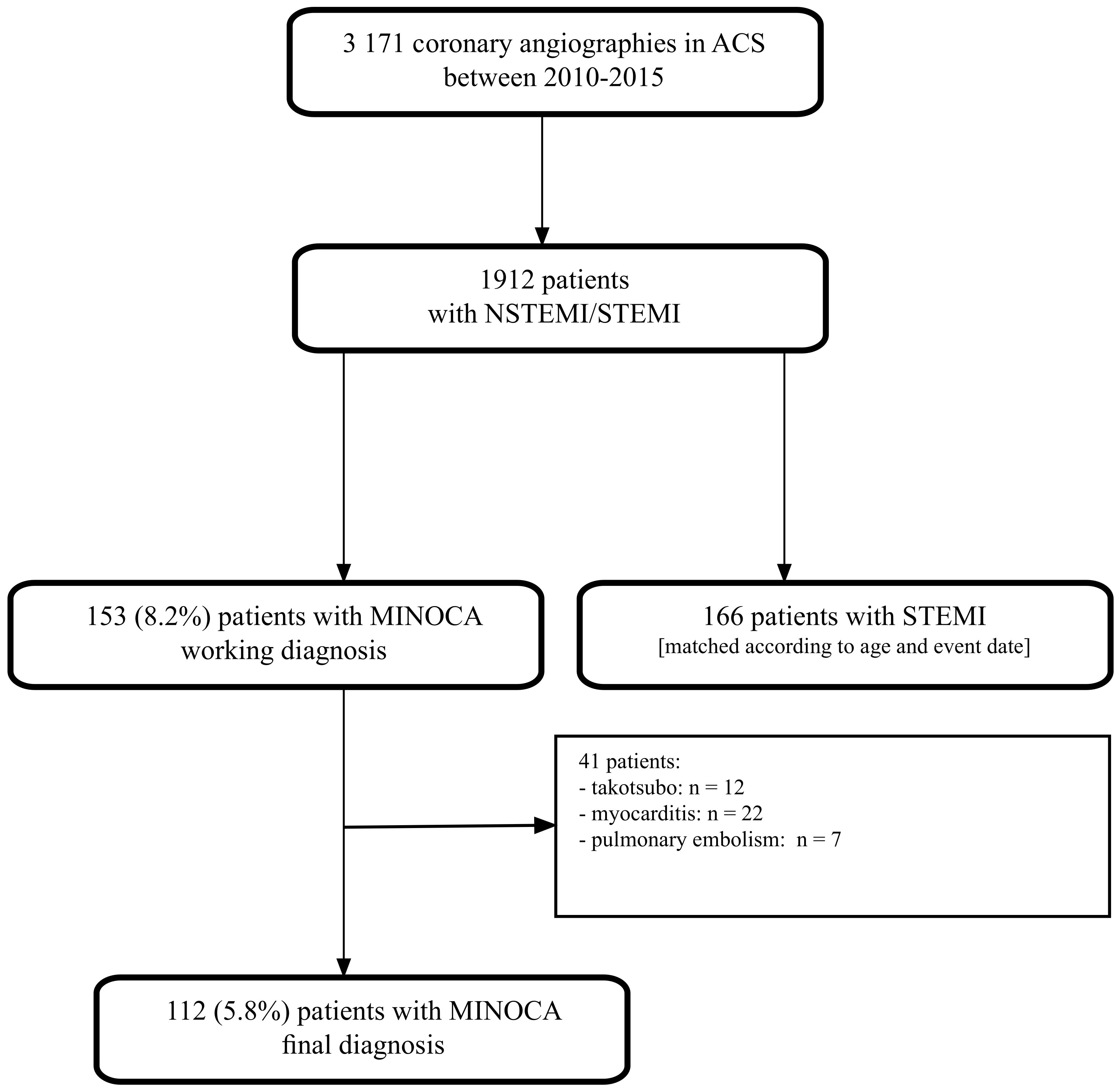
2.1. Study Design and Participants

The data were obtained retrospectively from the hospital database. First, we analyzed all patients who underwent coronary angiography due to myocardial infarction, i.e., NSTEMI or STEMI. We then identified patients with coronary angiography with non-obstructive coronary arteries (lesions < 50% of diameter stenosis) (MINOCA working diagnosis). For the final analysis, we included patients with a final diagnosis of MINOCA after excluding other potential causes such as pulmonary embolism or myocarditis. The definition of MINOCA was verified according to the diagnostic criteria enclosed in the European Society of Cardiology guidelines on NSTE-ACS [13]. Additionally, we identified a STEMI group with obstructive disease that were matched based on age and the event date.
This study compared various baseline demographic and clinical characteristics, laboratory data, and clinical outcomes at a 5-year follow-up between MINOCA and STEMI patients.

2.2. Data Collection

We retrieved demographic, clinical, periprocedural, and laboratory data from the hospital database. We took into consideration the following comorbidities: arterial hypertension, dyslipidemia, diabetes mellitus, peripheral artery disease, atrial fibrillation, chronic kidney disease (defined as eGFR < 60 mL/min/1.73 m2), prior coronary artery bypass grafting (CABG), prior PCI (percutaneous coronary intervention), prior MI, and clinical data associated with MI: MI type, disease severity, treatment strategy, and periprocedural complications. Additionally, we gathered information on echocardiographic parameters (left ventricular ejection fraction, left ventricular end-diastolic diameter, intraventricular septal diameter, left atrial diameter) and laboratory findings assessed at admission: complete blood count with differential (WBC—white blood cells, RBC—red blood cells, Hgb—hemoglobin, PLT—platelets), alanine aminotransferase (ALT), creatinine, troponin T, C-reactive protein (CRP), eGFR, glucose, glycated hemoglobin (HbA1c), lipid profile, N-terminal pro-B-type natriuretic peptide (NT-proBNP), thyroid-stimulating hormone (TSH), and uric acid. We also gathered information on medications at discharge.

2.3. Study Endpoints

The primary study endpoint was to compare the 5-year rate of major cardiovascular adverse events (MACE) defined as joined rates of cardiac death, MI, and recurrent hospitalization due to angina. The secondary endpoints included all-cause death, cardiac death, MI, PCI, and recurrent hospitalization due to angina rates at 1, 2, 3, 4, and 5 years.

2.4. Statistical Methods

Descriptive statistics were presented: mean, standard deviation, minimum, median, interquartile range, and maximum for continuous variables; count and percent for categorical variables. Pearson’s Chi-squared test or Fisher’s exact test was performed to compare categorical variables between the two groups (e.g., MINOCA and STEMI patients). Fisher’s exact test was used when at least one of the subgroups had a count = 0. The Wilcoxon rank sum test was performed to compare continuous variables between two groups (e.g., MINOCA and STEMI patients). A p-value < 0.05 was considered statistically significant.
Kaplan-Meier estimators with 95% CI were calculated to compare 5-year survival curves for various endpoints between groups (e.g., MINOCA and STEMI patients).
If a given endpoint occurred for a particular patient more than once in a 5-year follow-up period, then the survival time was assumed as the time to the first occurrence of this endpoint. Notably, in the case of MACE (a composite endpoint), survival time was assumed as the time to the first occurrence of either cardiac death, myocardial infarction, or angina pectoris hospitalization.
Univariable and multivariable Cox regression (the Cox proportional hazards model) was performed to compare survival rates between groups. The multivariable Cox regression model was chosen in stepwise selection with a backward elimination algorithm with a significance level = 0.3. Results regarding the Hazard Ratio (HR) and 95% confidence intervals for HR were presented.
Statistical analyses were performed using R software version 4.2.1 (23 June 2022 ucrt)—“Funny-Looking Kid” Copyright (C) 2022 The R Foundation for Statistical Computing Platforma: x86_64-w64-mingw32/x64 (64-bit).

3. Results

3.1. Baseline Characteristics

Between 2010 and 2015, we identified 3171 coronary angiography procedures performed due to acute coronary syndrome, of which 153 had a working MINOCA diagnosis, and the final diagnosis of MINOCA was ascribed to 112 (5.8%) patients (Figure 1). Additionally, we matched 166 patients with STEMI and obstructive coronary arteries as the reference group.
The baseline characteristics are presented in Table 1, and angiographic data are shown in Table 2. In MINOCA patients, there were more females (60% vs. 26%, p < 0.001), and patients presented most frequently with NSTEMI (83.9%). Patients with MINOCA had more frequent atrial fibrillation (22% vs. 5.4%, p < 0.001) and higher left ventricular ejection fraction (59 ± 10% vs. 54 ± 10%, p < 0.001) compared to STEMI patients. In STEMI patients, we observed higher rates of dyslipidemia (25% vs. 37%, p = 0.031) and prior MI (0 vs. 5.4%, p = 0.012). Most patients with MINOCA had no coronary lesions (45.5%) or lesions < 30% in diameter (36.6%). In STEMI patients, significant lesions (>50% in diameter) were present in only one artery in 42.2% of cases (one-vessel disease) and in two arteries in 37.3% of patients (two-vessel disease).
Table 3 presents laboratory findings at admission. Interestingly, MINOCA patients had lower glucose, creatine levels, LDL, and triglycerides, whereas they had higher levels of HDL. Also, MINOCA patients had lower levels of troponin T, mainly within the range of 0–500 ng/mL (59.8%), whereas STEMI patients had troponin T levels mainly within the range of 2501–10,000 ng/mL (54.8%), p < 0.001.

3.2. Management at Discharge

All included patients were discharged. Table 4 presents medications prescribed at discharge. STEMI patients received ASA (95% vs. 100%, p = 0.004), clopidogrel (73% vs. 100%), beta-blocker (79% vs. 90%, p = 0.009), ACE inhibitor (73% vs. 93%, p < 0.001), nitrates (55% vs. 77%, p < 0.001) and statins (91% vs. 99%, p < 0.001) more frequently. MINOCA patients received more frequent Ca-blockers (25% vs. 9.6%, p < 0.001).

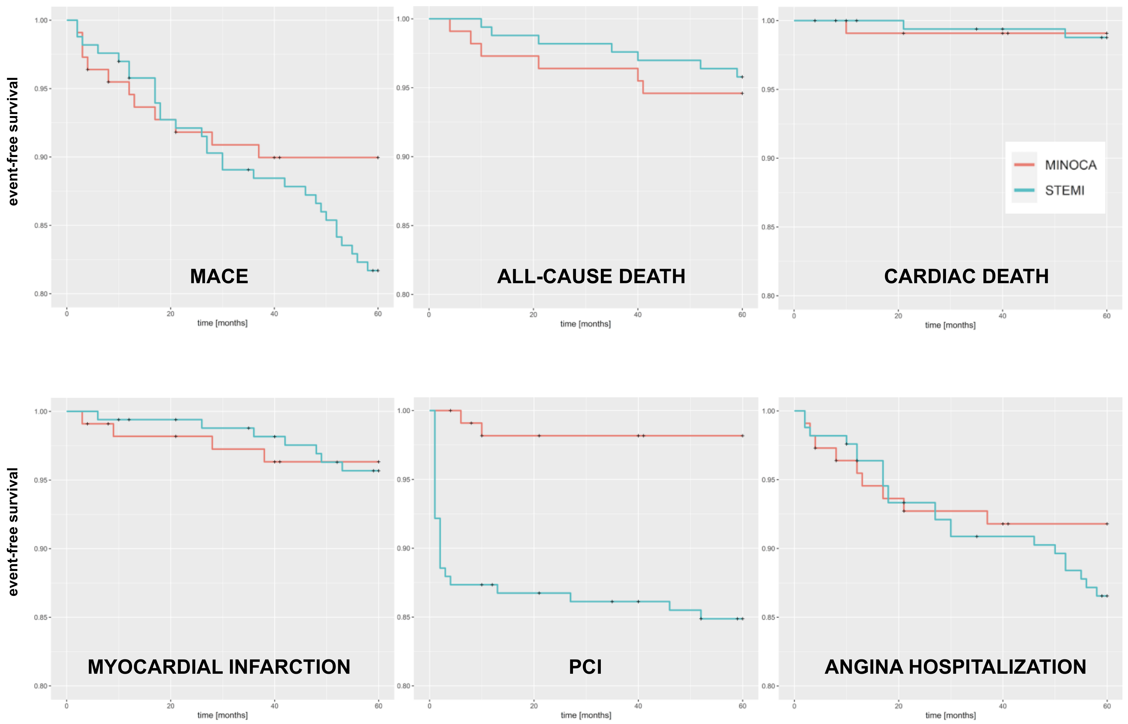
3.3. Outcomes at 5 Years

Survival rates at 5 years are presented in Table 5, and Kaplan-Meier curves are shown in Figure 2. We only observed a statistically significant difference for PCI, which was higher for STEMI patients (1.8% vs. 15.1%, HR 9.0, 95% CI 2.13–38.0, p = 0.003), and a trend for a higher rate of MACE in STEMI patients (11.6% vs. 18.7%, HR 1.82, 95% CI 0.91–3.63, p = 0.09). We provide the data on all-cause death, cardiac death, and myocardial infarction rates at the follow-up with a median of 9 years (Table S1 and Figure S1) in the Supplementary Material. We observed no statistically significant differences in terms of those outcomes between MINOCA and STEMI patients.

3.4. Predictor Factors for MACE at 5-Year Follow-Up

Additionally, we performed a Cox regression analysis to identify the predictive factors for MACE in MINOCA and STEMI patients separately. Table 6 shows the analysis for MINOCA patients. Here, only a beta blocker was a protective factor (a trend observed)—HR 0.33, 95% CI 0.10–1.15, p = 0.082. Table 7 shows the analysis for STEMI patients. Here, the use of an angiotensin receptor blocker (HR 9.35, 95% CI 1.03–84.6, p = 0.047) was the significant predictive factor, and a trend was observed for male sex HR 2.87, 95% CI 0.98–8.46, p = 0.056.

4. Discussion

We analyzed 3171 myocardial infarction cases and identified 112 patients with a final diagnosis of MINOCA. The mean MINOCA incidence was 5.8%. We observed similar 5-year outcomes between the MINOCA and STEMI patients. No statistically significant differences were observed in MACE rates.
The MINOCA incidence (5.8%) in our paper was similar to values provided in other reports, i.e., between 5% and 15% [10,14,15,16]. In addition, the recent paper on MINOCA incidence in Poland just before the COVID-19 pandemic (6.3%) and during the COVID-19 pandemic (5.9%) showed similar results [17]. The rate of MINOCA in the MINOCA-TR Registry (10.3%) was slightly higher, but similarly to ours, women dominated in the MINOCA population (our study—60%, MINOCA-TR—89%) [18].
In our study, MINOCA causes were rarely thoroughly investigated. This also reflected the approach to those patients a couple of years ago (2010–2015), when not much attention was provided to patients with no lesions in coronary arteries unless there was a suspicion of an underlying disease, e.g., myocarditis or a plaque rupture (suggestive coronary angiography view) [19]. Bucciarelli et al. recently showed that cardiac magnetic resonance (cMR) could really change the diagnosis [20]. They observed that 1/3 of ischemic MINOCA cases were initially treated as myocarditis, but after including cMR results, different management was adopted in 22% of subjects. Unfortunately, too few patients are still referred to cMR. This may be associated with costs issue as well as with cMR availability. Not every hospital has this imaging modality. Also, it was recently shown that other techniques could be helpful in the diagnosis of patients with ischemia and non-obstructive coronary arteries. We refer to intracoronary ECG [21] or comprehensive microcirculatory and epicardial vessel bed assessment as suggested by the recent European Society of Cardiology guidelines [13,22,23,24].
In our paper, we observed similar outcomes between MINOCA and STEMI at 5 years. One must also stress that the results were quite favorable, with low rates of all-cause death (5.4% vs. 4.2%), cardiac death (0.9% vs. 1.2%), recurrent myocardial infarction (3.6% vs. 4.2%) or MACE (11.6% vs. 18.7%). The rates of repeated hospitalizations due to angina in MINOCA and STEMI patients were 8.1% vs. 13.3%, respectively. The relative low incidence of comorbidities could have influenced this finding, with only 42% of patients having arterial hypertension, or 37% of patients having dyslipidemia in the STEMI group. However, as shown in the Supplementary Materials, the outcomes at the very long follow-up (median: MINOCA—9 years, STEMI—9.8 years) were much worse (all-cause death rate—17.9% vs. 24.1%, p = 0.61; cardiac death rate—9.8% vs. 16.9%, p = 0.31 and MI rate—14.3% vs. 21.1%, p = 0.41). Nevertheless, once again, no statistical difference was observed between the MINOCA and STEMI patients.
Interestingly, in the previously published study on MINOCA in Poland, no statistical difference in 12-month all-cause mortality in patients with MINOCA before and during the COVID-19 pandemic was observed, but the values were higher than in our study (total population 9.9%; 9.2% (2019) vs. 11.0% (2020), p = 0.09) [17]. Quesada et al. showed that at five years, the all-cause death rate was higher in ST-elevation MINOCA cases than in STEMI patients (18% vs. 15%, p = 0.033). In the propensity score-matched cohort, ST elevation MINOCA patients demonstrated a 1.4-fold (95%CI 1.04–1.89, p = 0.028) higher risk of 5-year all-cause death than STEMI patients [25]. However, this study gathered patients from 2003, and many advancements have been observed in pharmacotherapy and invasive treatment strategies since then which could have influenced the long-term outcomes. Recently, Zalewska-Adamiec et al. presented interesting results on MINOCA patients’ survival in a 3-year follow-up. The all-cause death rate of MINOCA patients was 16.85%. However, they differentiated two subgroups with eGFR < 60 and ≥ 60 mL/min/1.73 m2. At 12 months, the all-cause death was 26.42% vs. 7.20% (p = 0.0004), and after 36 months, it was 33.96% vs. 9.6% (p < 0.0001) [26]. In our study, only 3.6% of patients had chronic kidney disease. This can also be one of the factors explaining our favorable outcomes.
In our study, we reported that in MINOCA patients, only beta-blockers could potentially decrease the risk of MACE at 5 years (a trend observed: HR 0.33, 95% CI 0.10—1.15, p = 0.082). However, in STEMI patients, the potential predictor factors of MACE at 5 years were angiotensin receptor blocker use (HR 9.35, 95% CI 1.03—84.6, p = 0.047) and male sex (a trend: HR 2.87, 95% CI 0.98—8.46, p = 0.056). As mentioned earlier, Bucciarelli et al. showed that dyslipidemia, reduced left ventricular ejection fraction, ST-elevation at the hospitalization, and the late-gadolinium enhancement transmural pattern were the independent predictors of cardiac-related rehospitalizations in MINOCA patients [20]. In addition, complete blood count-derived indices could predict the diagnosis and outcomes, such as platelet distribution width [1]. Notably, Gabaldon-Perez et al. observed that elderly patients with MINOCA had a better prognosis than those with obstructive coronary arteries; however, this only applied to those with no lesions in the coronary arteries [11].
Chen et al. showed no differences in outcomes between non-ST-elevation or ST-elevation groups during a 4-year follow-up [27]. This was also true in our study. Chen et al. also identified multivariable predictors of MACE. In the non-ST-elevation group, there were Killip grades ≥ 2, reduced beta-blocker use during hospitalization, and higher levels of low-density lipoprotein cholesterol. The reduced beta-blocker use was the sole independent factor in the ST-elevation group [27]. We also observed that using beta-blockers in MINOCA patients had a beneficial effect in terms of MACE. Nevertheless, Li et al. recently showed that ST-elevation MINOCA patients demonstrated lower risks for MACE at a 2-year follow-up [7]. Here, we should also mention the paper by Jedrychowska et al., in which the authors designed a risk score for predicting MINOCA amid overall STEMI patients [28]. And, interestingly, Stepien et al. showed that the MINOCA treatment and outcomes were not affected whether patients were treated during the weekend (free days) or working days [10].

Study Limitations

This study has several limitations. First, this was a retrospective study; therefore, residual confounding factors may exist. Second, we could not systematically establish real MINOCA causes, since cardiac magnetic resonance or IVUS/OCT was rarely used. And, finally, we included all MINOCA patients we could identify; therefore, no sample size calculation was performed; however, relatively small populations might have caused no statistically significant evident differences in the outcomes between MINOCA and STEMI patients. Nevertheless, most papers on MINOCA present the results on 100–200 patient populations.

5. Conclusions

We observed the frequency of MINOCA patients at 5.8%. In addition, we observed comparable long-term outcomes between MINOCA and STEMI patients. No statistically significant differences were observed in MACE rates (11.6% vs. 18.7%, HR 1.82, 95% CI 0.91–3.63, p = 0.09).

Supplementary Materials

The following supporting information can be downloaded at: https://www.mdpi.com/article/10.3390/jpm13050856/s1, Figure S1: Kaplan-Meier curves on long-term outcomes (follow-up of median 9 years); Table S1: 9-year follow-up data.

Author Contributions

Conceptualization, P.B. and J.B.; methodology, P.B. and J.B.; investigation, P.B., M.T., W.R. and W.F.; data curation, A.K. and R.J.G.; writing—original draft preparation, P.B. and J.B.; writing—review and editing, M.T., W.R., W.F. and A.K.; supervision, R.J.G. All authors have read and agreed to the published version of the manuscript.

Funding

This research received no external funding.

Institutional Review Board Statement

The study protocol was reviewed and approved by the Institutional Review Board (or Ethics Committee) of the District Physician Chamber in Plock (No 1/2020).

Informed Consent Statement

Patient consent was waived due to the retrospective nature of the study.

Data Availability Statement

Raw data are available from the corresponding author upon request.

Conflicts of Interest

The authors declare no conflict of interest.

References

  1. Bil, J.; Pietraszek, N.; Gil, R.J.; Gromadzinski, L.; Onichimowski, D.; Jalali, R.; Kern, A. Complete Blood Count-Derived Indices as Prognostic Factors of 5-Year Outcomes in Patients with Confirmed Coronary Microvascular Spasm. Front. Cardiovasc. Med. 2022, 9, 933374. [Google Scholar] [CrossRef]
  2. Casolo, G.; Gabrielli, D.; Colivicchi, F.; Murrone, A.; Grosseto, D.; Gulizia, M.M.; Di Fusco, S.; Domenicucci, S.; Scotto di Uccio, F.; Di Tano, G.; et al. ANMCO Position Paper: Prognostic and therapeutic relevance of non-obstructive coronary atherosclerosis. Eur. Heart J. Suppl. 2021, 23 (Suppl. C), C164–C175. [Google Scholar] [CrossRef] [PubMed]
  3. Bil, J.; Mozenska, O.; Segiet-Swiecicka, A.; Gil, R.J. Revisiting the use of the provocative acetylcholine test in patients with chest pain and nonobstructive coronary arteries: A five-year follow-up of the AChPOL registry, with special focus on patients with MINOCA. Transl. Res. 2021, 231, 64–75. [Google Scholar] [CrossRef] [PubMed]
  4. Atri, D.; Siddiqi, H.K.; Lang, J.P.; Nauffal, V.; Morrow, D.A.; Bohula, E.A. COVID-19 for the Cardiologist: Basic Virology, Epidemiology, Cardiac Manifestations, and Potential Therapeutic Strategies. JACC Basic Transl. Sci. 2020, 5, 518–536. [Google Scholar] [CrossRef] [PubMed]
  5. Quesada, O.; Van Hon, L.; Yildiz, M.; Madan, M.; Sanina, C.; Davidson, L.; Htun, W.W.; Saw, J.; Garcia, S.; Dehghani, P.; et al. Sex Differences in Clinical Characteristics, Management Strategies, and Outcomes of STEMI with COVID-19: NACMI Registry. J. Soc. Cardiovasc. Angiogr. Interv. 2022, 1, 100360. [Google Scholar] [CrossRef]
  6. Magnani, G.; Bricoli, S.; Ardissino, M.; Maglietta, G.; Nelson, A.; Malagoli Tagliazucchi, G.; Disisto, C.; Celli, P.; Ferrario, M.; Canosi, U.; et al. Long-term outcomes of early-onset myocardial infarction with non-obstructive coronary artery disease (MINOCA). Int. J. Cardiol. 2022, 354, 7–13. [Google Scholar] [CrossRef]
  7. Li, M.; He, Y.; Cheang, I.; Zhang, Z.; Liu, Y.; Wang, H.; Kong, X. Clinical characteristics and outcome in patients with ST-segment and non-ST-segment elevation myocardial infarction without obstructive coronary artery: An observation study from Chinese population. BMC Cardiovasc. Disord. 2022, 22, 21. [Google Scholar] [CrossRef]
  8. Pasupathy, S.; Lindahl, B.; Litwin, P.; Tavella, R.; Williams, M.J.A.; Air, T.; Zeitz, C.; Smilowitz, N.R.; Reynolds, H.R.; Eggers, K.M.; et al. Survival in Patients with Suspected Myocardial Infarction With Nonobstructive Coronary Arteries: A Comprehensive Systematic Review and Meta-Analysis from the MINOCA Global Collaboration. Circ. Cardiovasc. Qual. Outcomes 2021, 14, e007880. [Google Scholar] [CrossRef]
  9. Racz, A.O.; Racz, I.; Szabo, G.T.; Uveges, A.; Koszegi, Z.; Penczu, B.; Kolozsvari, R. The Effects of Percutaneous Coronary Intervention on the Flow in Acute Coronary Syndrome Patients-Geometry in Focus. J. Pers. Med. 2022, 12, 1264. [Google Scholar] [CrossRef]
  10. Schmitz, K.; Groth, N.; Mullvain, R.; Renier, C.; Oluleye, O.; Benziger, C. Prevalence, Clinical Factors, and Outcomes Associated With Myocardial Infarction with Nonobstructive Coronary Artery. Crit. Pathw. Cardiol. 2021, 20, 108–113. [Google Scholar] [CrossRef]
  11. Gabaldon-Perez, A.; Bonanad, C.; Garcia-Blas, S.; Marcos-Garces, V.; D’Gregorio, J.G.; Fernandez-Cisnal, A.; Valero, E.; Minana, G.; Merenciano-Gonzalez, H.; Mollar, A.; et al. Clinical Predictors and Prognosis of Myocardial Infarction with Non-Obstructive Coronary Arteries (MINOCA) without ST-Segment Elevation in Older Adults. J. Clin. Med. 2023, 12, 1181. [Google Scholar] [CrossRef] [PubMed]
  12. Smilowitz, N.R.; Mahajan, A.M.; Roe, M.T.; Hellkamp, A.S.; Chiswell, K.; Gulati, M.; Reynolds, H.R. Mortality of Myocardial Infarction by Sex, Age, and Obstructive Coronary Artery Disease Status in the ACTION Registry-GWTG (Acute Coronary Treatment and Intervention Outcomes Network Registry-Get with the Guidelines). Circ. Cardiovasc. Qual. Outcomes 2017, 10, e003443. [Google Scholar] [CrossRef] [PubMed]
  13. Collet, J.P.; Thiele, H.; Barbato, E.; Barthelemy, O.; Bauersachs, J.; Bhatt, D.L.; Dendale, P.; Dorobantu, M.; Edvardsen, T.; Folliguet, T.; et al. 2020 ESC Guidelines for the management of acute coronary syndromes in patients presenting without persistent ST-segment elevation. Eur. Heart J. 2021, 42, 1289–1367. [Google Scholar] [CrossRef] [PubMed]
  14. Bil, J.; Pietraszek, N.; Pawlowski, T.; Gil, R.J. Advances in Mechanisms and Treatment Options of MINOCA Caused by Vasospasm or Microcirculation Dysfunction. Curr. Pharm. Des. 2018, 24, 517–531. [Google Scholar] [CrossRef] [PubMed]
  15. Bossard, M.; Gao, P.; Boden, W.; Steg, G.; Tanguay, J.F.; Joyner, C.; Granger, C.B.; Kastrati, A.; Faxon, D.; Budaj, A.; et al. Antiplatelet therapy in patients with myocardial infarction without obstructive coronary artery disease. Heart 2021, 107, 1739–1747. [Google Scholar] [CrossRef]
  16. Gasior, P.; Desperak, A.; Gierlotka, M.; Milewski, K.; Wita, K.; Kalarus, Z.; Fluder, J.; Kazmierski, M.; Buszman, P.E.; Gasior, M.; et al. Clinical Characteristics, Treatments, and Outcomes of Patients with Myocardial Infarction with Non-Obstructive Coronary Arteries (MINOCA): Results from a Multicenter National Registry. J. Clin. Med. 2020, 9, 2779. [Google Scholar] [CrossRef]
  17. Bil, J.; Kern, A.; Bujak, K.; Gierlotka, M.; Legutko, J.; Gasior, M.; Wanha, W.; Gromadzinski, L.; Gil, R.J. Clinical characteristics and 12-month outcomes in MINOCA patients before and during the COVID-19 pandemic. Pol. Arch. Intern. Med. 2023, 16405, ahead of print. [Google Scholar] [CrossRef]
  18. Gok, G.; Coner, A.; Cinar, T.; Kilic, S.; Yenercag, M.; Oz, A.; Ekmekci, C.; Ozluk, O.; Zoghi, M.; Ergene, A.O.; et al. Evaluation of demographic and clinical characteristics of female patients presenting with MINOCA and differences between male patients: A subgroup analysis of MINOCA-TR registry. Turk Kardiyol. Dern. Ars. 2022, 50, 4–13. [Google Scholar] [CrossRef]
  19. Bil, J.; Pawlowski, T.; Gil, R.J. Coronary spasm revascularized with a bioresorbable vascular scaffold. Coron. Artery Dis. 2015, 26, 634–636. [Google Scholar] [CrossRef]
  20. Bucciarelli, V.; Bianco, F.; Francesco, A.D.; Vitulli, P.; Biasi, A.; Primavera, M.; Belleggia, S.; Ciliberti, G.; Guerra, F.; Seferovic, J.; et al. Characteristics and Prognosis of a Contemporary Cohort with Myocardial Infarction with Non-Obstructed Coronary Arteries (MINOCA) Presenting Different Patterns of Late Gadolinium Enhancements in Cardiac Magnetic Resonance Imaging. J. Clin. Med. 2023, 12, 2266. [Google Scholar] [CrossRef]
  21. Bil, J.; Gil, R.J.; Vassilev, D.; Gromadzinski, L.; Onichimowski, D.; Jalali, R.; Kern, A. Intracoronary ECG monitoring during provocation acetylcholine test in chest pain patients with non-obstructive coronary artery disease: Results from the AChPOL Registry. Kardiol. Pol. 2022, 80, 1256–1259. [Google Scholar] [CrossRef] [PubMed]
  22. Bil, J.; Tyczynski, M.; Modzelewski, P.; Gil, R.J. Acetylcholine provocation test with resting full-cycle ratio, coronary flow reserve, and index of microcirculatory resistance give definite answers and improve health-related quality of life. Kardiol. Pol. 2020, 78, 1291–1292. [Google Scholar] [CrossRef] [PubMed]
  23. Fluder-Wlodarczyk, J.; Milewski, M.; Roleder-Dylewska, M.; Haberka, M.; Ochala, A.; Wojakowski, W.; Gasior, P. Underlying Causes of Myocardial Infarction with Nonobstructive Coronary Arteries: Optical Coherence Tomography and Cardiac Magnetic Resonance Imaging Pilot Study. J. Clin. Med. 2022, 11, 7495. [Google Scholar] [CrossRef] [PubMed]
  24. Kunadian, V.; Chieffo, A.; Camici, P.G.; Berry, C.; Escaned, J.; Maas, A.; Prescott, E.; Karam, N.; Appelman, Y.; Fraccaro, C.; et al. An EAPCI Expert Consensus Document on Ischaemia with Non-Obstructive Coronary Arteries in Collaboration with European Society of Cardiology Working Group on Coronary Pathophysiology & Microcirculation Endorsed by Coronary Vasomotor Disorders International Study Group. Eur. Heart J. 2020, 41, 3504–3520. [Google Scholar] [PubMed]
  25. Quesada, O.; Yildiz, M.; Henry, T.D.; Okeson, B.K.; Chambers, J.; Shah, A.; Stanberry, L.; Volpenhein, L.; Aziz, D.; Lantz, R.; et al. Characteristics and Long-term Mortality in Patients with ST-Segment Elevation Myocardial Infarction with Non-Obstructive Coronary Arteries (STE-MINOCA): A High Risk Cohort. medRxiv 2023. [Google Scholar] [CrossRef]
  26. Zalewska-Adamiec, M.; Malyszko, J.; Grodzka, E.; Kuzma, L.; Dobrzycki, S.; Bachorzewska-Gajewska, H. The outcome of patients with myocardial infarction with non-obstructive coronary arteries (MINOCA) and impaired kidney function: A 3-year observational study. Int. Urol. Nephrol. 2021, 53, 2557–2566. [Google Scholar] [CrossRef]
  27. Chen, L.; Fan, Y.; Fang, Z.; Liu, N. Long-term outcomes and predictors of patients with ST elevated versus non-ST elevated myocardial infarctions in non-obstructive coronary arteries: A retrospective study in Northern China. PeerJ 2023, 11, e14958. [Google Scholar] [CrossRef]
  28. Jedrychowska, M.; Siudak, Z.; Malinowski, K.P.; Zandecki, L.; Zabojszcz, M.; Kameczura, T.; Mika, P.; Bartus, K.; Wanha, W.; Wojakowski, W.; et al. ST-segment elevation myocardial infarction with non-obstructive coronary arteries: Score derivation for prediction based on a large national registry. PLoS ONE 2021, 16, e0254427. [Google Scholar] [CrossRef]
Figure 1. Study flowchart. ACS—acute coronary syndrome; NSTEMI—non-ST-elevation myocardial infarction; STEMI—ST-elevation myocardial infarction, MINOCA—myocardial infarction with non-obstructive coronary arteries.
Jpm 13 00856 g001
Figure 2. Kaplan-Meier curves for 5-year outcomes. PCI: percutaneous coronary intervention.
Jpm 13 00856 g002
Table 1. Baseline characteristics.
ParameterMINOCA
N = 112
STEMI
N = 166
p-Value
Females67 (60%)43 (26%)<0.001
Age [years]63 ± 1461 ± 110.2
Body mass index [kg/m2]27.9 ± 5.828.1 ± 4.80.3
Myocardial infarction type at presentation
  NSTEMI94 (83.9%)--
  STEMI18 (16.1%)166 (100)<0.001
Arterial hypertension59 (53%)69 (42%)0.068
Diabetes type 215 (13%)29 (17%)0.4
Dyslipidemia28 (25%)62 (37%)0.031
Prior myocardial infarction0 (0%)9 (5.4%)0.012
Prior PCI0 (0%)3 (1.8%)0.3
Prior CABG0 (0%)1 (0.6%)>0.9
Chronic kidney disease4 (3.6%)9 (5.4%)0.5
Atrial fibrillation25 (22%)9 (5.4%)<0.001
Peripheral artery disease1 (0.9%)5 (3.0%)0.4
Smoking13 (12%)27 (16%)0.3
Echocardiographic parameters
  LVEDd [mm]48.7 ± 6.051.4 ± 6.4<0.0001
  IVSd [mm]11.1 ± 2.611.3 ± 2.00.056
  LA [mm]39.8 ± 6.43.96 ± 5.6>0.9
  LVEF [%]59 ± 1054 ± 10<0.001
NSTEMI—non-ST-elevation myocardial infarction, STEMI—ST-elevation myocardial infarction, PCI—percutaneous coronary intervention, CABG—coronary artery bypass grafting, LVEDd—left ventricular end-diastolic diameter, IVSd—interventricular septal diameter; LA—left atrial diameter; LVEF—left ventricular ejection fraction.
Table 2. Angiographic data.
ParameterMINOCA
N = 112
STEMI
N = 166
Coronary lesions
  No lesions51 (45.5%)-
  <30%41 (36.6%)-
  30–50%20 (17.9%)-
Intravascular imaging use5 (4.5%)20 (12.1%)
Infarct-related artery
  LAD-79 (47.6%)
  LCx-19 (11.4%)
  RCA-68 (41.0%)
TIMI before PCI
  0-142 (85.5%)
  1-20 (12.1%)
  2-4 (2.4%)
  3-0
TIMI post PCI
  0-1 (0.6%)
  1-1 (0.6%)
  2-4 (2.4%)
  3-160 (96.4%)
Disease advancement
  1VD-70 (42.2%)
  2VD-62 (37.3%)
  3VD/LM-34 (20.5%)
Stent implanted
  BMS-117 (70.5%)
  DES-45 (27.1%)
  No stent-4 (2.4%)
LAD—left anterior descending artery; LCx—left circumflex artery; RCA—right coronary artery; PCI—percutaneous coronary intervention; VD—vessel disease; BMS—bare metal stent; DES—drug-eluting stent.
Table 3. Laboratory findings at admission.
ParameterMINOCA
N = 112
STEMI
N = 166
p-Value
White blood cells [109/L]9.8 ± 4.111.7 ± 3.8<0.001
Hemoglobin [g/dL]13.84 ± 1.4714.55 ± 1.40<0.001
Red blood cells [1012/L]4.55 ± 0.484.76 ± 0.470.002
Platelets [109/L]250 ± 80261 ± 720.13
Glucose [mmol/L]7.43 ± 2.489.08 ± 3.56<0.001
HbA1c [%]5.91 ± 0.377.99 ± 2.280.053
NT-proBNP [pg/mL]3747 ± 51495622 ± 53230.005
C-reactive protein3.5 ± 9.55.6 ± 7.10.3
AST [U/L]74 ± 128143 ± 1440.092
ALT [U/L]41 ± 2854 ± 300.13
Total cholesterol [mmol/L]4.78 ± 1.115.24 ± 1.130.004
HDL [mmol/L]1.58 ± 0.651.23 ± 0.35<0.001
LDL [mmol/L]2.59 ± 1.043.22 ± 1.00<0.001
Triglycerides [mmol/L]1.37 ± 0.601.72 ± 0.980.002
Creatine [µmol/l]86 ± 3291 ± 19<0.001
TSH [µU/mL]1.57 ± 1.421.50 ± 1.610.4
Uric acid [µmol/L]406 ± 111414 ± 980.6
Maximal troponin T [ng/mL]
  0–50067 (59.8%)16 (9.6%)<0.001
  501–250039 (34.8%)45 (27.2%)
  2501–10,0006 (5.4%)91 (54.8%)
  10,000+014 (8.4%)
ALT—alanine aminotransferase, AST—asparagine aminotransferase, HbA1c—glycated hemoglobin, NT-proBNP—N-terminal pro-B-type natriuretic peptide, TSH—thyroid-stimulating hormone, Troponin T reference values: 0—0.037 ng/mL, and the cut-off for MI: 0.1 ng/mL.
Table 4. Medications at discharge.
ParameterMINOCA
N = 112
STEMI
N = 166
p-Value
ASA105 (95%)166 (100%)0.004
Clopidogrel81 (73%)166 (100%)<0.001
Beta-blocker88 (79%)150 (90%)0.009
Ca-blocker28 (25%)16 (9.6%)<0.001
ACE inhibitor81 (73%)155 (93%)<0.001
Angiotensin receptor blocker5 (4.5%)2 (1.2%)0.12
Diuretic25 (23%)19 (11%)0.013
Trimetazidine2 (1.8%)4 (2.4%)>0.9
Nitrates61 (55%)127 (77%)<0.001
Vitamin K antagonist13 (12%)4 (2.4%)0.002
Novel oral anticoagulant4 (3.6%)0 (0%)0.025
Statin101 (91%)165 (99%)<0.001
ASA—aspirin, ACE—angiotensin-converting enzyme.
Table 5. 5-year outcomes in MINOCA vs. STEMI.
ParameterMINOCA
N = 112
STEMI
N = 166
HR95% CIp
All-cause death6 (5.4%)7 (4.2%)0.770.26–2.290.6
Cardiac death1 (0.9%)2 (1.2%)1.310.12–14.50.8
Myocardial infarction4 (3.6%)7 (4.2%)1.150.34–3.920.8
Percutaneous intervention2 (1.8%)25 (15.1%)9.02.13–38.00.003
Hospitalization due to angina9 (8.1%)22 (13.3%)1.620.75–3.530.2
MACE13 (11.6%)31 (18.7%)1.820.91–3.630.09
MACE—major adverse cardiovascular event.
Table 6. Univariable and multivariable Cox regression for MINOCA patients.
Univariable AnalysisMultivariable Analysis
CharacteristicHR95% CIpHR95% CIp
Sex
  Female
  Male0.550.15–2.070.375
Age1.010.97–1.050.762
Body mass index0.950.84–1.080.429
Coronary arteries lesions
  No lesions
  <30%0.780.19–3.270.735
  30–50%1.270.25–6.530.778
Final diagnosis
  STEMI
  NSTEMI0.870.19–4.040.862
Arterial hypertension2.520.67–9.490.1732.900.76–11.00.118
Dyslipidemia0.640.14–2.940.562
Diabetes2.380.63–8.980.201
Smoking0.700.09–5.500.737
Atrial fibrillation1.350.36–5.090.658
NT-proBNP1.001.00–1.000.397
LDL0.680.38–1.220.194
Creatine1.000.99–1.020.664
Left ventricular ejection fraction0.970.92–1.020.2390.970.92–1.020.221
Clopidogrel0.570.17–1.960.374
Beta-blocker0.430.12–1.460.1730.330.10–1.150.082
Ca blocker1.780.52–6.070.359
ACE inhibitor1.000.27–3.780.997
Angiotensin receptor blocker2.210.28–17.30.450
Statin0.950.12–7.440.963
NSTEMI—non-ST-elevation myocardial infarction, STEMI—ST-elevation myocardial infarction, N-terminal pro-B-type natriuretic peptide, ACE—angiotensin-converting enzyme.
Table 7. Univariable and multivariable Cox regression for STEMI patients.
Univariable AnalysisMultivariable Analysis
CharacteristicHR95% CIpHR95% CIp
Sex
  Female--
  Male2.380.83–6.820.1062.870.98–8.460.056
Age0.990.96–1.030.633
Body mass index0.980.90–1.060.602
IRA location
  LAD-- ---
  LCx2.190.77–6.210.1422.200.74–6.580.156
  RCA1.290.59–2.820.5291.350.58–3.160.490
Disease advancement
  1VD--
  2VD1.510.66–3.430.331
  3VD/LM1.530.58–4.030.385
Stent type
  BMS--
  DES0.600.25–1.470.2640.510.19–1.350.174
Arterial hypertension0.810.39–1.710.583
Dyslipidemia1.310.64–2.690.467
Diabetes0.980.38–2.570.973
Smoking0.760.26–2.170.606
Chronic kidney disease1.500.36–6.320.577
Peripheral artery disease1.300.18–9.550.797
Prior myocardial infarction0.670.09–4.940.697
NTproBNP1.001.00–1.000.852
LDL1.290.89–1.880.1811.220.85–1.770.281
Creatine1.000.98–1.020.803
Left ventricular ejection fraction1.020.98–1.060.431
Beta-blocker3.280.45–24.10.2433.780.50–28.40.197
Ca blocker0.660.16–2.770.569
ACE inhibitor2.100.29–15.40.466
Angiotensin receptor blocker4.880.66–35.90.1199.351.03–84.60.047
IRA: infarct-related artery; LAD: left anterior descending artery; LCx: left circumflex artery; RCA: right coronary artery; VD: vessel disease; LM: left main; BMS: bare metal stent; DES: drug-eluting stent; N-terminal pro-B-type natriuretic peptide, ACE: angiotensin-converting enzyme.
Disclaimer/Publisher’s Note: The statements, opinions and data contained in all publications are solely those of the individual author(s) and contributor(s) and not of MDPI and/or the editor(s). MDPI and/or the editor(s) disclaim responsibility for any injury to people or property resulting from any ideas, methods, instructions or products referred to in the content.

