
Open-Heart Cardio-Thoracic Biological Valve Replacement Following Complicated Transcatheter Aortic Valve Implantation
 and
and 
Abstract
:1. Introduction
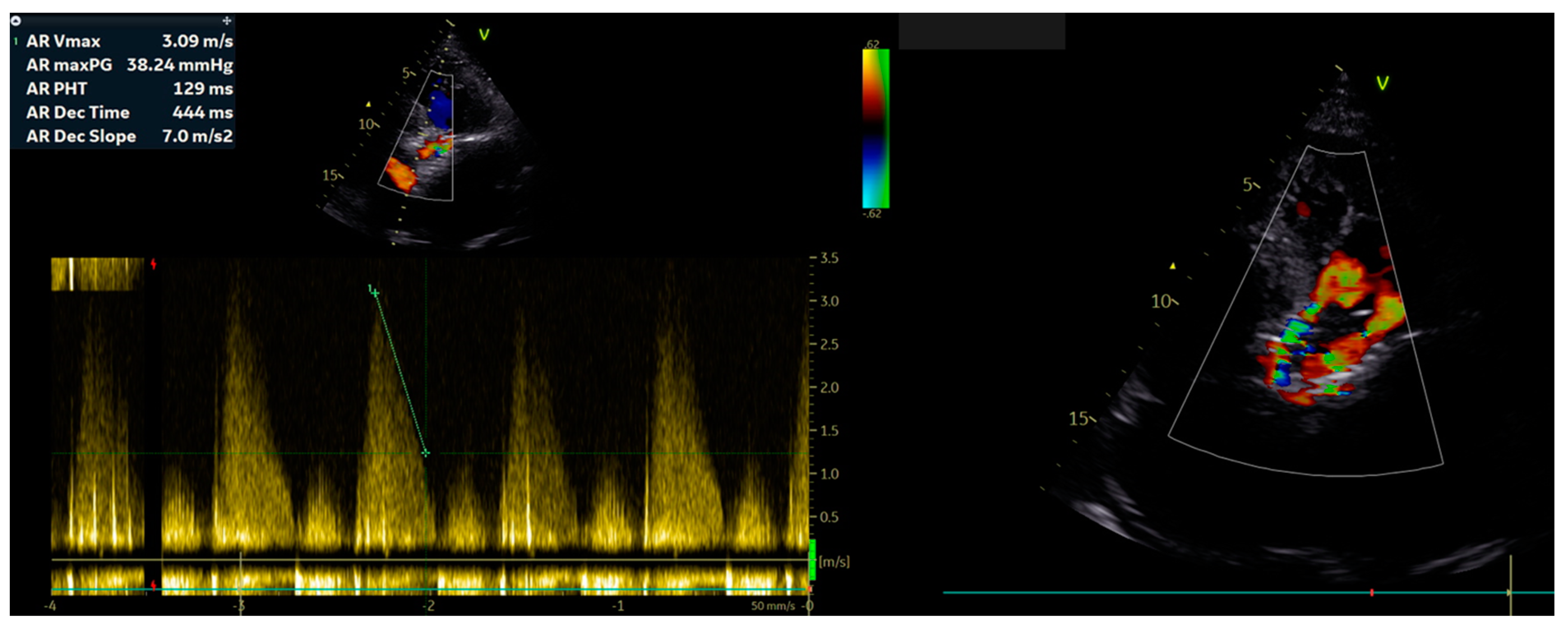
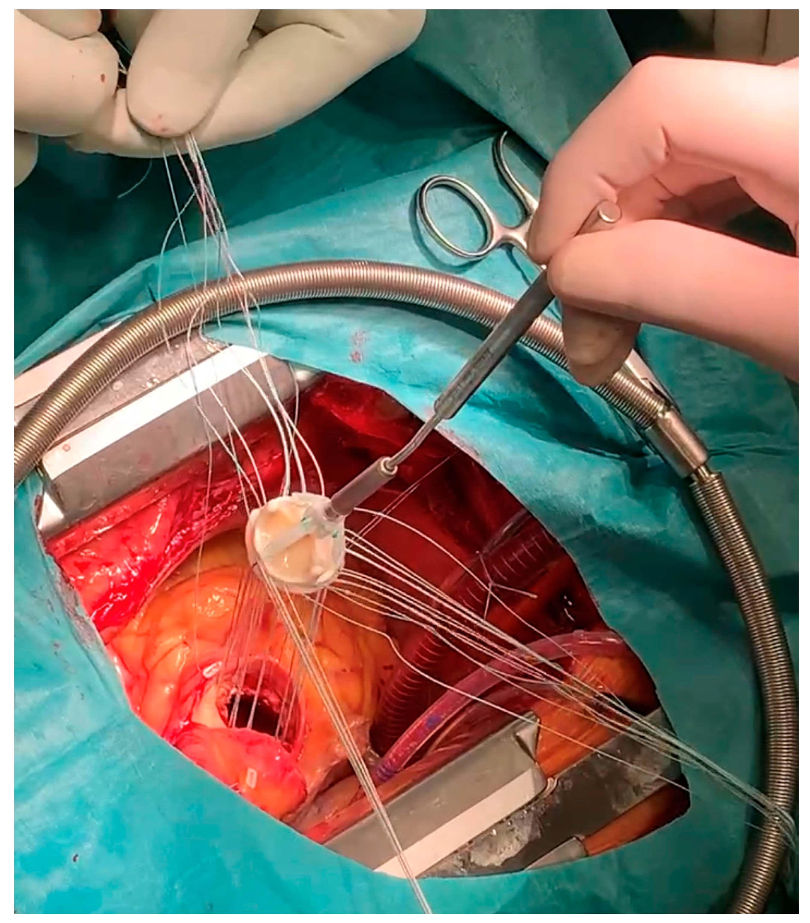
2. Case Report
3. Discussion
4. Conclusions
Author Contributions
Funding
Institutional Review Board Statement
Informed Consent Statement
Data Availability Statement
Conflicts of Interest
References
- Coffey, S.; Cox, B.; Williams, M.J.A. The Prevalence, Incidence, Progression, and Risks of Aortic Valve Sclerosis: A Systematic Review and Meta-Analysis. J. Am. Coll. Cardiol. 2014, 63, 2852–2861. [Google Scholar] [CrossRef] [PubMed]
- Lindroos, M.; Kupari, M.; Heikkilä, J.; Tilvis, R. Prevalence of aortic valve abnormalities in the elderly: An echocardiographic study of a random population sample. J. Am. Coll. Cardiol. 1993, 21, 1220–1225. [Google Scholar] [CrossRef] [PubMed]
- Gharacholou, S.M.; Karon, B.L.; Shub, C.; Pellikka, P.A. Aortic Valve Sclerosis and Clinical Outcomes: Moving toward a Definition. Am. J. Med. 2011, 124, 103–110. [Google Scholar] [CrossRef] [PubMed]
- Stewart, B.; Siscovick, D.; Lind, B.K.; Gardin, J.M.; Gottdiener, J.S.; Smith, V.E.; Kitzman, D.W.; Otto, C.M. Clinical Factors Associated With Calcific Aortic Valve Disease. J. Am. Coll. Cardiol. 1997, 29, 630–634. [Google Scholar] [CrossRef] [PubMed]
- Czarny, M.J.; Resar, J.R. Diagnosis and Management of Valvular Aortic Stenosis. Clin. Med. Insights Cardiol. 2014, 8, 15. [Google Scholar] [CrossRef] [PubMed]
- Grygier, M.; Olasińska-Wiśniewska, A.; Misterski, M.; Perek, B.; Urbanowicz, T.; Markiewicz, A.; Jemielity, M.; Lesiak, M. Navitor valve—A new TAVI solution for patients with aortic stenosis. Kardiol. Pol. 2021, 79, 1278–1279. [Google Scholar] [CrossRef] [PubMed]
- Steinvil, A.; Weissman, G.; Ertel, A.W.; Weigold, G.; Rogers, T.; Koifman, E.; Buchanan, K.D.; Shults, C.; Torguson, R.; Okubagzi, P.G.; et al. Accuracy of predicted orthogonal projection angles for valve deployment during transcatheter aortic valve replacement. J. Cardiovasc. Comput. Tomogr. 2018, 12, 398–403. [Google Scholar] [CrossRef] [PubMed]
- Bhushan, S.; Huang, X.; Li, Y.; He, S.; Mao, L.; Hong, W.; Xiao, Z. Paravalvular Leak After Transcatheter Aortic Valve Implantation Its Incidence, Diagnosis, Clinical Implications, Prevention, Management, and Future Perspectives: A Review Article. Curr. Probl. Cardiol. 2022, 47, 100957. [Google Scholar] [CrossRef] [PubMed]
- Leveille, L.; Jaussaud, N.; Theron, A.; Riberi, A.; Collart, F. Open-heart transcatheter aortic valve replacement in complex aortic valve reoperation: About a case series. Eur. Heart J. Case Rep. 2018, 2, yty064. [Google Scholar] [CrossRef] [PubMed]
- Hagar, A.; Li, Y.; Wei, X.; Peng, Y.; Xu, Y.; Ou, Y.; Wang, Z.; Wang, X.; Shah, J.-P.; Sihag, V.; et al. Incidence, Predictors, and Outcome of Paravalvular Leak after Transcatheter Aortic Valve Implantation. J. Interv. Cardiol. 2020, 2020, 1–11. [Google Scholar] [CrossRef] [PubMed]
- Eleid, M.F.; Nishimura, R.A.; Sorajja, P.; Borlaug, B.A. Systemic Hypertension in Low-Gradient Severe Aortic Stenosis With Preserved Ejection Fraction. Circulation 2013, 128, 1349–1353. [Google Scholar] [CrossRef] [PubMed]
- Pibarot, P.; Weissman, N.J.; Stewart, W.J.; Hahn, R.T.; Lindman, B.R.; McAndrew, T.; Kodali, S.K.; Mack, M.J.; Thourani, V.H.; Miller, D.C.; et al. Incidence and sequelae of prosthesis-patient mismatch in transcatheter versus surgical valve replacement in high-risk patients with severe aortic stenosis: A PARTNER trial cohort—A analysis. J. Am. Coll. Cardiol. 2014, 64, 1323–1334. [Google Scholar] [CrossRef] [PubMed]
- Musallam, A.; Buchanan, K.D.; Yerasi, C.; Dheendsa, A.; Zhang, C.; Shea, C.; Case, B.C.; Forrestal, B.J.; Satler, L.F.; Ben-Dor, I.; et al. Impact of Left Ventricular Outflow Tract Calcification on Outcomes Following Transcatheter Aortic Valve Replacement. Cardiovasc. Revasc. Med. 2022, 35, 1–7. [Google Scholar] [CrossRef] [PubMed]
- Ewe, S.H.; Ng, A.; Schuijf, J.; van der Kley, F.; Colli, A.; Palmen, M.; de Weger, A.; Marsan, N.A.; Holman, E.R.; de Roos, A.; et al. Location and severity of aortic valve calcium and implications for aortic regurgitation after transcatheter aortic valve implantation. Am. J. Cardiol. 2011, 108, 1470–1477. [Google Scholar] [CrossRef] [PubMed]
- Wilczek, K.; Bujak, K.; Reguła, R.; Chodór, P.; Osadnik, T. CARDIAC SURGERY Risk factors for paravalvular leak after transcatheter aortic valve implantation. Pol. J. Cardio-Thorac. Surg. 2015, 2, 89–94. [Google Scholar] [CrossRef] [PubMed]
- Kumar, A.; Sato, K.; Jobanputra, Y.; Betancor, J.; Halane, M.; George, R.; Banerjee, K.; Mohananey, D.; Menon, V.; Sammour, Y.M.; et al. Time-Integrated Aortic Regurgitation Index Helps Guide Balloon Postdilation During Transcatheter Aortic Valve Replacement and Predicts Survival. J. Am. Heart Assoc. 2019, 8, e012430. [Google Scholar] [CrossRef] [PubMed]
- Hovasse, T.; Tchétché, D. TAVR and bicuspid aortic valve. Ann. Cardiol. Angeiol. 2019, 68, 450–452. [Google Scholar] [CrossRef] [PubMed]
- Kleczyński, P.; Dziewierz, A.; Daniec, M.; Bagieński, M.; Rzeszutko, Ł.; Sorysz, D.; Trębacz, J.; Sobczyński, R.; Tomala, M.; Dudek, D. Impact of post-dilatation on the reduction of paravalvular leak and mortality after transcatheter aortic valve implantation. Kardiol. Pol. 2017, 75, 742–748. [Google Scholar] [CrossRef] [PubMed]
- Nalluri, N.; Atti, V.; Munir, A.B.; Karam, B.; Patel, N.J.; Kumar, V.; Vemula, P.; Edla, S.; Asti, D.; Paturu, A.; et al. Valve in valve transcatheter aortic valve implantation (ViV-TAVI) versus redo-Surgical aortic valve replacement (redo-SAVR): A systematic review and meta-analysis. J. Interv. Cardiol. 2018, 31, 661–671. [Google Scholar] [CrossRef] [PubMed]
- Al-Abcha, A.; Saleh, Y.; Boumegouas, M.; Prasad, R.; Herzallah, K.; Baloch, Z.Q.; Abdelkarim, O.; Rayamajhi, S.; Abela, G.S. Meta-Analysis of Valve-in-Valve Transcatheter Aortic Valve Implantation Versus Redo-surgical Aortic Valve Replacement in Failed Bioprosthetic Aortic Valve. Am. J. Cardiol. 2021, 146, 74–81. [Google Scholar] [CrossRef] [PubMed]


Disclaimer/Publisher’s Note: The statements, opinions and data contained in all publications are solely those of the individual author(s) and contributor(s) and not of MDPI and/or the editor(s). MDPI and/or the editor(s) disclaim responsibility for any injury to people or property resulting from any ideas, methods, instructions or products referred to in the content. |
© 2023 by the authors. Licensee MDPI, Basel, Switzerland. This article is an open access article distributed under the terms and conditions of the Creative Commons Attribution (CC BY) license (https://creativecommons.org/licenses/by/4.0/).
Share and Cite
Klotzka, A.; Woźniak, P.; Misterski, M.; Rodzki, M.; Puślecki, M.; Jemielity, M.; Grygier, M.; Araszkiewicz, A.; Iwańczyk, S.; Buczkowski, P. Open-Heart Cardio-Thoracic Biological Valve Replacement Following Complicated Transcatheter Aortic Valve Implantation. J. Pers. Med. 2023, 13, 838. https://doi.org/10.3390/jpm13050838
Klotzka A, Woźniak P, Misterski M, Rodzki M, Puślecki M, Jemielity M, Grygier M, Araszkiewicz A, Iwańczyk S, Buczkowski P. Open-Heart Cardio-Thoracic Biological Valve Replacement Following Complicated Transcatheter Aortic Valve Implantation. Journal of Personalized Medicine. 2023; 13(5):838. https://doi.org/10.3390/jpm13050838
Chicago/Turabian StyleKlotzka, Aneta, Patrycja Woźniak, Marcin Misterski, Michał Rodzki, Mateusz Puślecki, Marek Jemielity, Marek Grygier, Aleksander Araszkiewicz, Sylwia Iwańczyk, and Piotr Buczkowski. 2023. "Open-Heart Cardio-Thoracic Biological Valve Replacement Following Complicated Transcatheter Aortic Valve Implantation" Journal of Personalized Medicine 13, no. 5: 838. https://doi.org/10.3390/jpm13050838