Article

Stenting in Brain Hemodynamic Injury of Carotid Origin Caused by Type A Aortic Dissection: Local Experience and Systematic Literature Review

1
Department of Neuroradiology, APHM La Timone, 13005 Marseille, France
2
LIIE, Aix Marseille University, 13007 Marseille, France
3
CERIMED, Aix Marseille University, 13007 Marseille, France
4
Department of Medical Imaging, APHM La Timone, 13005 Marseille, France
5
Department of Cardiac Surgery, La Timone Hospital, La Timone Hospital 264, Rue Saint Pierre, 13005 Marseille, France
*
Author to whom correspondence should be addressed.
J. Pers. Med. 2023, 13(1), 58; https://doi.org/10.3390/jpm13010058
Submission received: 9 December 2022 / Revised: 17 December 2022 / Accepted: 21 December 2022 / Published: 27 December 2022
(This article belongs to the Special Issue Present and Future Perspectives of Vascular Interventional Radiology)

Abstract

:
In this study, we report our local experience of type A aortic dissections in patients with cerebral malperfusion treated with carotid stenting before or after aortic surgery, and present a systematic literature review on these patients treated either with carotid stenting (CS) before or after aortic surgery (AS) or with aortic and carotid surgery alone (ACS). We report on patients treated in our center with carotid stenting for brain hemodynamic injury of carotid origin caused by type A dissection since 2018, and a systematic review was conducted in PubMed for articles published from 1990 to 2021. Out of 5307 articles, 19 articles could be included with a total of 80 patients analyzed: 9 from our center, 29 patients from case reports, and 51 patients from two retrospective cohorts. In total, 8 patients were treated by stenting first, 72 by surgery first, and 7 by stenting after surgery. The mean age; initial NIHSS score; time from symptom onset to treatment; post-treatment clinical improvement; post-treatment clinical worsening; mortality rate; follow-up duration; and follow-up mRS were, respectively, for each group (local cohort, CS before AS, ACS, CS after AS): 71.2 ± 5.3 yo, 65.5 ± 11.0 yo; 65.3 ± 13.1 yo, 68.7 ± 5.8 yo; 4 ± 8.4, 11.3 ± 8.5, 14.3 ± 8.0, 0; 11.8 ± 14.3 h, 21 ± 39.3 h, 13.6 ± 17.8 h, 13 ± 17.2 h; 56%, 71%, 86%, 57%; 11%, 28%, 0%, 14%; 25%, 12.3%, 14%, 33%; 5.25 ± 2.9 months, 54 months, 6.8 ± 3.8 months, 14 ± 14.4 months; 1 ± 1; 0.25 ± 0.5, 1.3 ± 0.8, 0.68 ± 0.6. Preoperative carotid stenting for hemodynamic cerebral malperfusion by true lumen compression appears to be feasible, and could be effective and safe, although there is still a lack of evidence due to the absence of comparative statistical analysis. The literature, albeit growing, is still limited, and prospective comparative studies are needed.

1. Introduction

Type A aortic dissection (AAD) is a diagnostic and therapeutic emergency, always needing an urgent surgical repair.
Left untreated, mortality is 1–2% per hour, 25% the first day, 50% in 48 h and 75% in two weeks. Incidence is estimated at 3–6 people per 100,000 people per year [1].
Complications are related to extension of the dissection; retrograde extension to the aortic ring can lead to acute aortic insufficiency and to an intra-pericardial effusion, with cardiac tamponade, accounting for 70% of acute mortality. False lumen aneurysmal evolution may be complicated by aortic rupture [2].
Extension to visceral arteries, complicating 20–30% of AAD, may be responsible for a malperfusion syndrome in downstream organs, including cerebral ischemia.
“Radiographic” malperfusion (evidence of an organ’s vascular compromise) should be distinguished from “clinical” malperfusion or “malperfusion syndrome”, characterized by clinical symptoms, indicating ischemia of downstream organs: the latter carries a much worse prognosis [3].
Reperfusion can cause an increase in intracranial pressure. The brain, very sensitive to ischemia, is susceptible to reperfusion-related edema or hemorrhagic infarction and its complications. The longer the time to reperfusion, the more complications that may occur, thus minimizing the time to reperfusion is therefore essential [3].
Strokes are the major neurological complication, affecting up to one-third of AAD patients, occurring either initially or postoperatively; Zhao et al. reported an incidence of 36.7% of ischemic lesions on diffusion magnetic resonance imaging (MRI) [4]. Stroke is an independent risk factor for early and mid-term mortality [5].
Strokes complicating AAD are of ischemic type, most often multifactorial, due to:
An extension of the dissection to the supra-aortic branches, with carotid or vertebral occlusion, by compression of the true lumen, or thrombo-embolic mechanism arising from the false lumen;
Often coexisting with systemic hypotension, resulting in decreased cerebral perfusion [4,6].
Cerebral malperfusion (CM), to be distinguished from ischemic stroke, complicates 11% of AAD [3,7]. In the study by Geirsson et al., its presence in the preoperative period increased in-hospital mortality from 9.5 to 50%, the risk of postoperative stroke from 4.5 to 46.7%, the occurrence of coma from 4.5 to 40%, and confusion from 29.6 to 73.3% [8].
Extension of dissection to the common carotid arteries (CCA) has an incidence of 30%. It is associated with a significant increase in preoperative neurological deficits (23%, compared to 3% without extension) [9,10]. The right CCA is most frequently affected, in conjunction with the brachiocephalic artery [11].
Appearance of new deficits postoperatively may result from the increase in preoperative malperfusion, intraoperative hypoperfusion, or a perioperative embolic event [12]. A transient deficit may be observed in association with reperfusion edema.
Aortic and carotid surgery is the standard treatment in case of cerebral malperfusion.
Amr et al. noted that isolated hypoperfusion should not be a contraindication for surgery, regardless of its severity [13].
If CM occurs, the aim of surgery is to restore cerebral perfusion to minimize the risk of long-term complications. The usual attitude is to replace the arch and repair the dissected common carotid artery, hoping that repair of the dissection will correct the malperfusion [10].
There is no definitive recommendation regarding the place of carotid stenting (CS) in case of CM complicating an AAD extended to the carotid arteries [14].
Several authors have reported stenting management of the dissected carotid artery before surgery for downstream CM, but data in the literature remain limited.
Carotid angioplasty can be performed more rapidly than surgery, under local anesthesia. The goal is to cover the entire dissected portions with stents, allowing obliteration of the false lumen and re-expansion of the true lumen.
On the other hand, the benefit of CS in the aftermath of aortic surgery (AS) has also been reported, because of residual dissection with true lumen stenosis, resulting in symptomatic cerebral hypoperfusion.
Postoperative malperfusion syndrome is an independent predictor of early mortality according to the multicenter study by Czerny et al. [7,10]. Similarly, asymptomatic residual radiological malperfusion after surgical repair, has been associated with long-term neurological sequelae, with a fourfold relative risk of transient ischemic attack or stroke [3].
Our objective is to report our local experience of AAD patients with CM treated with CS before or after AS and to perform a systematic literature review of AAD patients with CM treated either with CS before or after AS or with aortic and carotid surgery alone (ACS).

2. Materials and Methods

2.1. Ethics

For the cohort study, as this was a non-interventional retrospective study of routinely acquired data, written informed consent for this study was not necessary. The manuscript was prepared following the Preferred Reporting Items for Systematic-Reviews and Meta-Analyses (PRISMA) guidelines.

2.2. Local Center Patients’ Selection

We conducted a retrospective search in our center’s PACS database, from January 2018 to June 2022.
The inclusion criteria were: (1) adult patients, aged 18 years old or more, (2) with uni- or bilateral carotid dissection, following acute type A aortic dissection, without intracranial vessel occlusion, (3) associated with brain hypoperfusion in the territory of the dissected vessels, attested by either hypoperfusion proved with perfusion–CT or perfusion–MRI sequence, or typical junctional stroke pattern on diffusion MRI sequence, (4) patients who underwent CS before or after AS.
Patients with carotid or large intracranial vessel occlusion due to an embolic mechanism, or who were managed conservatively or with thrombolysis were excluded.

2.3. Literature Review

A systematic review of the literature was performed in the PubMed database separately by two authors (J-FA and TA). The keyword associations used were “Aortic dissection AND Carotid stenting”, “Aortic dissection AND carotid angioplasty”, “aortic dissection AND carotid plasty”, “Aortic dissection AND stroke”, “Acute disease AND aneurysm, dissecting AND cerebrovascular disorders”. All articles dating from 1990 to the research date (February 2022) were included for screening.
The inclusion and exclusion criteria for selecting the articles were the same.
Titles and abstracts were screened. Studies meeting prespecified inclusion criteria were reviewed in full. PRISMA guidelines were strictly adhered. Two authors completed the quality assessment and evaluation of bias according to specific guidelines.
We evaluated the outcome of these patients in terms of mortality, residual disability, and intra- and post-procedural complications.
Risk of bias was assessed according to the Joanna Briggs Institute Critical Appraisal tools for case reports, and to the Cochrane Collaboration tool for cohort studies (details in online Supplementary Tables S1 and S2).

2.4. Data Extraction and Expression of Results
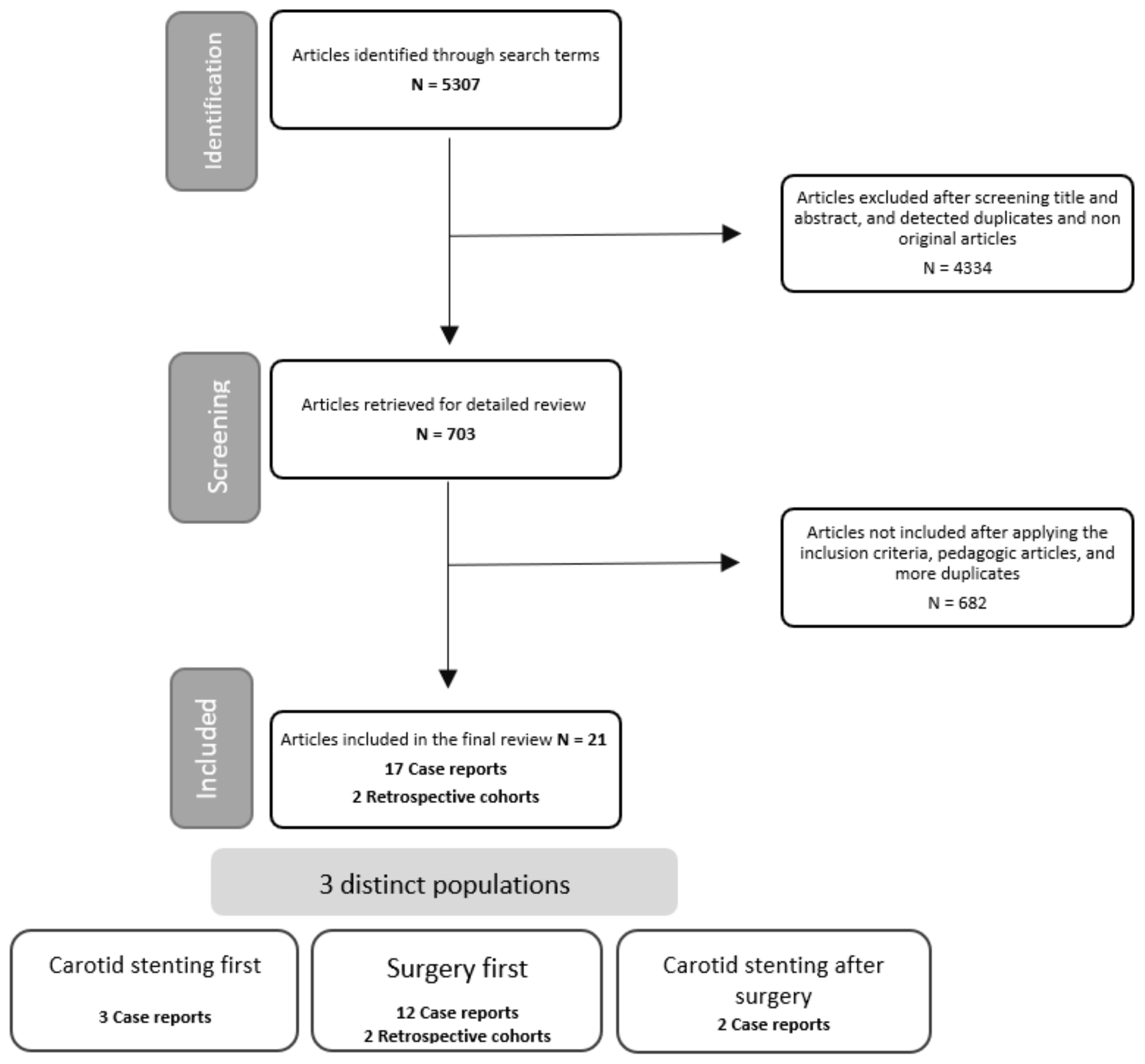
Baseline characteristics were displayed as absolute number (percentage) or mean (SD) or median (interquartile range [IQR], e.g., 25th–75th quantiles).
Extracted data included bibliographic information, type of paper, stated aim, topic/focus of systematic review, study/review methodology, description of reported involvement, baseline characteristics and results about treatment and outcome.

3. Results

3.1. Local Center Patients’ Selection (n = 9)

In total, nine patients were included in the study: four who underwent CS before AS, and five following AS, all of them because of neurologic deficits, attributed to a hemodynamic brain hypoperfusion. The characteristics of these patients are detailed in Table 1.
The nine patients managed in our center had a median age of 72 years (IQR 67.5–75.5), with a male predominance.
All patients had an mRS score of 0 before dissection. The median ASPECTS score was 9 (IQR 8–10) (n = 3). Following CS, the neurological deficit partially regressed in 66.7% of the patients. An increase in neurological deficit occurred in 11% for various reasons detailed in Table 1. During carotid stenting, three patients presented with distal emboli, e.g., a distal A2 embolus without endovascular rescue, a sylvian M2 embolus treated by thrombectomy; multiple distal emboli were not accessible by thrombectomy.
Following CS, three patients (33%) presented new ischemic lesion: a junctional infarct and a sylvian punctiform spot, distal diffuse ischemic lesions. Of these three patients, one patient presented with clinical worsening. All surviving patients had good stent permeability at follow-up.

3.2. Literature Review (n = 73)

A total of 5307 articles were identified. After exclusion based on the abstract, 703 articles were retained for detailed review. Finally, 19 articles matched all the criteria, 17 case reports, and 2 retrospective series were included. None of the studies were prospective; see flowchart of the included studies (Figure 1). The characteristics of included studies are shown in Table 2.
Patients had a median age of 64.5 years (IQR 57–70) for the case reports [11,13,14,15,16,17,18,19,20,21,22,23,24,25,26,27,28], and 60.5 years and 68 years in the cohorts [12,17]; 52% were female. A history of arterial hypertension (AH) was found in 83% of the cases.
All patients had an mRS score of 0 before dissection. The mean initial NIHSS score (all items combined) was 13.6 ± 11.2. The median time from symptom onset to treatment was 6 h (IQR 3.5–15).
Table 2. Summary of included studies.
StudyYearStudy TypeNumber of Patients IncludedFirst Treatment
Schönholz et al. [15]2008Case report1Surgery
Chahine et al. [16]2018Case report2Surgery
Morihara et al. [17]2016Case report1Surgery
Matsumoto et al. [18]2016Case report2Surgery
Amr et al. [13]2016Case report2Surgery
Hong et al. [19]2005Case report1Surgery
Karawabuki et al. [20]2006Case report1Surgery
Kim et al. [29]2006Case report1Surgery
Ueyama et al. [22]2007Case report1Surgery
Roseborough et al. [11]2006Case report1Surgery then stenting
Sakaguchi et al. [23]2005Case report1Surgery
Usui et al. [24]2021Case report1Surgery
Fukuhara et al. [25]2021Case report1Surgery
Funakoshi et al. [26]2020Case report2Stenting then surgery
Heran et al. [27]2019Case report1Stenting then surgery
Popovic et al. [14]2016Case report1Stenting then surgery
Casana et al. [28]2011Case report1Surgery then stenting
Fichadaya et al. [12]2022Cohort10Surgery
Morimoto et al. [30]2011Cohort41Surgery

3.3. Aortic and Carotid Surgery (ACS) Group (n = 72)

The mean age of the group was 65.5 ± 11.0 years; 53% were female.
The median initial NIHSS score was 16 (IQR 5–19) for the case reports and 8 for the Morimoto et al. cohort [31]. The mean NIHSS for all articles was 11.3 ± 8.5. The median time from symptom onset to treatment for the case reports was 5.5 h (IQR 3–12), and the mean time for all articles was 21 ± 39.3 h.
One patient died during surgery [15] of an uncontrollable hemorrhage complication. No other intraoperative complications were reported.
A new persistent neurological deficit occurred for 28% of the operated patients. A new ischemic lesion on postoperative DWI–MRI occurred for 49% of the patients.
Overall, 13% of the patients (15/53) died during hospitalization (mRS 6). For the other patients, the median mRS score was 3 (IQR 2–3) at discharge, and 0 (IQR 0–0.5) at last follow-up. The mean follow-up time was 53.4 months.
Individual patient data for this group are available in Supplementary Table S3A.

3.4. Carotid Stenting (CS) before Aortic Surgery (AS) Group (n = 8)

The mean age of the group was 65.3 ± 13.1 years; 25% were female. A history of AH was found in 66% of cases. All patients had an mRS score of 0 before dissection.
The median initial NIHSS score was 14 (IQR 11–21). The mean NIHSS was 14.3 ± 8.0. The median time from symptom onset to treatment was 5.5 h (IQR 3–18), with a mean duration of 13.6 ± 17.8 h.
No deaths during CS or surgery occurred. No patient had any intra-CS or intra-operative complication. Partial regression of neurological deficits was achieved in six patients (86%). The median post-CS NIHSS was 4 (n = 3 patients).
A new ischemic lesion on post-CS imaging occurred in 2 patients.
One patient died before aortic surgery, at day 2, of mesenteric ischemia; 1 patient did not undergo surgery because of a history of Bentall surgery. The other patients received aortic replacement afterwards.
None of the operated patients presented new neurological deficits or new ischemic lesions on postoperative imaging.
One patient died postoperatively during hospitalization from mediastinitis on day 35 [16]. For the other patients, the median mRS score was 2 at discharge, and 1 at follow-up. The mean follow-up time was 6.8 ± 3.6 months. All stents were patented at follow-up.
An example of one of our treated patients is shown in Figure 2.
Individual patient data for this group are available in Supplementary Table S3B.

3.5. Carotid Stenting (CS) after Aortic Surgery (AS) Group (n = 7)

The mean age of the group was 68.7 ± 5.8 years; 71% were female. A history of AH was found in 66% of cases.
All patients had an mRS score of 0 before dissection.
The initial NIHSS score of all patients was 0. The median time from symptom onset to surgery was 6 h (IQR 6–8), with a mean duration of 13 ± 17.2 h.
All patients presented a new neurological deficit postoperatively, with six of the seven patients presenting new lesions and a hypoperfused cerebral territory on imaging, justifying management by stenting.
The median surgery to stenting time was 3 h (IQR 1–14). During the procedure, three patients presented distal emboli despite the use of an FilterWire EZ (Boston Scientific, Marlborough, MA, USA) distal protection filter in all cases:
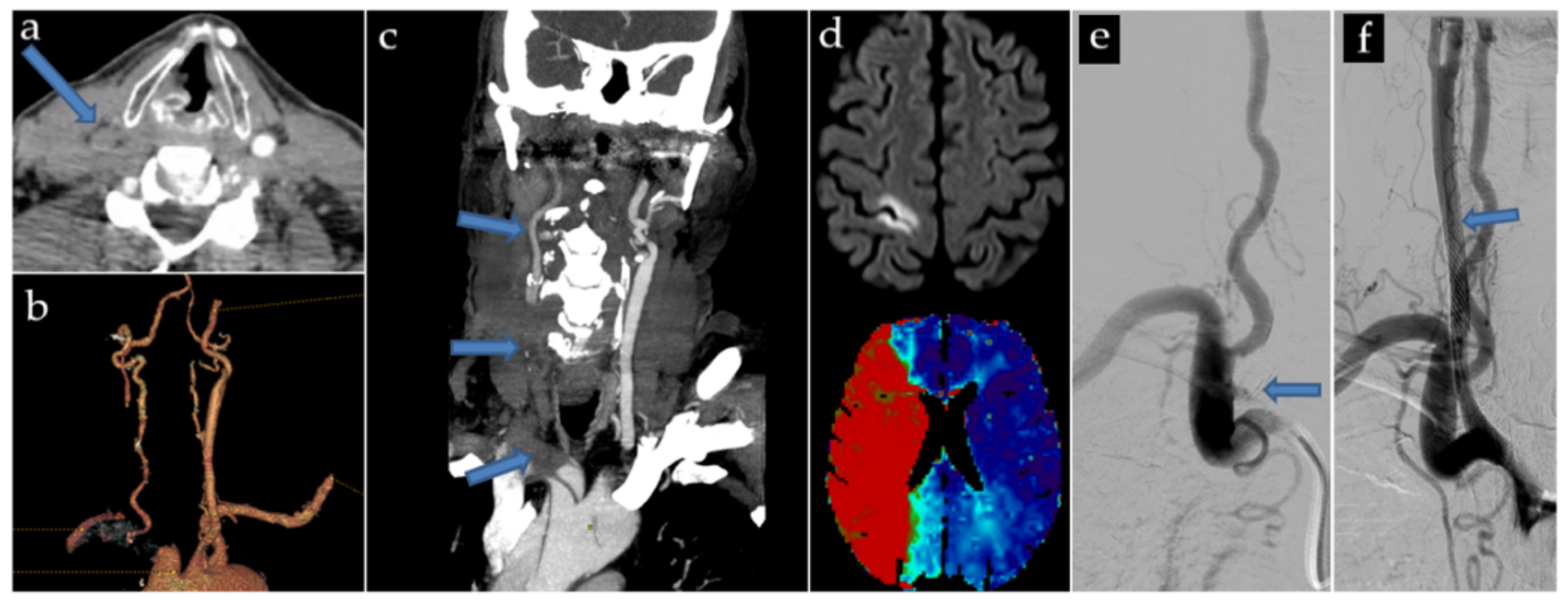
A 78 yo patient presented with a distal A2 embolus, too distant to be accessible by thrombectomy. Three 9 × 30 mm Carotid Wallstents were used, covering the entirety of the brachiocephalic trunk and right common carotid artery. After deployment of the first two Carotid Wallstents, an intra-stent thrombosis occurred, and was immediately and successfully treated by aspiration, and was not recurrent. Antiplatelet treatment was started the next day.
A 74 yo patient presented a sylvian M2 embolus at the end of the procedure, successfully treated by thrombectomy immediately after stenting with three Carotid Wallstent (7 × 40 mm, 9 × 50 mm and 5 × 30 mm) and a Smart Control 14 × 40 mm. Antiplatelet treatment and preventive low molecular weight Heparin were started the next day.
A 62 yo patient presented multiple distal emboli not accessible to thrombectomy. Antiplatelet treatment and curative low molecular weight Heparin were started 12 h post-procedure.
On postoperative imaging, the last two patients presented, respectively, a junctional infarct and distal embolic lesions.
Partial regression of neurological deficits was obtained in four patients (57%).
Only one patient presented a neurological worsening in the post-stenting period, related to reperfusion edema, which rapidly completely resolved.
One patient was still hospitalized at the time of writing, a few days after stenting, and the outcome could not be studied.
Two patients died during hospitalization. In the surviving patients, the median mRS score was 2 at discharge and 1 at follow-up.
Individual patient data for this group are available in Supplementary Table S3C.

4. Discussion

Regarding the patients from our local cohort (n = 9), the mean age was 71.2 ± 5.3 years, with a male predominance, a history of AH was found in 78%. The median ASPECTS score was 9 (IQR 8–10). Following CS, neurological deficits partially regressed in 0.67% (2/3) of the patients and remained stable in 0.33 (1/3) of the cases. During CS, three patients presented procedural complications: multiple distal emboli, an A2 emboli, and a homolateral sylvian M2 emboli, the latter being successfully treated by thrombectomy. The first patient had an immediate intra-stent thrombosis, successfully treated by aspiration. All surviving patients had good stent permeability at follow-up.
Experience in carotid stenting in the context of stroke derives from three situations: thrombectomy in thromboembolic stroke, urgent carotid repair in a traumatic setting, and isolated carotid artery stenosis or dissections (in rare cases).
The meta-analysis of Fabre et al. in the context of primitive carotid dissections, reported a technical success rate of 99.1% in 201 patients, with rare complications, including one embolic stroke, two subarachnoid hemorrhages, a transient vasospasm in two patients.
During follow-up, only 3.3% of patients developed intimal hyperplasia or intrastent stenosis; 2.1% had recurrent TIA in the territory of the stented vessels [32].
In the context of AAD extended to the carotid arteries, a limited number of case reports have been published in the literature, with stenting before [14,26,27], during [31,33,34], or after [11,21,28,34,35,36,37,38] aortic surgical repair: in all reported cases, technical success was achieved, with complete exclusion of false lumen, and stent patency at follow-up. All patients showed improvement or complete resolution of neurological symptoms. Only one case of death is reported, unrelated, from mediastinitis.
Recently, Mukherjee et al. reported three patients treated by retrograde carotid stenting after aortic repair, with residual stenosing carotid or brachial artery dissections. Venous stents (Boston Scientific VICI) were chosen because of their greater radial strength than the stents normally used, with the goal of a better obliteration of the false lumen [34].
Carotid angioplasty can be performed more rapidly than surgery, under local anesthesia.
Funakoshi et al. summarized the various challenges of performing carotid angioplasty-stenting preoperatively [26]:
(1) The need for double antiplatelet therapy in the immediate post-procedure period, which may lead to postpone the urgent aortic replacement surgery, which can only be carried out under mono-antiaggregation. (2) A risk of post-procedure reperfusion edema.
A prior perfusion imaging (CT or MRI) allows to evaluate this risk. (3) The technical difficulty of catheterizing the true lumen. (4) Restriction of future surgical options: when stents cover the origin of aortic branches total arch replacement is impossible. Both patients in the article were treated with hemi-arch replacement for this reason. (5) The risk of per-procedure distal emboli. However, according to a meta-analysis by Fabre et al., the risk of distal emboli is low for the treatment of dissections: for patients who benefited from a stenting treatment of primary internal carotid dissections, only 1 out of 201 treated patients presented an embolic stroke [32].
After the procedure, most patients are put on double antiplatelet therapy for 1–6 months, then on permanent single antiaggregant [32].
A summary of the benefits and drawbacks of the procedure is presented in Table 3.
The study by Fukuhara et al. found a significant difference in morbidity and mortality with ICA vs. CCA occlusion: every one of the ICA patients developed rapid cerebral edema with herniation, and died during hospitalization, whereas 79% of patients with occlusion limited to CCAs (uni- or bilateral) survived, and only one patient developed cerebral edema. These results suggest that occlusion of an ICA may be a marker at risk for cerebral edema and herniation [25].
The residual dissection of postoperative supra-aortic branches is associated with long-term neurological complications [11]. The prospective study by Neri et al., which followed 42 of these patients for a median of 3.17 years, found an incidence of neurological events of 30.9%, including stroke in 18 patients, with a relative risk of 3.99, all of which occurred in the territory of the initial dissected artery [39].
To the best of our knowledge, our study is the first aiming to synthesize the literature on carotid stenting for hemodynamic injury in the context of AAD.
Our study has many limitations. The included articles were mostly case reports, a source of important biases, notably publication bias. The patients’ selection from our center was retrospective, non-randomized, and monocentric. Due to the small number of patients included, statistics comparing the different groups could not be performed. All the articles studied did not evaluate the long-term outcome of patients and did not allow us to obtain any hindsight on mortality or recurrence of symptoms in the long term.

5. Conclusions

Preoperative carotid stenting for hemodynamic CM by true lumen compression appears to be feasible, and could be effective and safe, although there is still a lack of evidence, due to the absence of comparative statistical analysis. The literature, albeit growing, is still limited, and prospective comparative studies are needed.

Supplementary Materials

The following supporting information can be downloaded at: https://www.mdpi.com/article/10.3390/jpm13010058/s1, Table S1: Risk of bias of each case report study according to the The Joanna Briggs Institute Critical Appraisal tools; Table S2: Risk of bias of each individual non-randomised study according to the Cochrane Collaboration tool; Table S3A: Individual patients data from the patients treated by aortic and carotid surgery; Table S3B: Individual patients data from the patients treated by carotid stenting before aortic surgery; Table S3C: Individual patients data from the patients treated by carotid stenting after aortic surgery.

Author Contributions

Conceptualization, J.-F.H. and J.-F.A.; methodology, J.-F.H.; software, J.-F.H.; validation, J.-F.A., T.A., B.T., P.-A.B., A.J., A.R., N.G., F.T., P.H., V.G., F.C., A.B. and J.-F.H.; formal analysis, J.-F.H. and J.-F.A.; investigation, A.A., J.-F.A., T.A., B.T., P.-A.B., A.J., A.R., N.G., F.T., P.H., V.G., F.C., A.B. and J.-F.H.; resources, J.-F.H. and J.-F.A.; data curation, J.-F.A., T.A., B.T., P.-A.B., A.J., A.R., N.G., F.T., P.H., V.G., F.C., A.B. and J.-F.H.; writing—original draft preparation J.-F.A.; writing—review and editing, J.-F.H.; visualization, J.-F.H. and J.-F.A. supervision, J.-F.H.; project administration, A.J., J.-F.H. and J.-F.A. All authors have read and agreed to the published version of the manuscript.

Funding

This research received no external funding.

Institutional Review Board Statement

The study was conducted according to the guidelines of the Declaration of Helsinki and approved by the Ethics Committee of the CERIM (IRB: CRM-2209-303).

Informed Consent Statement

Patient consent was waived due to the fact that the study was a non-interventional retrospective study of routinely acquired data.

Data Availability Statement

The data presented in this study are available on request from the corresponding author.

Conflicts of Interest

The authors declare no conflict of interest.

References

  1. Sukockienė, E.; Laučkaitė, K.; Jankauskas, A.; Mickevičienė, D.; Jurkevičienė, G.; Vaitkus, A.; Stankevičius, E.; Petrikonis, K.; Rastenytė, D. Crucial role of carotid ultrasound for the rapid diagnosis of hyperacute aortic dissection complicated by cerebral infarction: A case report and literature review. Medicina 2016, 52, 378–388. [Google Scholar] [CrossRef] [PubMed]
  2. Sullivan, P.R.; Wolfson, A.B.; Leckey, R.D.; Burke, J.L. Diagnosis of acute thoracic aortic dissection in the emergency department. Am. J. Emerg. Med. 2000, 18, 46–50. [Google Scholar] [CrossRef] [PubMed]
  3. Goldberg, J.B.; Lansman, S.L.; Kai, M.; Tang, G.H.L.; Malekan, R.; Spielvogel, D. Malperfusion in Type A Dissection: Consider Reperfusion First. Semin. Thorac. Cardiovasc. Surg. 2017, 29, 181–185. [Google Scholar] [CrossRef] [PubMed]
  4. Zhao, H.; Ma, W.; Wen, D.; Duan, W.; Zheng, M. Computed tomography angiography findings predict the risk factors for preoperative acute ischaemic stroke in patients with acute type A aortic dissection. Eur. J. Cardio-Thorac. Surg. 2020, 57, 912–919. [Google Scholar] [CrossRef]
  5. Chemtob, R.A.; Fuglsang, S.; Geirsson, A.; Ahlsson, A.; Olsson, C.; Gunn, J.; Ahmad, K.; Hansson, E.C.; Pan, E.; O Arnadottir, L.; et al. Stroke in acute type A aortic dissection: The Nordic Consortium for Acute Type A Aortic Dissection (NORCAAD). Eur. J. Cardio-Thorac. Surg. 2020, 58, 1027–1034. [Google Scholar] [CrossRef]
  6. Okita, Y.; Okada, K. Treatment strategies for malperfusion syndrome secondary to acute aortic dissection. J. Card Surg. 2021, 36, 1745–1752. [Google Scholar] [CrossRef]
  7. Czerny, M.; Schoenhoff, F.; Etz, C.; Englberger, L.; Khaladj, N.; Zierer, A.; Weigang, E.; Hoffmann, I.; Blettner, M.; Carrel, T.P. The Impact of Pre-Operative Malperfusion on Outcome in Acute Type A Aortic Dissection. J. Am. Coll. Cardiol. 2015, 65, 2628–2635. [Google Scholar] [CrossRef] [Green Version]
  8. Geirsson, A.; Szeto, W.Y.; Pochettino, A.; McGarvey, M.L.; Keane, M.G.; Woo, Y.J.; Augoustides, J.G.; Bavaria, J.E. Significance of malperfusion syndromes prior to contemporary surgical repair for acute type A dissection: Outcomes and need for additional revascularizations. Eur. J. Cardio-Thorac. Surg. 2007, 32, 255–262. [Google Scholar] [CrossRef] [Green Version]
  9. Kreibich, M.; Desai, N.D.; Bavaria, J.E.; Szeto, W.Y.; Vallabhajosyula, P.; Beyersdorf, F.; Czerny, M.; Siepe, M.; Rylski, B.; Itagaki, R.; et al. Common carotid artery true lumen flow impairment in patients with type A aortic dissection. Eur. J. Cardio-Thorac. Surg. 2021, 59, 490–496. [Google Scholar] [CrossRef]
  10. Munir, W.; Chong, J.H.; Harky, A.; Bashir, M.; Adams, B. Type A aortic dissection: Involvement of carotid artery and impact on cerebral malperfusion. Asian Cardiovasc Thorac Ann. 2021, 29, 635–642. [Google Scholar] [CrossRef]
  11. Roseborough, G.S.; Murphy, K.P.; Barker, P.B.; Sussman, M. Correction of symptomatic cerebral malperfusion due to acute type I aortic dissection by transcarotid stenting of the innominate and carotid arteries. J. Vasc. Surg. 2006, 44, 1091–1096. [Google Scholar] [CrossRef] [PubMed] [Green Version]
  12. Fichadiya, A.; Menon, B.K.; Gregory, A.J.; Teleg, E.; Appoo, J.J. Neuroanatomy and severity of stroke in patients with type A aortic dissection. J. Card. Surg. 2022, 37, 339–347. [Google Scholar] [CrossRef] [PubMed]
  13. Amr, G.; Boulouis, G.; Bricout, N.; Modine, T.; Fayad, G.; Aguettaz, P.; Koussa, M. Stroke Presentation of Acute Type A Aortic Dissection with 100% Perfusion-Weighted Imaging–Diffusion-Weighted Imaging Mismatch: A Call for Urgent Action. J. Stroke Cerebrovasc. Dis. 2016, 25, 1280–1283. [Google Scholar] [CrossRef] [PubMed]
  14. Popovic, R.; Radovinovic-Tasic, S.; Rusovic, S.; Lepic, T.; Ilic, R.; Raicevic, R.; Obradovic, D. Urgent carotid stenting before cardiac surgery in a young male patient with acute ischemic stroke caused by aortic and carotid dissection. VSP 2016, 73, 674–678. [Google Scholar] [CrossRef] [Green Version]
  15. Schönholz, C.; Ikonomidis, J.S.; Hannegan, C.; Mendaro, E. Bailout Percutaneous External Shunt to Restore Carotid Flow in a Patient With Acute Type A Aortic Dissection and Carotid Occlusion. J. Endovasc. Ther. 2008, 15, 639–642. [Google Scholar] [CrossRef]
  16. Chahine, J.; Thapa, B.; Gajulapalli, R.D.; Kadri, A. Acute Aortic Dissection Presenting with a Headache: An Easily Missed Life-threatening Emergency. Cureus 2018, 10, e3531. [Google Scholar] [CrossRef] [Green Version]
  17. Morihara, R.; Yamashita, T.; Deguchi, K.; Tsunoda, K.; Manabe, Y.; Takahashi, Y.; Yunoki, T.; Sato, K.; Nakano, Y.; Kono, S.; et al. Successful Delayed Aortic Surgery for a Patient with Ischemic Stroke Secondary to Aortic Dissection. Int. Med. 2017, 56, 2343–2346. [Google Scholar] [CrossRef] [Green Version]
  18. Matsumoto, H.; Yoshida, Y.; Hirata, Y. Usefulness of cervical magnetic resonance imaging for detecting type A acute aortic dissection with acute stroke symptoms. Magn. Reson. Imaging 2016, 34, 902–907. [Google Scholar] [CrossRef] [PubMed]
  19. Hong, K.-S.; Park, S.-Y.; Whang, S.-I.; Seo, S.-Y.; Lee, D.-H.; Kim, H.-J.; Cho, J.-Y.; Cho, Y.-J.; Jang, W.-I.; Kim, C.Y. Intravenous Recombinant Tissue Plasminogen Activator Thrombolysis in a Patient with Acute Ischemic Stroke Secondary to Aortic Dissection. J. Clin. Neurol. 2009, 5, 49. [Google Scholar] [CrossRef] [Green Version]
  20. Kawarabuki, K.; Sakakibara, T.; Hirai, M.; Shirasu, M.; Kohara, I.; Tanaka, H.; Oyamada, M.; Takamatsu, T.; Murayama, Y.; Yamaki, T. Acute Aortic Dissection Presenting as a Neurologic Disorder. J. Stroke Cerebrovasc. Dis. 2006, 15, 26–29. [Google Scholar] [CrossRef]
  21. Kim, S.H.; Song, S.; Kim, S.-P.; Lee, J.; Lee, H.C.; Kim, E.S. Hybrid technique to correct cerebral malperfusion following repair of a type a aortic dissection. Korean J. Thorac. Cardiovasc. Surg. 2014, 47, 163–166. [Google Scholar] [CrossRef] [PubMed]
  22. Ueyama, K.; Otaki, K.; Koyama, M.; Kamiyama, H. Urgent simultaneous revascularization of the carotid artery and ascending aortic replacement for type A acute aortic dissection with cerebral malperfusion. Gen. Thorac. Cardiovasc. Surg. 2007, 55, 284–286. [Google Scholar] [CrossRef] [PubMed]
  23. Sakaguchi, G.; Komiya, T.; Tamura, N.; Obata, S.; Masuyama, S.; Kimura, C.; Kobayashi, T. Cerebral malperfusion in acute type A dissection: Direct innominate artery cannulation. J. Thorac. Cardiovasc. Surg. 2005, 129, 1190–1191. [Google Scholar] [CrossRef] [PubMed] [Green Version]
  24. Usui, T.; Suzuki, K.; Niinami, H.; Sakai, S. Aortic dissection diagnosed on stroke computed tomography protocol: A case report. J. Med. Case Rep. 2021, 15, 299. [Google Scholar] [CrossRef] [PubMed]
  25. Fukuhara, S.; Norton, E.L.; Chaudhary, N.; Burris, N.; Shiomi, S.; Kim, K.M.; Patel, H.J.; Deeb, G.M.; Yang, B. Type A Aortic Dissection With Cerebral Malperfusion: New Insights. Ann. Thorac. Surg. 2021, 112, 501–509. [Google Scholar] [CrossRef] [PubMed]
  26. Funakoshi, Y.; Imamura, H.; Tokunaga, S.; Murakami, Y.; Tani, S.; Adachi, H.; Ohara, N.; Kono, T.; Fukumitsu, R.; Sunohara, T.; et al. Carotid artery stenting before surgery for carotid artery occlusion associated with acute type A aortic dissection: Two case reports. Interv. Neuroradiol. 2020, 26, 814–820. [Google Scholar] [CrossRef]
  27. Heran, M.K.S.; Balaji, N.; Cook, R.C. Novel Percutaneous Treatment of Cerebral Malperfusion Before Surgery for Acute Type A Dissection. Ann. Thorac. Surg. 2019, 108, e15–e17. [Google Scholar] [CrossRef]
  28. Casana, R.; Tolva, V.; Majnardi, A.R.; Bianchi, P.G.; Addobati, L.; Bertoni, G.B.; Cireni, L.V.; Silani, V. Endovascular Management of Symptomatic Cerebral Malperfusion Due to Carotid Dissection After Type A Aortic Dissection Repair. Vasc Endovasc. Surg. 2011, 45, 641–645. [Google Scholar] [CrossRef]
  29. Sik Kim, Y.; Chernyshev, O.Y.; Alexandrov, A.V. Nonpulsatile Cerebral Perfusion in Patient With Acute Neurological Deficits. Stroke 2006, 37, 1562–1564. [Google Scholar] [CrossRef] [Green Version]
  30. Morimoto, N.; Okada, K.; Okita, Y. Lack of neurologic improvement after aortic repair for acute type A aortic dissection complicated by cerebral malperfusion: Predictors and association with survival. J. Thorac. Cardiovasc. Surg. 2011, 142, 1540–1544. [Google Scholar] [CrossRef]
  31. Igarashi, T.; Takahashi, S.; Takase, S.; Yokoyama, H. Intraoperative thrombectomy for occluded carotid arteries in patients with acute aortic dissection: Report of two cases. Surg. Today 2014, 44, 1177–1179. [Google Scholar] [CrossRef]
  32. Fabre, O.; Guesnier, L.; Renaut, C.; Gautier, L.; Geronimi, H.; Jasaitis, L.; Strauch, K. Prise en charge actuelle des dissections aortiques de type A. Traitement chirurgical et traitement des syndromes de malperfusion. Ann. De Cardiol. Et D’angéiologie 2005, 54, 332–338. [Google Scholar] [CrossRef] [PubMed]
  33. Lentini, S.; Tancredi, F.; Benedetto, F.; Gaeta, R. Type A aortic dissection involving the carotid arteries: Carotid stenting during open aortic arch surgery. Interact Cardiovasc. Thorac. Surg. 2009, 8, 157–159. [Google Scholar] [CrossRef] [PubMed] [Green Version]
  34. Mukherjee, D.; Lewis, E.; Spinosa, D.; Tang, D.; Ryan, L. Retrograde Carotid Stenting Using Newly Released Venous Stent for Cerebral Malperfusion in Type A Aortic Dissection. J. Endovasc. 2022, 29, 444–450. [Google Scholar] [CrossRef] [PubMed]
  35. Gao, P.; Wang, Y.; Chen, Y.; Jiao, L. Open retrograde endovascular stenting for left common carotid artery dissection secondary to surgical repair of acute aortic dissection: A case report and review of the literature. Ann. Vasc Surg. 2015, 29, e11–e15. [Google Scholar] [CrossRef]
  36. Tsai, K.-T.; Shen, T.-C. Challenging carotid intervention after total arch rerouting and hybrid zone 0 elephant trunk repair for a complicated type A aortic dissection. J. Endovasc. 2014, 21, 306–311. [Google Scholar] [CrossRef]
  37. Cardaioli, P.; Rigatelli, G.; Giordan, M.; Faggian, G.; Chinaglia, M.; Roncon, L. Multiple carotid stenting for extended thoracic aorta dissection after initial aortic surgical repair. Cardiovasc. Revasc. Med. 2007, 8, 213–215. [Google Scholar] [CrossRef]
  38. Pavkov, I.; Horner, S.; Klein, G.E.; Niederkorn, K. Percutaneous transluminal angioplasty with stenting in extended supra-aortic artery dissection. Croat. Med. J. 2004, 45, 217–219. [Google Scholar]
  39. Neri, E.; Sani, G.; Massetti, M.; Frati, G.; Buklas, D.; Tassi, R.; Giubbolini, M.; Benvenuti, A.; Sassi, C. Residual dissection of the brachiocephalic arteries: Significance, management, and long-term outcome. J. Thorac. Cardiovasc. Surg. 2004, 128, 303–312. [Google Scholar] [CrossRef]
Figure 1. Flowchart of included studies.
Jpm 13 00058 g001
Figure 2. Local cohort case of a 72-year-old, who suffered a brutal left hemicorporal deficit. CT scan showed an AAD extended to the LCCA, with a subocclusive compression of the true lumen (ac). Initial MRI showed a significant mismatch between the limited cytotoxic lesion of the rolandic area, and the hypoperfusion of the entire sylvian territory (d). Preoperative angioplasty was decided (e) The initial occlusion is shown. Stenting allowed to re-expand the true lumen, with coverage of the entire carotid dissection (f). The patient underwent aortic surgery 6 h later, without complications, and recovered gradually after. Post-surgical MRI (not shown) found no new cytotoxic lesion, and showed complete resolution of the hypoperfusion.
Jpm 13 00058 g002
Table 1. Local patients characteristics.
Patient123456789
TreatmentCarotid stenting before aortic surgeryCarotid stenting after aortic surgery
Age717172727864717462
SexMMMMFMFFF
Arterial hypertensionyesNoyesyesyesyesyesnoyes
Current smokingNoNoYesNoNoYesNoNoyes
Pre-stroke MRS000000000
NIHSS on presentation21NA103NA0000
ASPECTS score7108910108NANA
Supra-aortic branch involvedRCCAand ICA, LCCA and ICARCCA and ICARCCA and ICARCCA RCCA LCCARCCARCCA and ICA, LCCA RCCA
Per-procedure complicationsNoNoNoNoYes
(common carotid intra-stent thrombosis treated by aspiration, A2 distal emboli)
NoNoYes
M2 (sylvian emboli)
Yes
(distal emboli)
New lesion on post-procedure MRINoNoNoYes NoNoNoYes
(junctionnal infarct)
Yes
(diffuse distal ischemic lesions)
New neurological deficit post-procedureNoNoNoNoNoYes
(transient majoration due to reperfusion oedemea)
NoNoNo
Post-procedure neurologic deficit regressionPartialPartialPartialNoNoPartialPartialNoNo
SurvivedYesNo Mesenteric ischemia (day 2)YesYesNo
Neurologic degradation (day 8)
YesYesNo
Cardiac arrest (day 4)
NA
MRS at discharge2-4NA-14-NA
MRS after 90 days1-31-11-NA
Sent permeability at follow-upYesYesYesYesYesYesYesYesNA
Table 3. Comparison of benefits and drawbacks.
Carotid Stenting FirstAortic and Carotid Surgery First
Benefits
Local anesthesiaFaster repair of ascending aorta
Faster installation (15 min of installation on table)Correction of systemic hypotension
Treatment of distal internal carotid artery dissections
Complete exclusion of the false lumen
Drawbacks
Double antiaggregation post-procedure: postponed surgeryLonger preparation
Difficulty in catheterizing the true lumenGeneral anesthesia
Restriction of surgical options: impossibility of total arch replacement when stents cover the origin of
supra-aortic branches
Risk of intraoperative hypoperfusion under
cardiopulmonary bypass
Risk of distal embolization per procedureRisk of residual dissection and/or cerebral malperfusion if distal repair is impossible
Disclaimer/Publisher’s Note: The statements, opinions and data contained in all publications are solely those of the individual author(s) and contributor(s) and not of MDPI and/or the editor(s). MDPI and/or the editor(s) disclaim responsibility for any injury to people or property resulting from any ideas, methods, instructions or products referred to in the content.

