Review

Practical Aspects of Esophageal Pressure Monitoring in Patients with Acute Respiratory Distress Syndrome

Department of Anesthesiology and Intensive Care Medicine, Charles University, Faculty of Medicine in Hradec Kralove, University Hospital Hradec Kralove, Sokolska 581, 500 05 Hradec Kralove, Czech Republic
*
Author to whom correspondence should be addressed.
J. Pers. Med. 2023, 13(1), 136; https://doi.org/10.3390/jpm13010136
Submission received: 17 December 2022 / Revised: 9 January 2023 / Accepted: 10 January 2023 / Published: 10 January 2023
(This article belongs to the Special Issue Precision Medicine for Acute Respiratory Distress Syndrome (ARDS))

Abstract

:
Esophageal pressure (Pes) monitoring is a minimally invasive advanced respiratory monitoring method with the potential to guide ventilation support management. Pes monitoring enables the separation of lung and chest wall mechanics and estimation of transpulmonary pressure, which is recognized as an important risk factor for lung injury during both spontaneous breathing and mechanical ventilation. Appropriate balloon positioning, calibration, and measurement techniques are important to avoid inaccurate results. Both the approach of using absolute expiratory Pes values and the approach based on tidal Pes difference have shown promising results for ventilation adjustments, with the potential to decrease the risk of ventilator-induced lung injury.

1. Introduction

Esophageal pressure monitoring is a minimally invasive and clinically available method for estimating transpulmonary pressure [1], of which absolute values and changes are considered one of the main determinants of lung injury due to mechanical forces applied during mechanical ventilation [2]. The aim of this narrative review is to summarize the concept of esophageal pressure monitoring to assess transpulmonary pressures in patients with acute respiratory distress syndrome, its technical aspects of measurement, and its possible use for adjustment of the ventilator setting.

2. Transpulmonary Pressure, Pleural Pressure, and Esophageal Pressure

During mechanical ventilation, airway pressure distends the lung and chest wall in series. Increases in chest wall elastance, such as chest wall edema, kyphoscoliosis, and intraabdominal hypertension, are usually associated with increases in pleural pressure, as more force is necessary to distend the chest wall [3]; therefore, substantial and variable differences between airway and transpulmonary pressure may exist.
Transpulmonary pressure is a measure of lung stress, which is a distending force applied to lung structures. Changes in transpulmonary pressure are directly linked to changes in lung structure size—lung strain. If the stress and strain within the lung become unphysiological, the lung reacts by producing inflammatory cytokines and mediators, thereby initiating an inflammatory process within the lung parenchyma [4]. Although using a single value of transpulmonary pressure does not take into consideration lung inhomogeneity and ignores the role of respiratory rate and inspiratory flow in the development of ventilator-induced lung injury (VILI), transpulmonary pressure represents a physiologically sound safety limit for mechanical ventilation that should be measured and targeted, at least in patients with severe ARDS [2].
Transpulmonary pressure (PL) is defined as the pressure that corresponds to the pressure distending the lung, that is, the difference between airway pressure (Paw) and pleural pressure (Ppl). If this pressure is measured under static conditions during a sufficiently long inspiratory or expiratory pause, it corresponds to the difference between alveolar pressure (Palv) and Ppl.
Transpulmonary pressure changes during the respiratory cycle. Inspiratory and expiratory transpulmonary pressures are usually considered separately as safety limits or targets during mechanical ventilation [2,5]. Owing to gravitationally dependent pleural pressure differences (the so-called vertical pleural pressure gradient), there are also gravitationally dependent differences in transpulmonary pressures between different lung regions. During passive mechanical ventilation, lower pleural pressure values are expected in the non-dependent lung regions. Therefore, non-dependent lung regions are exposed to higher and dependent regions to lower inspiratory transpulmonary pressures. During spontaneous breathing, the respiratory muscles, mainly the diaphragm, generate negative pleural pressure, which is most negative close to the dorsal regions of the diaphragm [2]. Therefore, dependent lung regions are exposed to the highest inspiratory transpulmonary pressure during spontaneous breathing [6]. During pressure support ventilation, tidal volume distribution may be similar to spontaneous breathing; however, higher pressure support levels modify the distribution of ventilation and pleural pressure, which may be very close to the passive condition [7]. Pes is considered an estimate of the pleural pressure close to the balloon position in the chest. In humans, it approximates mid-thoracic pleural pressure [8]. Therefore, it underestimates pleural pressure in the most dependent lung regions and overestimates it in non-dependent lung regions. The vertical gradient of pleural pressure is significantly greater in injured versus normal lungs (1.8 times) [8] and could be more than 10 cm H2O in human cadavers in the supine position [6]. Consequently, PL calculated as the difference between Paw and Pes could be overestimated in the most dependent and underestimated in the most non-dependent lung regions by more than 5 cm H2O [8].
Direct subtraction of the absolute values of Paw and Pes (Table 1), the so-called directly measured PL, is currently considered an accurate estimation of PL in the mid-chest and dependent regions, with implications for the positive end-expiratory pressure (PEEP) setting [8] to decrease the risk of derecruitment in the dependent lung. As previously described, this method underestimates PL in non-dependent lung regions.
The release-derived transpulmonary pressure method (Table 1) is the reference method for calculating total end-inspiratory transpulmonary pressure, a surrogate of lung stress. Its calculation requires measuring Pes at a Paw equal to the atmospheric pressure, usually during disconnection from the ventilator. Although the PL calculated using the release-derived method probably reflects total lung stress better than other methods used to estimate PL [9], disconnection from the ventilator could be associated with sudden deterioration of respiratory function. Therefore, the release-derived calculation of transpulmonary pressure remains mainly a research tool.
The elastance-derived transpulmonary pressure method (Table 1) estimates the total end-inspiratory PL, multiplying the airway plateau pressure (Pplat) by the ratio between the lung and the total respiratory system elastance [9,10]. This method of PL calculation is currently considered the method of choice for the clinical estimation of inspiratory PL in non-dependent regions [8], and it is considered an indirect estimate of total lung strain and the risk of overdistension. A possible limitation of this method is the nonlinear behavior of lung and chest wall elastance at the extremes of the pressure/volume curve of the respiratory system [9].
The Pes value can also be used to calculate so-called lung or transpulmonary driving pressure, which is considered another important indicator of possible lung injury during both spontaneous and mechanical ventilation. Transpulmonary driving pressure is calculated either as the difference between end-inspiratory and end-expiratory transpulmonary pressures, or as the difference between airway driving pressure and the so-called chest wall driving pressure, which is the difference between the measured inspiratory and expiratory esophageal pressures (Table 1) [11]. Transpulmonary driving pressure is considered an indirect estimate of dynamic lung strain.
Table 1. Suggested methods to calculate inspiratory transpulmonary pressure, expiratory transpulmonary pressure, total transpulmonary pressure, and transpulmonary driving pressure. All pressures must be measured under static conditions after the airway occlusion maneuver. Adapted from Mieto et al. [9] and Grieco et al. [11].
ParameterMethodComputation
End-inspiratory PL, Elastance-derivedPL = Pplat x EL/Ers = Pplat x [(Pplat − Pesplat) − (PawPEEP − PesPEEP)]/(Pplat − PawPEEP)
End-inspiratory PL (reference method)Release-derivedPL = (Pplat − PawATM) − (Pesplat − PesATM)
End-expiratory PLDirect methodPL = PawPEEP − PesPEEP
Driving PLDirect methodDPL = (Pplat − Pesplat) − (PawPEEP − PesPEEP), or
DPL = (Pplat − PawPEEP) − (Pesplat − PesPEEP)
PL—transpulmonary pressure; Pplat—plateau pressure; PEEP—positive end-expiratory pressure; EL—lung elastance; Ers—respiratory system elastance; PesPEEP—esophageal pressure measured at PEEP; Pesplat—esophageal pressure measured at plateau pressure; PesATM—esophageal pressure at atmospheric pressure; PawPEEP—airway pressure at PEEP.

3. Technical Aspects of Esophageal Pressure Measurement

Pes measured using the balloon technique is considered the best noninvasive surrogate of pleural pressure available at the bedside in critically ill patients. As it is an indirect estimate of pleural pressure, possibly affected by some artifacts, the correct technique of catheter insertion, balloon positioning, filling, and validation testing are important for obtaining reliable results [12].

3.1. Ballon Catheter Insertion and Positioning

Some esophageal balloons are integrated into a traditional nasogastric feeding tube, whereas others are carried by a thin, dedicated tube [12]. The insertion of the catheter is similar to that of a nasogastric tube. The integrity of the catheter and balloon should be checked by inflation of the catheter using the specific recommended amount of air through the balloon port. Then, the catheter and its deflated balloon are lubricated and inserted through the nostril into the esophagus and stomach. Depth markers on the catheter aid orientation and assessment of the insertion depth. The intragastric position of the balloon may be confirmed after the balloon port is connected to the pressure monitoring device and inflation of the balloon with the recommended volume. The position of the balloon in the stomach can be confirmed by visualization of the positive deflections of the balloon pressure during gentle external compressions of the left upper abdominal quadrant [12]. Although the presence of a nasogastric tube does not affect the reliability of the measurement [13,14], insertion of a thin balloon catheter could be more difficult. If the nasogastric tube is already in place, it may be necessary to remove it before inserting the balloon catheter.
After insertion of the balloon catheter into the stomach and inflation with the volume of air recommended by the manufacturer, the catheter is slowly withdrawn into the mid-distal esophagus. Catheters usually have depth markers to aid in positioning the balloon, and the depth at which the balloon should be placed can be estimated on the basis of the distance from the nostril to the ear tragus to the xyphoid. In almost all cases, the correct distance between the nostril and distal end of the balloon ranges from 35 to 45 cm [12]. Changes in the Pes waveform are observed during the slow and stepwise withdrawal of the balloon catheter. A sudden change in baseline pressure is expected when the balloon transitions from the abdomen to the chest [12]. Positive deflections of Pes should be visible in patients on positive pressure ventilation, and negative deflections in spontaneously breathing patients. Typical cardiac artifacts usually appear in the Pes waveform, and their presence suggests the balloon’s position beneath the heart. If cardiac oscillations are excessive and prevent reliable assessment of the tidal swing of the Pes, the catheter can be carefully pulled further back, but the position of the balloon in the upper esophagus should be avoided [12]. An additional check on the correct positioning can be performed by examining the waveforms. The chest wall has linear elastic behavior; thus, a linear relationship between the volume and pleural pressure is expected. Therefore, if the esophageal balloon is surrounded by pleural pressure (i.e., it is in the correct position), the esophageal balloon pressure and volume curves should have a similar shape (not amplitude) on the monitoring device or ventilator screen [12].

3.2. Ballon Filling and Validation Testing

Balloon catheters produced by different producers differ in terms of the material used, volume, length, and elastance. Therefore, the use of balloon-specific filling volumes is required because underfilled balloons tend to underestimate and overfilled balloons tend to overestimate the real Pes value [13,15]. Larger balloons usually have a wider range of adequate filling volumes, at which point the balloon wall elastance does not affect the measured Pes values [13]. It enables adjustment of the filling volume for other important factors, such as esophageal elastance (Ees) and balloon-surrounding pressure. Higher surrounding pressures require higher filling volumes to reliably measure tidal Pes swings [13].
To overcome the problem of the sub- or supra-optimal filling volume of the balloon, inter-individual differences in esophageal elastance, and the effects of the surrounding pressure, a technique of in vivo calibration of Pes has recently been proposed [15]. Briefly, the balloon is inflated stepwise using small aliquots of air (Figure 1). Pesplat and PesPEEP are recorded at each step. Further balloon inflation is interrupted if a sudden substantial increase in the Pes baseline is observed. The smallest inflation volume with the largest tidal swing of Pes below the balloon volume, associated with a sudden baseline change in Pes, is considered the best volume (Vbest).
To compensate for the baseline changes of Pes at higher inflation volumes, it was suggested to subtract from the measured value of Pes the pressure generated by the distension of the esophageal wall—esophageal recoil pressure (Pew). This pressure can be calculated from the elastance of the linear part of the pressure-volume curve of the balloon: Pew = (Vbest − Vmin) × Ees, where Vbest in mL is the balloon volume with the largest tidal swing of Pes,Vmin in mL is the minimum filling volume of the balloon at the beginning of the linear part of the balloon pressure-volume curve (Figure 2), and Ees in cm H2O/mL is calculated using the least square fitting method from Pes values [15,16]. Ees can also be estimated using a simplified method based on the Pes values obtained at Vmin and Vmax [17]. Using this approach, Ees is calculated as the difference in Pes at Vmax and Vmin divided by the difference between Vmax and Vmin, and calibrated Pes is calculated as the difference between the measured Pes at Vbest and Pew at Vbest (Table 2, Figure 2). All calculations can be done using an appropriate table calculator.
To check the position of the balloon and its appropriate filling, a validation test, also called the occlusion test or Baydur test, should be performed during airway occlusion [12]. During the end-expiratory occlusion maneuver, the occluded airway impedes changes in the intrathoracic volume. Patient breathing effort, or gentle sternal compression, leads to changes in the intrathoracic pressure that are fully and equally transferred into the airways, pleural space, and esophagus. In the case of a properly placed and filled balloon catheter, the changes in Pes (ΔPes) are expected to be equal to the changes in Paw (ΔPaw). The target value of the ΔPes/ΔPaw ratio ranges from 0.9 to 1.1. If the ratio is outside this range, the balloon filling (first) and balloon positioning (second) should be rechecked [12].
There is ongoing discussion on whether balloon calibration is necessary for routine practice. Some authorities in the field of Pes monitoring state that using a balloon with a consistent working range of inflation volume helps to obtain consistent and accurate measurements. While the optimal inflation volume can be confirmed based on the pressure-volume characteristics of the balloon itself, calibration is time-consuming and not required in practice when using a balloon with a known working range. A simple observation of the Pes waveform should be sufficient to detect overinflation resulting in inaccurately high measured pressures secondary to the compliance of the balloon, whereas underinflation is associated with dampening of waveform variation [18]. The disadvantage of this simplified approach is that it does not correct for individual differences in esophageal elastance. Mojoli et al. [15] showed that the pressure generated by the esophageal wall could range from 0 to +6 cm H2O and that the optimal filling volume range was highly variable among different patients and conditions, ranging from 0.5 to 6 mL in their series. The ventilator setting and body mass index (BMI) modify the optimal filling volume [15,19]. This suggests that the esophageal catheter filling volume should be adapted to the intrathoracic pressure condition of any patient [15]. Using the simplified calibration method suggested by Sun et al. [17], the measured Pes value at Vbest is corrected to obtain the Pes value at Vmin. Therefore, if the value of the end-expiratory transpulmonary pressure is of interest, visual identification of Vmin from the balloon pressure-volume curve and using a corresponding Pes value is probably sufficient for clinical use. For a more reliable estimation of transpulmonary driving pressure and end-inspiratory transpulmonary pressure, identification of Vbest and the corresponding end-inspiratory and end-expiratory Pes values is necessary. The calibration procedure is not complicated and is clinically feasible [19].
In summary, stepwise inflation of the balloon with an appropriate amount of air (0.2–1.0 mL) according to the type of balloon catheter can be recommended. If the expiratory Pes value after the inflation step remains close to a previous value (difference within 1.0 cm H2O), the previous volume of the balloon is used to measure the expiratory Pes value, and no correction of the measured value is necessary. Adjustment of the balloon inflation is possible based on the results of a validation test during airway occlusion. Vbest estimation is necessary for a more accurate estimation of the inspiratory transpulmonary pressure in nondependent lung regions.

3.3. Artifacts, Errors, and Rules during the Pes Measurement

Esophageal pressure is affected by many factors, including the patient’s weight, body position, muscle activity, heart weight, lung weight, ventilator setting, and presence of fluid in the pleural cavity [3]. Any change in these factors, such as setting higher PEEP values, influences Pes. The effect of these factors cannot be considered artifacts or errors because they usually reflect either changed pleural pressure or a relative change in the esophageal position on a vertical thoracic axis.
The most important errors occur either because of improper measurement techniques or the effects of local factors, such as previously described cardiac oscillations or esophageal peristalsis, which could be detected as slow changes in the Pes baseline. The importance of optimal balloon positioning and calibration has been previously discussed. Possible overinflation due to previous insufficient balloon deflation may occur; therefore, complete and active deflation of the balloon with the syringe with the subsequent opening of the balloon port using a three-way stopcock to the atmosphere to allow equilibration of the atmospheric pressure and balloon pressure should be employed before balloon inflation. The mechanical properties of the balloon may change over time, and air leakage from the balloon or insufficient tightness of the connectors may occur. Incorrect calibration of the pressure transducer, particularly if an air-filled blood pressure transducer is used instead of a designated device, is also a potential source of error.
Pes should be measured at the body position where the patient is ventilated. Position changes are associated with changes in the Pes value, with high interpersonal variability [3]. The prone position is associated with lower Pes values in comparison with the supine position, possibly because of the diminished effect of mediastinum/heart weight, the more non-dependent position of the esophagus in the supine position, increased chest wall elastance, and a lower vertical pleural pressure gradient in the supine position. Changes in the setting of the ventilator, mainly the selection of a different PEEP level, require repeating the measurement of Pes [15,19,20].

4. Ventilatory Setting Based on Transpulmonary Pressure in Passive ARDS Patients

Esophageal pressure monitoring allows for the differentiation of chest wall, lung, and respiratory system mechanics, and there is ongoing interest in using Pes values for PEEP titration, monitoring of parenchymal lung stress, limiting peak end-inspiratory transpulmonary pressures, and monitoring ventilator synchrony [1,18].

4.1. PEEP Setting Targeting Expiratory Transpulmonary Pressure

Elevation of pleural pressure occurs in dependent regions due to increased chest wall weight in patients with obesity, chest wall edema, or pleural effusions. Increased lung weight or elevated intra-abdominal pressure may lead to lung derecruitment, mainly in dependent lung regions, increased lung elastance, and hypoxemia. It was hypothesized that, if the pleural pressure is greater than the airway/alveolar pressure, the application of PEEP may be helpful [18] in preventing lung derecruitment. The EPVent [5] and EPvent2 [21] studies investigated the use of esophageal manometry to titrate PEEP. Although the results of the EPvent study suggested a possible benefit of a strategy using a PEEP setting based on expiratory transpulmonary pressures, the EPvent2 study did not show a clear benefit compared to a high-PEEP strategy. Further analysis [22] of EPvent2 data suggested a benefit when end-expiratory PL was maintained in a tight physiological range of − 2 to + 2 cm H2O with PEEP adjustment. Based on the results of this mechanistic analysis, proponents of this approach suggest titrating PEEP to obtain expiratory PL close to zero [18,21]. The benefits of this approach should be expected in obese patients and patients with intra-abdominal hypertension. Although this approach is appealing, it has not been tested in a prospective randomized clinical trial; performed trials targeted an end-expiratory PL between 0 cm H2O and +6 cm H2O, depending on the inspiratory fraction of the oxygen (FiO2) requirement. Strong evidence from experimental and clinical trials on mechanical ventilation suggests that the use of higher levels of PEEP could be beneficial only if the increment in PEEP is associated with improved compliance of the respiratory system [23], reasonable CO2 elimination, and stable patient hemodynamics [24,25]. This information should always be considered when attempting to adjust PEEP levels according to expiratory PL. Until further information from clinical trials is available, the evaluation of expiratory transpulmonary pressure could be useful as an indicator of the safety of the ventilator setting.

4.2. PEEP Setting Targeting Inspiratory Transpulmonary Pressure

This approach to setting the PEEP was used in patients considered ECMO candidates owing to severe H1N1 influenza pneumonia [26]. If the inspiratory PL calculated using the release-derived method was below 25 cm H2O, using a constant tidal volume, PEEP was elevated to reach a so-called maximum physiological inspiratory PL of 25 cm H2O. The inspiratory PL value may be used either to limit the maximum value of PEEP or to personalize the targeting of the tidal volume. Recently, a safety limit for the maximum inspiratory transpulmonary pressure below 20–25 cm H2O was suggested [1,11], and a recent consensus statement suggested using inspiratory PL as an indicator of the safety of ventilator settings [1].

4.3. Transpulmonary Driving Pressure and Safety

Respiratory system driving pressure is an important indicator of mechanical ventilation safety [23,27,28]. The upper limits for tidal changes in the transpulmonary pressure for patients with ARDS 10 to 12 cm H2O have recently been recommended for personalized targeting of the tidal volume [1,28]. The suggested limits for transpulmonary pressure are presented in Table 3.

5. The Other Possible Use for Measuring Esophageal Pressure in ARDS Patients

Measuring transpulmonary pressure is only one of the possible clinical applications of Pes monitoring; it can also be used to assess respiratory efforts in spontaneously breathing patients, evaluate patient-ventilator asynchronies, and compute transmural vascular pressures [1]. A detailed description of these topics is beyond the scope of the present review.
Although there are other clinically available alternatives, Pes monitoring could be used to identify and manage patients with a substantial risk of so-called patient self-inflicted lung injury (P-SILI) [29,30]. Recently, a novel noninvasive method for detecting excessively high respiratory effort using expiratory airway occlusion was developed [31]. In patients with an estimated high dynamic transpulmonary pressure (>15 cm H2O), the authors recommend considering the use of Pes monitoring to adjust patient management. Pes recording allows the detection of inappropriate inspiratory efforts. An absolute value of ∆Pes lower than 2–3 cm H2O is usually considered a sign of over assistance. Conversely, a ∆Pes higher than 8–12 cm H2O is considered a marker of under assistance [29,30].

6. Conclusions

Esophageal pressure monitoring is a minimally invasive and clinically appealing tool for personalized mechanical ventilation in ARDS patients. Both the approach of using absolute expiratory Pes values and that in which a tidal Pes difference is considered have shown promising results for ventilation adjustments, with the potential to decrease the risk of ventilator-induced lung injury.

Author Contributions

Conceptualization, P.D. and V.D.; methodology, P.D.; writing—original draft preparation, P.D. and V.D.; writing—review and editing, P.D.; funding acquisition, P.D. All authors have read and agreed to the published version of the manuscript.

Funding

This work was supported by the Cooperatio Program, research area INCA.

Institutional Review Board Statement

Not applicable.

Informed Consent Statement

Not applicable.

Data Availability Statement

Not applicable.

Conflicts of Interest

The authors declare no conflict of interest.

References

  1. Mauri, T.; Yoshida, T.; Bellani, G.; Goligher, E.C.; Carteaux, G.; Rittayamai, N.; Mojoli, F.; Chiumello, D.; Piquilloud, L.; Grasso, S.; et al. PLeUral pressure working Group (PLUG—Acute Respiratory Failure section of the European Society of Intensive Care Medicine). Esophageal and transpulmonary pressure in the clinical setting: Meaning, usefulness and perspectives. Intensive Care Med. 2016, 42, 1360–1373. [Google Scholar] [CrossRef] [PubMed]
  2. Gattinoni, L.; Giosa, L.; Bonifazi, M.; Pasticci, I.; Busana, M.; Macri, M.; Romitti, F.; Vassalli, F.; Quintel, M. Targeting transpulmonary pressure to prevent ventilator-induced lung injury. Expert Rev. Respir. Med. 2019, 13, 737–746. [Google Scholar] [CrossRef] [PubMed]
  3. Sahetya, S.K.; Brower, R.G. The promises and problems of transpulmonary pressure measurements in acute respiratory distress syndrome. Curr. Opin. Crit. Care 2016, 22, 7–13. [Google Scholar] [CrossRef] [PubMed] [Green Version]
  4. Gattinoni, L.; Carlesso, E.; Caironi, P. Stress and strain within the lung. Curr. Opin. Crit. Care 2012, 18, 42–47. [Google Scholar] [CrossRef] [PubMed]
  5. Talmor, D.; Sarge, T.; Malhotra, A.; O’Donnell, C.R.; Ritz, R.; Lisbon, A.; Novack, V.; Loring, S.H. Mechanical ventilation guided by esophageal pressure in acute lung injury. N. Engl. J. Med. 2008, 359, 2095–2104. [Google Scholar] [CrossRef] [Green Version]
  6. Yoshida, T.; Grieco, D.L.; Brochard, L.; Fujino, Y. Patient self-inflicted lung injury and positive end-expiratory pressure for safe spontaneous breathing. Curr. Opin. Crit. Care 2020, 26, 59–65. [Google Scholar] [CrossRef]
  7. Mauri, T.; Bellani, G.; Confalonieri, A.; Tagliabue, P.; Turella, M.; Coppadoro, A.; Citerio, G.; Patroniti, N.; Pesenti, A. Topographic distribution of tidal ventilation in acute respiratory distress syndrome: Effects of positive end-expiratory pressure and pressure support. Crit. Care Med. 2013, 41, 1664–1673. [Google Scholar] [CrossRef]
  8. Yoshida, T.; Amato, M.B.P.; Grieco, D.L.; Chen, L.; Lima, C.A.S.; Roldan, R.; Morais, C.C.A.; Gomes, S.; Costa, E.L.V.; Cardoso, P.F.G.; et al. Esophageal Manometry and Regional Transpulmonary Pressure in Lung Injury. Am. J. Respir. Crit. Care Med. 2018, 197, 1018–1026. [Google Scholar] [CrossRef]
  9. Mietto, C.; Malbrain, M.L.; Chiumello, D. Transpulmonary pressure monitoring during mechanical ventilation: A bench-to-bedside review. Anaesthesiol. Intensive Ther. 2015, 8, 27–37. [Google Scholar] [CrossRef]
  10. Gattinoni, L.; Chiumello, D.; Carlesso, E.; Valenza, F. Bench-to-bedside review: Chest wall elastance in acute lung injury/acute respiratory distress syndrome patients. Crit. Care 2004, 8, 350–355. [Google Scholar] [CrossRef] [Green Version]
  11. Grieco, D.L.; Chen, L.; Brochard, L. Transpulmonary pressure: Importance and limits. Ann. Transl. Med. 2017, 5, 285. [Google Scholar] [CrossRef] [Green Version]
  12. Mojoli, F.; Torriglia, F.; Orlando, A.; Bianchi, I.; Arisi, E.; Pozzi, M. Technical aspects of bedside respiratory monitoring of transpulmonary pressure. Ann. Transl. Med. 2018, 6, 377. [Google Scholar] [CrossRef] [PubMed]
  13. Mojoli, F.; Chiumello, D.; Pozzi, M.; Algieri, I.; Bianzina, S.; Luoni, S.; Volta, C.A.; Braschi, A.; Brochard, L. Esophageal pressure measurements under different conditions of intrathoracic pressure. An in vitro study of second generation balloon catheters. Minerva Anestesiol. 2015, 81, 855–864. [Google Scholar] [PubMed]
  14. Niknam, J.; Chandra, A.; Adams, A.B.; Nahum, A.; Ravenscraft, S.A.; Marini, J.J. Effect of a nasogastric tube on esophageal pressure measurement in normal adults. Chest 1994, 106, 137–141. [Google Scholar] [CrossRef] [Green Version]
  15. Mojoli, F.; Iotti, G.A.; Torriglia, F.; Pozzi, M.; Volta, C.A.; Bianzina, S.; Braschi, A.; Brochard, L. In vivo calibration of esophageal pressure in the mechanically ventilated patient makes measurements reliable. Crit. Care 2016, 20, 98. [Google Scholar] [CrossRef] [PubMed] [Green Version]
  16. Yang, Y.L.; He, X.; Sun, X.M.; Chen, H.; Shi, Z.H.; Xu, M.; Chen, G.Q.; Zhou, J.X. Optimal esophageal balloon volume for accurate estimation of pleural pressure at end-expiration and end-inspiration: An in vitro bench experiment. Intensive Care Med. Exp. 2017, 5, 35. [Google Scholar] [CrossRef] [PubMed] [Green Version]
  17. Sun, X.M.; Chen, G.Q.; Huang, H.W.; He, X.; Yang, Y.L.; Shi, Z.H.; Xu, M.; Zhou, J.X. Use of esophageal balloon pressure-volume curve analysis to determine esophageal wall elastance and calibrate raw esophageal pressure: A bench experiment and clinical study. BMC Anesthesiol. 2018, 18, 21. [Google Scholar] [CrossRef]
  18. Kassis, E.B.; Talmor, D. Clinical application of esophageal manometry: How I do it. Crit. Care 2021, 25, 6. [Google Scholar] [CrossRef]
  19. Jiang, J.; Su, L.; Cheng, W.; Wang, C.; Rui, X.; Tang, B.; Zhang, H.; He, H.; Long, Y. The calibration of esophageal pressure by proper esophageal balloon filling volume: A clinical study. Front. Med. 2022, 9, 986982. [Google Scholar] [CrossRef]
  20. Kumaresan, A.; Gerber, R.; Mueller, A.; Loring, S.H.; Talmor, D. Effects of Prone Positioning on Transpulmonary Pressures and End-expiratory Volumes in Patients without Lung Disease. Anesthesiology 2018, 128, 1187–1192. [Google Scholar] [CrossRef]
  21. Beitler, J.R.; Sarge, T.; Banner-Goodspeed, V.M.; Gong, M.N.; Cook, D.; Novack, V.; Loring, S.H.; Talmor, D.; EPVent-2 Study Group. Effect of Titrating Positive End-Expiratory Pressure (PEEP) With an Esophageal Pressure-Guided Strategy vs an Empirical High PEEP-Fio2 Strategy on Death and Days Free From Mechanical Ventilation Among Patients With Acute Respiratory Distress Syndrome: A Randomized Clinical Trial. JAMA 2019, 321, 846–857. [Google Scholar] [CrossRef] [PubMed] [Green Version]
  22. Sarge, T.; Baedorf-Kassis, E.; Banner-Goodspeed, V.; Novack, V.; Loring, S.H.; Gong, M.N.; Cook, D.; Talmor, D.; Beitler, J.R.; EPVent-2 Study Group. Effect of Esophageal Pressure-guided Positive End-Expiratory Pressure on Survival from Acute Respiratory Distress Syndrome: A Risk-based and Mechanistic Reanalysis of the EPVent-2 Trial. Am. J. Respir. Crit. Care Med. 2021, 204, 1153–1163. [Google Scholar] [CrossRef] [PubMed]
  23. Amato, M.B.; Meade, M.O.; Slutsky, A.S.; Brochard, L.; Costa, E.L.; Schoenfeld, D.A.; Stewart, T.E.; Briel, M.; Talmor, D.; Mercat, A.; et al. Driving pressure and survival in the acute respiratory distress syndrome. N. Engl. J. Med. 2015, 372, 747–755. [Google Scholar] [CrossRef] [Green Version]
  24. Writing Group for the Alveolar Recruitment for Acute Respiratory Distress Syndrome Trial (ART) Investigators; Cavalcanti, A.B.; Suzumura, É.A.; Laranjeira, L.N.; Paisani, D.M.; Damiani, L.P.; Guimarães, H.P.; Romano, E.R.; Regenga, M.M.; Taniguchi, L.N.T.; et al. Effect of Lung Recruitment and Titrated Positive End-Expiratory Pressure (PEEP) vs Low PEEP on Mortality in Patients With Acute Respiratory Distress Syndrome: A Randomized Clinical Trial. JAMA 2017, 318, 1335–1345. [Google Scholar] [CrossRef]
  25. Gattinoni, L.; Marini, J.J. In search of the Holy Grail: Identifying the best PEEP in ventilated patients. Intensive Care Med. 2022, 48, 728–731. [Google Scholar] [CrossRef] [PubMed]
  26. Grasso, S.; Terragni, P.; Birocco, A.; Urbino, R.; Del Sorbo, L.; Filippini, C.; Mascia, L.; Pesenti, A.; Zangrillo, A.; Gattinoni, L.; et al. ECMO criteria for influenza A (H1N1)-associated ARDS: Role of transpulmonary pressure. Intensive Care Med. 2012, 38, 395–403. [Google Scholar] [CrossRef] [PubMed]
  27. Bugedo, G.; Retamal, J.; Bruhn, A. Driving pressure: A marker of severity, a safety limit, or a goal for mechanical ventilation? Crit. Care 2017, 21, 199. [Google Scholar] [CrossRef] [Green Version]
  28. Pelosi, P.; Ball, L.; Barbas, C.S.V.; Bellomo, R.; Burns, K.E.A.; Einav, S.; Gattinoni, L.; Laffey, J.G.; Marini, J.J.; Myatra, S.N.; et al. Personalized mechanical ventilation in acute respiratory distress syndrome. Crit. Care 2021, 25, 250. [Google Scholar] [CrossRef]
  29. Goligher, E.C.; Dres, M.; Patel, B.K.; Sahetya, S.K.; Beitler, J.R.; Telias, I.; Yoshida, T.; Vaporidi, K.; Grieco, D.L.; Schepens, T.; et al. Lung- and Diaphragm-Protective Ventilation. Am. J. Respir. Crit. Care Med. 2020, 202, 950–961. [Google Scholar] [CrossRef]
  30. Carteaux, G.; Parfait, M.; Combet, M.; Haudebourg, A.F.; Tuffet, S.; Mekontso Dessap, A. Patient-Self Inflicted Lung Injury: A Practical Review. J. Clin. Med. 2021, 10, 2738. [Google Scholar] [CrossRef]
  31. Bertoni, M.; Telias, I.; Urner, M.; Long, M.; Del Sorbo, L.; Fan, E.; Sinderby, C.; Beck, J.; Liu, L.; Qiu, H.; et al. A novel non-invasive method to detect excessively high respiratory effort and dynamic transpulmonary driving pressure during mechanical ventilation. Crit. Care 2019, 23, 346. [Google Scholar] [CrossRef] [PubMed] [Green Version]
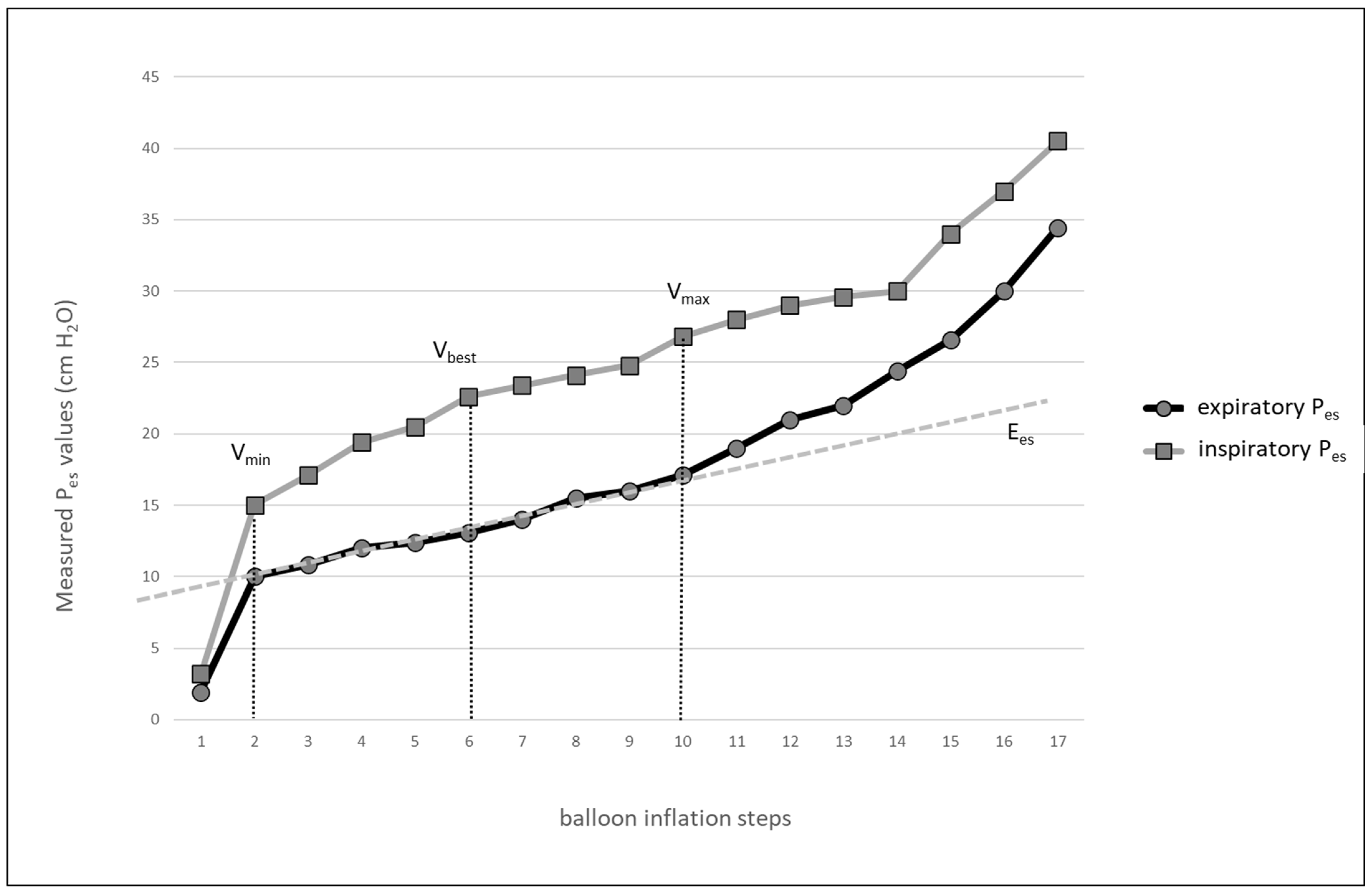
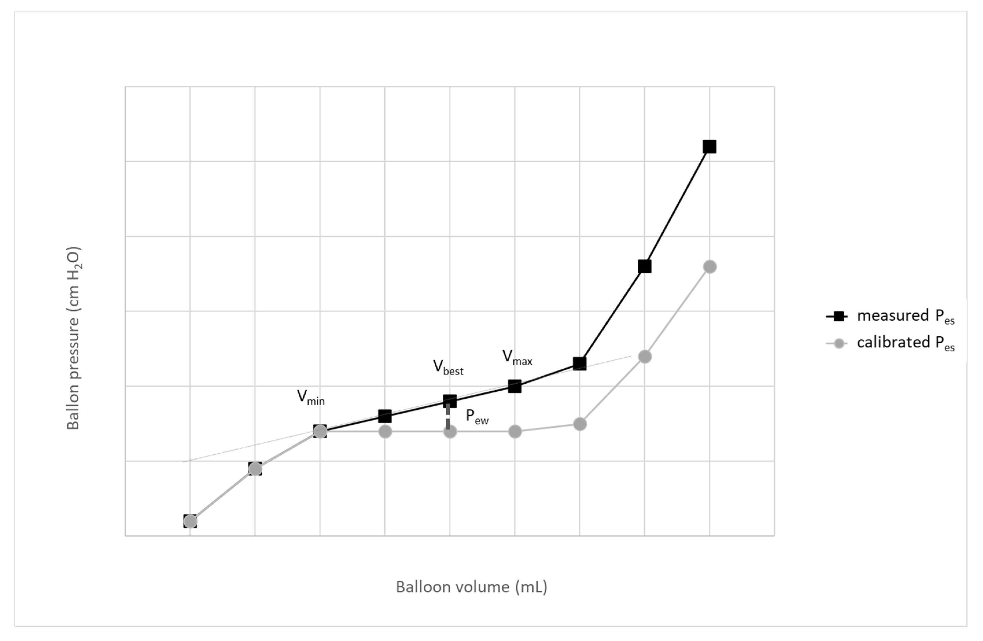
Figure 1. An example of the balloon pressure-volume curve obtained in an obese patient with influenza A pneumonia and congestive heart failure. Inflation steps are shown on x axis. Ees—esophageal elastance; Vmin—volume of the balloon at the beginning of the linear part of balloon pressure-volume curve; Vmax—volume of the balloon at the end of the linear part of balloon pressure-volume curve; Vbest—lowest volume of the balloon with largest tidal difference of esophageal pressure; Pes—esophageal pressure.
Jpm 13 00136 g001
Figure 2. Effect of correction of esophageal elastance. A simplified scheme; expiratory Pes values are shown only. Pew—pressure generated by the distension of esophageal wall; Vmin–volume of the balloon at the beginning of the linear part of the balloon pressure-volume curve; Vmax—volume of the balloon at the beginning of the linear part of the balloon pressure-volume curve.
Jpm 13 00136 g002
Table 2. Suggested steps to obtain a calibrated value of Pes. Adapted from Mojoli et al. [15] and Sun et al. [17].
StepsMethod
1st stepPerform a stepwise inflation of the balloon (0.2–1.0 mL steps, according to the balloon size), record Pes at different volumes of balloon.
2nd stepUsing an appropriate table calculator, create the balloon pressure-volume curve to identify a linear part of the curve with Vmin and Vmax, for further measurement select Vbest with largest tidal change of Pes.
3rd stepCalculate the esophageal wall elastance: Ees = (PesPEEPVmax − PesPEEPVmin)/(Vmax − Vmin)
4th stepCalculate the esophageal recoil pressure: PewVbest = (Vbest -Vmin) x Ees
5th stepCalculate the calibrated esophageal pressure: calPes = PesVbest − PewVbest
Pes—esophageal pressure; Vbest—balloon inflation volume with highest tidal change of Pes; Vmin—minimal balloon inflation volume on the linear part of esophageal pressure PV curve; Vmax—maximal balloon inflation volume on the linear part of esophageal pressure PV; Ees—esophageal wall elastance; PesPEEPVmax—expiratory value of Pes measured at Vmax; PesPEEPVmin—expiratory value of esophageal pressure measured at Vmin; PesVbest—esophageal pressure measured at Vbest; PewVbest—esophageal recoil pressure at Vbest; calPes—calibrated value of esophageal pressure.
Table 3. Suggested limits of transpulmonary pressures in patients with ARDS. Adapted from Mauri at al. [1], Bayerdorf Kassis et al. [15], and Pelosi et al. [28].
ParameterLimit
End-expiratory PL±2 cm H2O
End-inspiratory PL<20 cm H2O
Driving PL<10–12 cm H2O
End-inspiratory PL during recruitment maneuvers≤25 cm H2O
PL—transpulmonary pressure.
Disclaimer/Publisher’s Note: The statements, opinions and data contained in all publications are solely those of the individual author(s) and contributor(s) and not of MDPI and/or the editor(s). MDPI and/or the editor(s) disclaim responsibility for any injury to people or property resulting from any ideas, methods, instructions or products referred to in the content.

