Article

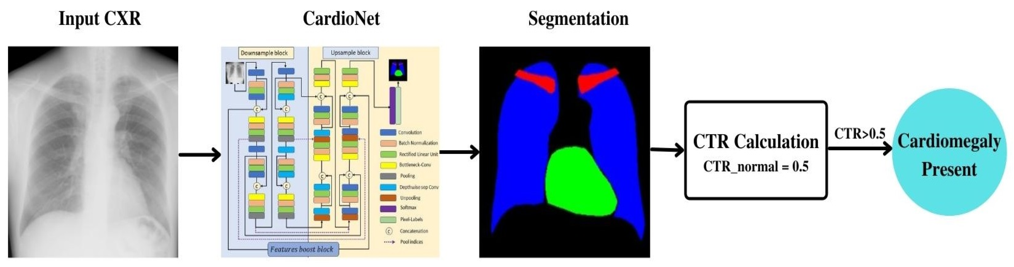
CardioNet: Automatic Semantic Segmentation to Calculate the Cardiothoracic Ratio for Cardiomegaly and Other Chest Diseases

1
Department of Computer Engineering, Myongji University, Yongin 03674, Korea
2
Department of Primary and Secondary Healthcare, Lahore 54000, Pakistan
3
Research and Development, AItheNutrigene, Seoul 06132, Korea
4
School of Intelligent Mechatronics Engineering, Sejong University, Seoul 05006, Korea
5
Department of Unmanned Vehicle Engineering, Sejong University, Seoul 05006, Korea
*
Authors to whom correspondence should be addressed.
These authors contributed equally to this work.
J. Pers. Med. 2022, 12(6), 988; https://doi.org/10.3390/jpm12060988
Submission received: 5 May 2022 / Revised: 2 June 2022 / Accepted: 13 June 2022 / Published: 17 June 2022
(This article belongs to the Special Issue The Challenges and Prospects in Cardiology)

Abstract

:
Semantic segmentation for diagnosing chest-related diseases like cardiomegaly, emphysema, pleural effusions, and pneumothorax is a critical yet understudied tool for identifying the chest anatomy. A dangerous disease among these is cardiomegaly, in which sudden death is a high risk. An expert medical practitioner can diagnose cardiomegaly early using a chest radiograph (CXR). Cardiomegaly is a heart enlargement disease that can be analyzed by calculating the transverse cardiac diameter (TCD) and the cardiothoracic ratio (CTR). However, the manual estimation of CTR and other chest-related diseases requires much time from medical experts. Based on their anatomical semantics, artificial intelligence estimates cardiomegaly and related diseases by segmenting CXRs. Unfortunately, due to poor-quality images and variations in intensity, the automatic segmentation of the lungs and heart with CXRs is challenging. Deep learning-based methods are being used to identify the chest anatomy segmentation, but most of them only consider the lung segmentation, requiring a great deal of training. This work is based on a multiclass concatenation-based automatic semantic segmentation network, CardioNet, that was explicitly designed to perform fine segmentation using fewer parameters than a conventional deep learning scheme. Furthermore, the semantic segmentation of other chest-related diseases is diagnosed using CardioNet. CardioNet is evaluated using the JSRT dataset (Japanese Society of Radiological Technology). The JSRT dataset is publicly available and contains multiclass segmentation of the heart, lungs, and clavicle bones. In addition, our study examined lung segmentation using another publicly available dataset, Montgomery County (MC). The experimental results of the proposed CardioNet model achieved acceptable accuracy and competitive results across all datasets.

1. Introduction

The most used and evaluated method of diagnosing chest-related pathologies such as pneumothorax, pulmonary cancer, congestive heart failure, lung nodule, and heart enlargement is the chest X-ray (CXRs) [1]. Heart enlargement is classified as cardiomegaly, one of the serious cardiovascular diseases among the general public [2]. Cardiomegaly can happen from different conditions such as cardiac insufficiency, blood pressure, hypertension, and coronary artery disease. These cardiac concerns affect patients’ health ranging from a high risk of heart failure to immediate death [3]. Therefore, the early diagnosis of cardiomegaly is critical; the disease can be diagnosed with edge detection of the size and shape of the chest and heart from posterior–anterior (PA) CXR. The cardiothoracic ratio (CTR) is a quantitative measure of heart enlargements in CXRs to detect cardiomegaly and the boundaries of other chest organs [4,5].
The CTR is the ratio between the maximum horizontal cardiac diameter and the maximum horizontal thoracic diameter, and the normal range is between 0.42 and 0.50. A value higher than normal (>0.50) is considered cardiomegaly [5]. The manual measurement of the CTR from CXR performed by medical experts requires the domain knowledge of chest physiologies. Cardiologists detect cardiomegaly by measuring the heart’s left distance, D L , and right distance, D R , boundaries from the central vertical line of the chest. M is the maximum horizontal distance between the left- and right-side boundaries of the respective lungs, as shown in Figure 1. The method of calculating the CTR for cardiomegaly is expressed in Equation (1). There is the possibility of observational error, and the process is time-consuming. This problem has motivated researchers to develop a computer-aided diagnosis- (CAD) based CTR measurement to diagnose cardiomegaly. Several researchers have automatically measured the CTR and other heart diseases [6,7]. Most of the techniques used to detect the boundaries and size of lungs and heart require the accurate segmentation of anatomical organs.
CTR = ( D L + D R ) / M
In medical imaging, segmentation is extracted with similar properties from the images. The areas of interest, like the lung and heart, are segmented with automated deep learning segmentation from the CXRs. The advancements in convolutional neural networks (CNN) have led to better performance in the image segmentation domain [8]. Multilayer CNNs [9] are used to detect different types of chest diseases and to segment medical tasks. A CNN-based automatic brain segmentation is performed on the MRI brain images to detect the tissues [10]. The authors of [11] proposed a semantic segmentation EG-CNN deep model to accurately detect the edges and boundaries of organs. Semantic segmentation is a pixel-wise classification that labels each pixel of a given class and groups the similar features. Medical images are complex, and this pixel-based semantic segmentation help with efficiently locating the infected areas in an image [12]. Due to low quality and pixel variations in the CXRs, the automatic semantic segmentation of the heart and other chest organs (lung and clavicle bones) is a challenging task. Previous studies addressed this issue and solved it by developing complex neural networks with higher computational resources [13].
In this study, we addressed this challenging issue and proposed a learning-based solution that performs accurate segmentation of lungs and heart to measure CTR from chest PA CXRs. CardioNet is the method proposed and used to determine the presence of cardiomegaly, and the graphical representation is discussed in Section 3. CardioNet is a semantic segmentation network that uses the dense identity features in the architecture to detect the edge properly within a few pixels. This deep model provides fine segmentation within fewer trainable parameters than the CNNs. CardioNet was trained on chest PA CXRs and provided the binary masks to compute the pixels and the positions of the anatomical organs. The semantic segmentation output and the calculated CTR decide the performance evaluation and make the final decision.
The paper’s organization is as follows: We review the related work about cardiomegaly and the concept of chest anatomy segmentation based on the conventional handcrafted and deep features in Section 2. In Section 3, we propose a deep methodology for performing the semantic segmentation. Section 4 provides the CXRs data for the experiment and the data generation approach; the training and the testing of the proposed CardioNet model are performed in this section, followed by the experimental evaluation results. The work concludes with challenges in Section 5.

2. Related Works

There are two methods of segmenting the chest-related organs: handmade and deep features. The handmade feature-based methods utilize various image-processing schemes, where in-depth features are the learned features that depend on deep learning-based semantic segmentation.

2.1. Chest Anatomy Segmentation Using Conventional Handmade Features

Handmade features are based on general image-processing approaches to segmenting the chest anatomy from the background. Most of the local feature-based methods do not consider multiclass segmentation of CXRs; rather, they focus on lung region segmentation. Peng et al. presented the Hull-CPLM method to detect the lung region of interest (ROI). The segmentation requires prior preprocessing for coarse segmentation, where the principle curve method is used to refine segmentation [14]. Candemir et al. proposed a non-grid registration-based lung segmentation method that performs the task in three steps: content-based image retrieval, sift-flow modeling for deformable registration, and graph cut optimization for boundary refinement [15]. Jaeger et al. computed three masks of lung segmentation, a probabilistic lung shape model and a Log Gabor mask, where segmentation was obtained by averaging and thresholding for the diagnostic purpose [16].
Jangam et al. presented a hybrid segmentation scheme that utilized an optimized clustering approach to exclude the lung field from the background in CXR images [17]. Vital et al. introduced an automatic system for lung field segmentation. A wavelet enhances the CXR images, and in the second step, OTSU thresholding is combined with mathematical morphology. For the third step, the active contour method improves the performance [18]. Ahmad et al. proposed a Gaussian derivative filter that considered seven different orientations of top segment fields. The segmentation task combines the Gaussian derivative filter with fuzzy c-mean clustering and thresholding [19]. Pattrapisetwong et al. used an unsupervised method for lung region exclusion from the background. They used preprocessing to enhance the image contrast, and then a shadow filter was applied to enhance the outline of the lungs; finally, multilevel thresholding segmented the resultant lung region [20].
Li et al. used graph-based lung segmentation. In detail, the CXR images are divided into several subregions and each region’s saliency value, where the cubic spline interpolation is used to obtain fine, smoother boundaries of the lung region [21]. Chen et al. proposed a system that estimates the effusion volume. The segmentation was performed using a 2-D image processing scheme, similar to the Harris corner detector, for enhancement using a convolutional process with a 2 × 2 mask to detect the lung contour [22]. Dawoud presented an iterative framework for lung field segmentation. The method calculates the intensity and shape information, where the main segmentation is handled with iterative thresholding [23]. Saad et al. showed that the edge detection from Sobel, Prewitt, and Laplacian could segment the lung fields; however, the accuracy decreases owing to the image noise. Therefore, combining these edge detectors with morphological operators can produce better results for lung segmentation in CXRs [24]. Chondro et al. proposed a low-order adaptive lung segmentation method based on the growing region. The ROI was obtained by brick–block binarization and morphology, where the boundary refinement was measured by statistical region growing and graph cutting [25]. Chung et al. considered lung segmentation a prerequisite task for diagnosis. The segmentation of the lungs performed by the Bayesian active contour model is an iterative process for segmentation [26].

2.2. Chest Anatomy Segmentation Using Deep Feature (CNN)

Conventional handcrafted features are based on specific local intensities; thus, these methods cannot perform multiclass segmentation at once. The performance parameters change from image to image. Therefore, automatic deep feature methods are an alternative to handmade features for evaluation. Long et al. used the first learning method for the multiclass chest anatomy segmentation. The encode-decoder is used for critical learning of higher-order structures. The encoder part extracts the features, and the decoder performs upsampling operations to obtain the final segmented output [27]. An adversarial network is proposed by Dong et al. to estimate the CTR. The produced model creates the predicted domain-independent output mask [28]. Tang et al. developed a transfer learning approach for lung segmentation, and it consists of two main modules. The first module is crisscross attention-wise responsible for the enriched global contextual information, whereas the second module, image-to-image translation, is used for data augmentation [29].
Souza et al. used a learning method to segment the lung regions. The original images were divided into patches, and those patches were classified into lung and non-lung classes by a neural network [30]. Kalinovsky et al. modified the encoder–decoder-based SegNet model for lung segmentation and achieved 96.2% accuracy [31]. This method worked on the limitation of the original SegNet model. It used the max-pooling features to upsample the features maps in the decoding layers, the LF-SegNet method developed by Mittal et al. [13] to perform lung segmentation. The lung segmentation was performed and the proposed model was evaluated on two famous chest X-ray datasets, JSRT and MC, and achieved 98.73% and 95.10% accuracy, respectively. Liu et al. [32] proposed a U-Net segmentation model on the JSRT dataset to extract the lung regions, and DenseNet is used to segment the lungs.
Venkataramani et al. developed ContextNets for semantic segmentation to adapt the target domain with fewer images [33]. Frid-Adar et al. considered an important application of semantic segmentation to detect the clavicle bone positioning using Chest X-rays. The modified architecture used to segment the clavicle bones and the weights of VGG16 used in the encoder [34]. Oliveira et al. presented a transfer learning-based approach f chest-related organs segmentation. The approach consists of pre-trained networks for semantic segmentation as; U-Net, fully connected network, and SegNet [35]. Wang et al. considered the instance segmentation to segment multiclass chest organs using chest X-ray images with Mask-RCNN [36]. Dong et al. presented a generative adversarial network (GAN) for a semantic segmentation purpose using CXRs [37]. Jiang et al. developed a CNN-based VGG16 segmentation model with prior weight initialization with fewer data. [38]. Most of the semantic segmentation from the radiographic images is performed by the UNet and encoder-decoder architectures or by the different variants of the followings; however, currently, the recurrent neural network (RNN) is also used for the segmentation purpose in radiology. Stollenga et al. [39] presented a segmentation 3D LSTM-RNN deep model to extract the brain features automatically. Chen et al. [40] used a semantic segmentation approach to 3D images by combining the LSTM-RNN and U-Net architecture.

3. Methodology

3.1. Proposed CardioNet

The flowchart of the proposed CardioNet model for the automatic semantic segmentation of chest-related organs is presented in Figure 2. An input chest of CXR images without any preprocessing is given to the CardioNet. CardioNet uses pixel-wise classification to segment the heart, lungs, and clavicle bones. The output of the model is the segmented masks for each class. The heart and lungs masks are utilized to calculate CTR to detect cardiomegaly, make accurate diagnostic decisions, and measure the model’s performance. The CardioNet is based on the dense connections between the downsample block, upsample block, and features boost block. Dense concatenation paths are used for the subsequent layers in the downsample and upsample block of the CardioNet and between the downsample and upsample block. The specific paths are introduced for the flow of information within the network to provide the edge information to the subsequent layers and the upsample block. Feature boost block is introduced to preserve minor features in the segmented image. It applies continuous convolutions without resizing the feature map size, which helps to premaintain needful spatial information to boost the segmentation performance.

3.2. Chest Anatomy Segmentation Using CardioNet Architecture

Semantic segmentation in CXRs is not an easy task. X-ray images have inferior quality, whereas the correct CTR computation is based on the true lungs and heart boundaries. Moreover, the conventional semantic segmentation architectures [27,41] lack spatial information loss compensation as they are not using any residual or dense connectivity to empower the feature after the continuous convolutional operation. The proposed CardioNet is a robust network that incorporates three principal parts: a downsample block (DSB), an upsample block (USB), and a features boost block (FBB).
Figure 3 shows the proposed CardioNet architecture for the CXR semantic segmentation. The DSB is an encoder part of the network that squeezes the vital information from the input images in combination with dense connectivity benefits. It consists of five 3 × 3 general convolutions and three depth-wise separable convolutions. The convolutional layers with many channels consume more trainable parameters. Therefore, the convolution in the deeper side of the DSB is replaced by depth-wise convolutions. The edge features in the CXR image can be very small, and those small features can be eliminated if the feature map size is greatly reduced within the network. It can be noticed from Table 1 that the smallest feature map size in the DSB is 21 × 21, which is not enough to represent the minor information available in the CXR; the minor features can be eliminated, and therefore, the FBB is used to apply continuous convolutions without resizing the feature map. The FBB retains the feature map on a flat feature map size of 350 × 350, which can contain the smaller and most valuable features. The USB is mainly based on three depth-wise separable convolutions combined with five 3 × 3 general convolutions and batch normalization. As mentioned previously, in the case of DSB, general convolutions with more channels are costly, so depth-wise separable convolutions are replacing those of the general convolution layers to reduce cost. Furthermore, softmax and pixel classification layers are used in the USB; where softmax layer works as the activation function and pixel classification layer provides a categorical label for each pixel in the image.
Moreover, the residual connections from the ResNet model were used to empower the features that degraded the contextual image information during the downsampling [41]. This problem is termed a vanishing gradient problem, and researchers attempted to address it using residual skip connections, but it still faces the information flow latency problem. We used the dense connectivity function from the DenseNet deep architecture to overcome this problem and concatenated the in-depth features [42]. DenseNet is a famous classification model that outperforms the previous networks while using fewer trainable parameters. The proposed architecture is entirely different from conventional networks such as the segmentation network (SegNet) [43], outer residual skip network (OR-Skip-Net) [44], and U-shaped network (U-Net) [45], which are deep neural networks with larger number of trainable parameters. The shallow architecture of CardioNet exhibits 1.72 million (M) trainable parameters, while the conventional networks such as SegNet, OR-Skip-Net, and U-Net, have 29.46 M, 09.70 M, and 31.03 M trainable parameters, respectively. The key architectural differences of the CardioNet with the conventional segmentation networks such as Seg-Net, OR-Skip-Net, and U-Net are listed in Table 2.
Figure 4 illustrates the in-depth view of the proposed CardioNet with dense connectivity feature concatenation. CardioNet accomplishes the chest X-ray image; the spatial loss is compensated by concatenating deep, dense features. The dense concatenation method transfers these enriched features from the DSB to the USB, as shown in the figure.
As shown in Figure 4, the first convolution layer in the downsample block, i.e., Conv - A , receives the   D i feature as input, and after applying the convolution operation, it outputs the   F i feature. This F i feature is rich with low-level spatial information, and it flows in two directions for concatenation. First, F i is provided to the second convolution (Conv-B), which converts into K i . K i is the output achieved after the two convolution layers, and spatial loss of these convolutional operations is recovered by dense feature concatenation. The first aggregated rich, dense concatenated feature, A Concat . 1 , is the output of F i and K i given by Equation (2):
A Concat 1 = F i   ©   K i
Here © shows the depth-wise concatenation of   F i and K i . F i is also directly transferred to the corresponding USB to transfer the low-level spatial information to the last layers of the network for better performance. A Concat 1 is the resulting dense feature after the depth-wise concatenation. This process increases the number of filters for the dense features, the parameters, and the training time. To overcome this problem, we introduced the Bottlenecki layer to decrease the number of filters with the combination of the BN and ReLU layers. The obtained feature after the Bottlenecki block is Δ A Concat 1 and given by Equation (3):
Δ A Concat . 1 = Δ ( F i   ©   K i )
Here, Δ represents the BN and ReLU operations in combination with the Bottlenecki layer that limit the filters. Similarly, Figure 4 shows that each USB receives the   U i feature as input, and after applying the first convolution (Conv-A), it outputs the   J i feature. J i has less spatial loss than the next convolution layer, and it flows in two directions for concatenation. First, J i is provided to the second convolution (Conv-B), producing L i . L i is the output obtained after two convolution operations that concatenate with J i from the first convolution (Conv-A) and   F i from the corresponding DSB, creating a second aggregated rich feature A Concat . 2 given by Equation (4):
A Concat . 2 = J i   ©   L i   ©   F i
Here, ©   shows the depth-wise concatenation of   J i , L i , and F i . A Concat . 2 is a powerful feature that contains rich information from the initial layers, resulting in better segmentation of the lung and heart region pixels. Again, to overcome the filter size problem, the Bottleneckj block was used with a combination of BN and ReLU operations. Δ A Concat . 2 is a bottleneck feature and is given by Equation (5):
Δ A Concat . 2 = Δ ( J i   ©   L i   ©   F i )
Here, Δ represents the combination of BN and ReLU operations with the Bottleneckj layer limiting the filter size. Both the bottleneck features ( Δ A Concat . 1 , Δ A Concat . 2 ) achieved from the downsample block or upsample block empowered the dense connectivity. The output A Concat . 2 of the last USB provided the Softmax and pixel classification.
The layer details in the booster block are shown in Table 3. The connectivity of the booster block in CardioNet and the dense feature aggregation principle are shown in Figure 5. The downsample–upsample block (DUB) takes the input feature, and this feature passes through several convolutional layers to extract the meaningful features for chest anatomy segmentation. This DUB provides the F d feature. F d is densely aggregated with the rich F b from the feature booster block (FBB). The features in FBB are without an extensive pooling operation and therefore contain the features that represent the low-level boundary information. Both F d and F b are aggregated with depth-wise concatenation to create S 1 , given by Equation (6):
S 1 = F b   ©   F d
Here, S 1 is the densely aggregated feature created by the depth-wise concatenation of F d   (feature coming from the DUB block) and F b (coming from FBB), representing the depth-wise concatenation.

4. Experiments

4.1. Data Augmentation

We investigated chest anatomy segmentation in multiple classes. To analyze the performance of CardioNet, the Japanese Society of Radiological Technology (JSRT), i.e., a publicly available multiclass dataset [46], is used. In the JSRT dataset, there are 247 chest radiograph images for the lungs, heart, and clavicle, each annotated at the pixel level. A total of 154 of the 247 images show nodules, whereas the remaining 94 do not. The pixel space of each image in the dataset is 0.175 mm, and the image size is 2048 × 2048 pixels. The provided data are divided into two folds with odd (123) and even (124) numbered images. Training is done one-fold, testing one on the other and vice versa. Considering the two-fold cross-evaluation criteria, the results are obtained by averaging both folds. Figure 6 shows sample chest radiograph images from the JSRT dataset with corresponding ground truth. In detail, the red, green, and blue pixels show the clavicle bone, heart, and lungs, respectively.
CardioNet is a pixel-wise classification network that requires large training data to classify the multiple classes. The original image size and labels of 2048 × 2048 pixels of JSRT are resized to 350 × 350 pixels to reduce the memory usage of the graphic processing unit (GPU) and the training time. Different data generation approaches are applied to increase the data size so the model will learn accurately. Acquiring enough medically labeled data is difficult, requiring an expert to label the data. The training of CardioNet is performed with various images, and images are generated with data augmentation. The image transformations create artificial images by cropping, horizontal flipping, translation, and horizontal flipping with translation. Figure 7 represents the proposed data augmentation for this task. The first step of augmentation, horizontal flipping (H-Flip), is applied to 124 original images, which results in 248 images. The 248 images are translated (X = 4, Y = −4) to produce 496 images. In the third step, horizontally flipping the 496 images makes 992 images. Using the 992 images from the previous step, the next step applies the translation of (X = 8, Y = 8) with an H-Flip and the translation of (X = −12, Y = −12) with an H-Flip, resulting in 1984 and 1984 images, respectively. Consequently, 3968 images (1984 + 1984) are obtained and used as training images by combining transformational images from step four.
In our experiments, CardioNet is trained from scratch using MATLAB R2021b [47] (without fine-tuning or a pre-trained model, such as DenseNet, ResNet, Inception, or GoogleNet). CardioNet is trained and tested on a desktop computer, i.e., Intel®® Core™ i7-8700 CPU. The system clock speed is 3.20 GHz, memory (RAM) is 16 GB, and NVIDIA GPU GeForce RTX 2060 (graphics memory of 8 GB) [48].

4.2. CardioNet Training

CardioNet contains several dense paths that allow the network to provide encoder–decoder connections both internally and externally, which is a useful tool to help the network converge quickly. As a result of the spatial edge information from the dense connectivity concatenation feature, the edges can be segmented finely without preprocessing. CardioNet training takes place from scratch, without any weight transfer or initialization. Since we designed CardioNet, fine-tuning is not required when training it.
The training of CardioNet involves the following considerations. The gradient of the network is optimized with a well-known training optimizer called Adam. Adam can efficiently scale gradients diagonally, is suitable for large datasets, and can even handle moving object classification problems [49]. Adam is adopted as an optimizer for CardioNet training due to its benefits. Considering all other parameters, the learning rate of 0.001 for 30 epochs (51,660 iterations) is used throughout the CardioNet training. CardioNet requires high memory, so a small batch size with 4 images is considered. To maintain the gradient threshold, the global L2 normalization method with an epsilon of 0.000001 is used during the training. The cross-entropy loss function with median frequency is used to make training faster and similar to frequency balancing and cross-entropy [43,44,50,51]. Figure 8a,b represent the relationships between training loss and training accuracy for the 2-across-validation. The loss curves are on the left side and the accuracy curves of training are on the right side of the figures. The epochs are represented on the x-axis.

4.3. CardioNet Testing

4.3.1. Chest Anatomy Segmentation Testing Using CardioNet

A CXR image without data preprocessing is given to CardioNet as an input image. This input image is passed through the CardioNet downsample block and upsample block to acquire the segmented output. The final output of the proposed network is a multiclass segmentation mask. These segmented masks further evaluate the final output by generating the chest-related automatic semantic segmentation results. The performance of CardioNet is evaluated and measured using different metrics. The following segmentation metrics are calculated: Accuracy (Acc), Jaccard index (J), and Dice coefficient(D). J is a mean intersection over union (IoU), and D is calculated as [1,36,52] for a JSRT dataset. Equations (7)–(9) give the formulas for evaluation metrics:
Acc = TP + TN TP   +   FP   + FN + TN
J = TP TP   +   FP   + FN
D = 2 TP P   +   FP   + FN
Here, TP = true positives, FP = false positives, and FN = false negatives. We are considering multiclass segmentation, considering lung class as an example: TP and FP are predicted as lung pixels and non-lung pixels in the ground truth, respectively. TP pixels are the pixels that are predicted as lung pixels and listed as lung pixels in the ground truth. FN are those lung pixels with ground truth predicted as non-lung pixels by our CardioNet model.
The segmented chest X-ray images with CardioNet using the JSRT dataset are shown in Figure 9. The FP convention is black; the FN convention is yellow; and the TP convention is shown in green, blue, and red for the heart, lung, and clavicle bone classes, respectively. While there are some bad segmentation cases, there is no significant segmentation error using our method for the test images.

4.3.2. Ablation Study

An ablation study of our proposed method was conducted to prove the efficiency of CardioNet. We considered two variants of CardioNet, one with the booster block, i.e., CardioNet-B (mentioned as CardioNet throughout the whole manuscript), and the second one without the booster block, referred to as CardioNet-X. Here, we compared CardioNet-B with CardioNet-X. The experiments showed that the effectiveness of the booster block in CardioNet-B provides superior results to those of CardioNet-X (without booster block). Table 4 shows that CardioNet-B with the booster block provides higher Acc, J, and D values than CardioNet-X with a minor increase in the number of trainable parameters. Booster block creates considerable performance differences owing to preserving spatial information to boost the segmentation performance.

4.3.3. Comparison of CardioNet with Deep Methods

This section compares CardioNet segmentation performance with other methods. The segmentation performance of the existing state-of-the-art methods compared with CardioNet is shown in Table 5. This comparison is based on the JSRT dataset. The local feature-based methods and the learned feature-based methods are presented separately in Table 5. Based on J and D, the study results demonstrate that CardioNet outperforms other studies for chest anatomy segmentation.

4.3.4. Lung Segmentation from MC Dataset Using CardioNet

After obtaining the valuable results from the JSRT image dataset, we evaluated the performance of our CardioNet model with the Montgomery County chest X-rays (MC) dataset. MC is a publicly available dataset published by the famous Montgomery County tuberculosis program, one of the most famous in the USA. It has 138 chest PA X-ray images with 80 normal and 58 TB cases. Figure 10 shows sample images from the MC data with lung contour binary masks, and the format of the X-ray images format is PNG. We divided the MC dataset into training and testing following [60], where the training set has 80 images, the validation set has 20, and the testing set consists of 38 images. The MC dataset is also augmented, and the image size was artificially increased using the same augmentation approach used for the JSRT dataset (Section 4.1). We only provided the ground truth masks for lungs. The CardioNet results for the MC dataset are shown in Figure 11. However, the experimental comparison of CardioNet with other existing models is shown in Table 6. The experimental results clearly show that CardioNet outperformed for lung segmentation and can be used in real-world medical applications for diagnostic purposes.

4.3.5. Automated Computation of CTR by the Proposed Method

As explained in the introduction section, medical practitioners can use CTR in diagnosing cardiomegaly, a heart enlargement disease. CTR is a quantitative measure of heart enlargements in CXRs to detect cardiomegaly and boundaries of other chest organs. CTR estimates the size of the heart. The cardiac health experts manually calculate it. Our proposed semantic segmentation network (CardioNet) can automate the computation of CTR by accurately segmenting the lungs and heart boundaries. As shown in Figure 12a, the heart boundary is critical in X-ray images due to a change in pixel values. The CTR can only be calculated by the proposed method once the heart and lungs are segmented properly. With our feature-boosting mechanism, we can achieve accurate boundaries of the heart and lungs even with minor changes in pixel values.
An example image from the JSRT dataset for calculating the CTR of the ground truth and the predicted mask (by CardioNet) using Equation (1) is shown in Figure 12b,c, respectively. For calculating the CTR, we need the ratio of distance D L +   D R and M . Here, D L +   D R is the distance between two extreme points of the heart. M is the maximum horizontal distance between two extreme outer points for both lungs. The distance D L +   D R by the predicted mask is 126 pixels, and the distance M is 304. Hence, using Equation (1), the predicted CTR (P-CTR) will be 0.4145. On the other hand, the distance DL + DR by the ground truth mask is 123 pixels, and the distance M is 303. Therefore, the ground truth CTR (G-CTR) calculated by Equation (1) is 0.4045. In JSRT, masks are prepared under the supervision of an expert radiologist. It can be determined from the obtained results that the proposed automatic method has predicted the CTR very efficiently. Hence, it can aid the medical practitioner in diagnosing using CTR and other chest anatomy segmentation as an alternative system.

5. Conclusions

Anatomical chest structures (lungs, hearts, and clavicle bones) were segmented using a residual connection-based semantic segmentation network (CardioNet) for diagnostic purposes. Even under nonideal situations and multiple classes, the method provides excellent segmentation accuracy. Since the pixel value of the heart is low and its boundary edges blend with the lungs, segmenting the heart is essential. CardioNet can segment the heart accurately, even in X-ray images of inferior quality. The segmentation accuracy of the heart and lung regions is directly related to the CTR. Conventional CNNs reduce the feature map size to classify classes, resulting in the depreciation of the minor information (clavicle bone and small-sized heart) due to the overuse of the max-pooling layers. The feature map size is not reduced in the network for classification; fewer pooling layers are used, and a large feature size helps restore the minor classes’ information. The direct, outer, dense-connectivity feature concatenation causes direct information transfer, enabling CardioNet to converge faster, as seen in the training accuracy and loss curves. In our work, the proposed CardioNet automatically detects the boundaries of the lungs and heart to accurately calculate the CTR. Multiple cardiac and lung diseases can be diagnosed using the CTR.

Author Contributions

Conceptualization, A.J., U.W. and R.A.N.; methodology, M.T.H. and N.A.; software, R.A.N.; validation, M.T.H., N.A. and H.S.K.; formal analysis, U.W.; investigation, R.A.N.; resources, H.S.K.; data curation, A.J.; writing—original draft preparation, A.J.; writing—review and editing, R.A.N.; visualization, M.T.H.; supervision, H.S.K.; project administration, R.A.N.; funding acquisition, R.A.N. All authors have read and agreed to the published version of the manuscript.

Funding

This work was supported by an national research foundation (NRF) grant funded by the Ministry of Science and ICT (MSIT), South Korea through the Development Research Program (NRF2022R1G1A101022611).

Institutional Review Board Statement

Not applicable.

Informed Consent Statement

Not applicable.

Data Availability Statement

Not applicable.

Acknowledgments

This work was supported by an NRF grant funded by the Ministry of Science and ICT (MSIT) through the Development Research Program (NRF2022R1G1A101022611).

Conflicts of Interest

The authors declare no conflict of interest.

References

  1. Novikov, A.A.; Lenis, D.; Major, D.; Hladůvka, J.; Wimmer, M.; Bühler, K. Fully Convolutional Architectures for Multi-Class Segmentation in Chest Radiographs. IEEE Trans. Med. Imaging 2018, 37, 1865–1876. [Google Scholar] [CrossRef] [Green Version]
  2. Semsarian, C.; Ingles, J.; Maron, M.S.; Maron, B.J. New Perspectives on the Prevalence of Hypertrophic Cardiomyopathy. J. Am. Coll. Cardiol. 2015, 65, 1249–1254. [Google Scholar] [CrossRef] [Green Version]
  3. Tavora, F.; Zhang, Y.; Zhang, M.; Li, L.; Ripple, M.; Fowler, D.; Burke, A. Cardiomegaly Is a Common Arrhythmogenic Substrate in Adult Sudden Cardiac Deaths, and Is Associated with Obesity. Pathology 2012, 44, 187–191. [Google Scholar] [CrossRef]
  4. Candemir, S.; Jaeger, S.; Lin, W.; Xue, Z.; Antani, S.; Thoma, G. Automatic Heart Localization and Radiographic Index Computation in Chest X-rays. In Proceedings of the Medical Imaging, San Diego, CA, USA, 28 February–2 March 2016; p. 978517. [Google Scholar]
  5. Dimopoulos, K.; Giannakoulas, G.; Bendayan, I.; Liodakis, E.; Petraco, R.; Diller, G.-P.; Piepoli, M.F.; Swan, L.; Mullen, M.; Best, N.; et al. Cardiothoracic Ratio from Postero-Anterior Chest Radiographs: A Simple, Reproducible and Independent Marker of Disease Severity and Outcome in Adults with Congenital Heart Disease. Int. J. Cardiol. 2013, 166, 453–457. [Google Scholar] [CrossRef]
  6. Hasan, M.A.; Lee, S.-L.; Kim, D.-H.; Lim, M.-K. Automatic Evaluation of Cardiac Hypertrophy Using Cardiothoracic Area Ratio in Chest Radiograph Images. Comput. Methods Programs Biomed. 2012, 105, 95–108. [Google Scholar] [CrossRef]
  7. Browne, R.F.J.; O’Reilly, G.; McInerney, D. Extraction of the Two-Dimensional Cardiothoracic Ratio from Digital PA Chest Radiographs: Correlation with Cardiac Function and the Traditional Cardiothoracic Ratio. J. Digit. Imaging 2004, 17, 120–123. [Google Scholar] [CrossRef] [Green Version]
  8. Chen, L.-C.; Papandreou, G.; Kokkinos, I.; Murphy, K.; Yuille, A.L. DeepLab: Semantic Image Segmentation with Deep Convolutional Nets, Atrous Convolution, and Fully Connected CRFs. arXiv 2017, arXiv:1606.00915. [Google Scholar] [CrossRef] [Green Version]
  9. Moeskops, P.; Viergever, M.A.; Mendrik, A.M.; de Vries, L.S.; Benders, M.J.N.L.; Išgum, I. Automatic Segmentation of MR Brain Images with a Convolutional Neural Network. IEEE Trans. Med. Imaging 2016, 35, 1252–1261. [Google Scholar] [CrossRef] [Green Version]
  10. Havaei, M.; Davy, A.; Warde-Farley, D.; Biard, A.; Courville, A.; Bengio, Y.; Pal, C.; Jodoin, P.-M.; Larochelle, H. Brain Tumor Segmentation with Deep Neural Networks. Med. Image Anal. 2017, 35, 18–31. [Google Scholar] [CrossRef] [Green Version]
  11. Hatamizadeh, A.; Terzopoulos, D.; Myronenko, A. Edge-Gated CNNs for Volumetric Semantic Segmentation of Medical Images. arXiv 2020, arXiv:2002.04207. [Google Scholar]
  12. Hwang, E.J.; Nam, J.G.; Lim, W.H.; Park, S.J.; Jeong, Y.S.; Kang, J.H.; Hong, E.K.; Kim, T.M.; Goo, J.M.; Park, S.; et al. Deep Learning for Chest Radiograph Diagnosis in the Emergency Department. Radiology 2019, 293, 573–580. [Google Scholar] [CrossRef]
  13. Mittal, A.; Hooda, R.; Sofat, S. LF-SegNet: A Fully Convolutional Encoder-Decoder Network for Segmenting Lung Fields from Chest Radiographs. Wirel. Pers. Commun. 2018, 101, 511–529. [Google Scholar] [CrossRef]
  14. Peng, T.; Wang, Y.; Xu, T.C.; Chen, X. Segmentation of Lung in Chest Radiographs Using Hull and Closed Polygonal Line Method. IEEE Access 2019, 7, 137794–137810. [Google Scholar] [CrossRef]
  15. Candemir, S.; Jaeger, S.; Palaniappan, K.; Musco, J.P.; Singh, R.K.; Xue, Z.; Karargyris, A.; Antani, S.; Thoma, G.; McDonald, C.J. Lung Segmentation in Chest Radiographs Using Anatomical Atlases With Nonrigid Registration. IEEE Trans. Med. Imaging 2014, 33, 577–590. [Google Scholar] [CrossRef]
  16. Jaeger, S.; Karargyris, A.; Antani, S.; Thoma, G. Detecting Tuberculosis in Radiographs Using Combined Lung Masks. In Proceedings of the 2012 Annual International Conference of the IEEE Engineering in Medicine and Biology Society, San Diego, CA, USA, 28 August 2012; pp. 4978–4981. [Google Scholar]
  17. Jangam, E.; Rao, A.C.S. Segmentation of Lungs from Chest X rays Using Firefly Optimized Fuzzy C-Means and Level Set Algorithm. In Recent Trends in Image Processing and Pattern Recognition; Santosh, K.C., Hegadi, R.S., Eds.; Springer: Singapore, 2019; pp. 303–311. [Google Scholar]
  18. Vital, D.A.; Sais, B.T.; Moraes, M.C. Robust Pulmonary Segmentation for Chest Radiography, Combining Enhancement, Adaptive Morphology, and Innovative Active Contours. Res. Biomed. Eng. 2018, 34, 234–245. [Google Scholar] [CrossRef]
  19. Wan Ahmad, W.S.H.M.; Zaki, W.M.D.W.; Ahmad Fauzi, M.F. Lung Segmentation on Standard and Mobile Chest Radiographs Using Oriented Gaussian Derivatives Filter. Biomed. Eng. Online 2015, 14, 20. [Google Scholar] [CrossRef] [Green Version]
  20. Pattrapisetwong, P.; Chiracharit, W. Automatic Lung Segmentation in Chest Radiographs Using Shadow Filter and Multilevel Thresholding. In Proceedings of the 2016 International Computer Science and Engineering Conference (ICSEC), Chiang Mai, Thailand, 14–17 December 2016; pp. 1–6. [Google Scholar]
  21. Li, X.; Chen, L.; Chen, J. A Visual Saliency-Based Method for Automatic Lung Regions Extraction in Chest Radiographs. In Proceedings of the 2017 14th International Computer Conference on Wavelet Active Media Technology and Information Processing (ICCWAMTIP), Chengdu, China, 15–17 December 2017; pp. 162–165. [Google Scholar]
  22. Chen, P.-Y.; Lin, C.-H.; Kan, C.-D.; Pai, N.-S.; Chen, W.-L.; Li, C.-H. Smart Pleural Effusion Drainage Monitoring System Establishment for Rapid Effusion Volume Estimation and Safety Confirmation. IEEE Access 2019, 7, 135192–135203. [Google Scholar] [CrossRef]
  23. Dawoud, A. Lung Segmentation in Chest Radiographs by Fusing Shape Information in Iterative Thresholding. IET Comput. Vis. 2011, 5, 185–190. [Google Scholar] [CrossRef]
  24. Saad, M.N.; Muda, Z.; Ashaari, N.S.; Hamid, H.A. Image Segmentation for Lung Region in Chest X-ray Images Using Edge Detection and Morphology. In Proceedings of the 2014 IEEE International Conference on Control System, Computing and Engineering (ICCSCE 2014), Penang, Malaysia, 28–30 November 2014; pp. 46–51. [Google Scholar]
  25. Chondro, P.; Yao, C.-Y.; Ruan, S.-J.; Chien, L.-C. Low Order Adaptive Region Growing for Lung Segmentation on Plain Chest Radiographs. Neurocomputing 2018, 275, 1002–1011. [Google Scholar] [CrossRef]
  26. Chung, H.; Ko, H.; Jeon, S.J.; Yoon, K.-H.; Lee, J. Automatic Lung Segmentation with Juxta-Pleural Nodule Identification Using Active Contour Model and Bayesian Approach. IEEE J. Transl. Eng. Health Med. 2018, 6, 1–13. [Google Scholar] [CrossRef]
  27. Long, J.; Shelhamer, E.; Darrell, T. Fully Convolutional Networks for Semantic Segmentation. In Proceedings of the IEEE Conference on Computer Vision and Pattern Recognition 2015, Boston, MA, USA, 7 June 2015. [Google Scholar]
  28. Dong, N.; Kampffmeyer, M.; Liang, X.; Wang, Z.; Dai, W.; Xing, E. Unsupervised Domain Adaptation for Automatic Estimation of Cardiothoracic Ratio. In Proceedings of the Medical Image Computing and Computer Assisted Intervention—MICCAI 2018, Granada, Spain, 16–20 September 2018; Frangi, A.F., Schnabel, J.A., Davatzikos, C., Alberola-López, C., Fichtinger, G., Eds.; Springer International Publishing: Cham, Switzerland, 2018; pp. 544–552. [Google Scholar]
  29. Tang, Y.; Tang, Y.; Xiao, J.; Summers, R.M. XLSor: A Robust and Accurate Lung Segmentor on Chest X-rays Using Criss-Cross Attention and Customized Radiorealistic Abnormalities Generation. In Proceedings of the International Conference on Medical Imaging with Deep Learning, London, UK, 8–10 July 2019; pp. 457–467. [Google Scholar]
  30. Souza, J.C.; Bandeira Diniz, J.O.; Ferreira, J.L.; França da Silva, G.L.; Corrêa Silva, A.; de Paiva, A.C. An Automatic Method for Lung Segmentation and Reconstruction in Chest X-ray Using Deep Neural Networks. Comput. Methods Programs Biomed. 2019, 177, 285–296. [Google Scholar] [CrossRef]
  31. Kalinovsky, A.; Kovalev, V. Lung Image Segmentation Using Deep Learning Methods and Convolutional Neural Networks. In Proceedings of the XIII International Conference on Pattern Recognition and Information Processing, PRIP-2016, Minsk, Belarus, 3–5 October 2016. [Google Scholar]
  32. Liu, H.; Wang, L.; Nan, Y.; Jin, F.; Wang, Q.; Pu, J. SDFN: Segmentation-Based Deep Fusion Network for Thoracic Disease Classification in Chest X-ray Images. Comput. Med. Imaging Graph. 2019, 75, 66–73. [Google Scholar] [CrossRef] [Green Version]
  33. Venkataramani, R.; Ravishankar, H.; Anamandra, S. Towards Continuous Domain Adaptation for Medical Imaging. In Proceedings of the 2019 IEEE 16th International Symposium on Biomedical Imaging (ISBI 2019), Venice, Italy, 8–11 April 2019; pp. 443–446. [Google Scholar]
  34. Frid-Adar, M.; Amer, R.; Greenspan, H. Endotracheal Tube Detection and Segmentation in Chest Radiographs Using Synthetic Data. arXiv 2019, arXiv:1908.07170. [Google Scholar]
  35. Oliveira, H.; dos Santos, J. Deep Transfer Learning for Segmentation of Anatomical Structures in Chest Radiographs. In Proceedings of the 2018 31st SIBGRAPI Conference on Graphics, Patterns and Images (SIBGRAPI), Foz do Iguaçu, Brazil, 29 October 2018; pp. 204–211. [Google Scholar]
  36. Wang, J.; Li, Z.; Jiang, R.; Xie, Z. Instance Segmentation of Anatomical Structures in Chest Radiographs. In Proceedings of the 2019 IEEE 32nd International Symposium on Computer-Based Medical Systems (CBMS), Cordoba, Spain, 5 June 2019. [Google Scholar]
  37. Dong, N.; Xu, M.; Liang, X.; Jiang, Y.; Dai, W.; Xing, E. Neural Architecture Search for Adversarial Medical Image Segmentation. In Proceedings of the Medical Image Computing and Computer Assisted Intervention—MICCAI 2019, Shenzhen, China, 13–17 October 2019; Shen, D., Liu, T., Peters, T.M., Staib, L.H., Essert, C., Zhou, S., Yap, P.-T., Khan, A., Eds.; Springer International Publishing: Cham, Switzerland, 2019; pp. 828–836. [Google Scholar]
  38. Jiang, F.; Grigorev, A.; Rho, S.; Tian, Z.; Fu, Y.; Jifara, W.; Adil, K.; Liu, S. Medical Image Semantic Segmentation Based on Deep Learning. Neural Comput. Appl. 2018, 29, 1257–1265. [Google Scholar] [CrossRef]
  39. Stollenga, M.F.; Byeon, W.; Liwicki, M.; Schmidhuber, J. Parallel Multi-Dimensional LSTM, with Application to Fast Biomedical Volumetric Image Segmentation. arXiv 2015, arXiv:1506.07452. [Google Scholar]
  40. Chen, J.; Yang, L.; Zhang, Y.; Alber, M.; Chen, D.Z. Combining Fully Convolutional and Recurrent Neural Networks for 3D Biomedical Image Segmentation. arXiv 2016, arXiv:1609.01006. [Google Scholar]
  41. He, K.; Zhang, X.; Ren, S.; Sun, J. Deep Residual Learning for Image Recognition. In Proceedings of the 2016 IEEE Conference on Computer Vision and Pattern Recognition (CVPR), Las Vegas, NV, USA, 27 June 2016; pp. 770–778. [Google Scholar]
  42. Huang, G.; Liu, Z.; van der Maaten, L.; Weinberger, K.Q. Densely Connected Convolutional Networks. In Proceedings of the 2017 IEEE Conference on Computer Vision and Pattern Recognition (CVPR), Honolulu, HI, USA, 21 July 2017; pp. 2261–2269. [Google Scholar]
  43. Badrinarayanan, V.; Kendall, A.; Cipolla, R. SegNet: A Deep Convolutional Encoder-Decoder Architecture for Image Segmentation. IEEE Trans. Pattern Anal. Mach. Intell. 2017, 39, 2481–2495. [Google Scholar] [CrossRef]
  44. Arsalan, M.; Kim, D.S.; Owais, M.; Park, K. OR-Skip-Net: Outer Residual Skip Network for Skin Segmentation in Non-Ideal Situations. Expert Syst. Appl. 2020, 141, 112922. [Google Scholar] [CrossRef]
  45. Ronneberger, O.; Fischer, P.; Brox, T. U-Net: Convolutional Networks for Biomedical Image Segmentation. In Proceedings of the Medical Image Computing and Computer-Assisted Intervention, Munich, Germany, 5–9 October 2015; pp. 234–241. [Google Scholar]
  46. Shiraishi, J.; Katsuragawa, S.; Ikezoe, J.; Matsumoto, T.; Kobayashi, T.; Komatsu, K.; Matsui, M.; Fujita, H.; Kodera, Y.; Doi, K. Development of a Digital Image Database for Chest Radiographs With and Without a Lung Nodule. Am. J. Roentgenol. 2000, 174, 71–74. [Google Scholar] [CrossRef]
  47. R2019a-Updates to the MATLAB and Simulink Product Families. Available online: https://ch.mathworks.com/products/new_products/latest_features.html (accessed on 4 July 2019).
  48. GeForce GTX TITAN X Graphics Card. Available online: https://www.nvidia.com/en-us/geforce/graphics-cards/geforce-gtx-titan-x/specifications/ (accessed on 20 April 2022).
  49. Kingma, D.P.; Ba, J. Adam: A Method for Stochastic Optimization. In Proceedings of the 3rd International Conference on Learning Representations, ICLR 2015, San Diego, CA, USA, 7–9 May 2015; pp. 1–15. [Google Scholar]
  50. Arsalan, M.; Naqvi, R.A.; Kim, D.S.; Nguyen, P.H.; Owais, M.; Park, K.R. IrisDenseNet: Robust Iris Segmentation Using Densely Connected Fully Convolutional Networks in the Images by Visible Light and Near-Infrared Light Camera Sensors. Sensors 2018, 18, 1501. [Google Scholar] [CrossRef] [Green Version]
  51. Arsalan, M.; Kim, D.S.; Lee, M.B.; Owais, M.; Park, K. FRED-Net: Fully Residual Encoder-Decoder Network for Accurate Iris Segmentation. Expert Syst. Appl. 2019, 122, 217–241. [Google Scholar] [CrossRef]
  52. Solovyev, R.; Melekhov, I.; Pesonen, T.; Vaattovaara, E.; Tervonen, O.; Tiulpin, A. Bayesian feature pyramid networks for automatic multi-label segmentation of chest X-rays and assessment of cardiothoracic ratio. In Proceedings of the International Conference on Advanced Concepts for Intelligent Vision Systems, Auckland, New Zealand, 10–14 February 2020; pp. 117–130. [Google Scholar]
  53. Coppini, G.; Miniati, M.; Monti, S.; Paterni, M.; Favilla, R.; Ferdeghini, E.M. A Computer-Aided Diagnosis Approach for Emphysema Recognition in Chest Radiography. Med. Eng. Phys. 2013, 35, 63–73. [Google Scholar] [CrossRef] [PubMed]
  54. Van Ginneken, B.; Stegmann, M.B.; Loog, M. Segmentation of Anatomical Structures in Chest Radiographs Using Supervised Methods: A Comparative Study on a Public Database. Med. Image Anal. 2006, 10, 19–40. [Google Scholar] [CrossRef] [PubMed] [Green Version]
  55. Pan, X.; Li, L.; Yang, D.; He, Y.; Liu, Z.; Yang, H. An Accurate Nuclei Segmentation Algorithm in Pathological Image Based on Deep Semantic Network. IEEE Access 2019, 7, 110674–110686. [Google Scholar] [CrossRef]
  56. Dai, W.; Dong, N.; Wang, Z.; Liang, X.; Zhang, H.; Xing, E.P. SCAN: Structure Correcting Adversarial Network for Organ Segmentation in Chest X-rays. In Deep Learning in Medical Image Analysis and Multimodal Learning for Clinical Decision Support; Stoyanov, D., Taylor, Z., Carneiro, G., Syeda-Mahmood, T., Martel, A., Maier-Hein, L., Tavares, J.M.R.S., Bradley, A., Papa, J.P., Belagiannis, V., et al., Eds.; Lecture Notes in Computer Science; Springer International Publishing: Cham, Switzerland, 2018; Volume 11045, pp. 263–273. [Google Scholar]
  57. Gaggion, N.; Mansilla, L.; Mosquera, C.; Milone, D.H.; Ferrante, E. Improving Anatomical Plausibility in Medical Image Segmentation via Hybrid Graph Neural Networks: Applications to Chest X-ray Analysis. arXiv 2022, arXiv:2203.10977. [Google Scholar]
  58. Lyu, Y.; Huo, W.-L.; Tian, X.-L. RU-Net for Heart Segmentation from CXR. J. Phys. Conf. Ser. 2021, 1769, 012015. [Google Scholar] [CrossRef]
  59. Multi-Path Aggregation U-Net for Lung Segmentation in Chest Radiographs. Available online: https://www.researchsquare.com/article/rs-365278/v1 (accessed on 20 May 2022).
  60. Jaeger, S.; Candemir, S.; Antani, S.; Wáng, Y.-X.J.; Lu, P.-X.; Thoma, G. Two Public Chest X-ray Datasets for Computer-Aided Screening of Pulmonary Diseases. Quant. Imaging Med. Surg. 2014, 4, 475–477. [Google Scholar]
  61. Vajda, S.; Karargyris, A.; Jaeger, S.; Santosh, K.C.; Candemir, S.; Xue, Z.; Antani, S.; Thoma, G. Feature Selection for Automatic Tuberculosis Screening in Frontal Chest Radiographs. J. Med. Syst. 2018, 42, 146. [Google Scholar] [CrossRef]
  62. Santosh, K.C.; Antani, S. Automated Chest X-ray Screening: Can Lung Region Symmetry Help Detect Pulmonary Abnormalities? IEEE Trans. Med. Imaging 2018, 37, 1168–1177. [Google Scholar] [CrossRef]
Figure 1. Chest anatomy segmentation to calculate cardiomegaly: (a) original CXR PA image, (b) segmented image by CardioNet, (c) maximum width of heart and thorax to calculate the CTR.
Jpm 12 00988 g001
Figure 2. Flowchart of the proposed CardioNet methodology.
Jpm 12 00988 g002
Figure 3. The proposed CardioNet architecture for CXR semantic segmentation.
Jpm 12 00988 g003
Figure 4. The CardioNet dense connectivity feature concatenation method.
Jpm 12 00988 g004
Figure 5. CardioNet features boost block concatenation.
Jpm 12 00988 g005
Figure 6. Sample CXR images from the JSRT dataset with corresponding ground truth.
Jpm 12 00988 g006
Figure 7. The proposed data augmentation approach increases the data size; the horizontal flip is represented as H-Flip.
Jpm 12 00988 g007
Figure 8. The training loss curves are on the left, and the training accuracy curves are on the right sides of the figures. The two-fold training is d on the number of epochs: (a) first fold, (b) second fold.
Jpm 12 00988 g008
Figure 9. Examples of chest-related organs by CardioNet: (a) original chest PA X-ray image; (b) image with a ground-truth mask; (c) CardioNet predicted mask.
Jpm 12 00988 g009
Figure 10. Examples of MC chest X-ray images with ground truths.
Jpm 12 00988 g010
Figure 11. Examples of CardioNet lung semantic segmentation for MC data: (a) represents the best and most accurate segmentation (right image) and (b) shows the worst lung segmentation (right image) by proposed CardioNet.
Jpm 12 00988 g011
Figure 12. Example image from the JSRT dataset for calculating CTR using the proposed CardioNet: (a) original image; (b) ground truth image annotated with the cardiothoracic ratio (G-CTR); (c) predicted mask annotated with the cardiothoracic ratio (P-CTR).
Jpm 12 00988 g012
Table 1. Cardio-Net with feature concatenation, where the Downsample and Upsample blocks include Convolution (Conv), Bottleneck convolution (Bottleneck-C), Depth-wise separable convolution (DW-Sep-Conv), Concatenation, and Pool. Batch normalization and ReLU layers are used with convolutions and denoted as “**”.
BlockLayer NameLayer Size
(Height × Width × Number of Channels), (Stride)
Filters/GroupsOutput
Downsample
block
Conv-1-1 **3 × 3 × 64 (S = 1)64350 × 350 × 64
Conv-1-23 × 3 × 64 (S = 1)64350 × 350 × 64
Concatenation-1350 × 350 × 128
Bottleneck-C-1 **1 × 1 (S = 1)64350 × 350 × 64
Pool-1 2 × 2 (S = 2)175 × 175 × 64
Conv-2-1 **3 × 3 × 64 (S = 1)128175 × 175 × 128
Conv-2-23 × 3 × 128 (S = 1)128175 × 175 × 128
Concatenation-2175 × 175 × 256
Bottleneck-C-2 **1 × 1 (S = 1)128175 × 175 × 128
Pool-2 2 × 2 (S = 2)87 × 87 × 128
Conv-3-1 **3 × 3 × 128 (S = 1)25687 × 87 × 256
DW-Sep-Conv-3-23 × 3 × 256 (S = 1)25687 × 87 × 256
Concatenation-387 × 87 × 512
Bottleneck-C-3 **1 × 1 (S = 1)12887 × 87 × 256
Pool-3 2 × 2 (S = 2)43 × 43 × 256
DW-Sep-Conv-4-1 **3 × 3 × 256 (S = 1)25643 × 43 × 256
DW-Sep-Conv-4-23 × 3 × 256 (S = 1)25643 × 43 × 256
Concatenation-443 × 43 × 512
Bottleneck-C-4 **1 × 1 (S = 1)25643 × 43 × 256
Pool-4 2 × 2 (S = 2)21 × 21 × 256
Upsample
block
UnPool-4 2 × 2 (S = 2)43 × 43 × 256
DW-Sep-Conv-4-2 **3 × 3 × 256 (S = 1)25643 × 43 × 256
DW-Sep-Conv-4-13 × 3 × 256 (S = 1)25643 × 43 × 256
Concatenation-543 × 43 × 512
Bottleneck-C-5 **1 × 1 (S = 1)25643 × 43 × 256
UnPool-3 2 × 2 (S = 2)87 × 87 × 256
DW-Sep-Conv-3-2 **3 × 3 × 256 (S = 1)25687 × 87 × 256
Conv-3-13 × 3 × 256 (S = 1)12887 × 87 × 128
Concatenation-687 × 87 × 640
Bottleneck-C-6 **1 × 1 (S = 1)12887 × 87 × 128
UnPool-2 2 × 2 (S = 2)175 × 175 × 128
Conv-2-2 **3 × 3 × 128 (S = 1)128175 × 175 × 128
Conv-2-13 × 3 × 128 (S = 1)64175 × 175 × 64
Concatenation-7175 × 175 × 320
Bottleneck-C-7 **1 × 1 (S = 1)64175 × 175 × 64
UnPool-1 2 × 2 (S = 2)350 × 350 × 64
Conv-1-2 **3 × 3 × 64 (S = 1)64350 × 350 × 64
Conv-1-13 × 3 × 64 (S = 1)64350 × 350 × 64
Concatenation-8350 × 350 × 160
Bottleneck-C-8 **1 × 1 (S = 1)2350 × 350 × 2
Table 2. An architectural comparison of CardioNet with famous segmentation methods.
MethodOther ArchitecturesCardioNet
SegNet [43]26 convolutional layers (3 × 3)16 convolutional layers (3 × 3)
No depth-wise separable convolution6 depth-wise separable convolutions are involved in reducing the number of trainable parameters
No skip connections are used.Dense skip paths are used.
Each block has a different number of convolutional layersEach block has the same number of convolutions (2 convolutions)
No booster blockBooster block.
5 pooling layers4 pooling layers
The number of trainable parameters is 29.46 M.The number of trainable parameters is 1.72 M.
OR-Skip-Net [44]There is no internal connectivity between the convolutional layers in the encoder and decoder.Both internal and external connectivities are used.
Residual connectivity is used.Dense connectivity is used.
16 convolutional layers (3 × 3)16 convolution layers (3 × 3) including 6 layers of booster block (max. depth 32)
No depth-wise separable convolution6 depth-wise separable convolution is involved in reducing the number of trainable parameters
Bottleneck layers are not used.Bottleneck layers are used to reduce the number of channels.
The number of trainable parameters is 09.70 MThe number of trainable parameters is 1.72 M
U-Net [45]23 convolutional layers are used16 convolutional layers (3 × 3)
No depth-wise separable convolution6 depth-wise separable convolution is involved in reducing the number of trainable parameters
Up convolutions are used in the expansive part for upsamplingUnpooling layers are used for upsampling
External dense connectivity is used from encoder to decoder.Both internal and external dense connectivity in downsampling and upsampling block
Cropping is required owing to border pixel loss during convolutionCropping is not required
The number of trainable parameters is 31.03 MThe number of trainable parameters is 1.72 M
Table 3. Feature map size details for features boost block. Batch normalization and ReLU layers are used with bottleneck convolution layers as a unit and denoted as “**”. Stride is 1 throughout the features boost block.
BlockLayer NameLayer Size
(Height × Width × Number of channels)
Filters/GroupsOutput
Features boost block (FBB)Bottleneck-C **1 × 1 × 88350 × 350 × 8
Boost-Conv-1-1 **3 × 3 × 8 8350 × 350 × 8
Boost-Conv-1-2 **3 × 3 × 8 8350 × 350 × 8
Boost-Conv-2-1 **3 × 3 × 16 16350 × 350 × 16
Boost-Conv-2-2 **3 × 3 × 16 16350 × 350 × 16
Boost-Conv-3-1 **3 × 3 × 32 32350 × 350 × 32
Boost-Conv-3-2 **3 × 3 × 32 32350 × 350 × 32
Table 4. The ablation study-based comparison of CardioNet-B and CardioNet-X on the JSRT dataset.
MethodsSegmentation RegionsNumber of Trainable ParametersNumber of 3 × 3 Convolution LayersAccJD
CardioNet-XLungs1.57 M1098.0893.0496.38
Heart98.9188.7093.84
Clavicle bone97.8185.9991.53
CardioNet-BLungs1.72 M1699.2497.2898.61
Heart99.0890.4294.76
Clavicle bone99.7686.7492.74
Table 5. Accuracies of CardioNet and existing methods for the JSRT dataset (unit: %).
TypeMethodLungsHeartClavicle Bone
AccJDAccJDAccJD
Local feature-based methodsCoppini et al. [53]-92.795.5------
Jangam et al. [17]-95.697.6------
ASM default [54]-90.3--79.3--69.0-
Chondro et al. [25]-96.3-------
Candemir et al. [15]-95.496.7------
Dawoud [23]-94.0-------
Peng et al. [55]97.093.696.7 ------
Wan Ahmed et al. [19]95.77--------
Deep feature-based methodsDai et al. FCN [56]-94.797.3-86.692.7---
Oliveira et al. FCN [35] 95.05 97.45 89.2594.24 75.5285.90
OR-Skip-Net [44]98.9296.1498.0298.9488.894.0199.7083.7991.07
ResNet101 [36] 95.397.6 90.494.9 85.292.0
ContextNet-2 [33]-96.5- ----
BFPN [52]-87.093.0-82.090.0---
InvertedNet [1] 94.997.4 88.894.1 83.391.0
HybridGNet [57] 97.43 93.34
RU-Net [58] 85.57
MPDC DDLA U-Net [59] 95.6197.90
CardioNet
(Average of Fold 1
and Fold 2)
99.2497.2898.6199.0890.4294.7699.7686.7492.74
Table 6. The comparison of proposed CardioNet and other state-of-the-art methods for the MC dataset (unit: %).
TypeMethodAccuracyJaccard IndexDice Coefficient
Handcrafted feature-based methodsVajda et al. [61]69.0--
Candemir et al. [4]-94.196.0
Peng et al. [55]97.0--
Deep feature-based methodsFeature selection and Vote [62]83.0--
Feature selection with BN [62]77.0--
Bayesian feature pyramid network [52]-87.093.0
Souza et al. [30]96.9788.0796.97
HybridGNet [57] 95.4
MPDC DDLA U-Net [59] 94.8396.53
CardioNet (proposed method)98.9295.6197.75
Publisher’s Note: MDPI stays neutral with regard to jurisdictional claims in published maps and institutional affiliations.

