Article

Predictive Assessment of Quantitative Ultra-Widefield Angiographic Features for Future Need for Anti-VEGF Therapy in Diabetic Eye Disease

1
The Tony and Leona Campane Center for Excellence in Image-Guided Surgery and Advanced Imaging Research, Cole Eye Institute, Cleveland Clinic, Cleveland, OH 44195, USA
2
School of Medicine, Case Western Reserve University, Cleveland, OH 44195, USA
3
Vitreoretinal Service, Cole Eye Institute, Cleveland Clinic, Cleveland, OH 44195, USA
4
Department of Quantitative Health Sciences, Cleveland Clinic, Cleveland, OH 44195, USA
*
Author to whom correspondence should be addressed.
These authors contributed equally to this work.
J. Pers. Med. 2022, 12(4), 608; https://doi.org/10.3390/jpm12040608
Submission received: 29 December 2021 / Revised: 29 March 2022 / Accepted: 4 April 2022 / Published: 10 April 2022
(This article belongs to the Special Issue Towards Precision Medicine in Diabetes and Related Complications)

Abstract

:
The objective of this study was to identify biomarkers that predict a future need for anti-VEGF therapy in diabetic retinopathy (DR). Eyes with DR that underwent ultra-widefield angiography (UWFA) and had at least a 1 year follow-up were grouped based on future anti-VEGF treatment requirements: (1) not requiring treatment, (2) immediate treatment (within 3 months of UWFA), and (3) delayed treatment (after 3 months of UWFA). Quantitative UWFA features and clinical factors were evaluated. Random forest models were built to differentiate eyes requiring immediate and delayed treatment from the eyes not requiring treatment. A total of 173 eyes were included. The mean follow-up was 22 (range: 11–43) months. The macular leakage index, panretinal leakage index, presence of DME, and visual acuity were significantly different in eyes requiring immediate (n = 38) and delayed (n = 34) treatment compared to eyes not requiring treatment (n = 101). Random forest model differentiating eyes requiring immediate treatment from eyes not requiring treatment demonstrated an AUC of 0.91 ± 0.07. Quantitative angiographic features have potential as important predictive biomarkers of a future need for anti-VEGF therapy in DR and may serve to guide the frequency of a follow-up.

1. Introduction

Diabetic macular edema (DME) and proliferative diabetic retinopathy (PDR) are the leading causes of vision loss in diabetic eye disease [1]. High blood glucose levels and advanced glycosylation end products cause vascular damage and result in the upregulation of vascular endothelial growth factor (VEGF) and breakdown of the blood-retinal barrier. This leads to increased retinal vascular permeability and subsequent intraretinal fluid accumulation and macular thickening.
Anti-VEGF injections have become the gold standard therapy for DME, and work by stabilizing damaged blood vessels and promoting fluid reabsorption [2,3,4]. Randomized controlled trials have shown that anti-VEGF therapy can significantly improve macular edema, enhance visual acuity, and reduce diabetic retinopathy severity [5,6]. Recent data has also shown anti-VEGF therapy to be highly effective in regressing retinal neovascularization in PDR [7]. However, predicting who will need anti-VEGF treatment and the timing of the need for intervention remains uncertain when managing diabetic eye disease. This prognostication can play an important role in the frequency and timing of follow-up.
Imaging with optical coherence tomography (OCT) and ultra-widefield fluorescein angiography (UWFA) is critical for the identification and quantification of PDR and macular edema. OCTs provide cross-sectional data, enabling objective monitoring of intraretinal and subretinal fluid in DME. Ultra-widefield imaging has demonstrated its utility to detect more peripheral lesions than the standard 7-field ETDRS imaging [8]. Further, the presence of these peripheral lesions is associated with a 4.7 fold increased risk of progression to proliferative DR [8,9]. Imaging features, such as leakage, microaneurysms, and ischemia on UWFA can aid in determining disease severity, and with emerging technologies can now be objectively quantified [10]. Identifying imaging biomarkers could provide predictive information regarding disease progression and be used in risk stratification for requiring anti-VEGF therapy in the future.
Indications for anti-VEGF therapy in diabetic eye disease include DME, severe nonproliferative DR without DME, and PDR. However, factors that predict future disease progression or the need for anti-VEGF therapy are limited. This study seeks to identify angiographic features in patients with DR that may help to predict future need for anti-VEGF therapy.

2. Materials and Methods

An IRB-approved retrospective review of subjects with DR who underwent UWFA using Optos 200Tx or California systems (Optos, Scotland) was performed. The study was approved by the Cleveland Clinical Institutional Review Board, which adhered to the Declaration of Helsinki. Inclusion criteria included the presence of DR and UWFA imaging with at least 1 year (+/− 1 month) of follow-up. Exclusion criteria included prior panretinal laser photocoagulation, intravitreal pharmacotherapy within the last six months, concurrent retinal disease, and poor UWFA image quality [e.g., severe artifacts (e.g., lashes), poor field of view, limited contrast]. The treatment decision was made individually by Cole Eye Institute retina specialists. Clinical features, including age, gender, systolic and diastolic blood pressure, HgA1c at baseline, treatment with anti-VEGF therapy, and follow-up duration were recorded. OCT scans were reviewed for mean central subfield thickness (CST) and the presence of diabetic macular edema (DME).

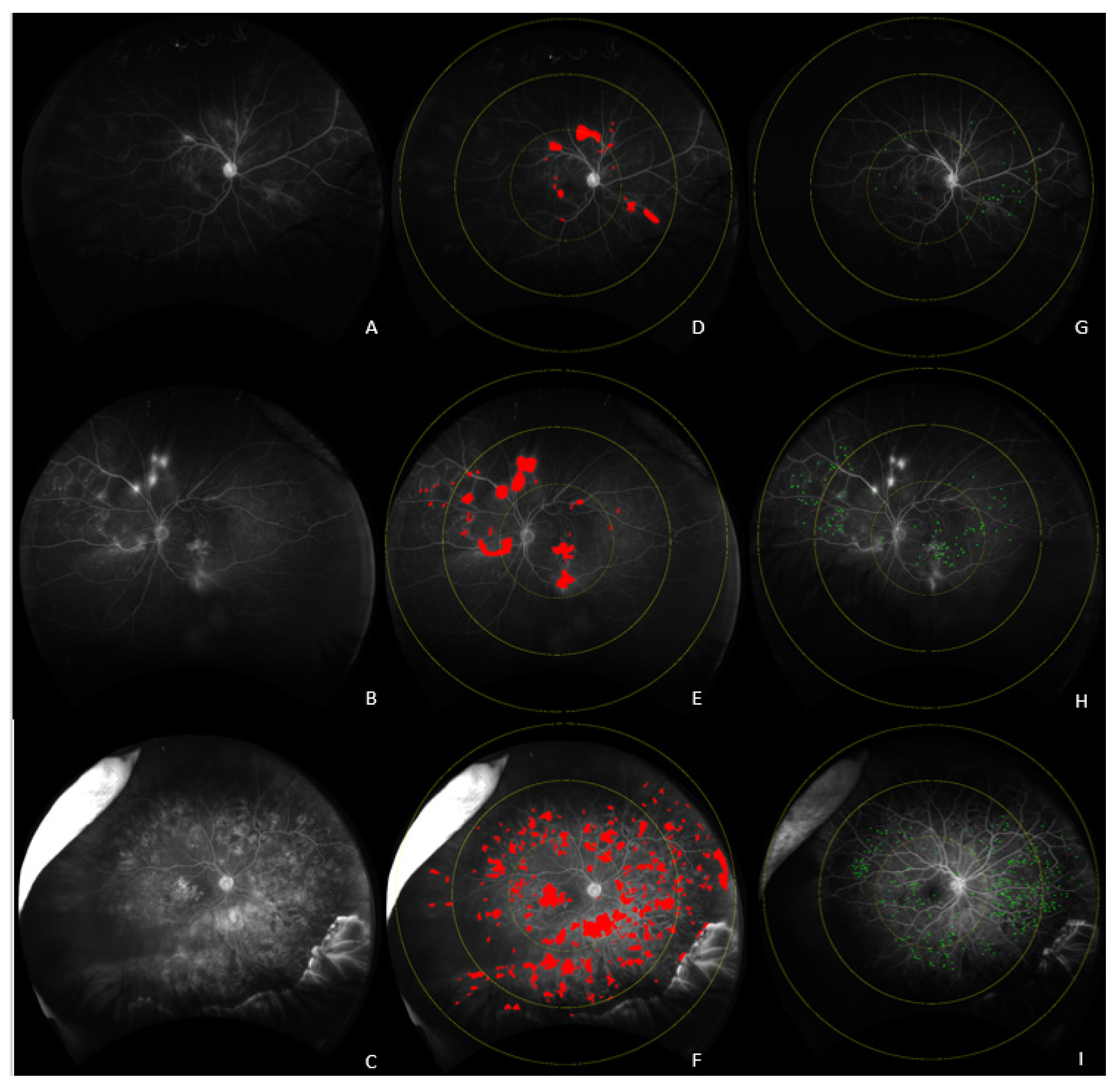
2.1. UWFA Image Analysis

Two images with optimum quality determined by the analyst were selected for each subject, one in the early–mid phase and one in the late phase. Images taken with the Optos 200Tx system were processed by dewarping to correct for peripheral distortion, as previously described [11]. Images from the California system were dewarped through the native software platform.
Ischemia analysis on UWFA was performed through segmentation of areas of capillary nonperfusion in early–mid phase FA images using a previously described image analysis platform [10]. Each image segmentation was reviewed for accuracy by a trained image analyst and sequentially reviewed by an additional expert reader. The total retinal analyzable area was defined as the peripheral edge of the visible retinal vasculature. The segmented areas of ischemia were calculated by summing the area of all pixels. The ischemia index was calculated as the area of ischemia expressed as a percentage out of the total retinal area. Microaneurysm (MA) and leakage segmentation on UWFA were first performed by an automatic feature extraction platform and then reviewed by a trained image analyst, and manually corrected, as needed. A second expert reader sequentially reviewed the final segmentation results for consistency [10,12,13]. MAs were defined as small circular spots of hyperfluorescence compared to surrounding choroidal background in early–mid phase UWFA images. Leakage was defined as a region of increasing hyperfluorescence in size and intensity in the late phase angiogram compared to the early–mid phase FA. Leakage index was calculated as the area of leakage expressed as a percentage out of the total analyzable retina. To assess the regional distribution of MAs and leakage, a mask of three concentric circles of increasing size (macular, midperiphery, and far periphery) was overlaid onto the image and centered around the macula, as previously described. MA count and leakage index were calculated zonally and panretinally [10]. In addition to the zonal indices, distribution of leakage and MA were assessed as the percentage of panretinal leakage and MA count located in the macula and in the periphery.
The retinal vasculature was extracted from the early phase frames using a deep learning algorithm [14,15,16]. Vascular parameters, including panretinal vessel area, length and localized vessel density features were calculated. Localized vessel density was calculated by dividing the vasculature mask into forty by forty pixel squares and measuring the percentage of the areas occupied with retinal vessels in each square. The mean, median, variance, skewness, and kurtosis values of localized retinal vessel density panretinally were included in the analysis.

2.2. Statistical Analysis

Eyes were grouped into three categories: (1) eyes not requiring anti-VEGF treatment during the entire follow-up period, (2) eyes requiring anti-VEGF injection within 3 months of the analyzed UWFA, and (3) eyes requiring anti-VEGF after 3 months of an UWFA imaging session. All statistical analyses were conducted using R version 3.6.1 (R Project for Statistical Computing). Statistical analyses were performed for the following clinical variables: age, gender, follow-up period, HbA1c at baseline, systolic and diastolic blood pressure closest to the baseline UWFA date, visual acuity at baseline, DME presence on OCT, and CST. Variables extracted from UWFA images included panretinal, macular, midperipheral and far peripheral leakage index, number of leakage areas and MA count, percentage of leakage and MA distribution in posterior pole, midperiphery and far periphery, panretinal ischemic index, panretinal, macular and midperipheral vessel area and vessel length, vessel area index, mean, median, variance, skewness, and kurtosis values of panretinal localized retinal vessel density and tortuosity. Distribution of normality of the continuous variables was assessed using the Shapiro and Wilk’s normality test. The generalized linear mixed effect model was used to compare each variable individually between the eyes that required anti-VEGF within 3 months, eyes that required anti-VEGF after 3 months, and eyes that did not required any anti-VEGF treatment, while considering intereye correlation. A p-value of less than 0.05 was considered statistically significant.
A random forest predictive modeling was used to predict the early and late need for anti-VEGF treatment as their performance is not affected by multicollinearity. Two random forest classifiers using 5 randomly sampled features out of all available imaging and clinical variables (n = 45) were used as candidates at each split grown with 1000 trees to differentiate eyes that required anti-VEGF within 3 months (including on the day of UWFA) and those that required it after 3 months of the imaging visit from eyes that did not require anti-VEGF during the follow-up period.

3. Results

A total of 173 eyes from patients with diabetic retinopathy were reviewed. The mean follow-up time was 22 (range: 11–43) months. Thirty-eight eyes (22%) required early anti-VEGF treatment within 3 months of the UWFA. Thirty-four eyes (20%) required late anti-VEGF treatment at least 3 months after the UWFA imaging. One hundred and one eyes did not require any anti-VEGF treatment during the follow-up time. There were no significant differences in age, gender, follow-up period, and HbA1c (Table 1).

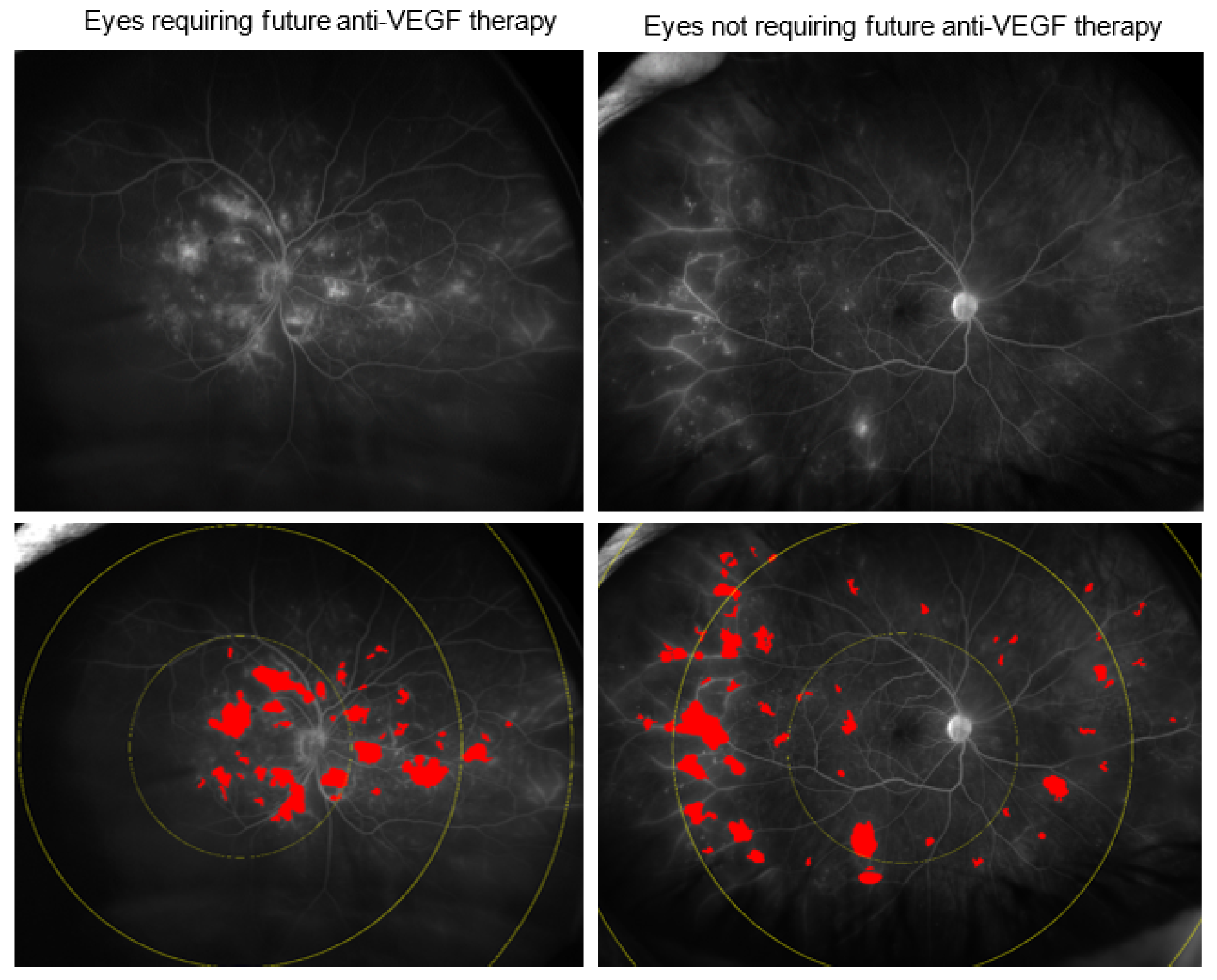
3.1. Early Anti-VEGF Intervention Requirement

At baseline, there were significant differences in multiple imaging biomarkers across the groups. Eyes requiring anti-VEGF treatment within 3 months demonstrated significant differences in the following parameters (Figure 1): panretinal leakage index (4.1% vs. 2.0%, p = 0.006), macular leakage index (11.5% vs. 4.3%, p = 0.002) (Figure 2), peripheral MA count (167 ± 320 vs. 108 ± 102 p = 0.005) (Figure 3), median vessel density (16.6 ± 2.7% vs. 15.1 ± 2.5%, p = 0.013), macular vessel area (17.1 ± 2.4 vs. 16.5 ± 2.1 mm2, p = 0.035), midperipheral vessel area (61.8 ± 16.0 vs. 57.2 ±12 mm2, p = 0.021), CST (415 ± 134 µm vs. 263 ± 57 µm, p = 0.002), DME presence (92% vs. 27%, p < 0.001), and visual acuity (20/80 vs. 20/25, p = 0.037).

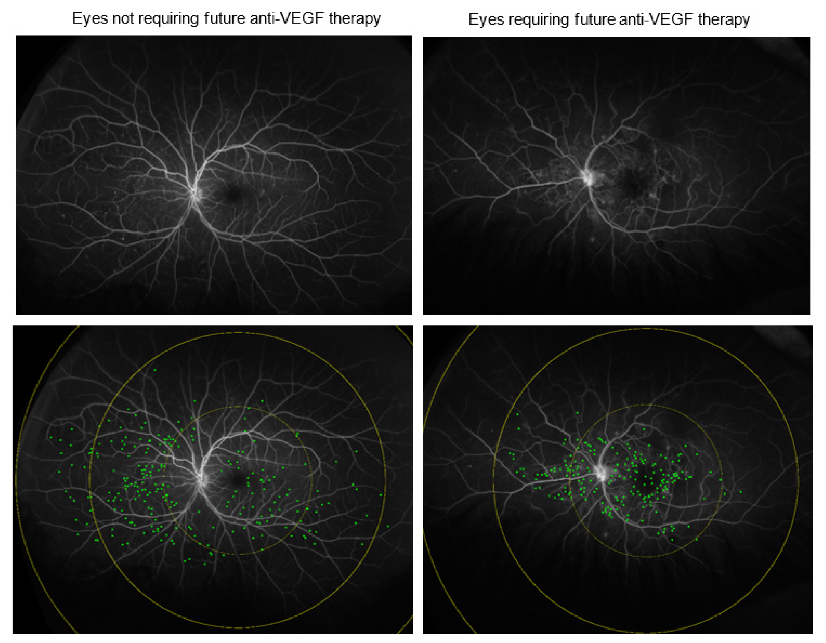
3.2. Delayed Anti-VEGF Treatment Requirement

Eyes requiring anti-VEGF treatment after 3 months were also significantly different from eyes not requiring anti-VEGF treatment in the following parameters: macular leakage index (12.9% vs. 4.3%, p = 0.007), panretinal leakage index (4.4% vs. 2.0%, p = 0.022), visual acuity (20/50 vs. 20/25, p < 0.001), and DME presence (65% vs. 27%, p < 0.001).

3.3. Comparative Assessment of Eyes Requiring Early vs. Deferred Anti-VEGF Treatment

Eyes requiring anti-VEGF treatment within 3 months and after 3 months had significantly different CST (415 ± 134 µm vs. 314 ± 112 µm, p = 0.040), DME presence (92% vs. 65%, p = 0.011), macular MA count (88 ± 64 vs. 57 ± 46, p = 0.044), and macular vessel area (17.1 ± 2.4 vs. 15.5 ± 2.9 mm2, p = 0.025). This suggests that the primary initial reason for early treatment was the presence/severity of DME.

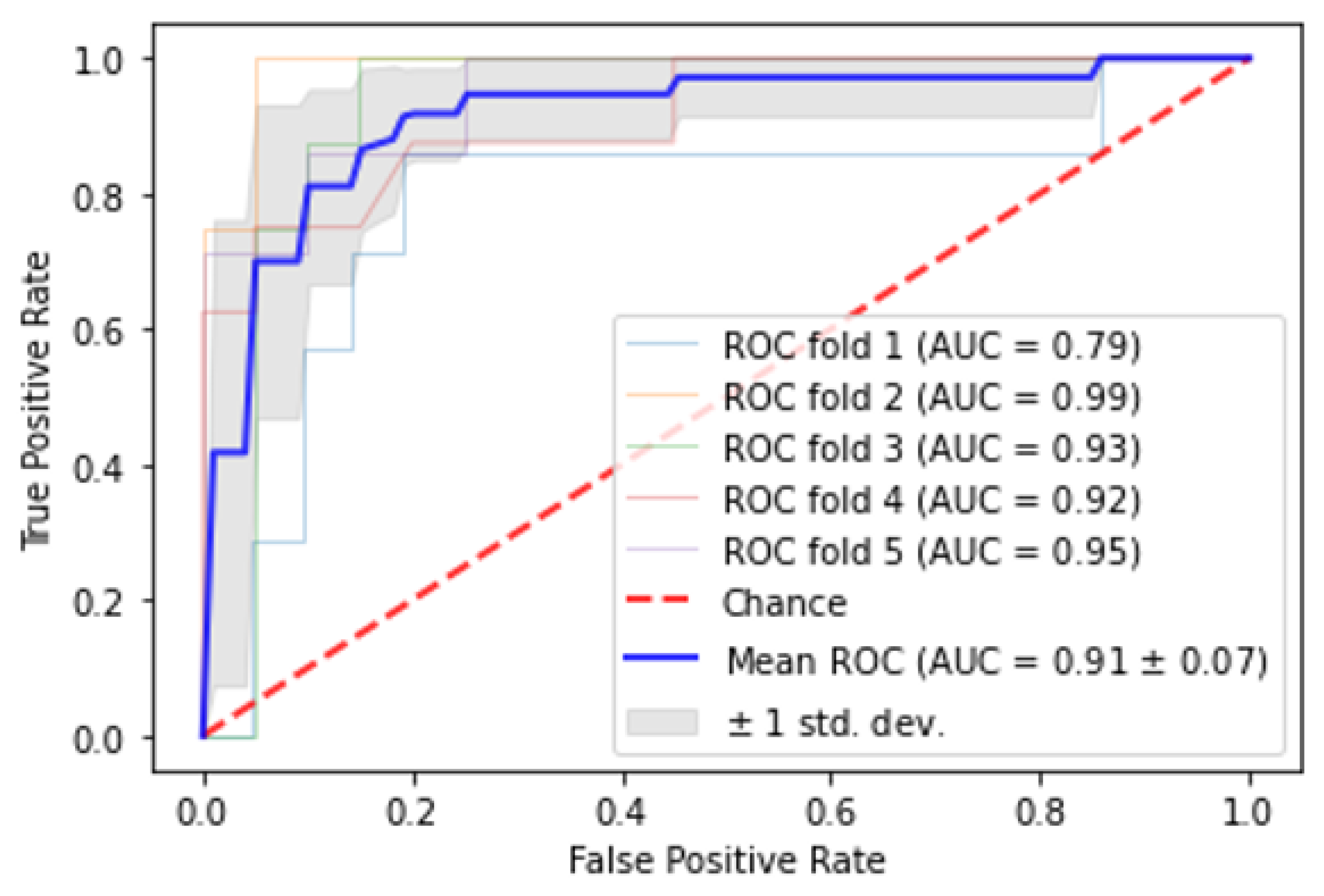
3.4. Automated Classification of Eyes Based on Need for Early or Late Anti-VEGF Therapy

A random forest model differentiating eyes requiring treatment within 3 months from the eyes not requiring future treatment identified macular leakage index, presence of DME, CST, visual acuity, and macular distribution of leakage as the top five most important features, and demonstrated an AUC of 0.91 ± 0.07 (Figure 4). Random forest model differentiating eyes requiring treatment after at least 3 months from eyes not requiring future treatment demonstrated the top five most important features to be macular leakage index, visual acuity, systolic blood pressure, panretinal leakage index, and follow-up duration and had an AUC of 0.77 ± 0.04.

4. Discussion

Anti-VEGF therapy in diabetic eye disease has been shown to improve visual acuity, reduce macular edema, and improve DRSS severity [17,18]. However, it is currently unknown which features predict whether a patient will require anti-VEGF injections. Early stratification of patients at risk for requiring anti-VEGF therapy in the future can help minimize unnecessary office visits and allow initiation of potential interventions sooner to prevent vision-threatening complications of diabetic eye disease.
The present study evaluated the utility of the imaging features in predicting the need for anti-VEGF treatment for the treatment of PDR with or without DME. OCT features including CST and DME presence were significantly different across all groups. This study demonstrated that there is a significant difference in the panretinal and macular leakage index for eyes that required anti-VEGF treatment compared to eyes that did not. These results suggest that the extent of leakage, especially in the posterior pole, may be an important marker for the need for future anti-VEGF treatments, and could potentially be used to modulate follow-up regimens, particularly in eyes without DME. Significantly higher DME presence was observed in eyes requiring immediate treatment compared to delayed treatment, suggesting that the treatment decision was driven by DME. Macular vessel area and macular MA count were also higher in eyes requiring immediate VEGF treatment compared to delayed treatment. The associations between the disease progression to treatment threshold and the angiographic features including macular vessel area and MA count should be further investigated.
Along with the intraretinal microvascular abnormalities, MAs are among the vascular abnormalities associated with DR severity. The number and turnover rate of MAs have previously been considered an important biomarker for the progression of DME [19]. The findings in this analysis indicate that MA count is associated with requiring anti-VEGF therapy. MA count in the posterior pole and macular vessel length were both significantly higher in eyes that required treatment within 3 months. MAs are hypothesized to be focal areas of permeability in the retinal vasculature that may give rise to leakage [20]. Anti-VEGF treatments for DME have been shown to decrease diffuse leakage but have relatively little effect on focal leakage as assessed by FA [21]. Another study found a significant decrease in microaneurysms after anti-VEGF therapy [22]. The relationship between anti-VEGF therapy and MA count requires additional investigation.
Larger vessel area measured in the macular zones of eyes requiring immediate treatment may be due to active vascular remodeling. In a recent study, increased macular vessel area was extracted from the UWFA frames of moderate and severe NPDR eyes compared to PDR eyes [23]. Another study demonstrated a higher risk of DME development in moderate and severe NPDR compared to PDR [24]. Further studies investigating the integrative impact of DR severity compared or combined with quantitative angiographic features on future treatment should be explored. The current study also demonstrated higher panretinal vascular density measures in eyes requiring immediate anti-VEGF treatment. Based on current vascular extraction platforms, increased vascular remodeling in the setting of increased disease activity often leads to greater contrast and enhanced visualization of individual vessels and may account for the higher vessel density measurements.
The present study did not demonstrate a difference in ischemia on UWFA in eyes with DR with different anti-VEGF needs. Previous studies have found variable results on the association of peripheral retinal ischemia with the presence of DME [9,25,26]. UWFA images from eyes with DR before and after anti-VEGF injections suggest that areas of ischemia do not demonstrate significant reperfusion following anti-VEGF therapy despite an improvement in the DRSS score based on color fundus photographs [7,12,17,27]. The findings from our study suggest that ischemia may not be a key predictor for the anti-VEGF therapy requirement in DR; but rather that the leakage index may reflect more current vascular disease activity and may be more indicative of a future need for anti-VEGF therapy, whereas ischemia may be more reflective of overall disease effects and not current disease activity.
Random forest analysis demonstrated that macular leakage index, visual acuity, CST, and DME were the most important features differentiating the three groups. The most successful model differentiating eyes requiring anti-VEGF treatment in the near future from eyes not requiring treatment demonstrated an AUC of 0.91. The utility of these models as clinical decision-making tools should be further investigated, particularly in relation to follow-up time and risk stratification.
The strengths of this study include the use of UWFA to capture both peripheral and nonperipheral angiographic features implicated in DR. The panretinal leakage index and peripheral MA count, which could not be visualized by standard FA, were significantly higher in eyes that required anti-VEGF treatment within 3 months. Moreover, the panretinal leakage index was an important variable predicting delayed anti-VEGF treatment. Semiautomated quantitative analysis of these imaging features provides a novel way to assess the extent of leakage, MAs, ischemia and vascular features on UWFA with improved objectivity. Several studies have reported therapeutic effects in the fellow eye following unilateral anti-VEGF injections [28,29,30]. For subjects that had two eyes included in the study, a linear mixed effects model was used to correct for dependence. Random forest predictive modeling was used to predict the early or delayed anti-VEGF needs for its ability to handle complex dependency patterns between correlated covariates. A limitation of this study included that the software used to segment areas of ischemia was unable to provide detailed analysis into the distribution of ischemia regions (e.g., in the posterior pole). Future studies should explore the regional differences in ischemia in DR. Additionally, DM duration, pharmacotherapy 6 months prior to the baseline visit, and axial length measurements of the patients were not available. Pixel to mm2 conversions were not adjusted to actual axial length. Another limitation of this study was a relatively small sample size when stratifying the timing of future anti-VEGF injections. A larger sample size would allow further delineation of the temporal relation between angiographic imaging features and future therapies in DR. Lastly, there were no preset indications guiding the treatment decision in this retrospective study, and retina specialists individually determined the appropriate treatment for each patient. This study identified quantitative imaging features on UWFA that predict the need for future anti-VEGF therapy; however, it was not possible to delineate the clinical indication for the injection (e.g., DME, DR, or both). Future studies should also investigate whether specific imaging features are predictive of the development of DME, PDR, or both.
In conclusion, quantitative leakage on UWFA along with OCT features are useful for identifying eyes with DR that likely need future anti-VEGF therapy. Additional research is needed to determine the underlying mechanisms allowing the angiographic prediction of intravitreal treatment for DR and treatment response.

Author Contributions

Conceptualization, D.D.S., A.C.J., S.K.S., K.E.T., J.L.R. and J.P.E.; Methodology, A.C.J., D.D.S., S.K.S., K.E.T. and J.P.E.; Software, D.D.S. and J.W.; Validation, M.H., D.D.S., A.C.J., C.M., J.W., S.K.S., K.E.T., J.L.R. and J.P.E.; Formal Analysis, D.D.S., A.C.J., M.H. and C.M.; Resources, S.K.S., K.E.T., J.L.R. and J.P.E.; Data Curation, D.D.S., A.C.J., M.H., C.M., J.W., S.K.S., K.E.T., J.L.R. and J.P.E.; Writing—Original Draft Preparation, A.C.J. and D.D.S.; Writing—Review and Editing, M.H., C.M., J.W., S.K.S., K.E.T., J.L.R. and J.P.E.; Visualization, D.D.S. and A.C.J.; Supervision, S.K.S., K.E.T., J.L.R. and J.P.E.; Project Administration, J.L.R. and J.P.E.; Funding Acquisition, J.P.E. All authors have read and agreed to the published version of the manuscript.

Funding

This research was supported by NIH/NEI K23-EY022947-01A1 (JPE) and the Research to Prevent Blindness (Cole Eye Institutional).

Institutional Review Board Statement

The study was conducted in accordance with the Declaration of Helsinki, and approved by the Institutional Review Board of the Cleveland Clinic with most recent ongoing renewal on 11 December 2021.

Informed Consent Statement

Patient consent was waived due to the retrospective nature and minimal risk nature of this research.

Data Availability Statement

Additional data available upon request.

Conflicts of Interest

The authors declare no conflict of interest specifically related to this article. The funders had no role in the design of the study; in the collection, analyses, or interpretation of data; in the writing of the manuscript, or in the decision to publish the results. The authors have the following financial disclosures: A.C.J.: None; D.D.S.: None; C.M.: None; J.W.: None, S.K.S.: Allergan (R, C); Leica (P), Zeiss (C); Regeneron (R, C); Eyepoint (R, C), Eyevensys (R, C), Novartis (C), Abbvie (C); K.E.T.: Zeiss (R); M.H.: None; J.L.R.: None; J.P.E.: Leica (C, P), Oxurion (C, R), Genentech (C, R), Roche (C, R), Zeiss (C), Alcon (C, R), Novartis (C, R), Aerpio (C, R), Allergan (C, R), Allegro (C), Regeneron (C, R), Alcon (C, R), Boehringer-Ingelheim (C, R), Stealth (C, R), Adverum (C, R).

References

  1. Bresnick, G.H. Diabetic Macular Edema: A Review. Ophthalmology 1986, 93, 989–997. [Google Scholar] [CrossRef]
  2. Photocoagulation For Diabetic Macular Edema: Early Treatment Diabetic Retinopathy Study Report Number 1 Early Treatment Diabetic Retinopathy Study Research Group. Arch. Ophthalmol. 1985, 103, 1796–1806. [CrossRef]
  3. Jiang, S.; Barner, J.C.; Park, C.; Ling, Y.-L. Treatment Patterns of Anti-Vascular Endothelial Growth Factor and Laser Therapy Among Patients with Diabetic Macular Edema. J. Manag. Care Spec. Pharm. 2015, 21, 735–741. [Google Scholar] [CrossRef] [PubMed] [Green Version]
  4. Mitchell, P.; Bandello, F.; Schmidt-Erfurth, U.; Lang, G.E.; Massin, P.; Schlingemann, R.O.; Sutter, F.; Simader, C.; Burian, G.; Gerstner, O.; et al. The RESTORE Study: Ranibizumab Monotherapy or Combined with Laser versus Laser Monotherapy for Diabetic Macular Edema. Ophthalmology 2011, 118, 615–625. [Google Scholar] [CrossRef] [PubMed] [Green Version]
  5. Wells, J.A.; Glassman, A.R.; Ayala, A.R.; Jampol, L.M.; Bressler, N.M.; Bressler, S.B.; Brucker, A.J.; Ferris, F.L.; Hampton, G.R.; Jhaveri, C.; et al. Aflibercept, Bevacizumab, or Ranibizumab for Diabetic Macular Edema: Two-Year Results from a Comparative Effectiveness Randomized Clinical Trial. Ophthalmology 2016, 123, 1351–1359. [Google Scholar] [CrossRef] [Green Version]
  6. Nguyen, Q.D.; Brown, D.M.; Marcus, D.M.; Boyer, D.S.; Patel, S.; Feiner, L.; Gibson, A.; Sy, J.; Rundle, A.C.; Hopkins, J.J.; et al. Ranibizumab for Diabetic Macular Edema: Results from 2 Phase III Randomized Trials: RISE and RIDE. Ophthalmology 2012, 119, 789–801. [Google Scholar] [CrossRef]
  7. Levin, A.M.; Rusu, I.; Orlin, A.; Gupta, M.P.; Coombs, P.; D’Amico, D.J.; Kiss, S. Retinal Reperfusion in Diabetic Retinopathy Following Treatment with Anti-VEGF Intravitreal Injections. Clin. Ophthalmol. 2017, 11, 193–200. [Google Scholar] [CrossRef] [Green Version]
  8. Silva, P.S.; Dela Cruz, A.J.; Ledesma, M.G.; van Hemert, J.; Radwan, A.; Cavallerano, J.D.; Aiello, L.M.P.; Sun, J.K.; Aiello, L.M.P. Diabetic Retinopathy Severity and Peripheral Lesions Are Associated with Nonperfusion on Ultrawide Field Angiography. Ophthalmology 2015, 122, 2465–2472. [Google Scholar] [CrossRef]
  9. Wessel, M.M.; Nair, N.; Aaker, G.D.; Ehrlich, J.R.; D’Amico, D.J.; Kiss, S. Peripheral Retinal Ischaemia, as Evaluated by Ultra-Widefield Fluorescein Angiography, Is Associated with Diabetic Macular Oedema. Br. J. Ophthalmol. 2012, 96, 694–698. [Google Scholar] [CrossRef]
  10. Ehlers, J.P.; Jiang, A.C.; Boss, J.D.; Hu, M.; Figueiredo, N.; Babiuch, A.; Talcott, K.; Sharma, S.; Hach, J.; Le, T.; et al. Quantitative Ultra-Widefield Angiography and Diabetic Retinopathy Severity. Ophthalmology 2019, 126, 1527–1532. [Google Scholar] [CrossRef]
  11. Croft, D.E.; van Hemert, J.; Wykoff, C.C.; Clifton, D.; Verhoek, M.; Fleming, A.; Brown, D.M. Precise Montaging and Metric Quantification of Retinal Surface Area from Ultra-Widefield Fundus Photography and Fluorescein Angiography. Ophthalmic Surg. Lasers Imaging Retina 2014, 45, 312–317. [Google Scholar] [CrossRef] [PubMed]
  12. Wykoff, C.C.; Nittala, M.G.; Zhou, B.; Fan, W.; Velaga, S.B.; Lampen, S.I.R.; Rusakevich, A.M.; Ehlers, J.P.; Babiuch, A.; Brown, D.M.; et al. Intravitreal Aflibercept for Retinal Non-Perfusion in Proliferative Diabetic Retinopathy: Outcomes from the RECOVERY Randomized Trial. Ophthalmol. Retin. 2019, 3, 1076–1086. [Google Scholar] [CrossRef] [PubMed]
  13. Automated Quantitative Characterisation of Retinal Vascular Leakage and Microaneurysms in Ultra-Widefield Fluorescein Angiography|Enhanced Reader. Available online: Chrome-extension://dagcmkpagjlhakfdhnbomgmjdpkdklff/enhanced-reader.html?openApp&pdf=https%3A%2F%2Fbjo.bmj.com%2Fcontent%2Fbjophthalmol%2F101%2F6%2F696.full.pdf (accessed on 20 May 2021).
  14. Damla Sevgi, D.; Scott, A.W.; Martin, A.; Mugnaini, C.; Patel, S.; Linz, M.O.; Nti, A.A.; Reese, J.; Ehlers, J.P. Longitudinal Assessment of Quantitative Ultra-Widefield Ischaemic and Vascular Parameters in Sickle Cell Retinopathy. Br. J. Ophthalmol. 2022, 106, 251–255. [Google Scholar] [CrossRef] [PubMed]
  15. Automated Quality Optimized Phase Selection in Ultra-Widefield Angiography Using Retinal Vessel Segmentation with Deep Neural Networks|IOVS|ARVO Journals. Available online: https://iovs.arvojournals.org/article.aspx?articleid=2770382 (accessed on 20 May 2021).
  16. Longitudinal Precision of Vasculature Parameter Assessment on Ultra-Widefield Fluorescein Angiography Using a Deep-Learning Model for Vascular Segmentation in Eyes without Vascular Pathology|IOVS|ARVO Journals. Available online: https://iovs.arvojournals.org/article.aspx?articleid=2769705 (accessed on 20 May 2021).
  17. Bonnin, S.; Dupas, B.; Lavia, C.; Erginay, A.; Dhundass, M.; Couturier, A.; Gaudric, A.; Tadayoni, R. Anti–vascular endothelial growth factor therapy can improve diabetic retinopathy score without change in retinal perfusion. Retina 2019, 39, 426–434. [Google Scholar] [CrossRef]
  18. Heier, J.S.; Korobelnik, J.F.; Brown, D.M.; Schmidt-Erfurth, U.; Do, D.V.; Midena, E.; Boyer, D.S.; Terasaki, H.; Kaiser, P.K.; Marcus, D.M.; et al. Intravitreal Aflibercept for Diabetic Macular Edema: 148-Week Results from the VISTA and VIVID Studies. Ophthalmology 2016, 123, 2376–2385. [Google Scholar]
  19. Nunes, S.; Pires, I.; Rosa, A.; Duarte, L.; Bernardes, R.; Cunha-Vaz, J. Microaneurysm Turnover Is a Biomarker for Diabetic Retinopathy Progression to Clinically Significant Macular Edema: Findings for Type 2 Diabetics with Nonproliferative Retinopathy. Ophthalmologica 2009, 223, 292–297. [Google Scholar] [CrossRef] [PubMed]
  20. Najeeb, B.H.; Simader, C.; Deak, G.; Vass, C.; Gamper, J.; Montuoro, A.; Gerendas, B.S.; Schmidt-Erfurth, U. The Distribution of Leakage on Fluorescein Angiography in Diabetic Macular Edema: A New Approach to Its Etiology. Investig. Ophthalmol. Vis. Sci. 2017, 58, 3986–3990. [Google Scholar] [CrossRef] [PubMed] [Green Version]
  21. Allingham, M.J.; Mukherjee, D.; Lally, E.B.; Rabbani, H.; Mettu, P.S.; Cousins, S.W.; Farsiu, S. A Quantitative Approach to Predict Differential Effects of Anti-VEGF Treatment on Diffuse and Focal Leakage in Patients with Diabetic Macular Edema: A Pilot Study. Transl. Vis. Sci. Technol. 2017, 6, 7. [Google Scholar] [CrossRef] [Green Version]
  22. Couturier, A.; Rey, P.-A.; Erginay, A.; Lavia, C.; Bonnin, S.; Dupas, B.; Gaudric, A.; Tadayoni, R. Widefield OCT-Angiography and Fluorescein Angiography Assessments of Nonperfusion in Diabetic Retinopathy and Edema Treated with Anti–Vascular Endothelial Growth Factor. Ophthalmology 2019, 126, 1685–1694. [Google Scholar] [CrossRef]
  23. Sevgi, D.D.; Srivastava, S.K.; Whitney, J.; O’Connell, M.; Kar, S.S.; Hu, M.; Reese, J.; Madabhushi, A.; Ehlers, J.P. Characterization of Ultra-Widefield Angiographic Vascular Features in Diabetic Retinopathy with Automated Severity Classification. Ophthalmol. Sci. 2021, 1, 100049. [Google Scholar] [CrossRef]
  24. Moshfeghi, A.; Garmo, V.; Sheinson, D.; Ghanekar, A.; Abbass, I. Five-Year Patterns of Diabetic Retinopathy Progression in Us Clinical Practice. Clin. Ophthalmol. 2020, 14, 3651–3659. [Google Scholar] [CrossRef]
  25. Sim, D.A.; Keane, P.A.; Rajendram, R.; Karampelas, M.; Selvam, S.; Powner, M.B.; Fruttiger, M.; Tufail, A.; Egan, C.A. Patterns of Peripheral Retinal and Central Macula Ischemia in Diabetic Retinopathy as Evaluated by Ultra-Widefield Fluorescein Angiography. Am. J. Ophthalmol. 2014, 158, 144–153.e1. [Google Scholar] [CrossRef] [PubMed]
  26. Patel, R.D.; Messner, L.V.; Teitelbaum, B.; Michel, K.A.; Hariprasad, S.M. Characterization of Ischemic Index Using Ultra-Widefield Fluorescein Angiography in Patients with Focal and Diffuse Recalcitrant Diabetic Macular Edema. Am. J. Ophthalmol. 2013, 155, 1038–1044.e2. [Google Scholar] [CrossRef] [PubMed]
  27. Figueiredo, N.; Srivastava, S.K.; Singh, R.P.; Babiuch, A.; Sharma, S.; Rachitskaya, A.; Talcott, K.; Reese, J.; Hu, M.; Ehlers, J.P. Longitudinal Panretinal Leakage and Ischemic Indices in Retinal Vascular Disease Following Aflibercept Therapy: The PERMEATE Study; Elsevier: Amsterdam, The Netherlands, 2019. [Google Scholar]
  28. Calvo, C.M.; Sridhar, J.; Shahlaee, A.; Ho, A.C. Reduction of Diabetic Macular Edema in the Untreated Fellow Eye Following Intravitreal Injection of Aflibercept. Ophthalmic Surg. Lasers Imaging Retin. 2016, 47, 474–476. [Google Scholar] [CrossRef] [PubMed] [Green Version]
  29. Isildak, H.; Schwartz, S.G.; Flynn, H.W. Therapeutic Effect of Anti-VEGF for Age-Related Macular Degeneration in the Untreated Fellow Eye. Case Rep. Ophthalmol. Med. 2018, 2018, 1–4. [Google Scholar] [CrossRef] [Green Version]
  30. Sharma, N.S.; Ong, J.M.; Ooi, J.L. Re: “Fellow Eye Effect of Unilateral Intravitreal Bevacizumab Injection in Eyes with Diabetic Macular Edema”. Eye 2015, 29, 291–292. [Google Scholar] [CrossRef] [Green Version]
Figure 1. Late UWFA images of eyes not requiring treatment (A), requiring delayed treatment (B) requiring immediate treatment (C), and their corresponding leakage (DF), segmentation, and microaneurysm (GI) segmentations.
Jpm 12 00608 g001
Figure 2. Leakage index in eyes requiring future anti-VEGF therapy. Total leakage index and posterior pole leakage index on UWFA were significantly higher in eyes requiring future anti-VEGF therapy compared to eyes not requiring future injections.
Jpm 12 00608 g002
Figure 3. MA count in eyes requiring future anti-VEGF therapy. Total MA count and posterior pole MA count on UWFA were significantly higher in eyes requiring future anti-VEGF therapy compared to eyes not requiring future injections.
Jpm 12 00608 g003
Figure 4. Area under the curve graph for random forest model trained in 5-fold cross-validation setting differentiating eyes requiring immediate anti-VEGF treatment from eyes not requiring treatment.
Jpm 12 00608 g004
Table 1. Summary of select characteristics and imaging features for each treatment group.
Not Requiring Treatment
(n = 101)
Anti-VEGF Injection
within 3 Months
(n = 38)
Anti-VEGF Injection
after 3 Months
(n = 34)
Age63 ± 1366 ± 1362 ± 13
Gender
Female482017
Male531817
Follow-up time (months)21.6 ± 8.321.0 ± 10.325.7 ± 10.8
Systolic Blood Pressure134.6 ± 15.6139.5 ± 19.8144.6 ± 20.2
Hypertension97.0%100%97.1%
Presence of DME on OCT *27%92%65%
HbA1c8.0 ± 1.88.4 ± 2.38.4 ± 2.3
Visual acuity *20/2520/8020/50
CST (µm) *263 ± 57415 ± 134314 ± 112
Panretinal LI (%) *2.0 ± 2.84.1 ± 3.44.4 ± 3.7
Macular LI (%) *4.3 ± 5.011.5 ± 7.612.9 ± 11.3
Panretinal ischemia index (%)2.1 ± 1.43.1 ± 4.14.7 ± 4.8
Panretinal MA count161 ± 131260 ± 385156 ± 125
Macular MA count *51 ± 4688 ± 6457 ± 46
Peripheral MA Count *108 ± 102167 ± 32097 ± 93
Panretinal vessel area (mm2)86.2 ± 19.892.2 ± 25.390.6 ± 25.0
Macular vessel area (mm2) *16.5 ± 2.117.1 ± 2.415.5 ± 2.9
Median vessel density (%) *15.1 ± 2.516.6 ± 2.716.1 ± 2.6
Abbreviations: CST: central subfield thickness, DME: diabetic macular edema, LI: leakage index, MA: microaneurysm. * significantly different among categories.
Publisher’s Note: MDPI stays neutral with regard to jurisdictional claims in published maps and institutional affiliations.

