Article

Polymorphisms and Gene-Gene Interaction in AGER/IL6 Pathway Might Be Associated with Diabetic Ischemic Heart Disease

1
Beijing Municipal Key Laboratory of Clinical Epidemiology, Department of Epidemiology and Health Statistics, School of Public Health, Capital Medical University, Beijing 100069, China
2
Department of Biostatistics, FMD K & L, Fort Washington, PA 19034, USA
*
Author to whom correspondence should be addressed.
J. Pers. Med. 2022, 12(3), 392; https://doi.org/10.3390/jpm12030392
Submission received: 23 December 2021 / Revised: 10 February 2022 / Accepted: 22 February 2022 / Published: 4 March 2022
(This article belongs to the Special Issue Non-communicable Diseases, Big Data and Artificial Intelligence)

Abstract

:
Background: Although the genetic susceptibility to diabetes and ischemic heart disease (IHD) has been well demonstrated, studies aimed at exploring gene variations associated with diabetic IHD are still limited; Methods: Our study included 204 IHD cases who had been diagnosed with diabetes before the diagnosis of IHD and 882 healthy controls. Logistic regression was used to find the association of candidate SNPs and polygenic risk score (PRS) with diabetic IHD. The diagnostic accuracy was represented with AUC. Generalized multifactor dimensionality reduction (GMDR) was used to illustrate gene-gene interactions; Results: For IL6R rs4845625, the CT and TT genotypes were associated with a lower risk of diabetic IHD than the CC genotype (OR = 0.619, p = 0.033; OR = 0.542, p = 0.025, respectively). Haplotypes in the AGER gene (rs184003-rs1035798-rs2070600-rs1800624) and IL6R gene (rs7529229-rs4845625-rs4129267-rs7514452-rs4072391) were both significantly associated with diabetic IHD. PRS was associated with the disease (OR = 1.100, p = 0.005) after adjusting for covariates, and the AUC were 0.763 (p < 0.001). The GMDR analysis suggested that rs184003 and rs4845625 were the best interaction model after permutation testing (p = 0.001) with a cross-validation consistency of 10/10; Conclusions: SNPs and haplotypes in the AGER and IL6R genes and the interaction of rs184003 and rs4845625 were significantly associated with diabetic IHD.

Graphical Abstract

1. Introduction

Ischemic heart disease (IHD) remains the leading global cause of death and lost life years in adults, and it is also the leading cause of mortality in people with type 2 diabetes mellitus (T2DM). Approximately 68% of deaths in type 2 diabetic patients are caused by cardiac complications [1,2]. It has been demonstrated that the advanced glycation end products (AGER)/interleukin-6 (IL-6) pathway plays an important role in the physiological mechanism of diabetic cardiovascular complications [3,4,5]; however, whether gene polymorphisms in this pathway can influence the disease susceptibility are still unknown.
Several single nucleotide polymorphisms (SNPs) in the AGER gene have been reported to be associated with diabetes or its complications [6,7,8]. A meta-analysis including 27 original articles showed that AGER genetic polymorphisms with CAD were potentiated in patients with diabetes mellitus disease, but the association was not consistently significant [9,10,11]. A Mendelian randomization study also showed that the C allele in rs2228145 was associated with a lower risk of coronary heart disease [12], but a meta-analysis of three GWA scans with 4107 type 2 diabetes cases and 5187 controls in Caucasians found no evidence that IL6R variants were associated with type 2 diabetes [13]. Due to the different effects of the IL6R gene on diabetes and coronary heart disease, further research is still needed to demonstrate the association between IL6R variants and diabetic macrovascular complications. Gene-gene interactions can explain the missing heritability of cardiometabolic disease and better reflect the complex pathophysiological process of disease [14]. However, few studies have focused on the influence of the gene-gene interactions among several SNPs on CVD susceptibility until now [15,16,17]. Machine learning methods may effectively reduce Type I and II errors and increase robustness, of which the generalized multifactor dimensionality reduction (GMDR) method has remained popular in detecting the interaction effect since its appearance [18,19].
The current study aimed to illustrate the association of AGER and IL6R gene polymorphisms with the risk for diabetic ischemic heart disease (IHD) and to assess the modulatory effect of gene-gene interactions between these variants on disease risk. SNPs that were previously reported to be associated with cardiometabolic disease, inflammatory disease, or located in miRNA binding sites were selected for the analysis (see Supplementary Materials Table S1).

2. Materials and Methods

2.1. Study Design and Population

A total of 204 diabetic ischemic heart disease cases and 882 healthy controls were enrolled from communities in Beijing. All subjects gave written informed consent. This study was approved by the Ethics Committee of Capital Medical University (No: 2016SY24).
The inclusion criteria for the cases were as follows: (1) Self-reported or physician-diagnosed diabetes according to the American Diabetes Association Criteria [20]; (2) Ischemic heart disease defined by clinical history, including acute myocardial infarction, angina pectoris, non-ST-elevation acute coronary syndromes, and/or ischemic electrocardiographic alterations; (3) T2DM was diagnosed earlier than ischemic heart disease for at least one year; and (4) Medical records or copies should be provided to verify the diagnosis of diseases.
The exclusion criteria for the cases were as follows: (1) Other diabetic complications: including diabetic nephropathy, diabetic foot, diabetic retinopathy, and diabetic neuropathy; (2) Ischemic cerebrovascular disease or cerebral hemorrhage; (3) Pregnant or lactating women; and (4) Critical physical disability or mental disorder and could not cooperate with the survey.
The inclusion criteria for the controls were as follows: (1) Subjects had not been diagnosed with T2DM before, and fasting blood glucose was less than 5.6 mmol/L in the current survey; (2) Subjects did not have ischemic heart disease, ischemic stroke, or cerebral hemorrhage; (3) Subjects did not have chronic kidney disease; and (4) Subjects were not in the acute phase of infection.
The exclusion criteria for the controls were as follows: (1) Pregnant or lactating women; and (2) Critical physical disability or mental disorder and could not cooperate with the survey.

2.2. Measurements

Lifestyle risk factors were obtained from a structured questionnaire. Smoking status was categorized as “currently smoking” and “past/never smoking”. Current smoking was defined as at least 1 cigarette per day, lasting for more than 1 year. Those who had never smoked before or had not smoked for at least 3 months were defined as past/never smoking. Alcohol drinking was categorized as “current alcohol drinking” and “past/never alcohol drinking”. Current drinking was defined as drinking at least once per week and still drinking at that frequency in the previous month. Those who never drank alcohol or had not consumed alcohol for at least one month were defined as never/past alcohol drinking.
Blood pressure (BP) was measured in the morning before participants used antihypertensive medication. Participants were asked to rest for at least 30 min before BP measurement if they had just smoked or had caffeinated products. BP (mmHg) was measured three times at sitting positions by a mercury sphygmomanometer. The average of the last two measurements was used for data analysis.

2.3. Serum Markers

After overnight fasting, all participants underwent fasting blood sampling. Fasting blood samples were collected and restored in a 2% EDTA vacutainer for each participant. After centrifugation, the plasma and blood cell samples were separated into two cryovials. Fasting plasma glucose (FPG), total cholesterol (TC), triglycerides (TG), high-density lipoprotein cholesterol (HDLC), and low-density lipoprotein cholesterol (LDLC) were tested using the Beckman Coulter chemistry analyzer AU5800 in the clinical laboratory of Beijing Hepingli Hospital.
Venous blood samples were obtained and stored in a 4 °C refrigerator. All biochemical analyses were performed within 8 h. Serum glucose and biochemical determinations were measured by an enzymatic method using a chemistry analyzer (Beckman LX20, Beckman, Brea, CA, USA) at the central laboratory of the hospital. Highly sensitive C-reactive protein was assessed using a Beckman Coulter chemistry analyzer AU5800 and white blood cell counts were obtained using an AcT5diff cell counter (Beckman Coulter®).

2.4. Genotyping

Important functional SNPs and previously reported susceptible SNPs were selected as candidate SNPs. Five SNPs (rs1035798, rs1800624, rs1800625, rs184003, and rs2070600) in the AGER gene and seven SNPs (rs2228144, rs4072391, rs4129267, rs4537545, rs4845625, rs7514452, and rs7529229) in the IL6R gene were selected in the current study.
Genomic DNA was extracted from 1 mL of peripheral blood cells using a TIANGEN DNA kit (TIANGEN Biotech, China, DP319-01) according to the manufacturer’s protocol. The primers were designed by AssayDesigner3.1 software and they were synthesized by Shanghai Thermo Fisher Scientific Co., Ltd., in China. Detailed information on the primers is shown in Supplementary Materials Table S1. A Sequenom MassARRAY® matrix-assisted laser desorption/ionization-time of flight mass spectrometry (MALDI-TOF MS) platform (Sequenom Inc., San Diego, CA, USA) was used to genotype SNPs.

2.5. Definition of Diseases and Recommendation Level of Their Risk Factors

T2DM was defined as FPG ≥ 7.0 mmol/L or self-reported physician-diagnosed diabetes and/or the use of antidiabetic agents, according to the American Diabetes Association Criteria [21]. Ischemic heart disease (IHD) includes non-fatal acute myocardial infarction [22], angina pectoris [23], acute coronary syndromes [24], and/or ischemic electrocardiographic alterations. Ischemic heart disease in T2DM patients was defined as diabetic ischemic heart disease. Hypertension was defined as systolic blood pressure (SBP) ≥ 140 mmHg and/or diastolic blood pressure (DBP) ≥ 90 mmHg and/or on current antihypertensive medication. Participants with TG ≥ 2.3 mmol/L, TC ≥ 6.2 mmol/L, LDLC ≥ 4.1 mmol/L, or HDLC ≤ 1.0 mmol/L were defined as having dyslipidemia according to the criteria of the 2016 Chinese guidelines for the management of dyslipidemia in adult [25]. The acute phase of infection was defined by hsCRP > 10 mg/L or white blood cell counts > 10.0 × 10⁹. Kidney disease was defined as self-report of diagnosed chronic kidney disease or GFR ≤ 90 mL/min over 3 months. The CKD-EPI equations were used to estimate the glomerular filtration rate [26].

2.6. Statistical Analysis

Each continuous variable was tested for normality by the Shapiro-Wilk test. The continuous variables with a normal distribution are expressed as the means ± standard deviations (SD), and the mean difference between groups was tested by Student’s t-test. The continuous variables that were non-normally distributed are displayed as the median (interquartile range), and the difference between groups was tested by the Mann-Whitney U test. The categorical variables are expressed as numbers (percentages). The polygenic risk score (PRS) was calculated by summing the number of risk alleles of all candidate SNPs. Logistic regression was used to evaluate the association of diabetic ischemic heart disease with candidate SNPs and PRS, and the diagnostic accuracy was quantified with the area under the ROC curve (AUC). SPSS 25.0 software (SPSS Inc., Chicago, IL, USA) was used for all abovementioned statistical analyses. The generalized multifactor dimensionality reduction (GMDR) method was used to estimate the gene-gene interactions. For the adjustment for multiple testing, a permutation test with 1000 replications was performed. Haplotypes were identified and visualized by Haploview software. The association between haplotypes and diabetic ischemic heart disease and Hardy-Weinberg equilibrium (HWE) was demonstrated by using Plink software (version 1.07). All the SNPs were in HWE (p > 0.05). We also performed subgroup analysis by considering FPG and ICVD risk scores. The “10-year ICVD Risk Assessment Form” applicable to the “Cardiovascular Disease Prevention Guidelines in China” was used to estimate ICVD risk score [27]. A two-sided p ≤ 0.05 was considered statistically significant.

3. Results

3.1. General Characteristics of the Studied Participants

A total of 882 healthy controls and 204 diabetic ischemic heart disease cases were included in the current study. The levels of AGEs, TG, FPG, and DBP were significantly higher in the ischemic heart disease cases than in the controls (p < 0.001). Serum IL-6 was also higher in the case group, but the difference was not statistically significant. The levels of TC, LDLC, HDLC, and SBP were significantly higher in the controls than in the cases (p < 0.001). According to the recommendation of the “2017 Guidelines for the prevention and treatment of type 2 diabetes in China”, the percentages of SBP, DBP, HDLC, LDLC, TG, and TC in the ideal range were significantly higher in the control group than in the case group (p < 0.001), see Supplementary Materials Table S2. In people with diabetic ischemic heart disease, the proportion of current smokers or alcohol drinkers was significantly lower than in the controls (p < 0.001). The details are shown in Table 1.

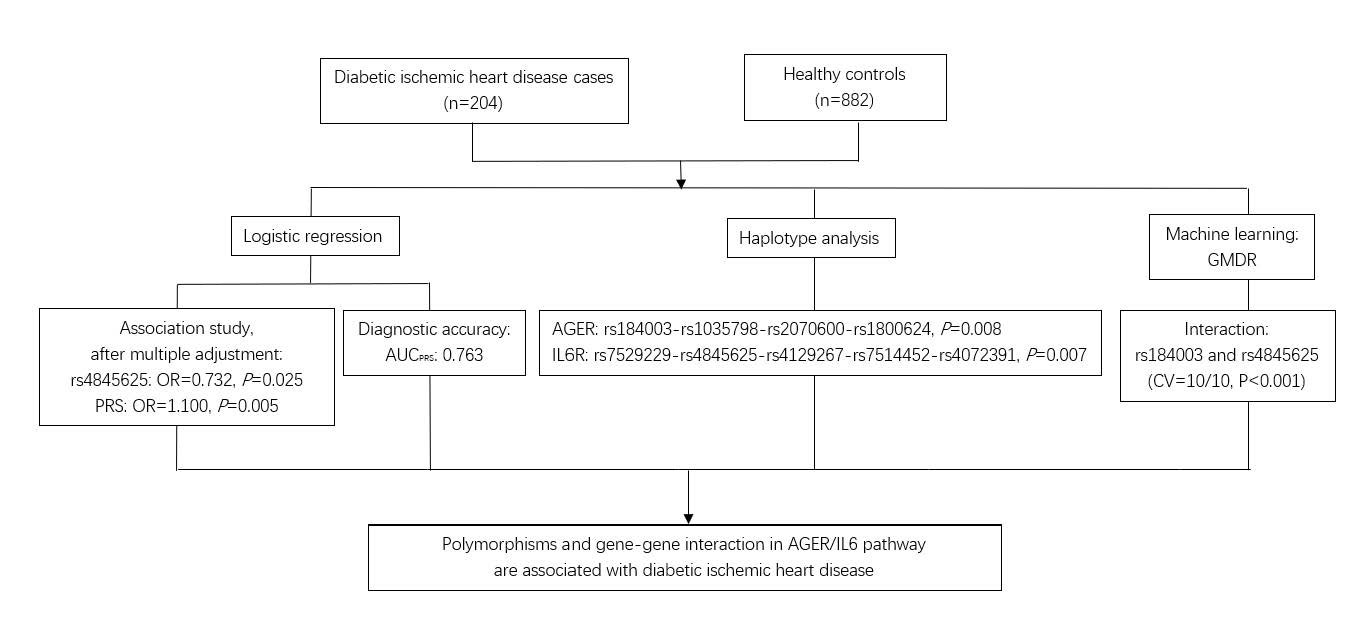
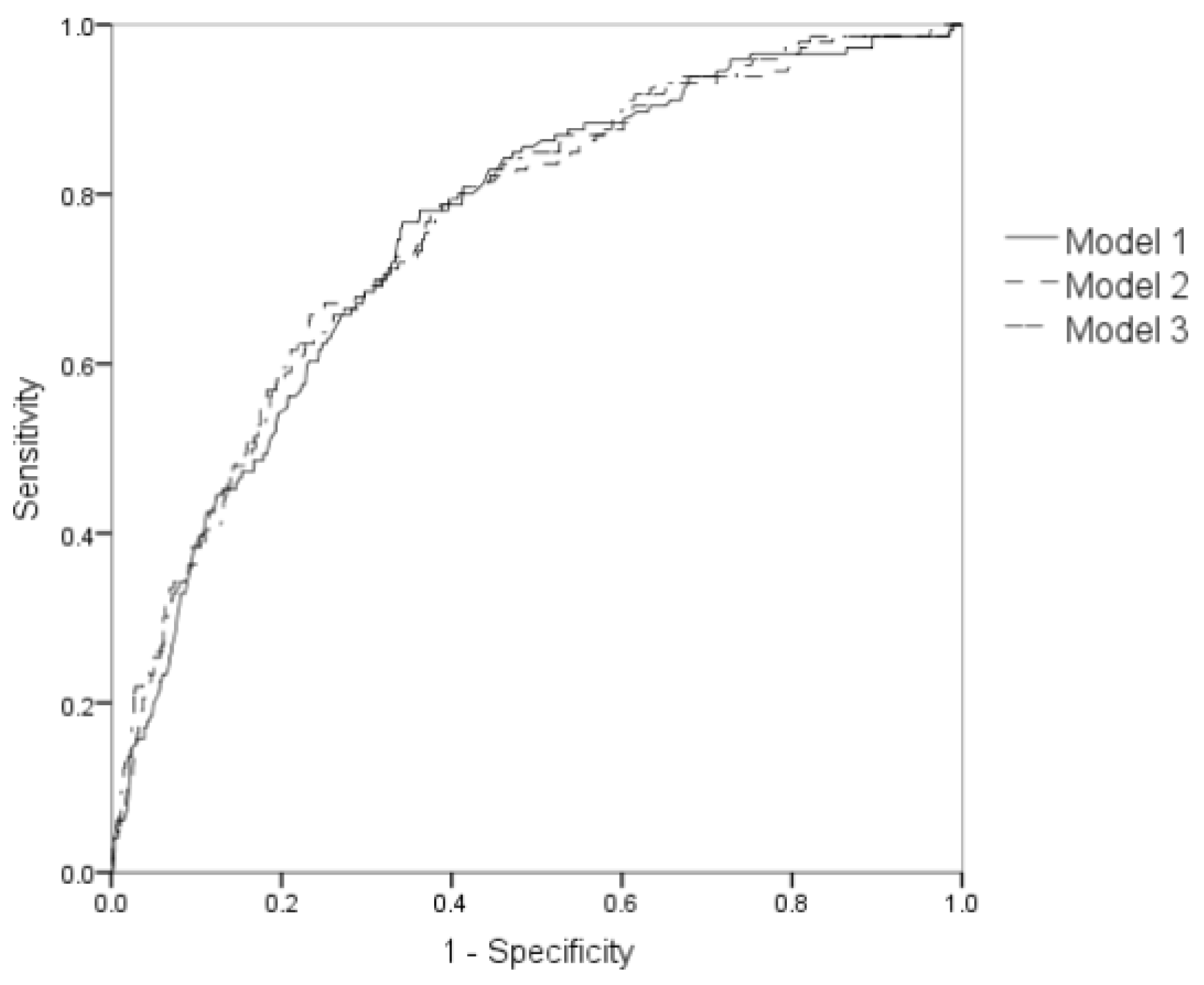
3.2. Association of AGER and IL6R Polymorphisms with Diabetic Ischemic Heart Disease

All polymorphisms were in Hardy-Weinberg equilibrium (all p-values were greater than 0.05). For AGER rs184003, participants with the GT and TT genotypes had a significantly higher risk of diabetic ischemic heart disease than those with the CC genotype (OR = 1.435, p = 0.039; OR = 2.525, p = 0.030, respectively). The T allele was associated with an increased risk of diabetic ischemic heart disease by 50% in additive and dominant models (p = 0.005; p = 0.012, respectively). For AGER rs2070600, the T allele was associated with about a 30% lower risk of diabetic ischemic heart disease in the additive and dominant models (p = 0.025; p = 0.030, respectively). However, after adjusting for potential confounders, the association between the above two SNPs and disease was null. The details are shown in Table 2.
For IL6R rs4845625, participants with the CT and TT genotypes had a significantly lower risk of diabetic ischemic heart disease than those with the CC genotype (OR = 0.692, p = 0.045; OR = 0.503, p = 0.003, respectively). The T allele significantly decreased the risk of diabetic ischemic heart disease in additive and dominant models (OR = 0.707, p = 0.003; OR = 0.632, p = 0.008, respectively). The association between rs4845625 and disease was still significant after adjusting for potential confounders. The details are shown in Table 2. The association between other SNPs and disease were shown in Supplementary Materials Table S3. The polygenic risk score was also associated with an increased risk of diabetic ischemic heart disease by 10% (OR = 1.101, 95% CI: 1.042–1.162, p = 0.001). After adjusting for dyslipidemia, hypertension, smoking, and drinking status, PRS was consistently associated with the disease (OR = 1.100, 95% CI: 1.029–1.176, p = 0.005).
Compared with models containing only traditional risk factors (AUC = 0.756; 95% CI: 0.714–0.798; p < 0.001), the diagnostic accuracy of models containing traditional risk factors together with IL6R and AGER polymorphisms (AUC = 0.759; 95% CI: 0.716–0.801; p < 0.001) and traditional risk factors together with PRS (AUC = 0.763; 95% CI: 0.72–0.80; p < 0.001) had slightly higher diagnostic accuracies. However, models containing genetic markers did not improve the diagnostic accuracy significantly (p > 0.05). The details were shown in Figure 1.

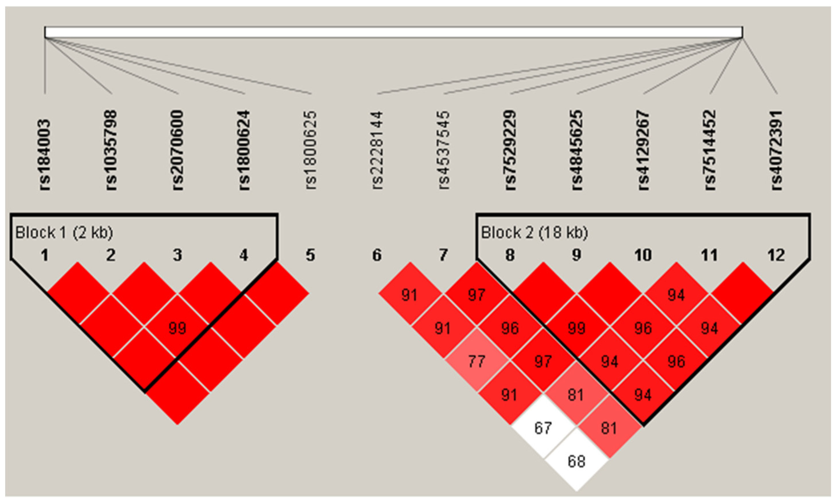
3.3. Association between Haplotypes and Diabetic Ischemic Heart Disease

Four out of five SNPs in the AGER gene (Block 1: rs184003-rs1035798-rs2070600-rs1800624) and five out of seven SNPs in the IL6R gene (Block 2: rs7529229-rs4845625-rs4129267-rs7514452-rs4072391) showed linkage disequilibrium (see Figure 2). These two blocks were both significantly associated with diabetic ischemic heart disease (Block 1: p = 0.008; Block 2: p = 0.007). Four haplotypes were constructed in block 1, and two of them were associated with diabetic ischemic heart disease (C-G-T-A: p = 0.018; A-G-C-A: p = 0.004). Four haplotypes were constructed in block 2, and two of them were associated with diabetic ischemic heart disease (T-C-C-T-C: p = 0.033; T-T-C-T-C: p = 0.001). The details of the haplotype analysis are shown in Table 3.

3.4. The Effect of Gene-Gene Interactions on Diabetic Ischemic Heart Disease

GMDR analysis was performed to assess the effect of gene-gene interactions on diabetic ischemic heart disease risk after adjustment for dyslipidemia, hypertension, smoking, and drinking. The GMDR analysis suggested that rs184003 in the AGER gene and rs4845625 in the IL6R gene were the best models in terms of statistical significance after permutation testing (p = 0.001). The two-locus models had a cross-validation consistency of 10/10 and a testing accuracy of 0.597. Logistic regression was subsequently used to obtain the odds ratios (ORs) and 95% confidence intervals (CIs) for the interaction between rs184003 and rs4845625. In the additive model, the joint effect of rs184003 and rs4845625 was associated with an increased risk of diabetic ischemic heart disease by 38% (OR = 1.38, 95% CI: 1.13–1.69, p = 0.002). The GeneMANIA was subsequently used to construct a gene network and predict gene function. As shown in Figure 3, IL6R and AGER have physical interactions with each other.

3.5. Sensitivity Analysis and Subgroup Analysis

The results of sensitivity analysis showed that both AGER and IL6R polymorphisms were still significantly associated with disease after the adjustment of blood glucose or other potential confounding factors. The interaction between rs4845625 and rs184003 turned to be null after the adjustment of FPG (OR = 1.166, 95% CI: 0.893–1.521, p = 0.259). Details were shown in Supplementary Materials Table S4.
Subgroup analysis was performed by considering FPG and ICVD risk scores. The results of subgroup analysis showed that rs184003 was significantly associated with disease in higher FPG subgroup (OR = 0.496, 95% CI: 0.288–0.856, p = 0.012), and rs4845625 was significantly associated with diabetic IHD in normal ICVD risk score group (OR = 0.389, 95% CI: 0.197–0.768, p = 0.007). Details were shown in Supplementary Materials Table S5. Subsequently, we found that AGER and IL6R polymorphisms were not associated with FPG and ICVD risk score, see Supplementary Materials Table S6.

4. Discussion

Individuals with T2DM have an increased risk of CVD, which cannot be fully explained by elevated glucose [28]. Genetic risk factors contribute greatly to the pathogenesis of diabetic macrovascular complications, but their role has not yet been fully illustrated. In the present community-based case-control study, rs4845625 in the IL6R gene and the interaction of rs184003 in the AGER gene and rs4845625 in IL6R were significantly associated with diabetic ischemic heart disease. The polygenic risk score calculated by summing the number of risk alleles of the SNPs located in the above two genes was also associated with an elevated risk of diabetic ischemic heart disease.
AGER is a multiligand cell surface receptor. Advanced glycation end products (AGEs), which are produced after high glucose exposure, can bind to AGER. Their interaction has been implicated in the pathogenesis of atherosclerosis. In addition, HMGB1 (high-mobility group protein 1) and neutrophil-derived S100 calcium-binding family members (S100A8/A9/A11/A12 and S100B) are ligands of AGER. After ligand binding, pro-inflammatory and pro-coagulant pathways are activated. Rs2070600 was found to be significantly associated with diabetic ischemic heart disease in the current study. However, after adjustments for covariates, the associations became null. Rs2070600 is located in the ligand-binding V domain of the AGER gene, often referred to as Gly82Ser [29]. Genome-wide association studies (GWAS) showed that rs2070600 was strongly and dose-dependently correlated with sRAGE levels in whites and blacks from the Atherosclerosis Risk in Communities Study and a Chinese population [30,31]. Interestingly, although soluble RAGE levels were found to be associated with diabetic complications in many studies, the association between rs2070600 and ischemic heart disease or other diabetic complications was not consistent. In the Atherosclerosis Risk in Communities Study, rs2070600 was not significantly associated with incident coronary heart disease or diabetes in either whites or blacks with a median follow-up of 20 years [30]. Chinese researchers [32] found a significant association between rs2070600 and coronary arterial disease in 175 cases and 170 controls. A meta-analysis found that the discrepancy may be attributable to ethnicity, and subjects with the rs2070600 risk allele were at higher risk of coronary arterial disease (CAD) in the Chinese population than in the non-Chinese population [11]. However, our study found that the association between rs2070600 and diabetic ischemic heart disease was null. Another study also found that rs2070600 was associated with the circulating levels of esRAGE but not with CAD in Chinese patients with T2DM [33]. These results might indicate that the association between rs2070600 and CAD may also be different in the general population and T2DM patients.
Only a few studies have demonstrated the association between rs184003 and ischemic heart disease. In the current study, we also found that the haplotypes C-G-T-A and A-G-C-A in the AGER gene (rs184003-rs1035798-rs2070600-rs1800624) were significantly associated with diabetic ischemic heart disease. Additionally, the association between haplotypes and potential confounders were null (Supplementary Materials Table S7). A hospital-based case-control study involving 1142 patients diagnosed with CAD and 1106 age- and sex-matched controls in a Chinese population was better powered and designed. In this study, the T allele in rs184003 and haplotypes in the AGER gene (rs1800625-rs1800624-rs2070600-rs184003, C-T-G-G and T-A-G-T) were also found to be associated with an increased risk of CAD [34]. In addition to the significant association with CAD, the result of a meta-analysis also showed that the homogeneity of the rs184003 polymorphism with the T allele conferred an increased risk of diabetes mellitus in East Asians (OR = 1.21; 95% CI: 1.04–1.40; I2 = 0) [35]. A previous study conducted in a Chinese population involving 200 gastric cancer patients and 207 cancer-free controls showed that subjects carrying the rs184003 T variant allele had an increased ability to produce soluble RAGE (sRAGE) [36]. Given that the T allele in rs184003 was associated with a higher risk of both diabetes and CAD, sRAGE might act on the common pathogenetic pathways of cardiovascular and metabolic diseases. Soluble RAGE levels were found to be significantly associated with CAD and diabetes [37,38,39] in many studies, and the association between haplotypes in the AGER gene and diabetic ischemic heart disease in the current study indicated that sRAGE levels could serve as a marker of diabetic ischemic disease. A recent review demonstrated that RAGE signaling contributed to vascular calcification in diabetic and nondiabetic subjects, presumably on account of the generation of RAGE ligands such as AGEs and other proinflammatory/pro-oxidative ligands [40]. The current study also found that the level of AGEs was elevated in the diabetic ischemic disease group, which supports the above hypothesis. To our knowledge, few studies have illustrated the relationship between rs184003 and diabetic macrovascular complications. Although the findings suggest potential benefits with RAGE antagonism both in the causes and consequences of diabetes and its macrovascular complications, more research is still needed to validate our results.
Mendelian randomization analysis illustrated that IL6R signaling might have a causal role in the development of coronary heart disease [12]. A previous meta-analysis demonstrated that the C allele in rs7529229 IL6R was associated with a lower risk of coronary heart disease [12,41]. Although the meta-analysis included a large sample size and better designed original studies, the populations of the studies were all Caucasian, so the evidence from Asian populations was still insufficient. In the current study, the association between rs7529229 and diabetic ischemic heart disease was null in the Chinese population. Chen et al. also did not find a significant effect of rs7529229 on coronary stenosis or acute myocardial infarction in the Chinese Han population with a sample size of 187 patients and 231 controls [42]. However, according to the sample size, allele frequency, and OR reported in the above study, the statistical power was relatively low and might lead to false negative results. Thus, studies with larger sample sizes are still needed to replicate the above findings. Likewise, He et al. conducted a hospital-based case-only study in 402 patients with left main coronary artery disease (LMCAD) and 804 patients with more peripheral coronary artery disease (MPCAD) in a Chinese population, and the results showed that rs7529229 CC or TC/CC genotypes were associated with an increased risk of LMCAD compared with MPCAD [43]. The haplotype T-T-C-T-C (rs7529229-rs4845625-rs4129267-rs7514452-rs4072391) in the IL6R gene and rs4845625 were associated with diabetic ischemic heart disease in our study, and the association held after adjusting for potential confounders. In addition, haplotypes T-T-C-C-T were significantly associated with TC (Supplementary Materials Table S8). Rs4845625 was found to be significantly associated with hypertriglyceridemia in the Japanese population [44], and the T allele was associated with a lower serum concentration of creatinine and increased EGFR [45]. Hypertriglyceridemia and chronic kidney disease (CKD) have common pathways, such as endothelial dysfunction, dyslipidemia, and inflammation, leading to metabolic cardiovascular disease [20]. Although few studies have focused on the association between rs4845625 and diabetic heart disease, its association with triglycerides and kidney function might indicate the potential mechanisms of rs4645625 in diabetic ischemic heart disease.
In response to hyperglycemia, AGER is activated by S100A8/A9 on hepatic Kupffer cells, leading to the secretion of IL-6. IL-6 subsequently binds to its receptor (IL6R) on hepatocytes to enhance the production of thrombopoietin, thereby regulating platelet production and resulting in diabetes-induced thrombocytosis [46]. In the current study, we found that the gene-gene interactions between AGER and IL6R increased the risk of diabetic ischemic heart disease. We subsequently used GeneMANIA to construct a gene network and predict gene function. IL6R and AGER have physical interactions with each other, and several pathways, including NF-kB/RelA and JAK/STAT, are involved in these interactions (Figure 3). The function of AGER and IL6R polymorphisms were listed in Supplementary Materials Table S9. These interactions illustrated that the interaction of SNPs in IL6R and AGER was not only a statistical interaction but also a biological interaction. To our knowledge, this is the first study aimed at identifying the interaction of the AGER and IL6R genes, and our results provide genetic evidence on the physiological mechanism of diabetic macrovascular complications. Whether the main effect and gene-gene interactions in these two genes could be used to predict the risk of diabetic macrovascular complications still needs to be validated by cohort studies in the future. Although we found a significant interaction between the AGER gene and the IL6R gene, the association between circulating IL-6 and diabetic ischemic heart disease was null. This result indicated that IL6R polymorphisms still need to be further demonstrated. The most common hypothesis is that IL-6 in hematopoietic cells, but not circulating IL-6, were more likely to affect TPO production and macrovascular complications [46,47].
The results of the sensitivity analysis showed that both AGER and IL6R polymorphisms were still significantly associated with disease after the adjustment of different risk factors, respectively. In the current study, SBP, TC, and LDLC levels and the proportion of people with smoking and drinking habits were significantly lower in the cases than in the controls, which is not consistent with other studies. According to the “2017 Guidelines for the prevention and treatment of type 2 diabetes in China”, diabetes patients have more stringent standards on blood pressure (BP) and blood lipids than the healthy population, and diabetes patients with ischemic heart disease should quit smoking and drinking [48]. Diabetes patients might change their lifestyles and medication to maintain their BP or blood lipids at a lower level. Due to the case-control study design of the current study, we were not able to collect lifestyle risk factors and blood samples before the incidence of diabetic ischemic heart disease. However, the percentages of SBP, DBP, HDLC, LDLC, TG, and TC in the ideal range were significantly higher in the control group than in the case group (p < 0.001, Supplementary Materials Table S2). Due to the above limitation of our study, more longitudinal studies are still needed to demonstrate whether genetic variants will increase the incidence of diabetic macrovascular complications. In addition, we did not collect any information about diabetic ketoacidosis in the current study. Since diabetic ketoacidosis predisposes individuals to ischemic heart disease, our study might induce bias to a certain extent. Moreover, medication information was not included in the investigation. Given that some antidiabetic medications, such as SGLT-2 inhibitors [49], will reduce the risk of ischemic heart disease in diabetes patients, future studies considering antidiabetic medication are still needed to validate the genetic effect on diabetic macrovascular complications. Since we did not recruit participants who only have diabetes or only have ICH, we performed subgroup analysis by considering FPG and ICVD risk scores. The ICVD risk scores were simple used as a tool to reflect the cumulative risk factors of ICVD in order to rule out the possibility of candidate SNPs that affect IHD. The subgroup analysis helps to rule out the possibility that diabetes or the ICVD risk score are confounders of the current study. Future research, including T2DM group without complications and ischemic heart disease (IHD) group without diabetes, would provide additional information in clarifying the roles of the AGER gene and IL6R gene.

5. Conclusions

Haplotypes in the AGER gene (C-G-T-A and A-G-C-A) were risk factors for diabetic ischemic disease, and rs4845625 and haplotypes in the IL6R gene (T allele and T-T-C-T-C) were associated with a lower risk of diabetic ischemic heart disease. The gene-gene interactions between rs184003 in AGER and rs4845625 in IL6R were associated with a higher risk of diabetic ischemic heart disease.

Supplementary Materials

The following supporting information can be downloaded at: https://www.mdpi.com/article/10.3390/jpm12030392/s1, Table S1: General information of the candidate SNPs and their primer sequences; Table S2: Recommendation cutoffs and proportions according to cutoffs in healthy people and ischemic heart disease patients with diabetes; Table S3: Associations of gene polymorphisms with the risk of diabetic cardiovascular disease; Table S4: Sensitivity analysis for the association between AGER and IL6R gene polymorphisms and diabetic ischemic heart disease; Table S5: Subgroup analysis for the association between candidate SNPs and diabetic IHD; Table S6: The association between candidate SNPs and FPG or ICVD risk scores; Table S7: The association of traits and haplotypes constituted by rs184003-rs1035798-rs2070600-rs1800624; Table S8: The association of traits and haplotypes constituted by rs7529229-rs4845625-rs4129267-rs7514452-rs4072391; Table S9: The function of AGER and IL6R polymorphisms. References [50,51,52,53,54,55,56,57,58,59,60,61,62,63,64,65,66] are cited in the supplementary materials.

Author Contributions

K.L. designed the study and wrote the manuscript, Y.X. analyzed data and visualized the interaction diagram. Q.Z. provided the statistical plan and helped to revise the manuscript. W.P. contributed to the verification of diabetic ischemic heart diseases in case group. C.G. and J.Z. contributed to the management of blood sample and DNA extraction. L.Z. contributed to the collection of controls and participated in the study design. All authors have read and agreed to the published version of the manuscript.

Funding

This study was funded by National Science Foundation of China, grant number 81602908, and National key research and development program of China, grant number 2016YFC0900600/2016YFC0900603.

Institutional Review Board Statement

This study was approved by the Ethics Committee of Capital Medical University (No:2016SY24).

Informed Consent Statement

Informed consent was obtained from all subjects involved in the study.

Data Availability Statement

Research could contact communication author to access data.

Acknowledgments

The authors thank all the participants and community health workers for their participation in this research effort.

Conflicts of Interest

The authors declare no conflict of interest.

References

  1. Shah, A.D.; Langenberg, C.; Rapsomaniki, E.; Denaxas, S.; Pujades-Rodriguez, M.; Gale, C.P.; Deanfield, J.; Smeeth, L.; Timmis, A.; Hemingway, H. Type 2 diabetes and incidence of cardiovascular diseases: A cohort study in 1.9 million people. Lancet Diabetes Endocrinol. 2015, 3, 105–113. [Google Scholar] [CrossRef] [Green Version]
  2. GBD 2015 Mortality and Causes of Death Collaborators. Global, regional, and national life expectancy, all-cause mortality, and cause-specific mortality for 249 causes of death, 1980–2015: A systematic analysis for the Global Burden of Disease Study 2015. Lancet 2016, 388, 1459–1544. [Google Scholar] [CrossRef] [Green Version]
  3. Maugeri, N.; Malato, S.; Femia, E.A.; Pugliano, M.; Campana, L.; Lunghi, F.; Rovere-Querini, P.; Lussana, F.; Podda, G.; Cattaneo, M.; et al. Clearance of circulating activated platelets in polycythemia vera and essential thrombocythemia. Blood 2011, 118, 3359–3366. [Google Scholar] [CrossRef] [PubMed]
  4. Zegeye, M.M.; Lindkvist, M.; Falker, K.; Kumawat, A.K.; Paramel, G.; Grenegard, M.; Sirsjö, A.; Ljungberg, L.U. Activation of the JAK/STAT3 and PI3K/AKT pathways are crucial for IL-6 trans-signaling-mediated pro-inflammatory response in human vascular endothelial cells. Cell Commun. Signal. 2018, 16, 55. [Google Scholar] [CrossRef]
  5. Grozovsky, R.; Giannini, S.; Falet, H.; Hoffmeister, K.M. Novel mechanisms of platelet clearance and thrombopoietin regulation. Curr. Opin. Hematol. 2015, 22, 445–451. [Google Scholar] [CrossRef] [Green Version]
  6. Serveaux-Dancer, M.; Jabaudon, M.; Creveaux, I.; Belville, C.; Blondonnet, R.; Gross, C. Pathological Implications of Receptor for Advanced Glycation End-Product (AGER) Gene Polymorphism. Dis. Markers 2019, 2019, 2067353. [Google Scholar] [CrossRef] [Green Version]
  7. Li, J.; Cai, W.; Zhang, W.; Zhu, W.F.; Liu, Y.; Yue, L.X.; Zhu, L.Y.; Xiao, J.R.; Liu, J.Y.; Xu, J.X. Polymorphism 2184A/G in the AGER gene is not associated with diabetic retinopathy in Han Chinese patients with type 2 diabetes. J. Int. Med. Res. 2016, 44, 520–528. [Google Scholar] [CrossRef] [Green Version]
  8. Fan, W.Y.; Gu, H.; Yang, X.F.; She, C.Y.; Liu, X.P.; Liu, N.P. Association of candidate gene polymorphisms with diabetic retinopathy in Chinese patients with type 2 diabetes. Int. J. Ophthalmol. 2020, 13, 301–308. [Google Scholar] [CrossRef]
  9. Peng, F.; Hu, D.; Jia, N.; Li, X.; Li, Y.; Chu, S.; Zhu, D.; Shen, W.; Lin, J.; Niu, W. Association of four genetic polymorphisms of AGER and its circulating forms with coronary artery disease: A meta-analysis. PLoS ONE 2013, 8, e70834. [Google Scholar] [CrossRef]
  10. Lu, W.; Feng, B. The -374A allele of the RAGE gene as a potential protective factor for vascular complications in type 2 diabetes: A meta-analysis. Tohoku J. Exp. Med. 2010, 220, 291–297. [Google Scholar] [CrossRef] [Green Version]
  11. Ma, W.Q.; Qu, Q.R.; Zhao, Y.; Liu, N.F. Association of RAGE gene Gly82Ser polymorphism with coronary artery disease and ischemic stroke: A systematic review and meta-analysis. Medicine 2016, 95, e5593. [Google Scholar] [CrossRef] [PubMed]
  12. Interleukin-6 Receptor Mendelian Randomisation Analysis (IL6R MR) Consortium; Swerdlow, D.I.; Holmes, M.V.; Kuchenbaecker, K.B.; Engmann, J.E.; Shah, T.; Sofat, R.; Guo, Y.; Chung, C.; Peasey, A.; et al. The interleukin-6 receptor as a target for prevention of coronary heart disease: A mendelian randomisation analysis. Lancet 2012, 379, 1214–1224. [Google Scholar] [PubMed] [Green Version]
  13. Rafiq, S.; Melzer, D.; Weedon, M.N.; Lango, H.; Saxena, R.; Scott, L.J.; DIAGRAM Consortium; Palmer, C.N.; Morris, A.D.; McCarthy, M.I.; et al. Gene variants influencing measures of inflammation or predisposing to autoimmune and inflammatory diseases are not associated with the risk of type 2 diabetes. Diabetologia 2008, 51, 2205–2213. [Google Scholar] [CrossRef] [PubMed] [Green Version]
  14. Manolio, T.A.; Collins, F.S.; Cox, N.J.; Goldstein, D.B.; Hindorff, L.A.; Hunter, D.J.; McCarthy, M.I.; Ramos, E.M.; Cardon, L.R.; Chakravarti, A.; et al. Finding the missing heritability of complex diseases. Nature 2009, 461, 747–753. [Google Scholar] [CrossRef] [Green Version]
  15. Peng, D.Q.; Zhao, S.P.; Nie, S.; Li, J. Gene-gene interaction of PPARgamma and ApoE affects coronary heart disease risk. Int. J. Cardiol. 2003, 92, 257–263. [Google Scholar] [CrossRef]
  16. Carty, C.L.; Heagerty, P.; Heckbert, S.R.; Enquobahrie, D.A.; Jarvik, G.P.; Davis, S.; Tracy, R.P.; Reiner, A.P. Association of genetic variation in serum amyloid-A with cardiovascular disease and interactions with IL6, IL1RN, IL1beta and TNF genes in the Cardiovascular Health Study. J. Atheroscler. Thromb. 2009, 16, 419–430. [Google Scholar] [CrossRef] [Green Version]
  17. Carty, C.L.; Heagerty, P.; Heckbert, S.R.; Jarvik, G.P.; Lange, L.A.; Cushman, M.; Tracy, R.P.; Reiner, A.P. Interaction between fibrinogen and IL-6 genetic variants and associations with cardiovascular disease risk in the Cardiovascular Health Study. Ann. Hum. Genet. 2010, 74, 1–10. [Google Scholar] [CrossRef] [Green Version]
  18. Zhu, Z.; Tong, X.; Zhu, Z.; Liang, M.; Cui, W.; Su, K.; Li, M.D.; Zhu, J. Development of GMDR-GPU for gene-gene interaction analysis and its application to WTCCC GWAS data for type 2 diabetes. PLoS ONE 2013, 8, e61943. [Google Scholar] [CrossRef] [Green Version]
  19. Xu, H.M.; Xu, L.F.; Hou, T.T.; Luo, L.F.; Chen, G.B.; Sun, X.W.; Lou, X.Y. GMDR: Versatile Software for Detecting Gene-Gene and Gene-Environment Interactions Underlying Complex Traits. Curr. Genom. 2016, 17, 396–402. [Google Scholar] [CrossRef] [Green Version]
  20. Gajjala, P.R.; Sanati, M.; Jankowski, J. Cellular and Molecular Mechanisms of Chronic Kidney Disease with Diabetes Mellitus and Cardiovascular Diseases as Its Comorbidities. Front. Immunol. 2015, 6, 340. [Google Scholar] [CrossRef] [Green Version]
  21. American Diabetes Association. Diagnosis and classification of diabetes mellitus. Diabetes Care 2014, 37, S81–S90. [Google Scholar] [CrossRef] [PubMed] [Green Version]
  22. Thygesen, K.; Alpert, J.S.; Jaffe, A.S.; Chaitman, B.R.; Bax, J.J.; Morrow, D.A.; White, H.D.; Executive Group on behalf of the Joint European Society of Cardiology (ESC)/American College of Cardiology (ACC)/American Heart Association (AHA)/World Heart Federation (WHF) Task Force for the Universal Definition of Myocardial Infarction. Fourth Universal Definition of Myocardial Infarction. J. Am. Coll. Cardiol. 2018, 72, 2231–2264. [Google Scholar] [CrossRef]
  23. Luepker, R.V.; Apple, F.S.; Christenson, R.H.; Crow, R.S.; Fortmann, S.P.; Goff, D.; Goldberg, R.J.; Hand, M.M.; Jaffe, A.S.; Julian, D.G.; et al. Case definitions for acute coronary heart disease in epidemiology and clinical research studies: A statement from the AHA Council on Epidemiology and Prevention; AHA Statistics Committee; World Heart Federation Council on Epidemiology and Prevention; the European Society of Cardiology Working Group on Epidemiology and Prevention; Centers for Disease Control and Prevention; and the National Heart, Lung, and Blood Institute. Circulation 2003, 108, 2543–2549. [Google Scholar]
  24. Amsterdam, E.A.; Wenger, N.K.; Brindis, R.G.; Casey, D.E., Jr.; Ganiats, T.G.; Holmes, D.R., Jr.; Jaffe, A.S.; Jneid, H.; Kelly, R.F.; Kontos, M.C.; et al. 2014 AHA/ACC Guideline for the Management of Patients with Non-ST-Elevation Acute Coronary Syndromes: A report of the American College of Cardiology/American Heart Association Task Force on Practice Guidelines. J. Am. Coll. Cardiol. 2014, 64, e139–e228. [Google Scholar] [CrossRef] [PubMed] [Green Version]
  25. Liu, L.S. Joint committee for guideline revision. 2016 Chinese guidelines for the management of dyslipidemia in adults. J. Geriatr. Cardiol. 2018, 15, 1–29. [Google Scholar]
  26. Stevens, L.A.; Schmid, C.H.; Greene, T.; Zhang, Y.L.; Beck, G.J.; Froissart, M.; Hamm, L.L.; Lewis, J.B.; Mauer, M.; Navis, G.J.; et al. Comparative performance of the CKD Epidemiology Collaboration (CKD-EPI) and the Modification of Diet in Renal Disease (MDRD) Study equations for estimating GFR levels above 60 mL/min/1.73 m2. Am. J. Kidney Dis. 2010, 56, 486–495. [Google Scholar] [CrossRef] [PubMed] [Green Version]
  27. Zhang, M.; Huang, Z.J.; Li, Y.C.; Wang, L.M.; Jiang, Y.; Zhao, W.H. Prediction of 10-year risk for ischemic cardiovascular disease in adults aged ≥35 years in China. Zhonghua Liu Xing Bing Xue Za Zhi 2016, 37, 689–693. [Google Scholar] [PubMed]
  28. Kirkman, M.S.; Mahmud, H.; Korytkowski, M.T. Intensive Blood Glucose Control and Vascular Outcomes in Patients with Type 2 Diabetes Mellitus. Endocrinol. Metab. Clin. N. Am. 2018, 47, 81–96. [Google Scholar] [CrossRef] [PubMed]
  29. Lu, W.; Feng, B.; Xie, G.; Liu, F. Association of AGER gene G82S polymorphism with the severity of coronary artery disease in Chinese Han population. Clin. Endocrinol. 2011, 75, 470–474. [Google Scholar] [CrossRef]
  30. Maruthur, N.M.; Li, M.; Halushka, M.K.; Astor, B.C.; Pankow, J.S.; Boerwinkle, E.; Coresh, J.; Selvin, E.; Kao, W.H. Genetics of Plasma Soluble Receptor for Advanced Glycation End-Products and Cardiovascular Outcomes in a Community-based Population: Results from the Atherosclerosis Risk in Communities Study. PLoS ONE 2015, 10, e0128452. [Google Scholar]
  31. Lim, S.C.; Dorajoo, R.; Zhang, X.; Wang, L.; Ang, S.F.; Tan, C.S.H.; Yeoh, L.Y.; Ng, X.W.; Li, N.; Su, C.; et al. Genetic variants in the receptor for advanced glycation end products (RAGE) gene were associated with circulating soluble RAGE level but not with renal function among Asians with type 2 diabetes: A genome-wide association study. Nephrol. Dial. Transpl. 2017, 32, 1697–1704. [Google Scholar] [CrossRef] [PubMed] [Green Version]
  32. Gao, J.; Shao, Y.; Lai, W.; Ren, H.; Xu, D. Association of polymorphisms in the RAGE gene with serum CRP levels and coronary artery disease in the Chinese Han population. J. Hum. Genet. 2010, 55, 668–675. [Google Scholar] [CrossRef] [PubMed]
  33. Peng, W.H.; Lu, L.; Wang, L.J.; Yan, X.X.; Chen, Q.J.; Zhang, Q.; Zhang, R.Y.; Shen, W.F. RAGE gene polymorphisms are associated with circulating levels of endogenous secretory RAGE but not with coronary artery disease in Chinese patients with type 2 diabetes mellitus. Arch. Med. Res. 2009, 40, 393–398. [Google Scholar] [CrossRef] [PubMed]
  34. Yu, X.; Liu, J.; Zhu, H.; Xia, Y.; Gao, L.; Li, Z.; Jia, N.; Shen, W.; Yang, Y.; Niu, W. An interactive association of advanced glycation end-product receptor gene four common polymorphisms with coronary artery disease in northeastern Han Chinese. PLoS ONE 2013, 8, e76966. [Google Scholar]
  35. Niu, W.; Qi, Y.; Wu, Z.; Liu, Y.; Zhu, D.; Jin, W. A meta-analysis of receptor for advanced glycation end products gene: Four well-evaluated polymorphisms with diabetes mellitus. Mol. Cell Endocrinol. 2012, 358, 9–17. [Google Scholar] [CrossRef]
  36. Li, T.; Qin, W.; Liu, Y.; Li, S.; Qin, X.; Liu, Z. Effect of RAGE gene polymorphisms and circulating sRAGE levels on susceptibility to gastric cancer: A case-control study. Cancer Cell Int. 2017, 17, 19. [Google Scholar] [CrossRef] [Green Version]
  37. Ligthart, S.; Sedaghat, S.; Ikram, M.A.; Hofman, A.; Franco, O.H.; Dehghan, A. EN-RAGE: A novel inflammatory marker for incident coronary heart disease. Arterioscler. Thromb. Vasc. Biol. 2014, 34, 2695–2699. [Google Scholar] [CrossRef] [Green Version]
  38. Reichert, S.; Triebert, U.; Santos, A.N.; Hofmann, B.; Schaller, H.G.; Schlitt, A.; Schulz, S. Soluble form of receptor for advanced glycation end products and incidence of new cardiovascular events among patients with cardiovascular disease. Atherosclerosis 2017, 266, 234–239. [Google Scholar] [CrossRef]
  39. Heier, M.; Margeirsdottir, H.D.; Gaarder, M.; Stensæth, K.H.; Brunborg, C.; Torjesen, P.A.; Seljeflot, I.; Hanssen, K.F.; Dahl-Jørgensen, K. Soluble RAGE and atherosclerosis in youth with type 1 diabetes: A 5-year follow-up study. Cardiovasc. Diabetol. 2015, 14, 126. [Google Scholar] [CrossRef] [Green Version]
  40. Egaña-Gorroño, L.; López-Díez, R.; Yepuri, G.; Ramirez, L.S.; Reverdatto, S.; Gugger, P.F.; Shekhtman, A.; Ramasamy, R.; Schmidt, A.M. Receptor for Advanced Glycation End Products (RAGE) and Mechanisms and Therapeutic Opportunities in Diabetes and Cardiovascular Disease: Insights from Human Subjects and Animal Models. Front. Cardiovasc. Med. 2020, 7, 37. [Google Scholar] [CrossRef]
  41. IL6R Genetics Consortium Emerging Risk Factors Collaboration; Sarwar, N.; Butterworth, A.S.; Freitag, D.F.; Gregson, J.; Willeit, P.; Gorman, D.N.; Gao, P.; Saleheen, D.; Rendon, A.; et al. Interleukin-6 receptor pathways in coronary heart disease: A collaborative meta-analysis of 82 studies. Lancet 2012, 379, 1205–1213. [Google Scholar]
  42. Chen, Z.; Qian, Q.; Tang, C.; Ding, J.; Feng, Y.; Ma, G. Association of two variants in the interleukin-6 receptor gene and premature coronary heart disease in a Chinese Han population. Mol. Biol. Rep. 2013, 40, 1021–1026. [Google Scholar] [CrossRef] [PubMed]
  43. He, F.; Teng, X.; Gu, H.; Liu, H.; Zhou, Z.; Zhao, Y.; Hu, S.; Zheng, Z. Interleukin-6 receptor rs7529229 T/C polymorphism is associated with left main coronary artery disease phenotype in a Chinese population. Int. J. Mol. Sci. 2014, 15, 5623–5633. [Google Scholar] [CrossRef] [PubMed] [Green Version]
  44. Abe, S.; Tokoro, F.; Matsuoka, R.; Arai, M.; Noda, T.; Watanabe, S.; Horibe, H.; Fujimaki, T.; Oguri, M.; Kato, K.; et al. Association of genetic variants with dyslipidemia. Mol. Med. Rep. 2015, 12, 5429–5436. [Google Scholar] [CrossRef]
  45. Horibe, H.; Fujimaki, T.; Oguri, M.; Kato, K.; Matsuoka, R.; Abe, S.; Tokoro, F.; Arai, M.; Noda, T.; Watanabe, S.; et al. Association of a polymorphism of the interleukin 6 receptor gene with chronic kidney disease in Japanese individuals. Nephrology 2015, 20, 273–278. [Google Scholar] [CrossRef] [PubMed]
  46. Kraakman, M.J.; Lee, M.K.; Al-Sharea, A.; Dragoljevic, D.; Barrett, T.J.; Montenont, E.; Basu, D.; Heywood, S.; Kammoun, H.L.; Flynn, M.; et al. Neutrophil-derived S100 calcium-binding proteins A8/A9 promote reticulated thrombocytosis and atherogenesis in diabetes. J. Clin. Investig. 2017, 127, 2133–2147. [Google Scholar] [CrossRef] [PubMed] [Green Version]
  47. Qu, D.; Liu, J.; Lau, C.W.; Huang, Y. IL-6 in diabetes and cardiovascular complications. Br. J. Pharmacol. 2014, 171, 3595–3603. [Google Scholar] [CrossRef] [Green Version]
  48. Society, C.D. Guidelines for the prevention and control of type 2 diabetes in China (2017 Edition). Chin. J. Pract. Int. Med. 2018, 38, 292–344. [Google Scholar]
  49. Scheen, A.J. Cardiovascular Effects of New Oral Glucose-Lowering Agents: DPP-4 and SGLT-2 Inhibitors. Circ. Res. 2018, 122, 1439–1459. [Google Scholar] [CrossRef]
  50. Olsson, S.; Jood, K. Genetic variation in the receptor for advanced glycation end-products (RAGE) gene and ischaemic stroke. Eur. J. Neurol. 2013, 20, 991–993. [Google Scholar] [CrossRef]
  51. Wang, Z.T.; Wang, L.Y.; Wang, L.; Cheng, S.; Fan, R.; Zhou, J.; Zhong, J. Association between RAGE gene polymorphisms and ulcerative colitis susceptibility: A case-control study in a Chinese Han population. Genet. Mol. Res. 2015, 14, 19242. [Google Scholar] [CrossRef]
  52. Kang, P.; Tian, C.; Jia, C. Association of RAGE gene polymorphisms with type 2 diabetes mellitus, diabetic retinopathy and diabetic nephropathy. Gene 2012, 500, 1–9. [Google Scholar] [CrossRef] [PubMed]
  53. Niu, H.; Niu, W.; Yu, T.; Dong, F.; Huang, K.; Duan, R.; Qumu, S.; Lu, M.; Li, Y.; Yang, T.; et al. Association of RAGE gene multiple variants with the risk for COPD and asthma in northern Han Chinese. Aging 2019, 11, 3220–3237. [Google Scholar] [CrossRef] [PubMed]
  54. Wadén, J.M.; Dahlström, E.H.; Elonen, N.; Thorn, L.M.; Wadén, J.; Sandholm, N.; Forsblom, C.; Groop, P.H.; FinnDiane Study Group. Soluble receptor for AGE in diabetic nephropathy and its progression in Finnish individuals with type 1 diabetes. Diabetologia 2019, 62, 1268–1274. [Google Scholar] [CrossRef] [PubMed] [Green Version]
  55. Kim, D.H.; Yoo, S.D.; Chon, J.; Yun, D.H.; Kim, H.S.; Park, H.J.; Kim, S.K.; Chung, J.H.; Kang, J.K.; Lee, S.A. Interleukin-6 Receptor Polymorphisms Contribute to the Neurological Status of Korean Patients with Ischemic Stroke. J. Korean Med. Sci. 2016, 31, 430–434. [Google Scholar] [CrossRef] [PubMed] [Green Version]
  56. He, F.; Yang, R.; Li, X.Y.; Ye, C.; He, B.C.; Lin, T.; Xu, X.Q.; Zheng, L.L.; Luo, W.T.; Cai, L. Single nucleotide polymorphisms of the NF-κB and STAT3 signaling pathway genes predict lung cancer prognosis in a Chinese Han population. Cancer Genet. 2015, 208, 310–318. [Google Scholar] [CrossRef]
  57. Key, K.V.; Mudd-Martin, G.; Moser, D.K.; Rayens, M.K.; Morford, L.A. Inflammatory Genotype Moderates the Association Between Anxiety and Systemic Inflammation in Adults at Risk for Cardiovascular Disease. J. Cardiovasc. Nurs. 2022, 37, 64–72. [Google Scholar] [CrossRef]
  58. Arguinano, A.A.; Naderi, E.; Ndiaye, N.C.; Stathopoulou, M.; Dadé, S.; Alizadeh, B.; Visvikis-Siest, S. IL6R haplotype rs4845625*T/rs4537545*C is a risk factor for simultaneously high CRP, LDL and ApoB levels. Genes Immun. 2017, 18, 163–169. [Google Scholar] [CrossRef]
  59. Tabassum, R.; Mahendran, Y.; Dwivedi, O.P.; Chauhan, G.; Ghosh, S.; Marwaha, R.K.; Tandon, N.; Bharadwaj, D. Common variants of IL6, LEPR, and PBEF1 are associated with obesity in Indian children. Diabetes 2012, 61, 626–631. [Google Scholar] [CrossRef] [Green Version]
  60. Van Dongen, J.; Jansen, R.; Smit, D.; Hottenga, J.J.; Mbarek, H.; Willemsen, G.; Kluft, C.; Penninx, B.W.; Ferreira, M.A.; Boomsma, D.I.; et al. The contribution of the functional IL6R polymorphism rs2228145, eQTLs and other genome-wide SNPs to the heritability of plasma sIL-6R levels. Behav. Genet. 2014, 44, 368–382. [Google Scholar] [CrossRef]
  61. Walston, J.D.; Matteini, A.M.; Nievergelt, C.; Lange, L.A.; Fallin, D.M.; Barzilai, N.; Ziv, E.; Pawlikowska, L.; Kwok, P.; Cummings, S.R.; et al. Inflammation and stress-related candidate genes, plasma interleukin-6 levels, and longevity in older adults. Exp. Gerontol. 2009, 44, 350–355. [Google Scholar] [CrossRef] [PubMed] [Green Version]
  62. Naitza, S.; Porcu, E.; Steri, M.; Taub, D.D.; Mulas, A.; Xiao, X.; Strait, J.; Dei, M.; Lai, S.; Busonero, F.; et al. A genome-wide association scan on the levels of markers of inflammation in Sardinians reveals associations that underpin its complex regulation. PLoS Genet. 2012, 8, e1002480. [Google Scholar] [CrossRef] [PubMed]
  63. Rafiq, S.; Frayling, T.M.; Murray, A.; Hurst, A.; Stevens, K.; Weedon, M.N.; Henley, W.; Ferrucci, L.; Bandinelli, S.; Corsi, A.M.; et al. A common variant of the interleukin 6 receptor (IL-6r) gene increases IL-6r and IL-6 levels, without other inflammatory effects. Genes Immun. 2007, 8, 552–559. [Google Scholar] [CrossRef] [PubMed]
  64. Webb, T.R.; Erdmann, J.; Stirrups, K.E.; Stitziel, N.O.; Masca, N.G.; Jansen, H.; Kanoni, S.; Nelson, C.P.; Ferrario, P.G.; König, I.R.; et al. Systematic Evaluation of Pleiotropy Identifies 6 Further Loci Associated with Coronary Artery Disease. J. Am. Coll. Cardiol. 2017, 69, 823–836. [Google Scholar] [CrossRef]
  65. Christiansen, M.K.; Larsen, S.B.; Nyegaard, M.; Neergaard-Petersen, S.; Ajjan, R.; Würtz, M.; Grove, E.L.; Hvas, A.M.; Jensen, H.K.; Kristensen, S.D. Coronary artery disease-associated genetic variants and biomarkers of inflammation. PLoS ONE 2017, 12, e0180365. [Google Scholar]
  66. Gigante, B.; Strawbridge, R.J.; Velasquez, I.M.; Golabkesh, Z.; Silveira, A.; Goel, A.; Baldassarre, D.; Veglia, F.; Tremoli, E.; Clarke, R.; et al. Analysis of the role of interleukin 6 receptor haplotypes in the regulation of circulating levels of inflammatory biomarkers and risk of coronary heart disease. PLoS ONE 2015, 10, e0119980. [Google Scholar]
Figure 1. ROC curve of different statistical models. Models were built by logistic regression, variables contained in each model were as follows: Model 1: Age, sex, hyperlipidaemia, hypertension, smoking, and alcohol drinking behavior. Model 2: Variables in model 1 and rs184003 and rs4845625. Model 3: Variables in model 1 and PRS.
Jpm 12 00392 g001
Figure 2. Haplotypes in AGER gene and IL6R gene. Two haplotypes were identified by haploview software. AGER gene: rs184003-rs1035798-rs2070600-rs180062; IL6R gene: rs7529229-rs4845625-rs4129267-rs7514452-rs4072391.
Jpm 12 00392 g002
Figure 3. The gene network between AGER and IL6R. According to the gene network constructed by GeneMANIA, IL6R and AGER have physical interactions with each other.
Jpm 12 00392 g003
Table 1. Demographic and biochemical characteristics of the participants.
ControlsT2DM + IHDp Value
Age (years) 164.00 ± 11.2565 ± 11.000.126
Male (n, %)488 (55.3)119 (58.3)0.436
SBP (mmHg) 1136.00 (24.00)130.00 (19.50)<0.001 **
DBP (mmHg) 179.00 (14.00)80 (12.00)0.001 **
BMI (kg/m2) 125.64 (4.46)25.36 (3.53)0.578
TC (mmol/L) 15.12 (1.41)4.68 (1.53)<0.001 **
HDLC (mmol/L) 11.34 (0.48)1.22 (0.45)<0.001 **
LDLC (mmol/L) 13.02 (1.17)2.68 (1.04)<0.001 **
TG (mmol/L) 11.40 (0.90)2.35 (1.57)<0.001 **
FPG (mmol/L) 15.60 (0.93)7.07 (3.53)<0.001 **
Current smoking (n, %)194 (22.0)18 (8.8)<0.001 **
Current drinking (n, %)295 (33.5)32 (15.7)<0.001 **
AGEs (mmol/L) 131.05 (15.94)38.07 (16.82)<0.001 **
IL-6 (mmol/L) 1133.08 (49.32)136.83 (40.18)0.353
1 Variables, which have non-normal distribution, were displayed as median (interquartile range), and were tested by Mann-Whitney U test. T2DM: type 2 diabetes, IHD: ischemic heart disease, BMI: body mass index, SBP: systolic blood pressure, DBP: diastolic blood pressure, FPG: fasting plasma glucose, TG: triglyceride, TC: total cholesterol, LDL-C: low density lipoprotein cholesterol, HDL-C: high density lipoprotein cholesterol, AGEs: advanced glycation end products, IL-6: interleukin 6. ** p < 0.01.
Table 2. Associations of rs184003, rs2070600, and rs4845625 with the risk of diabetic cardiovascular disease.
GenotypeCrude OR 1 (95%CI)Crude p ValueAdjusted OR¤ (95%CI)Adjusted p Value
rs184003GGRefRefRefRef
GT1.435 (1.019, 2.020)0.039 *1.223 (0.797, 1.878)0.357
TT2.525 (1.092, 5.837)0.030 *1.651 (0.580, 4.702)0.348
additive1.491 (1.125, 1.976)0.005 **1.247 (0.880, 1.767)0.215
dominant1.518 (1.093, 2.017)0.012 *1.265 (0.839, 1.905)0.261
recessive2.282 (0.993, 5.241)0.0461.571 (0.555, 4.449)0.395
rs2070600CCRefRefRefRef
CT0.713 (0.496, 1.024)0.0670.843 (0.550, 1.294)0.435
TT0.536 (0.237, 1.211)0.1340.611 (0.206, 1.807)0.373
additive0.721 (0.542, 0.960)0.025 *0.819 (0.578, 1.162)0.264
dominant0.684 (0.485, 0.964)0.030 *1.399 (0.914, 2.140)0.122
recessive0.587 (0.261, 1.320)0.1982.204 (0.493, 9.851)0.301
rs4845625CCRefRefRefRef
CT0.692 (0.483, 0.991)0.045 *0.619 (0.398, 0.961)0.033
TT0.503 (0.318, 0.795)0.003 **0.542 (0.318, 0.924)0.025
additive0.707 (0.563, 0.888)0.003 **0.732 (0.557, 0.961)0.025
dominant0.632 (0.448, 0.889)0.008 **0.594 (0.392, 0.902)0.014
recessive0.644 (0.434, 0.955)0.0280.757 (0.481, 1.191)0.229
1 No variables were adjusted in logistic regression model ¤ Dyslipidemia, hypertension, smoking, and drinking were adjusted in the logistic regression model. Adjusted p-values shown in the table are adjusted only by covariates. * p < 0.05, ** p < 0.01.
Table 3. Haplotype analysis for blocks in AGER and IL6R genes.
HaplotypesF_U 1F_A¤Chi-SquareOR (95%CI)p Value
Block 1 2Omnibus test--11.750 0.008 **
C-A-C-T0.1620.1700.1621.049 (0.830, 1.327)0.687
C-G-T-A0.2020.1505.5750.743 (0.580, 0.951)0.018 *
A-G-C-A0.1400.1978.2291.407 (1.114, 1.777)0.004 **
C-G-C-A0.4970.4820.2470.970 (0.859, 1.094)0.620
Block 2 3Omnibus test--11.99 0.007 **
T-T-C-C-T0.0930.1000.2271.075 (0.798, 1.449)0.634
C-C-T-T-C0.3870.4312.6391.114 (0.978, 1.268)0.104
T-C-C-T-C0.0950.1314.5511.379 (1.026, 1.853)0.033 *
T-T-C-T-C0.4260.33810.320.793 (0.689, 0.914)0.001 **
1 F_U: minor allele frequency in controls; ¤ F_A: minor allele frequency in cases; 2 Block1: rs184003-rs1035798-rs2070600-rs1800624; 3 Block2: rs7529229-rs4845625-rs4129267-rs7514452-rs4072391; * p < 0.05; ** p < 0.01.
Publisher’s Note: MDPI stays neutral with regard to jurisdictional claims in published maps and institutional affiliations.

