Article

Risk Stratification of Local Flaps and Skin Grafting in Skin Cancer-Related Facial Reconstruction: A Retrospective Single-Center Study of 607 Patients

by
Frederik Penzien Wainer Mamsen
1,*,
Claes Hannibal Kiilerich
1,
Jørgen Hesselfeldt-Nielsen
1,
Iselin Saltvig
1,
Celine Lund-Nielsen Remvig
1,
Hannah Trøstrup
1,† and
Volker-Jürgen Schmidt
1,2,†
1
Department of Plastic Surgery and Breast Surgery, Zealand University Hospital (SUH), University of Copenhagen, 4000 Roskilde, Denmark
2
Department of Clinical Medicine, University of Copenhagen, 2200 Copenhagen, Denmark
*
Author to whom correspondence should be addressed.
These authors contributed equally to this work.
J. Pers. Med. 2022, 12(12), 2067; https://doi.org/10.3390/jpm12122067
Submission received: 17 October 2022 / Revised: 10 December 2022 / Accepted: 10 December 2022 / Published: 15 December 2022

Abstract

:
Background: Non-melanoma skin cancer (NMSC) takes up a substantial fraction of dermatological and plastic surgical outpatient visits and surgeries. NMSC develops as an accumulated exposure to UV light with the face most frequently diagnosed. Method: This retrospective study investigated the risk of complications in relation to full-thickness skin grafts (FTSG) or local flaps in 607 patients who underwent facial surgery and reconstruction at a high-volume center for facial cancer surgery at a tertiary university hospital. Results: Between 01.12.2017 and 30.11.2020, 304 patients received reconstructive flap surgery and 303 received FTSG following skin cancer removal in the face. Flap reconstruction was predominantly performed in the nasal region (78%, n = 237), whereas FTSG reconstruction was performed in the nasal (41,6%, n = 126), frontal (19.8%, n = 60), and temporal areas (19.8%, n = 60), respectively. Patients undergoing FTSGs had a significantly higher risk of hematoma (p = 0.003), partial necroses (p < 0.001), and total necroses (p < 0.001) compared to flap reconstruction. Age and sex increased the risk of major complications (hematoma, partial or total necrosis, wound dehiscence, or infection) for FTSG, revealing that men exhibited 3.72 times increased risk of major complications compared to women reconstructed with FTSG. A tumor size above 15 mm increased the risk of hematoma and necrosis significantly. In summary, local flaps for facial reconstruction after skin cancer provide lower complication rate compared with FTSGs, especially in elderly and/or male patients. The indication for FTSG should be considered critically if the patient’s tumor size and location allow for both procedures.

1. Introduction

Skin cancer diagnoses are frequent and predominantly affect light-skinned individuals. It is the leading type of cancer globally [1,2] and takes up around 50% of all diagnoses in dermatological outpatient clinics [3]. Malignant skin cancer divides into melanoma- and non-melanoma skin cancer (NMSC), with NMSC being the most frequent, especially in the head and neck region of the elderly. NMSC comprises multiple types but predominantly consists of basal cell carcinomas (BCC) and squamous cell carcinomas (SCC) [2].
Facial cancer surgery and reconstruction pose a significant surgical challenge. The four pillars of reconstructive oncology, namely, clearance, form, function, and patient satisfaction, must be considered to ensure the tumor removal with consideration of the aesthetics subunits [4]. The decision on which surgical reconstructive technique to use is challenging but can be mastered by a skilled and knowledgeable surgeon. Often, simple suture is preferred if the tumor is small or placed in an easy-to-excise location. For tumors deemed too large for simple suture either due to location, tumor size, or aesthetic outcome, the defect can be reconstructed using full-thickness skin graft (FTSG), local flap, split-thickness skin grafts, or distant flaps. The decision is not clear-cut but includes patient health and comorbidities, tumor location and size, published data based on surgical empiricism, etc. [2,5,6]. The surgeon must therefore consider every surgical treatment to ensure the lowest risk of complications with regard to the patient’s wishes for a satisfying outcome [4].
This present study compares FTSGs and local flaps as treatment options in terms of complications after surgical removal of facial NMSC. By dividing the face into seven regions, the study further investigates surgical preferences according to distinct facial regions and the impact of patient demographics on complications. Additionally, we propose data to guide the surgeon in choosing and managing the best reconstructive technique in the complex field of facial reconstruction.

2. Materials and Methods

The retrospective quality assurance and optimization study is approved by the local authorities (approval number REG-098-2020). The study complies with the patient data protection regulation in Denmark. Data was collected between 01.12.2021 and 25.04.2022 and included patients undergoing facial surgery for BCC and SCC from 01.12.2017 to 01.12.2020 at the Department of Plastic and Breast Surgery, Zealand University Hospital, Roskilde, Denmark. The follow-up period for complications was one year. Data was obtained digitally through Sundhedsplatformen by Epic to a prefabricated database in REDCap (Research Electronic Data Capture) by Vanderbilt University. The database includes three categories: demographics, surgery, and complications. All investigators were employed at Zealand University Hospital, Roskilde. All surgeries were performed by different plastic surgeons from resident to attending level at the department of Plastic Surgery. The university department is considered as a high-volume center for skin cancer treatment. Initial data collection included 1312 patients receiving five different surgical interventions: simple primary wound closure, secondary healing, flap reconstruction, FTSG, and split-thickness skin grafts. After reviewing patient records and removing duplicates and uncompleted records, the total number of patients was 1283. The aimed patient population presented in this study is a group of 607 patients undergoing autologous flap- and FTSG. Reconstructions using a combination of flap and FTSG were excluded.
Data were collected uniformly by clearly written definitions of measurements susceptible to interpretation, such as definitions of comorbidities and complications (Table 1). Instructions in data collection and transfer were given both in writing and orally to the data collectors. Due to sensitive data, the records are not made public. Data was transferred from REDCap to R-studio for analysis. Data were analyzed using logistic regression for combinations of categoric and numeric predictors with categoric outcome variables. The Chi-square test was used to analyze categoric outcome variables with categoric predictors. An odds ratio was performed to analyze the association between risk factors and complications. A p-value of 0.05 or less was considered statistically significant, and a p-value of less than 0.001 was highly significant.
Definition of data item. The table shows short definitions of data susceptible to interpretation.

3. Results

3.1. Patient Demographics

The investigated group consisted of 607 patients surgically treated for BCC or SCC of the face, including 304 (49.9%) reconstructive flap surgeries and 303 (50.1%) FTSG procedures. Mean age at the time of surgery was 73.4 years (SD = 10.0) for flap and 76.7 years (SD = 10.1) for FTSGs reconstruction. Women were predominantly reconstructed using flaps (52.6%), and men were predominantly reconstructed by means of FTSG (53.1%). Mean body mass index (BMI) for both groups amounted 26.7 ± 4.8 for flap and 26.2 ± 4.9 for FTSG reconstruction. Comorbidities and group demographics are summarized in Table 2.
The groups carried most demographics without statistical differences. However, a few factors differed significantly. Patients undergoing FTSGs were significantly older, 76.7 years (SD = 10.1) vs. flaps 73.4 years (SD = 10.0, p =< 0.001), had significantly higher numbers of other cancers, 72 (23.8%) vs. 43 (14.1%, p = 0.003), and a higher fraction received blood thinners 148 (49%) vs. 129 (42%, p = 0.043).

3.2. Facial Distribution of Surgery

The study divided the face into seven distinct regions: frontal, temporal, periorbital, nasal, zygomatic/buccal, periocular, and mental. Flaps were distributed as follows: 5 in the frontal region (1.6%), 2 in the temporal region (0.7%), 15 in the periorbital region (4.9%), 25 around the cheeks (zygomatic region and buccal region, 8.2%), 237 in the nasal region (78.0%), 17 in the periocular region (5.6%) and 3 in the mental region (1.0%, see Figure 1a). The distribution of FTSG by region was: 60 in the frontal region (19.8%), 60 in the temporal region (19.8%), 31 in the periorbital region (10.2%), 22 around the cheeks (zygomatic region and buccal region) (7.3%), 126 in the nasal region (41.6%), 3 in the periocular region (1.0%) and 1 in the mental region (0.3%, see Figure 1b).
The most frequent site of surgery was the nasal region, (n = 363, 59.8%), where the defects were reconstructed approximately two-thirds as often with flaps (n = 237, 65.3%) as compared to FTSGs (n = 126, 34.7,3%). In the frontal (n = 60, 19.8%) and temporal regions (n = 60, 19.8%), FTSGs were used predominantly compared to flap surgery.

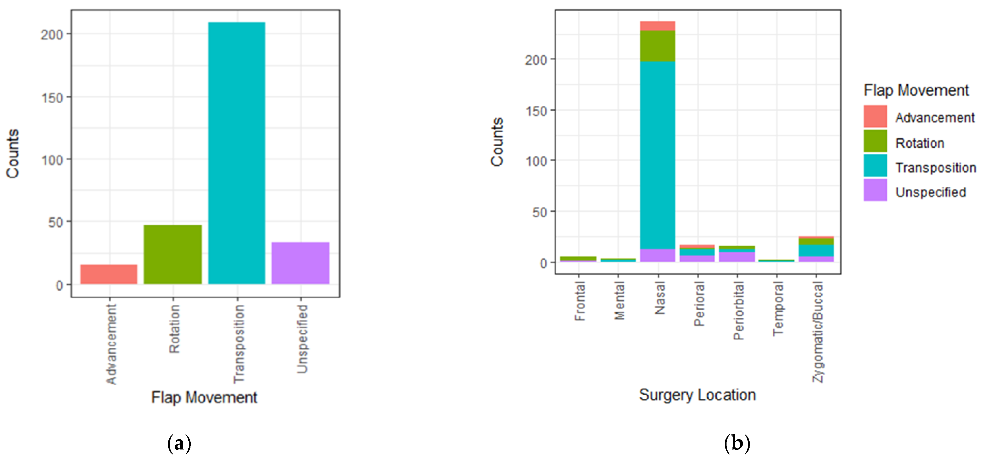
3.3. Facial Flap Variability

The variety of flaps consists of 13 different types utilized at least more than three times. Flap types performed less than three times were pooled in the “unspecified” category Table 3.
The 13 flap types were pooled based on tissue movement in rotation flaps, transposition flaps, and advancement flaps. Flaps without specification were designated to the group “unspecified flaps” (Figure 2a). Transposition flaps were the most frequent (n = 209, 69%), followed by rotational (n = 47, 15%), unspecified (n = 33, 11%), and advancement flaps (n = 15, 5%).
The most frequently used facial flaps were nasolabial flaps (n = 90, 29.6%), followed by bilobed flaps (n = 58, 19%), others (n = 37, 12.2%), rhomboid flaps (n = 35, 11.5%), and nasofrontal flaps (n = 25, 8.2%). Other specific flap types represented less than 5% of the combined flap surgeries. The facial distribution of flap types is presented in Figure 2b.

3.4. Complications

3.4.1. Flap Surgery and FTSG

Patients undergoing FTSGs had a significantly higher incidence of hematomas, (n = 32, 10.5%) compared to flaps (n = 13, 4.2%, p = 0.003). Necrosis occurred more often in the FTSG-group (n = 56, 18.5%) compared to local flaps (n = 11, 3.7%, p < 0.001). When subgrouping the degree of necrosis, edge necrosis was not significantly different between the groups p = 0.56), but partial necrosis (p < 0.001) and total necrosis (p < 0.001) were both highly significant, favoring flaps (see definition of complications in Table 1). Other complications investigated were not significantly different between groups (Table 4).
The FTSG group, presented in Table 4, received significantly more blood thinners than the flap group. A subgroup analysis of this patient group clarified that it was not the underlying cause of the increased hematomas of FTSGs as the fraction of hematomas among patients receiving blood thinners was 13.5% (n = 20) and 4% (n = 5) in flaps (p = 0.21) and therefore did not present with statistically more hematomas than for their entire group population. Nevertheless, the difference was still statistically significant, favoring flaps (p = 0.003). As hematoma under the local flap or skin graft can cause tension of the overlying tissue and, in the worst case, lead to necrosis [7], a correlation test was made to predict the causality of hematoma on necrosis. The analysis found that hematomas significantly increase the incidence of necrosis for both FTSGs and flaps (p < 0.001).

3.4.2. Flap Type-Associated Complications

The complication profile of the three different flap fashions, namely, transposition, rotation, and advancement, was investigated, but none of the flap designs was related to particularly increased complications (see Table 5).

3.4.3. Patient Age and Complications

Patients were grouped as ≤75 years (younger) and > 75 years (older). The younger group consisted of 320 individuals with a mean age of 66.8 ± 7.9 years. The older group consisted of 287 patients with a mean age of 82.4 ± 5.1 years. The older group had significantly lower BMI, 25.9 ± 7.7, than the older group, 27.1 ± 4.8 (p = 0.005); significantly more frequent hypertension, 66.2% vs. 44.9% (p < 0.001); significantly more other cancers, 22.5% vs. 15.0% (p = 0.022); significantly more heart diseases, 45.9% vs. 23.3% (p < 0.001); and received more blood thinners 57.7% vs. 33.4% (p < 0.001). In summary, the older group had significantly more comorbidities, but none of the investigated distinct complications was found to differ significantly, necrosis (p = 0.16), hematoma (p = 0.39), infection (p = 0.20), cellulitis (p = 1.00), wound dehiscence (p = 0.65). However, the odds ratio for major complications found that age significantly increase the risk of major complications for FTSGs, (OR =1.06 [CI 1.02, 1.11], p = 0.007) but not for local flaps (OR = 1.02 [CI 0.98, 1.06], p = 0.4). The risk of major complications increases with age, but the data did not provide a specific age cut off to define a high-risk group.

3.4.4. Facial Region and Complications

The seven defined facial regions, as depicted in Figure 1, were analyzed according distribution of either partial necrosis, total necrosis, or hematoma (see Table 6). Surgeries in the mental region were too few to be included in the analysis.
The risk of partial necrosis, total necrosis, or hematoma was highest in the temporal region (35.5%), followed by frontal region (26.1%), zygomatic/buccal region (19.2%), nasal region (13.0%), perioral region (10.0%) and lastly the periorbital region (8.7%). In the three regions with the most major complications, FTSG was performed predominantly (78.7% of the reconstructions). A total of 61% of surgeries were performed with flaps for the three regions with the fewest complications.

3.4.5. Complications Related to Patient Demographics and Comorbidities

Possible impact of patient demographics and comorbidities on major complications was investigated for flaps and FTSG (see Table 7). Comorbidity had no significant impact on the development of major complications in both reconstruction groups. For FTSG, male sex and age were associated with a significantly increased risk of major complications.

3.4.6. Tumor Size

The median tumor size of NMSCs amounted 10 mm (IQR = 7.0–17.0 mm, Table 5). FTSGs were applied to reconstruct significantly larger tumors (14.0 mm, IQR = 9.0–20.0 mm) compared to flaps (9.0 mm, IQR = 6.0–13.0 mm, p < 0.001). Data revealed that surgeons preferred flaps to reconstruct smaller defects in regions with low tissue mobility, such as the nasal region. Flaps were rarely preferred in areas with a better possibility of tissue recruitment compared to FTSGs (Table 8).
Tumor diameter was analyzed for its impact on complications. At tumor diameters > 15 mm, the risk of hematoma significantly increases from 6.0% to 12.5% (p = 0.01) for both FTSG and flaps. At tumor diameter > 20 mm, the risk of hematoma increases even further to 17.9% (p < 0.001). The risk of any kind of necrosis increases from 9.8% to 20.2% at tumor diameters > 20 mm (p = 0.014). No significant differences were found with increased tumor size for other investigated complications.

3.4.7. Level of Surgical Experience

The study investigates the outcome of being operated by board-certified plastic surgeons compared to a surgical trainee to uncover if the level of training may affect complication rates between FTSG and flaps. The analysis did not find any significant difference in any singular complication or the body encompassing major complications for FTSGs (p = 0.66) or flaps (p = 0.15). The tumor sizes of both groups (FTSGs and flaps) were equal for board-certified plastic surgeons and surgical trainees.

4. Discussion

This single-center study of 607 patients that received either local flaps or FTSG in order to restore skin cancer-related facial defects revealed that flaps were predominantly chosen to reconstruct defects in the nasal region, with a transpositional flap design most frequently used. In other facial regions, FTSGs were applied more often. The single-center study was conducted at a large high-volume center for plastic surgery, where all kinds of reconstructive procedures including advanced flap surgery were available at any time. At regions that tends to have better skin mobility and tissue recruitment, we argue that more significant defects that do not allow primary wound closure may be reconstructed with flaps instead of FTSGs, possibly lessening the patient’s risk of major complications according to the present results. Further, due to the support of subcutaneous tissue that provides better skin function and decreases the risk of aesthetically disturbing skin elevation changes, local flaps allow reconstructions with a good color match [7,8]. Supportively, a study by Schnabl et al., including over 1800 patients, found that patient-reported outcomes were significantly better for flaps than for FTSGs [9].
However, functional, aesthetical and quality-of-life-associated outcomes are not the main focus here. In the present study, we wanted to clearly figure out the risk for minor and major complications related to local flaps and FTSG used for facial reconstruction after skin cancer. According to the present data, the complication profile favors flaps over FTSGs in terms of major complications. From the perspective of the current literature, Leibovitch et al. investigated the rate of necrosis of FTSGs of the head, neck, genitals and upper limps in 2673 patients. They found that 3.7% had partial or total graft necrosis [10], though the study did not provide a definition of necrosis. However, it is approximately 10% less than in the present cohort and a cohort by Keh et al., where 13.6% of 128 FTSG reconstructions of the face developed graft necrosis of more than 60% reconstructed surface [11], resembling the data presented in this study. The most noticeable difference between the studies is the age difference in the patient populations. In the large study by Leibovitch, mean patient age was 65 years [10] compared to 75 years in the current study and 71 years in the study by Keh et al. [11]. None of the abovementioned studies investigated the impact of age on complications.
This study found that for FTSG (n = 303 patients), age significantly increased the risk of major complications, which may explain part of the difference between study complications. The patient-related factors found to increase the risk of major complications indicate that selecting patients for FTSG must be made with more caution than flaps.
The present data also highlights that men exhibited a 3.72 times increased risk for major complication compared to women. It is known that men tend to have higher risk of complications [12,13], which may explain why recommendations of postoperative bolus care may not have been met to the same degree for each gender resulting in increased complications. Although this hypothesis is highly speculative, it offers the local flap procedure more straight forward and less demanding postoperative care compared to FTSG.
The rate of partial or total necrosis in patients receiving local flaps was low (0.7%). Compared to a similar study by Rustemeyer et al., this appears to be very low [14]. One explanation might be that these procedures were solely performed in a plastic surgery high-volume center for facial reconstruction, providing skilled surgeons and an academic program for reconstruction. This might be a single-center bias.
The present findings do not favor any flap-type regarding complications.
FTSGs presented significantly more major complications in the studied cohort, among others [11,14] which caused additional or prolonged hospital visits and nursing hours [8] The wound dressing applied to FTSGs can also be challenging to manage by the patients or caretakers. On the other hand, certain flap surgeries demand multiple-step procedures, such as the median forehead flap (the Indian method). However, they represent a minor fraction of the total body of flaps used in the clinical praxis for facial reconstruction following skin cancer. The pre-operational preparation of flaps is considerable as each flap must be designed based on the precise tumor location [15], whereas FTSGs require less preparation and planning time. It requires plastic surgical experience to design and to choose the appropriate flap for at given or expected defect. Therefore, the organizational requirements and a certain case load are crucial to provide a facial reconstruction program that enables the best patient care and training by means of experienced reconstructive senior surgeons. The complication rates were equal when comparing less experienced and skilled surgeons in the present study. Similar findings have previously been reported in NMSC surgery of the face.
Hematomas significantly increased the risk of necrosis for FTSGs and flaps, especially for larger tumors. A fraction of minor hematomas may be underreported after flap reconstruction as it might be clinically invisible due to coverage of the “thicker” flap and can usually be handled with elevation and pressure. Patients receiving FTSGs are potentially more predisposed to hematoma-related reconstructive failure, as FTSGs demand diffusion to prevent necrosis and to enable take and healing. Thus, a hematoma layer can directly act as a diffusion-barrier [7].
Patients undergoing FTSG reconstruction always demand a post-operational control with bolus unwrapping and clinical evaluation.
There are several limitations of the present study. First, it is a single-center study that uses retrospective data. Second, the study did not assess the prognostic, aesthetic, functional or quality-of-life outcome after local flap and FTSG reconstruction, limiting the overall evaluation of the surgeries [9]. Our research group has initiated a prospective study to investigate patient-reported satisfaction and quality of life measurements to gather information on the patient’s surgical journey and self-assessed outcome. By means of this prospective approach, we expect to gain a better and fuller picture of the local flaps and FTSGs in facial reconstruction after skin cancer. Other studies investigating patient-reported outcomes after facial reconstruction suggest that flap reconstructions are associated with superior patient-reported aesthetic outcomes [8,14,16,17].
In conclusion, we still believe that reconstructive planning in patients suffering from skin cancer that demands reconstruction should be a highly individualized process that addresses the individual patient need and wishes in terms of personalized medicine. Data from 607 patients revealed that both local flaps and FTSGs are safe procedures that can be offered to patients with demand for facial reconstruction after cancer removal. Patients undergoing FTSGs for facial reconstruction have a higher risk of hematoma (p = 0.003) and partial- (p < 0.001) or total (p < 0.001) necrosis compared to flap reconstructions. With regard to risk stratification, the present study indicates that clinical recommendations for FTSGs should be made with caution especially in elderly patients and/or male patients due to a significant and distinctive higher risk for major complications. As expected, large tumors should be considered at high risk of hematoma and necrosis after reconstruction.

Author Contributions

Conceptualization, F.P.W.M., C.H.K., V.-J.S. and H.T.; methodology, F.P.W.M., C.H.K. and H.T.; software, C.H.K.; validation, F.P.W.M., C.H.K. and H.T.; formal analysis, F.P.W.M., C.H.K. and H.T.; investigation, F.P.W.M., C.H.K., J.H.-N., I.S., C.L.-N.R., V.-J.S. and H.T.; resources, C.H.K., I.S. and H.T.; writing—original draft preparation, F.P.W.M. and V.-J.S.; writing—review and editing, F.P.W.M., C.H.K., J.H.-N., I.S., C.L.-N.R., V.-J.S. and H.T.; visualization, I.S.; supervision, V.-J.S. and H.T.; project administration, F.P.W.M., C.H.K., V.-J.S. and H.T.; funding acquisition, V.-J.S. All authors have read and agreed to the published version of the manuscript.

Funding

This research received no external funding.

Institutional Review Board Statement

The study was conducted in accordance with the Declaration of Helsinki, and approved by the local authorities (approval number REG-098-2020).

Informed Consent Statement

Patient consent was waived due the study being a quality assessment study and no patients can be identified from the obtained data.

Acknowledgments

Thanks to the Department of Plastic Surgery management at Zealand University Hospital Roskilde for finding resources to conduct this study and establishing a scientific group to facilitate the development of scientific projects. A great appreciation is made to the data collection team of Zealand University Hospital Roskilde, Department of Plastic Surgery and Breast Surgery, including Caroline Skaarup, Christina Alexandersen, Huda Ateya, Kathrine Borres Jensen, Leeann Shariat, and Maria Snog. Thanks to Laura Anna Mamsen Wainer for drafting and drawing Figure 1.

Conflicts of Interest

The authors declare no conflict of interest. The funders had no role in the study’s design, in the collection, analyses, or interpretation of data, in the writing of the manuscript, or in the decision to publish the results.

References

  1. Lomas:, A.; Leonardi-Bee, J.; Bath-Hextall, F. A systematic review of worldwide incidence of nonmelanoma skin cancer. Br. J. Dermatol. 2012, 166, 1069–1080. [Google Scholar] [CrossRef] [PubMed]
  2. Badash, I.; Shauly, O.; Lui, C.G.; Gould, D.J.; Patel, K.M. Nonmelanoma Facial Skin Cancer: A Review of Diagnostic Strategies, Surgical Treatment, and Reconstructive Techniques. Clin. Med. Insights Ear Nose Throat 2019, 12, 117955061986527. [Google Scholar] [CrossRef] [PubMed]
  3. Martin-Gorgojo, A.; Descalzo-Gallego, M.Á.; Arias-Santiago, S.; Molina-Leyva, A.; Gilaberte, Y.; Fernández-Crehuet, P.; Husein-ElAhmed, H.; Viera-Ramírez, A.; Fernández-Peñas, P.; Taberner, R.; et al. What Proportion of the Caseload at Dermatology Outpatient Clinics in Spain Do Skin Tumors Account for? Results from the DIADERM National Random Sampling Project. Actas Dermo-Sifiliogr. (Engl. Ed. ) 2021, 112, 632–639. [Google Scholar] [CrossRef]
  4. Mahadevan, K.; Sruthi, S.; Sridevi, S.; Vivek, R. Fourth dimension in reconstruction of defects following excision of basal cell carcinoma of head and neck! J. Cutan. Aesthet. Surg. 2018, 11, 110. [Google Scholar] [CrossRef] [PubMed]
  5. Russo, F.; Linares, M.; Iglesias, M.E.; Martínez-Amo, J.L.; Cabo, F.; Tercedor, J.; Costa-Vieira, R.; Toledo-Pastrana, T.; Ródenas, J.M.; Leis, V. Reconstruction Techniques of Choice for the Facial Cosmetic Units. Actas Dermo-Sifiliogr. (Engl. Ed. ) 2017, 108, 729–737. [Google Scholar] [CrossRef]
  6. Eskiizmir, G.; Baker, S.; Cingi, C. Nonmelanoma Skin Cancer of the Head and Neck. Facial Plast. Surg. Clin. N. Am. 2012, 20, 493–513. [Google Scholar] [CrossRef] [PubMed]
  7. Ramsey, M.; Walker, B.; Patel, B. Full Thickness Skin Grafts; StatPearls Publishing: Treasure Island, FL, USA, 2022. [Google Scholar]
  8. Ebrahimi, A.; Ashayeri, M.; Rasouli, H. Comparison of local flaps and skin grafts to repair cheek skin defects. J. Cutan. Aesthet. Surg. 2015, 8, 92. [Google Scholar] [CrossRef] [PubMed]
  9. Schnabl, S.M.; Breuninger, H.; Iordanou, E.; Scheu, A.; Kofler, L.; Häfner, H.-M.; Eberle, F.C. Patient satisfaction in 1,827 patients following various methods of facial reconstruction based on age, defect size and site: Patient satisfaction after facial reconstruction. JDDG J. Dtsch. Dermatol. Ges. 2018, 16, 426–433. [Google Scholar] [CrossRef] [PubMed]
  10. Leibovitch, I.; Huilgol, S.C.; Richards, S.; Paver, R.; Selva, D. The Australian Mohs Database: Short-Term Recipient-Site Complications in Full-Thickness Skin Grafts. Derm. Surg 2006, 32, 1364–1368. [Google Scholar] [CrossRef]
  11. Keh, S.M.; Giblett, N.; Ahsan, S.F. Through-and-Through Mattress Suturing Versus Tie-Over Dressing in Full-Thickness Skin Graft Reconstruction. Turk. Arch. Otorhinolaryngol. 2017, 55, 119–124. [Google Scholar] [CrossRef] [PubMed]
  12. Aggarwal, P.; Knabel, P.; Fleischer, A.B. United States burden of melanoma and non-melanoma skin cancer from 1990 to 2019. J. Am. Acad. Dermatol. 2021, 85, 388–395. [Google Scholar] [CrossRef] [PubMed]
  13. Aghdassi, S.J.S.; Schröder, C.; Gastmeier, P. Gender-related risk factors for surgical site infections. Results from 10 years of surveillance in Germany. Antimicrob. Resist. Infect. Control 2019, 8, 95. [Google Scholar] [CrossRef] [PubMed] [Green Version]
  14. Rustemeyer, J.; Günther, L.; Bremerich, A. Complications after nasal skin repair with local flaps and full-thickness skin grafts and implications of patients’ contentment. Oral Maxillofac. Surg. 2009, 13, 15–19. [Google Scholar] [CrossRef] [PubMed]
  15. Erkul, E.; Patel, K.G.; Day, T. Surgical Planning for Resection and Reconstruction of Facial Cutaneous Malignancies. Int. J. Head Neck Surg. 2016, 7, 149–164. [Google Scholar] [CrossRef]
  16. Lee, K.S.; Kim, J.O.; Kim, N.G.; Lee, Y.J.; Park, Y.J.; Kim, J.S. A Comparison of the Local Flap and Skin Graft by Location of Face in Reconstruction after Resection of Facial Skin Cancer. Arch. Craniofac. Surg. 2017, 18, 255–260. [Google Scholar] [CrossRef] [Green Version]
  17. Yoon, S.; Kim, Y.; Choi, Y. Locoregional flaps versus skin grafts in the nose: Aesthetic considerations after cancer ablation. Arch. Aesthetic. Plast. Surg. 2022, 28, 31–35. [Google Scholar] [CrossRef]
Figure 1. Distribution of flaps and full thickness skin graft (FTSG) according to distinct facial units. (a) Distribution of flaps according to the facial region. (b) Distribution of FTSG by the facial regions.
Jpm 12 02067 g001
Figure 2. Distribution of flap types and facial regions. (a) Distribution of flap types and (b) flap design used according to distinct facial region.
Jpm 12 02067 g002
Table 1. Definition of terminology.
TermDefinition of the Collected Data
BMIBody mass index measured ≤30 days before surgery.
Autoimmune diseaseHas an autoimmune diagnosis code in their medical journal.
Including the following:
-
Inflammatory bowel disease
-
Musculoskeletal autoimmune disease
-
Skin autoimmune disease
-
Infectiously initiated autoimmune disease
-
Autoimmune organ failure
-
Combinatory autoimmune diseases
HypertensionHas the diagnosis code in their medical journal both well-regulated and unregulated?
Including the following:
-
White coat hypertension
-
Hypertension of unknown origin
-
Hypertensio arterialis essentialis
-
Hypertention secundary to other disease
-
Hypertensio renovascularis
-
Hypertensio arterialis by other renal diseases
-
Hypertensio arterialis by endocrine diseases
Other cancerHas a cancer diagnosis or previous cancer diagnosis, and still in follow-up, other than NMSC.
Heart diseaseHas a heart disease diagnosis code in their medical journal, both well treated and untreated.
Including following:
-
Ischemic heart diseases
-
Acute coronary syndrome
-
Acute myocardial infarction
-
Chronic ischemic heart diseases
-
Pulmonary heart diseases
-
Heart valve disease
-
Heart failure
-
Atherosclerosis
Blood thinnersTake blood thinners regularly. The patients are advised to pause their medication prior to surgery. However, comorbidities may have led to the patients continuing due to the risk of pause.
Alcoholic drinkOne substance contains 8 g of alcohol.
Previous smokerQuit smoking ≥30 days before the surgery.
Infection Clinically observed and required antibiotic treatment.
CellulitisClinically observed.
Minor hematomaNo surgical revision required.
Major hematomaRequired surgical revision.
Edge necrosis≤5% of the transplant or flap.
Partial necrosis>5% to 66% of the transplant or flap.
Total necrosis>66.6% of the transplant or flap.
Major complicationHematoma, partial necrosis, total necrosis, wound dehiscence, and infection.
Table 2. Patient demographics and comorbidities.
FTSG (n)Flap (n)p-Value
n303304
Age, (mean [a SD])76.7 (10.1)73.4 (10.0)<0.001
Sex = Male (%)161 (53.1)144 (47.4)0.168
BMI (mean [a SD])26.24 (4.9)26.74 (4.8)0.263
Autoimmune disease (%)31 (10.2)27 (8.9)0.584
Hypertension (%)171 (56.4)170 (55.9)0.935
Other cancer (%)72 (23.8)43 (14.1)0.003
Diabetes (%)40 (13.2)42 (13.8)0.906
Heart condition (%)107 (35.3)107 (35.2)1.000
Blood thinners (%) 0.043
  No155 (51.2)178 (58.6)
  Acetylic Acid48 (15.8)29 (9.5)
  Other blood thinners100 (33.0)97 (31.9)
b Alcohol (%) 0.736
  <7 units per week142 (46.9)154 (50.7)
  7–14 units per week 43 (14.2)41 (13.5)
  >14 units per week16 (5.3)18 (5.9)
  Not reported102 (33.7)91 (29.9)
Smoking (%) 0.950
  No127 (41.9)124 (40.8)
  Active smoker40 (13.2)39 (12.8)
  Former smoker56 (18.5)62 (20.4)
  Not reported80 (26.4)79 (26.0)
a SD = standard deviation, b alcohol, n = one drink containing 8 g of alcohol.
Table 3. Variety of utilized flaps.
Flap TypeNumber = n
Nasolabial90
Hatchet5
Frontonasal25
Unspecified transposition6
Paramedian Forehead 9
Random 3
Rhomboid 35
Unspecified rotation 14
Shark island3
Spear 3
Bilobed58
Trilobed4
V-Y12
Unspecified 37
Total304
Table 4. Complications related to FTSGs and local flaps.
ComplicationFTSGFlapsp-Value
Infection (%)25 (8.3)28 (9.2)0.774
Cellulitis (%)7 (2.3)5 (1.6)0.577
Hematoma (%) 0.003
  No271 (89.4)291 (95.7)
  Minor hematoma31 (10.2)12 (3.9)
  Major hematoma1 (0.3)1 (0.3)
Wound dehiscence (%)28 (9.2)20 (6.6)0.233
Necrosis (%) <0.001
  No247 (81.5)293 (96.4)
  Edge necrosis11 (3.6)9 (3.0)
  Partial necrosis22 (7.3)2 (0.7)
  Total necrosis23 (7.6)0 (0.0)
Table 5. Complications related to principle flap design.
AdvancementRotationTranspositionUnspecifiedp-Value
n154720933
Infection (%)1 (6.7)4 (8.5)20 (9.6)3 (9.1)1.000
Cellulitis (%)0 (0.0)0 (0.0)3 (1.4)2 (6.1)0.259
Hematoma (%) 0.436
  No15 (100.0)45 (95.7)200 (95.7)31 (93.9)
  Minor hematoma0 (0.0)2 (4.3)9 (4.3)1 (3.0)
  Major hematoma0 (0.0)0 (0.0)0 (0.0)1 (3.0)
Wound dehiscence (%)1 (6.7)2 (4.3)15 (7.2)2 (6.1)0.971
Necrosis (%) 0.893
  No15 (100.0)45 (95.7)200 (95.7)33 (100.0)
  Edge necrosis0 (0.0)2 (4.3)7 (3.3)0 (0.0)
  Partial necrosis0 (0.0)0 (0.0)2 (1.0)0 (0.0)
  Total necrosis0 (0.0)0 (0.0)0 (0.0)0 (0.0)
Table 6. Partial necrosis, total necrosis, or hematoma by facial region.
RegionFrontalMentalNasalPerioralPeriorbitalTemporalZygomatic/Buccal
n65-36320466247
Risk of partial necrosis, total necrosis, or hematoma26.1%-13%10.0%8.7%35.5%19.2%
Table 7. Impact of patient demographics on major complications.
CharacteristicFlapsFTSG
OR 195% CI 1p-ValueOR 195% CI 1p-Value
Age1.020.98, 1.060.401.061.02, 1.110.007
Sex
  Female
  Male1.330.60, 2.960.53.721.72, 8.430.001
BMI0.930.84, 1.020.120.960.88, 1.040.4
Autoimmune disease
  No
  Yes2.130.57, 7.200.21.900.60, 5.910.3
Hypertension
  No
  Yes0.570.24, 1.370.21.920.80, 4.820.2
Other cancer
  No
  Yes0.390.08, 1.420.20.870.36, 2.010.7
Diabetes
  No
  Yes2.380.80, 6.860.110.540.18, 1.540.3
Heart disease
  No
  Yes2.670.93, 7.970.0710.590.23, 1.460.3
Blood thinners
  ASA
  Others0.460.11, 2.190.30.800.26, 2.430.7
  No0.740.18, 3.580.70.610.18, 2.050.4
1 OR = Odds Ratio, CI = Confidence Interval.
Table 8. Tumor size.
FlapsFTSGTotal
Size of the tumors, mm (median, [IQR])9.0 [6.0, 13.0]14.0 [9.0, 20.0]10.0 [7.0, 17.0]
Publisher’s Note: MDPI stays neutral with regard to jurisdictional claims in published maps and institutional affiliations.

