
In Vitro Differentiation of Myoblast Cell Lines on Spider Silk Scaffolds in a Rotating Bioreactor for Vascular Tissue Engineering
Abstract
:1. Introduction
2. Methods
2.1. Silk Rearing, Scaffold Construction and Cell Seeding
2.2. Cell Culture
2.3. Bioreactor Construction and Setup
2.4. Live/Dead Assay
2.5. Scanning Electron Microscopy
2.6. Western Blot
2.7. qPCR
2.8. Tensile Strength Test
2.9. Statistical Analysis
3. Results
3.1. Scaffold Construction
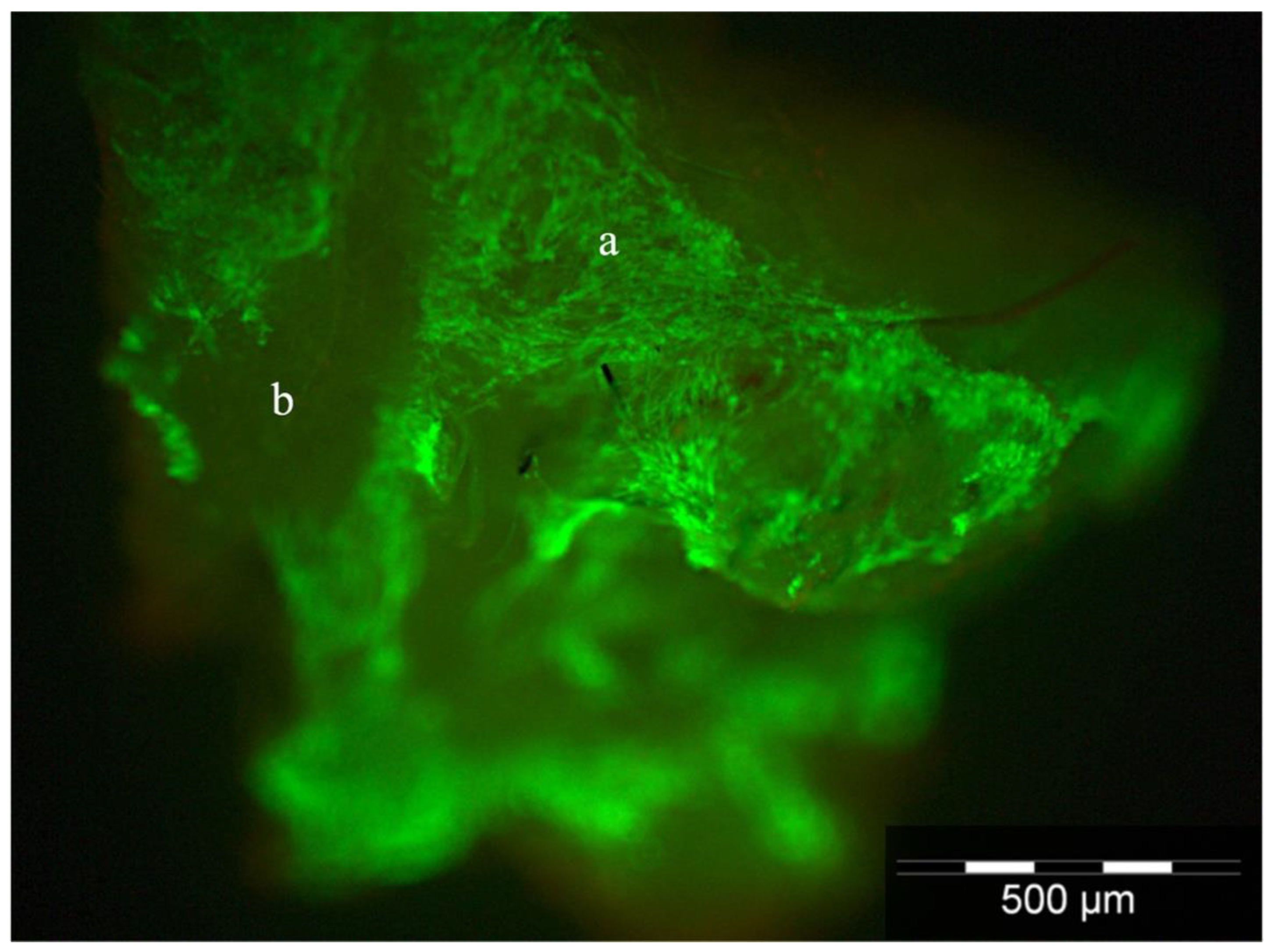
3.2. Cell Attachment and Viability
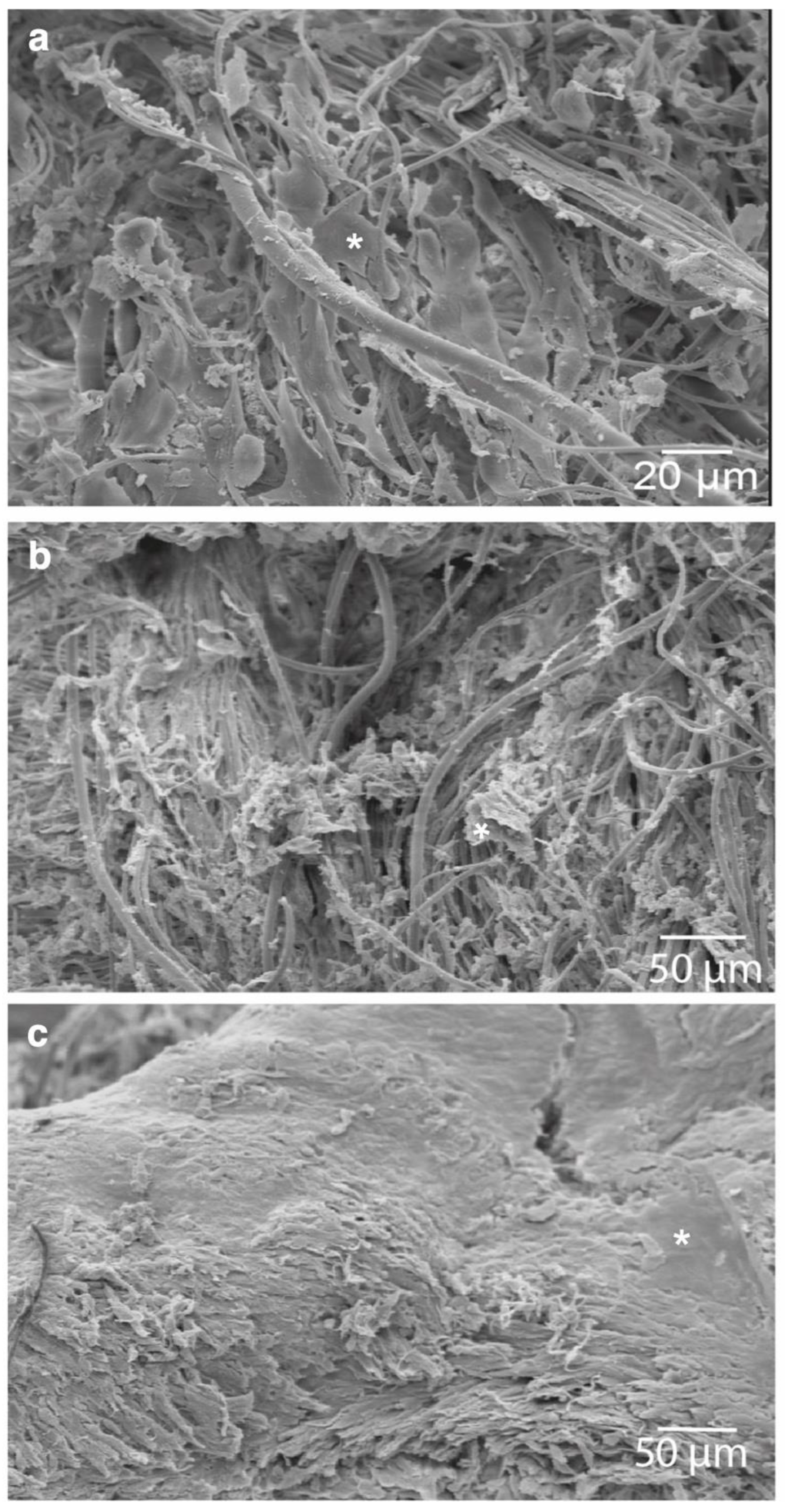
3.3. Scaffold Morphology and Cellular Properties
3.4. Western Blot
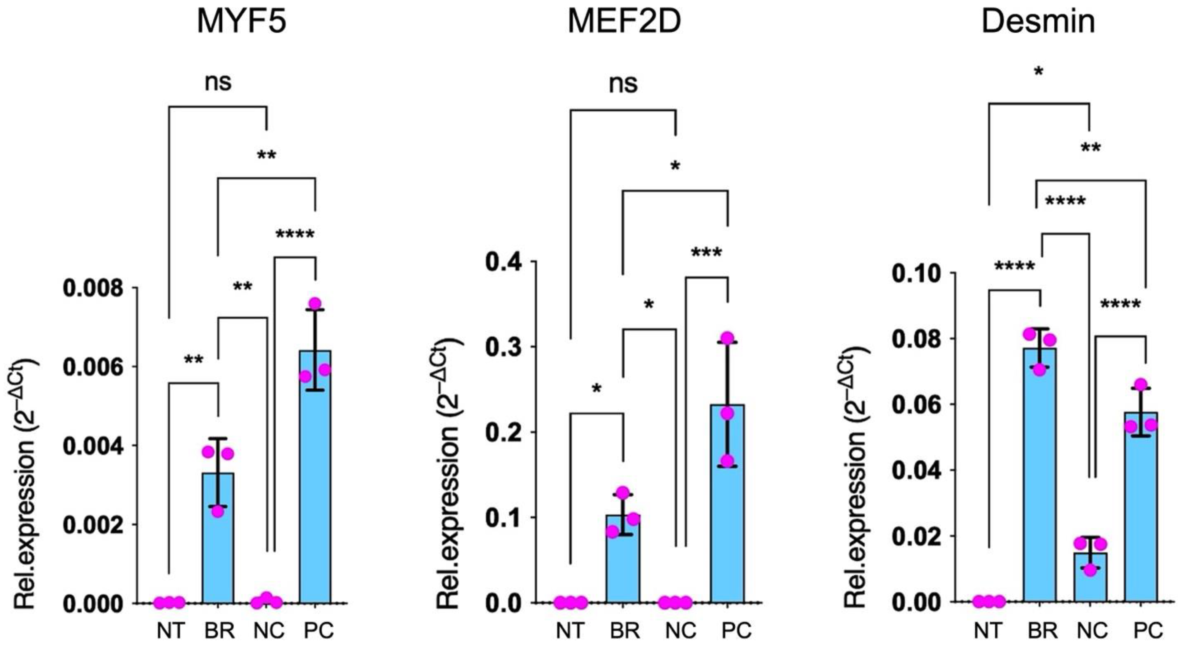
3.5. qPCR
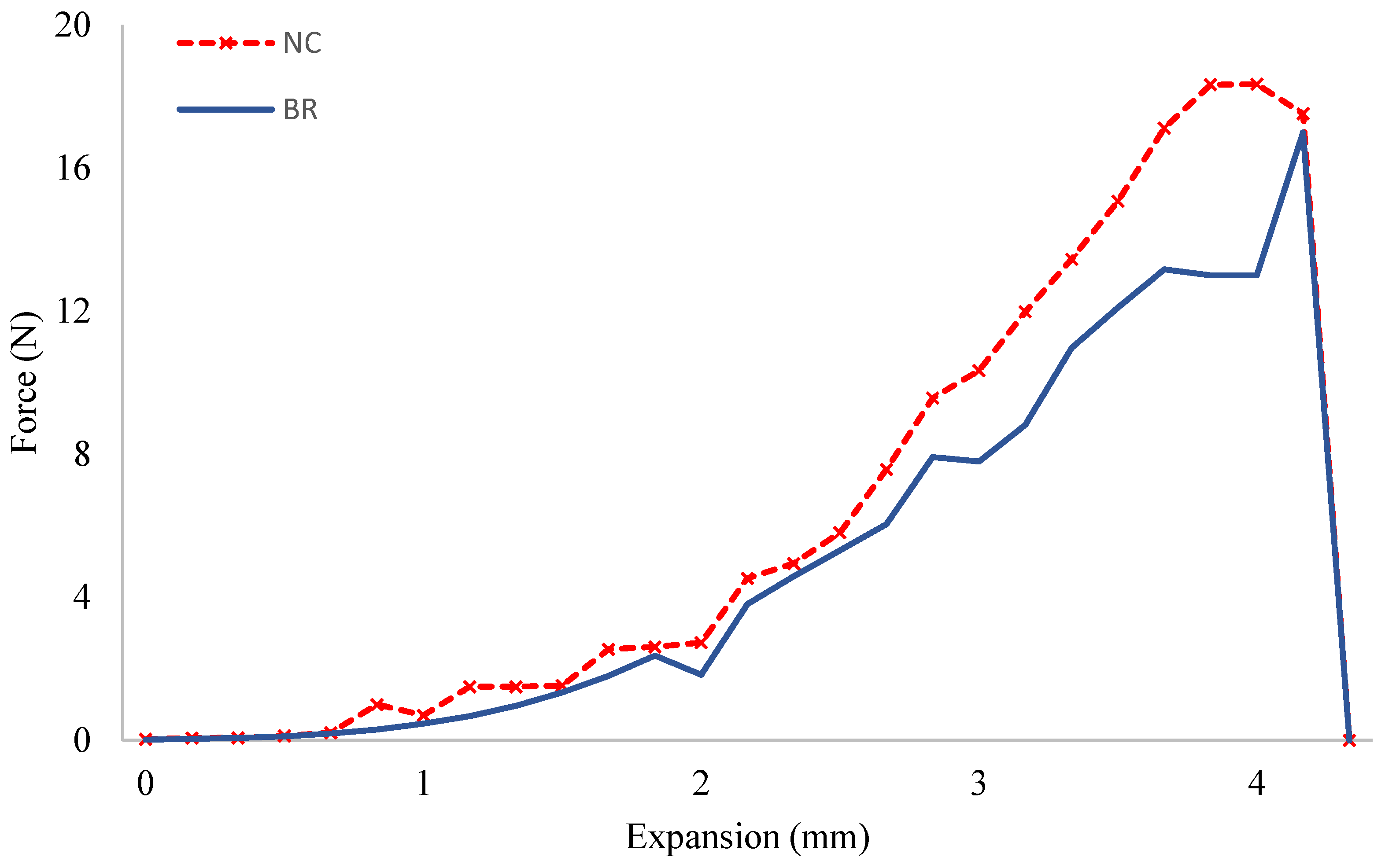
3.6. Tensile Strength Test
4. Discussion
5. Conclusions
Author Contributions
Funding
Institutional Review Board Statement
Informed Consent Statement
Data Availability Statement
Acknowledgments
Conflicts of Interest
References
- Conte, M.S.; Bandyk, D.F.; Clowes, A.W.; Moneta, G.L.; Seely, L.; Lorenz, T.J.; Namini, H.; Hamdan, A.D.; Roddy, S.P.; Belkin, M.; et al. Results of PREVENT III: A Multicenter, Randomized Trial of Edifoligide for the Prevention of Vein Graft Failure in Lower Extremity Bypass Surgery. J. Vasc. Surg. 2006, 43, 742–751, discussion 751. [Google Scholar] [CrossRef] [PubMed] [Green Version]
- Hoerstrup, S.P.; Cummings MRCS, I.; Lachat, M.; Schoen, F.J.; Jenni, R.; Leschka, S.; Neuenschwander, S.; Schmidt, D.; Mol, A.; Günter, C.; et al. Functional Growth in Tissue-Engineered Living, Vascular Grafts: Follow-Up at 100 Weeks in a Large Animal Model. Circulation 2006, 114, 159–166. [Google Scholar] [CrossRef] [PubMed] [Green Version]
- Teebken, O.E.; Bader, A.; Steinhoff, G.; Haverich, A. A new concept for substitutes in vascular surgery. Langenbecks Arch. Chir. Suppl. Kongr. Dtsch. Ges. Chir. Kongr. 1998, 115, 1256–1259. [Google Scholar]
- Shinoka, T.; Shum-Tim, D.; Ma, P.X.; Tanel, R.E.; Isogai, N.; Langer, R.; Vacanti, J.P.; Mayer, J.E. Creation of Viable Pulmonary Artery Autografts through Tissue Engineering. J. Thorac. Cardiovasc. Surg. 1998, 115, 536–545, discussion 545–546. [Google Scholar] [CrossRef] [Green Version]
- Niklason, L.E.; Gao, J.; Abbott, W.M.; Hirschi, K.K.; Houser, S.; Marini, R.; Langer, R. Functional Arteries Grown in Vitro. Science 1999, 284, 489–493. [Google Scholar] [CrossRef] [PubMed] [Green Version]
- Catto, V.; Farè, S.; Freddi, G.; Tanzi, M.C. Vascular Tissue Engineering: Recent Advances in Small Diameter Blood Vessel Regeneration. ISRN Vasc. Med. 2014, 2014, 923030. [Google Scholar] [CrossRef] [Green Version]
- Vepari, C.; Kaplan, D.L. Silk as a Biomaterial. Prog. Polym. Sci. 2007, 32, 991–1007. [Google Scholar] [CrossRef]
- Dastagir, K.; Dastagir, N.; Limbourg, A.; Reimers, K.; Strauß, S.; Vogt, P.M. In Vitro Construction of Artificial Blood Vessels Using Spider Silk as a Supporting Matrix. J. Mech. Behav. Biomed. Mater. 2020, 101, 103436. [Google Scholar] [CrossRef]
- Schäfer-Nolte, F.; Hennecke, K.; Reimers, K.; Schnabel, R.; Allmeling, C.; Vogt, P.M.; Kuhbier, J.W.; Mirastschijski, U. Biomechanics and Biocompatibility of Woven Spider Silk Meshes during Remodeling in a Rodent Fascia Replacement Model. Ann. Surg. 2014, 259, 781–792. [Google Scholar] [CrossRef]
- Martin, I.; Wendt, D.; Heberer, M. The Role of Bioreactors in Tissue Engineering. Trends Biotechnol. 2004, 22, 80–86. [Google Scholar] [CrossRef]
- Kuhbier, J.W.; Reimers, K.; Kasper, C.; Allmeling, C.; Hillmer, A.; Menger, B.; Vogt, P.M.; Radtke, C. First Investigation of Spider Silk as a Braided Microsurgical Suture. J. Biomed. Mater. Res. B Appl. Biomater. 2011, 97B, 381–387. [Google Scholar] [CrossRef] [PubMed]
- Liebsch, C.; Fliess, M.; Kuhbier, J.W.; Vogt, P.M.; Strauss, S. Nephila Edulis-Breeding and Care under Laboratory Conditions. Dev. Genes Evol. 2020, 230, 203–211. [Google Scholar] [CrossRef] [PubMed] [Green Version]
- Wan, S.; George, S.J.; Berry, C.; Baker, A.H. Vein Graft Failure: Current Clinical Practice and Potential for Gene Therapeutics. Gene Ther. 2012, 19, 630–636. [Google Scholar] [CrossRef] [PubMed] [Green Version]
- Schoen, F.J.; Mitchell, R.N.; Jonas, R.A. Pathological Considerations in Cryopreserved Allograft Heart Valves. J. Heart Valve Dis. 1995, 4 (Suppl. S1), S72–S75, discussion S75–S76. [Google Scholar]
- Shin’oka, T.; Imai, Y.; Ikada, Y. Transplantation of a Tissue-Engineered Pulmonary Artery. N. Engl. J. Med. 2001, 344, 532–533. [Google Scholar] [CrossRef]
- Shin’oka, T.; Matsumura, G.; Hibino, N.; Naito, Y.; Watanabe, M.; Konuma, T.; Sakamoto, T.; Nagatsu, M.; Kurosawa, H. Midterm Clinical Result of Tissue-Engineered Vascular Autografts Seeded with Autologous Bone Marrow Cells. J. Thorac. Cardiovasc. Surg. 2005, 129, 1330–1338. [Google Scholar] [CrossRef] [Green Version]
- Poh, M.; Boyer, M.; Solan, A.; Dahl, S.L.M.; Pedrotty, D.; Banik, S.S.R.; McKee, J.A.; Klinger, R.Y.; Counter, C.M.; Niklason, L.E. Blood Vessels Engineered from Human Cells. Lancet Lond. Engl. 2005, 365, 2122–2124. [Google Scholar] [CrossRef]
- Aper, T.; Wilhelmi, M.; Gebhardt, C.; Hoeffler, K.; Benecke, N.; Hilfiker, A.; Haverich, A. Novel Method for the Generation of Tissue-Engineered Vascular Grafts Based on a Highly Compacted Fibrin Matrix. Acta Biomater. 2016, 29, 21–32. [Google Scholar] [CrossRef]
- Helms, F.; Lau, S.; Aper, T.; Zippusch, S.; Klingenberg, M.; Haverich, A.; Wilhelmi, M.; Böer, U. A 3-Layered Bioartificial Blood Vessel with Physiological Wall Architecture Generated by Mechanical Stimulation. Ann. Biomed. Eng. 2021, 49, 2066–2079. [Google Scholar] [CrossRef]
- Niu, Y.; Galluzzi, M. Hyaluronic Acid/Collagen Nanofiber Tubular Scaffolds Support Endothelial Cell Proliferation, Phenotypic Shape and Endothelialization. Nanomaterials 2021, 11, 2334. [Google Scholar] [CrossRef]
- Zhang, F.; Bambharoliya, T.; Xie, Y.; Liu, L.; Celik, H.; Wang, L.; Akkus, O.; King, M.W. A Hybrid Vascular Graft Harnessing the Superior Mechanical Properties of Synthetic Fibers and the Biological Performance of Collagen Filaments. Mater. Sci. Eng. C 2021, 118, 111418. [Google Scholar] [CrossRef] [PubMed]
- Liu, K.; Wang, N.; Wang, W.; Shi, L.; Li, H.; Guo, F.; Zhang, L.; Kong, L.; Wang, S.; Zhao, Y. A Bio-Inspired High Strength Three-Layer Nanofiber Vascular Graft with Structure Guided Cell Growth. J. Mater. Chem. B 2017, 5, 3758–3764. [Google Scholar] [CrossRef] [PubMed]
- Wang, D.; Xu, Y.; Li, Q.; Turng, L.-S. Artificial Small-Diameter Blood Vessels: Materials, Fabrication, Surface Modification, Mechanical Properties, and Bioactive Functionalities. J. Mater. Chem. B 2020, 8, 1801–1822. [Google Scholar] [CrossRef] [PubMed]
- Grenier, G.; Rémy-Zolghadri, M.; Guignard, R.; Bergeron, F.; Labbé, R.; Auger, F.A.; Germain, L. Isolation and Culture of the Three Vascular Cell Types from a Small Vein Biopsy Sample. Vitr. Cell Dev. Biol. Anim. 2003, 39, 131–139. [Google Scholar] [CrossRef]
- Zhang, W.J.; Liu, W.; Cui, L.; Cao, Y. Tissue Engineering of Blood Vessel. J. Cell Mol. Med. 2007, 11, 945–957. [Google Scholar] [CrossRef] [PubMed]
- Hamilton, D.W.; Maul, T.M.; Vorp, D.A. Characterization of the Response of Bone Marrow-Derived Progenitor Cells to Cyclic Strain: Implications for Vascular Tissue-Engineering Applications. Tissue Eng. 2004, 10, 361–369. [Google Scholar] [CrossRef]
- Stegemann, J.P.; Hong, H.; Nerem, R.M. Mechanical, Biochemical, and Extracellular Matrix Effects on Vascular Smooth Muscle Cell Phenotype. J. Appl. Physiol. 2005, 98, 2321–2327. [Google Scholar] [CrossRef] [PubMed] [Green Version]
- Williams, C.; Wick, T.M. Perfusion Bioreactor for Small Diameter Tissue-Engineered Arteries. Tissue Eng. 2004, 10, 930–941. [Google Scholar] [CrossRef]
- Engbers-Buijtenhuijs, P.; Buttafoco, L.; Poot, A.A.; Dijkstra, P.J.; de Vos, R.A.I.; Sterk, L.M.T.; Geelkerken, R.H.; Vermes, I.; Feijen, J. Biological Characterisation of Vascular Grafts Cultured in a Bioreactor. Biomaterials 2006, 27, 2390–2397. [Google Scholar] [CrossRef]
- Webb, A.R.; Macrie, B.D.; Ray, A.S.; Russo, J.E.; Siegel, A.M.; Glucksberg, M.R.; Ameer, G.A. In Vitro Characterization of a Compliant Biodegradable Scaffold with a Novel Bioreactor System. Ann. Biomed. Eng. 2007, 35, 1357–1367. [Google Scholar] [CrossRef]
- Arrigoni, C.; Chittò, A.; Mantero, S.; Remuzzi, A. Rotating versus Perfusion Bioreactor for the Culture of Engineered Vascular Constructs Based on Hyaluronic Acid. Biotechnol. Bioeng. 2008, 100, 988–997. [Google Scholar] [CrossRef]
- Hahn, M.S.; McHale, M.K.; Wang, E.; Schmedlen, R.H.; West, J.L. Physiologic Pulsatile Flow Bioreactor Conditioning of Poly(Ethylene Glycol)-Based Tissue Engineered Vascular Grafts. Ann. Biomed. Eng. 2007, 35, 190–200. [Google Scholar] [CrossRef] [PubMed]
- Sterpetti, A.V.; Cucina, A.; Santoro, L.; Cardillo, B.; Cavallaro, A. Modulation of Arterial Smooth Muscle Cell Growth by Haemodynamic Forces. Eur. J. Vasc. Surg. 1992, 6, 16–20. [Google Scholar] [CrossRef] [PubMed]
- Lee, A.A.; Graham, D.A.; Dela Cruz, S.; Ratcliffe, A.; Karlon, W.J. Fluid Shear Stress-Induced Alignment of Cultured Vascular Smooth Muscle Cells. J. Biomech. Eng. 2002, 124, 37–43. [Google Scholar] [CrossRef]
- Niu, D.; Wei, H.-J.; Lin, L.; George, H.; Wang, T.; Lee, I.-H.; Zhao, H.-Y.; Wang, Y.; Kan, Y.; Shrock, E.; et al. Inactivation of Porcine Endogenous Retrovirus in Pigs Using CRISPR-Cas9. Science 2017, 357, 1303–1307. [Google Scholar] [CrossRef] [PubMed] [Green Version]
- Takahashi, K.; Yamanaka, S. Induction of Pluripotent Stem Cells from Mouse Embryonic and Adult Fibroblast Cultures by Defined Factors. Cell 2006, 126, 663–676. [Google Scholar] [CrossRef]


| Primer | Forward′➔3′ | Reverse 5′➔3′ | NCBI Accession No. |
|---|---|---|---|
| MYF5 | GCTTTCGAGACGCTCAAGAG | GGACAAGCAATCCAAGCTG | NM_008656 |
| MEF2D | CAAGCTGTTCCAGTATGCCAG | AAGGGATGATGTCACCAGGG | NM_001310587 |
| Desmin | AGAAGCCGATCCAGGCAAAA | AAGGGATGATGTCACCAGGG | NM_010043 |
| B2M | ATGAGTATGCCTGCCGTGTGGA | GGCATCTTGCAAACCTCCATG | NM_004048 |
| RPL37 | GCGTGATATAGCGGAAGTGC | ACTTCTGAAGGTGGTAGGCC | NM_009097 |
| TBP | GCAGTGCCCAGCATCACTAT | CACAAGGCCTTCCAGCCTTA | NM_013684.3 |
Publisher’s Note: MDPI stays neutral with regard to jurisdictional claims in published maps and institutional affiliations. |
© 2022 by the authors. Licensee MDPI, Basel, Switzerland. This article is an open access article distributed under the terms and conditions of the Creative Commons Attribution (CC BY) license (https://creativecommons.org/licenses/by/4.0/).
Share and Cite
Obed, D.; Dastagir, N.; Liebsch, C.; Bingoel, A.S.; Strauss, S.; Vogt, P.M.; Dastagir, K. In Vitro Differentiation of Myoblast Cell Lines on Spider Silk Scaffolds in a Rotating Bioreactor for Vascular Tissue Engineering. J. Pers. Med. 2022, 12, 1986. https://doi.org/10.3390/jpm12121986
Obed D, Dastagir N, Liebsch C, Bingoel AS, Strauss S, Vogt PM, Dastagir K. In Vitro Differentiation of Myoblast Cell Lines on Spider Silk Scaffolds in a Rotating Bioreactor for Vascular Tissue Engineering. Journal of Personalized Medicine. 2022; 12(12):1986. https://doi.org/10.3390/jpm12121986
Chicago/Turabian StyleObed, Doha, Nadjib Dastagir, Christina Liebsch, Alperen S. Bingoel, Sarah Strauss, Peter M. Vogt, and Khaled Dastagir. 2022. "In Vitro Differentiation of Myoblast Cell Lines on Spider Silk Scaffolds in a Rotating Bioreactor for Vascular Tissue Engineering" Journal of Personalized Medicine 12, no. 12: 1986. https://doi.org/10.3390/jpm12121986