
Clinical Significance of the Histone Deacetylase 2 (HDAC-2) Expression in Human Breast Cancer

 , , , , ,
Abstract
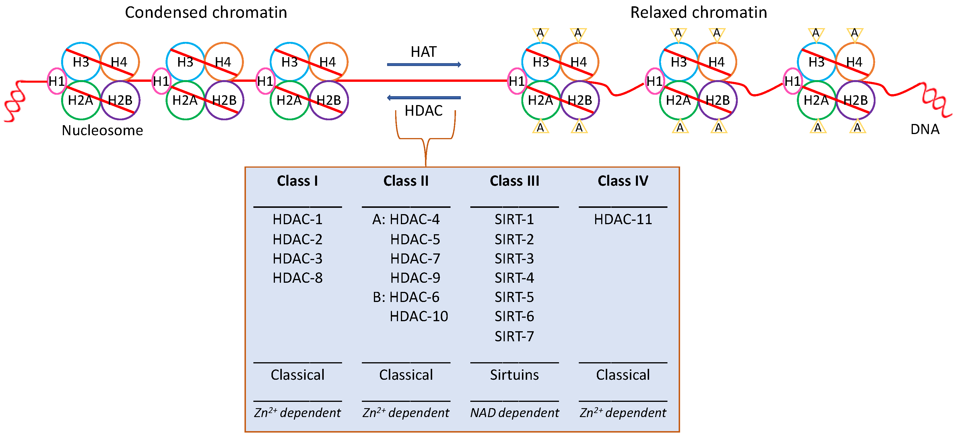
:1. Introduction
2. Materials and Methods
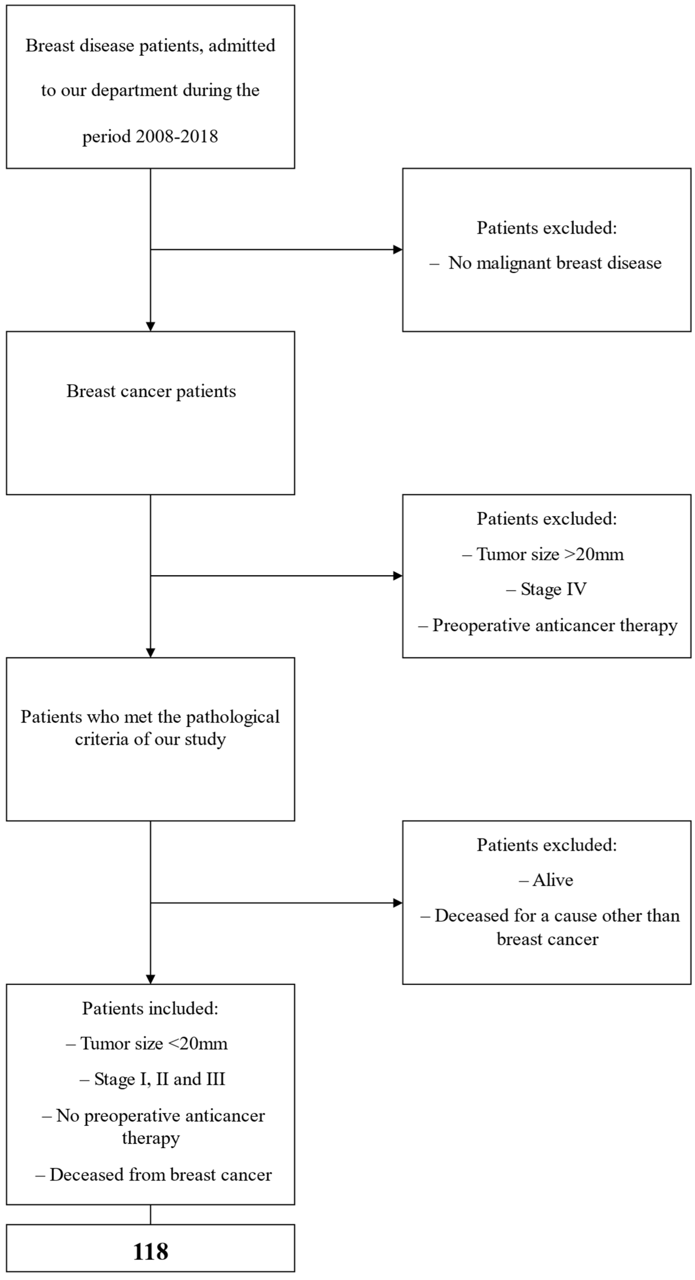
2.1. Clinical Material
2.2. Immunohistochemistry
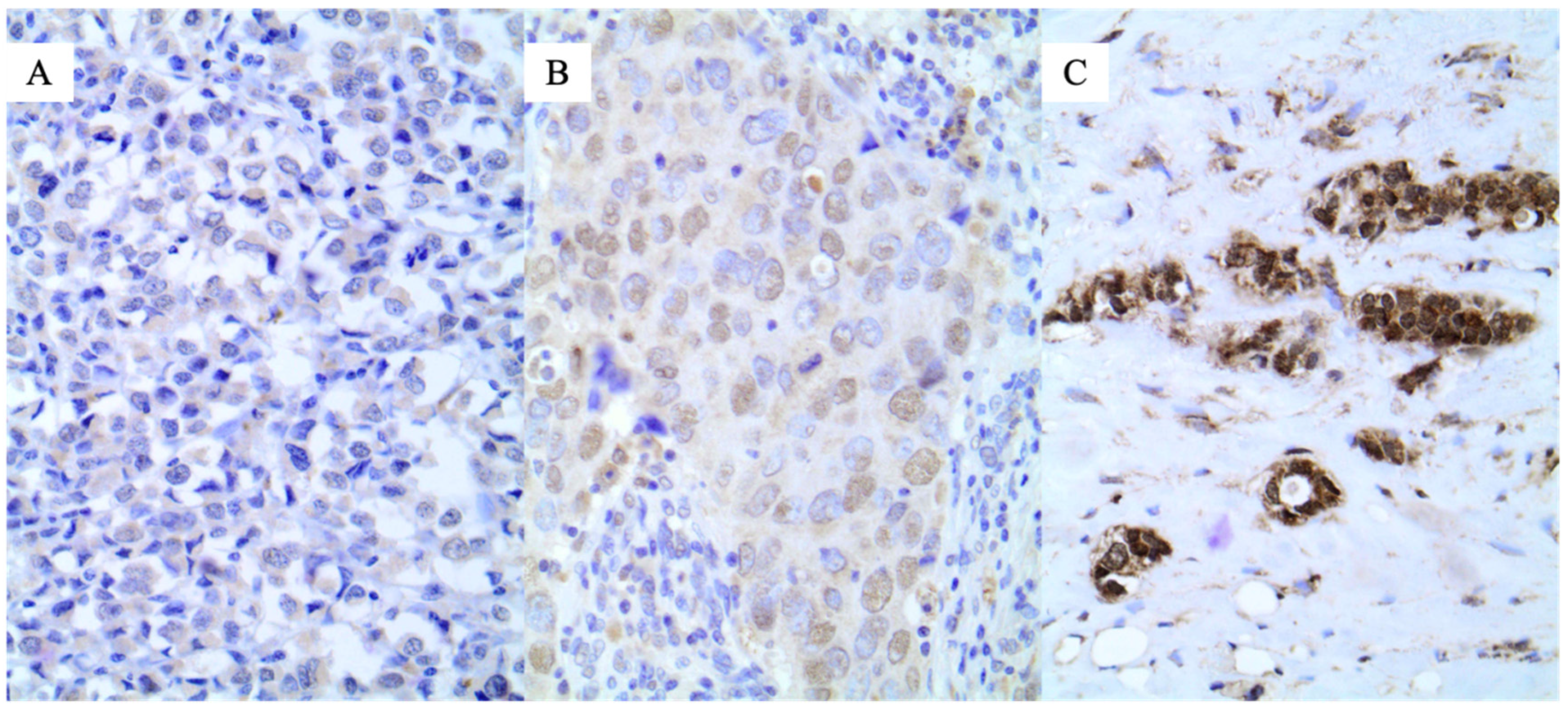
2.3. Evaluation of Immunohistochemistry
2.4. Statistical Analysis
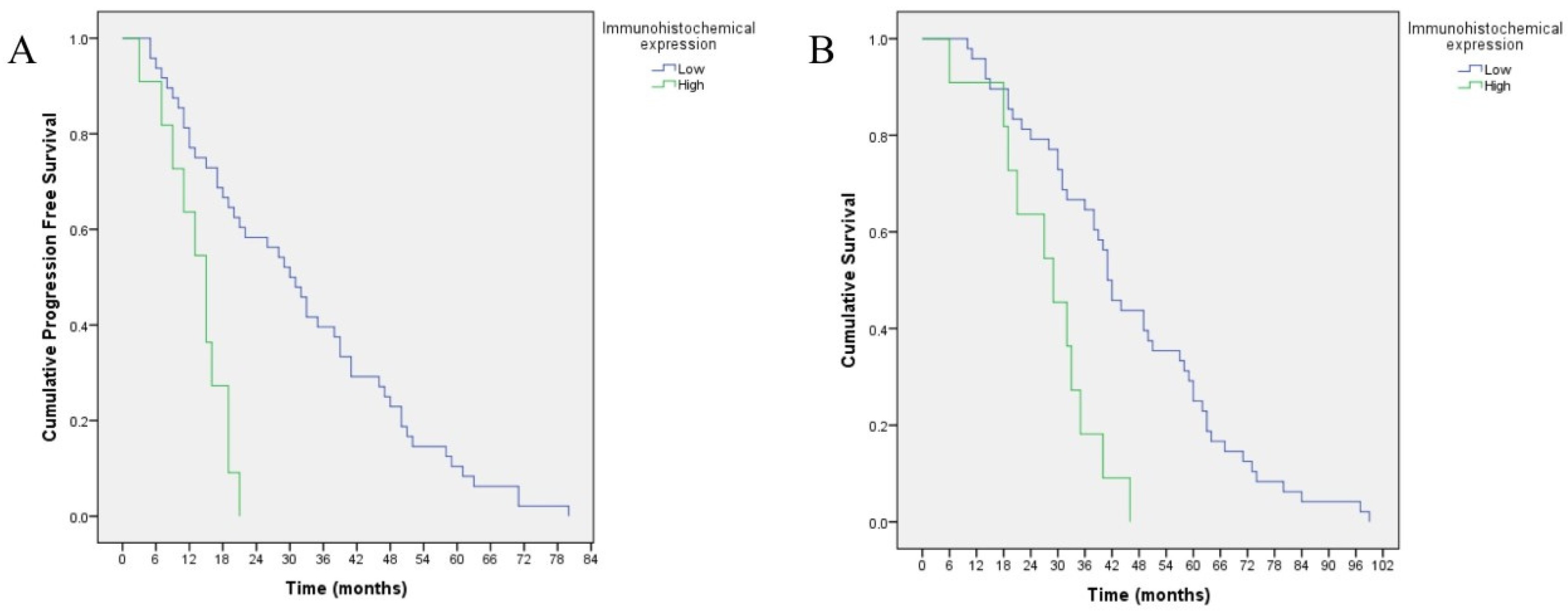
3. Results
4. Discussion
5. Conclusions
Author Contributions
Funding
Institutional Review Board Statement
Informed Consent Statement
Data Availability Statement
Conflicts of Interest
References
- Allfrey, V.G.; Faulkner, R.; Mirsky, A.E. Acetylation and methylation of histones and their possible role in the regulation of RNA synthesis. Proc. Natl. Acad. Sci. USA 1964, 51, 786–794. [Google Scholar] [CrossRef] [Green Version]
- Grunstein, M. Histone acetylation in chromatin structure and transcription. Nature 1997, 389, 349–352. [Google Scholar] [CrossRef] [PubMed]
- Haberland, M.; Montgomery, R.L.; Olson, E.N. The many roles of histone deacetylases in development and physiology: Implications for disease and therapy. Nat. Rev. Genet. 2009, 10, 32–42. [Google Scholar] [CrossRef] [PubMed] [Green Version]
- Trapp, J.; Jung, M. The role of NAD+ dependent histone deacetylases (sirtuins) in ageing. Curr. Drug Targets 2006, 7, 1553–1560. [Google Scholar] [CrossRef] [PubMed]
- Garmpi, A.; Garmpis, N.; Damaskos, C.; Valsami, S.; Spartalis, E.; Lavaris, A.; Patelis, N.; Margonis, G.A.; Apostolou, K.G.; Spartalis, M.; et al. Histone deacetylase inhibitors as a new anticancer option: How far can we go with expectations? J. BUON 2018, 23, 846–861. [Google Scholar] [PubMed]
- Olzscha, H.; Sheikh, S.; La Thangue, N.B. Deacetylation of chromatin and gene expression regulation: A new target for epigenetic therapy. Crit. Rev. Oncog. 2015, 20, 1–17. [Google Scholar] [CrossRef]
- Garmpi, A.; Damaskos, C.; Garmpis, N.; Kaminiotis, V.-V.; Georgakopoulou, V.E.; Spandidos, D.A.; Papalexis, P.; Diamantis, E.; Patsouras, A.; Kyriakos, G.; et al. Role of histone deacetylase inhibitors in diabetic cardiomyopathy in experimental models (Review). Med. Int. 2022, 2, 26. [Google Scholar] [CrossRef]
- Liu, Y.-R.; Wang, J.-Q.; Huang, Z.-G.; Chen, R.-N.; Cao, X.; Zhu, D.-C.; Yu, H.-X.; Wang, X.-R.; Zhou, H.-Y.; Xia, Q.; et al. Histone deacetylase-2: A potential regulator and therapeutic target in liver disease (Review). Int. J. Mol. Med. 2021, 48, 131. [Google Scholar] [CrossRef] [PubMed]
- Marks, P.A.; Richon, V.M.; Rifkind, R.A. Histone Deacetylase Inhibitors: Inducers of Differentiation or Apoptosis of Transformed Cells. JNCI J. Natl. Cancer Inst. 2000, 92, 1210–1216. [Google Scholar] [CrossRef] [PubMed] [Green Version]
- Archer, S.Y.; Hodin, R.A. Histone acetylation and cancer. Curr. Opin. Genet. Dev. 1999, 9, 171–174. [Google Scholar] [CrossRef]
- Glozak, M.A.; Seto, E. Histone deacetylases and cancer. Oncogene 2007, 26, 5420–5432. [Google Scholar] [CrossRef] [PubMed] [Green Version]
- Büchler, P.; Gazdhar, A.; Schubert, M.; Giese, N.; Reber, H.A.; Hines, O.J.; Giese, T.; Ceyhan, G.O.; Müller, M.; Büchler, M.W.; et al. The Notch Signaling Pathway Is Related to Neurovascular Progression of Pancreatic Cancer. Ann. Surg. 2005, 242, 791–800, discussion 800–801. [Google Scholar] [CrossRef] [PubMed] [Green Version]
- Huang, Y.; Myers, S.J.; Dingledine, R. Transcriptional repression by REST: Recruitment of Sin3A and histone deacetylase to neuronal genes. Nat. Neurosci. 1999, 2, 867–872. [Google Scholar] [CrossRef]
- Giaginis, C.; Alexandrou, P.; Delladetsima, I.; Giannopoulou, I.; Patsouris, E.; Theocharis, S. Clinical significance of histone deacetylase (HDAC)-1, HDAC-2, HDAC-4, and HDAC-6 expression in human malignant and benign thyroid lesions. Tumor Biol. 2013, 35, 61–71. [Google Scholar] [CrossRef] [PubMed]
- Damaskos, C.; Karatzas, T.; Nikolidakis, L.; Kostakis, I.D.; Karamaroudis, S.; Boutsikos, G.; Damaskou, Z.; Kostakis, A.; Kouraklis, G. Histone Deacetylase (HDAC) Inhibitors: Current Evidence for Therapeutic Activities in Pancreatic Cancer. Anticancer Res. 2015, 35, 3129–3135. [Google Scholar] [PubMed]
- Marks, P.A. The clinical development of histone deacetylase inhibitors as targeted anticancer drugs. Expert Opin. Investig. Drugs 2010, 19, 1049–1066. [Google Scholar] [CrossRef] [PubMed]
- Ruefli, A.A.; Ausserlechner, M.J.; Bernhard, D.; Sutton, V.R.; Tainton, K.M.; Kofler, R.; Smyth, M.J.; Johnstone, R.W. The histone deacetylase inhibitor and chemotherapeutic agent suberoylanilide hydroxamic acid (SAHA) induces a cell-death pathway characterized by cleavage of Bid and production of reactive oxygen species. Proc. Natl. Acad. Sci. USA 2001, 98, 10833–10838. [Google Scholar] [CrossRef] [PubMed] [Green Version]
- Richon, V.M.; Sandhoff, T.W.; Rifkind, R.A.; Marks, P.A. Histone deacetylase inhibitor selectively induces p21WAF1 expression and gene-associated histone acetylation. Proc. Natl. Acad. Sci. USA 2000, 97, 10014–10019. [Google Scholar] [CrossRef] [PubMed] [Green Version]
- Dokmanovic, M.; Clarke, C.; Marks, P.A. Histone Deacetylase Inhibitors: Overview and Perspectives. Mol. Cancer Res. 2007, 5, 981–989. [Google Scholar] [CrossRef] [PubMed] [Green Version]
- Qian, D.Z.; Kachhap, S.K.; Collis, S.J.; Verheul, H.M.; Carducci, M.A.; Atadja, P.; Pili, R. Class II Histone Deacetylases Are Associated with VHL-Independent Regulation of Hypoxia-Inducible Factor 1α. Cancer Res. 2006, 66, 8814–8821. [Google Scholar] [CrossRef]
- Kano, Y.; Akutsu, M.; Tsunoda, S.; Izumi, T.; Kobayashi, H.; Mano, H.; Furukawa, Y. Cytotoxic effects of histone deacetylase inhibitor FK228 (depsipeptide, formally named FR901228) in combination with conventional anti-leukemia/lymphoma agents against human leukemia/lymphoma cell lines. Investig. New Drugs 2006, 25, 31–40. [Google Scholar] [CrossRef] [PubMed]
- Wardell, S.E.; Ilkayeva, O.R.; Wieman, H.L.; Frigo, D.E.; Rathmell, J.C.; Newgard, C.B.; McDonnell, D.P. Glucose Metabolism as a Target of Histone Deacetylase Inhibitors. Mol. Endocrinol. 2009, 23, 388–401. [Google Scholar] [CrossRef] [PubMed] [Green Version]
- Barbarotta, L.; Hurley, K. Romidepsin for the Treatment of Peripheral T-Cell Lymphoma. J. Adv. Pract. Oncol. 2015, 6, 22–36. [Google Scholar]
- Libby, E.N.; Becker, P.S.; Burwick, N.; Green, D.J.; Holmberg, L.; Bensinger, W.I. Panobinostat: A review of trial results and future prospects in multiple myeloma. Expert Rev. Hematol. 2014, 8, 9–18. [Google Scholar] [CrossRef] [PubMed]
- Damaskos, C.; Garmpis, N.; Valsami, S.; Kontos, M.; Spartalis, E.; Kalampokas, T.; Kalampokas, E.; Athanasiou, A.; Moris, D.; Daskalopoulou, A.; et al. Histone Deacetylase Inhibitors: An Attractive Therapeutic Strategy Against Breast Cancer. Anticancer Res. 2017, 37, 35–46. [Google Scholar] [CrossRef] [Green Version]
- Siegel, R.L.; Miller, K.D.; Jemal, A. Cancer statistics, 2018. CA Cancer J. Clin. 2018, 68, 7–30. [Google Scholar] [CrossRef] [PubMed]
- Chen, W.; Zheng, R.; Baade, P.D.; Zhang, S.; Zeng, H.; Bray, F.; Jemal, A.; Yu, X.Q.; He, J. Cancer statistics in China, 2015. CA Cancer J. Clin. 2016, 66, 115–132. [Google Scholar] [CrossRef] [Green Version]
- Torre, L.A.; Bray, F.; Siegel, R.L.; Ferlay, J.; Lortet-Tieulent, J.; Jemal, A. Global cancer statistics, 2012. CA Cancer J. Clin. 2015, 65, 87–108. [Google Scholar] [CrossRef] [Green Version]
- Veronesi, U.; Boyle, P.; Goldhirsch, A.; Orecchia, R.; Viale, G. Breast cancer. Lancet 2005, 365, 1727–1741. [Google Scholar] [CrossRef]
- Bray, F.; Ferlay, J.; Soerjomataram, I.; Siegel, R.L.; Torre, L.A.; Jemal, A. Global cancer statistics 2018: GLOBOCAN estimates of incidence and mortality worldwide for 36 cancers in 185 countries. CA Cancer J. Clin. 2018, 68, 394–424. [Google Scholar] [CrossRef] [Green Version]
- Siegel, R.L.; Miller, K.D.; Fuchs, H.E.; Jemal, A. Cancer statistics, 2022. CA Cancer J. Clin. 2022, 72, 7–33. [Google Scholar] [CrossRef] [PubMed]
- Feng, Y.; Spezia, M.; Huang, S.; Yuan, C.; Zeng, Z.; Zhang, L.; Ji, X.; Liu, W.; Huang, B.; Luo, W.; et al. Breast cancer development and progression: Risk factors, cancer stem cells, signaling pathways, genomics, and molecular pathogenesis. Gene Funct. Dis. 2018, 5, 77–106. [Google Scholar] [CrossRef] [PubMed]
- Carlsson, M.; Hamrin, E. Psychological and psychosocial aspects of breast cancer and breast cancer treatment. A literature review. Cancer Nurs. 1994, 17, 418–428. [Google Scholar] [CrossRef] [PubMed]
- Chambers, M.S.; Rugo, H.S.; Litton, J.K.; Meiller, T.F. Stomatitis associated with mammalian target of rapamycin inhibition: A review of pathogenesis, prevention, treatment, and clinical implications for oral practice in metastatic breast cancer. JADA 2018, 149, 291–298. [Google Scholar]
- Ariana, M.; Arabi, N.; Pornour, M.; Vaseghi, H.; Ganji, S.M.; Alivand, M.R.; Salari, M.; Akbari, M.E. The diversity in the expression profile of caveolin II transcripts, considering its new transcript in breast cancer. J. Cell. Biochem. 2017, 119, 2168–2178. [Google Scholar] [CrossRef]
- Rich, J.N.; Bao, S. Chemotherapy and Cancer Stem Cells. Cell Stem Cell 2007, 1, 353–355. [Google Scholar] [CrossRef] [Green Version]
- Li, Y.; Seto, E. HDACs and HDAC Inhibitors in Cancer Development and Therapy. Cold Spring Harb. Perspect. Med. 2016, 6, a026831. [Google Scholar] [CrossRef] [Green Version]
- Xu, P.; Xiong, W.; Lin, Y.; Fan, L.; Pan, H.; Li, Y. Histone deacetylase 2 knockout suppresses immune escape of triple-negative breast cancer cells via downregulating PD-L1 expression. Cell Death Dis. 2021, 12, 779. [Google Scholar] [CrossRef]
- Xie, Y.; Shi, Z.; Qian, Y.; Jiang, C.; Liu, W.; Liu, B.; Jiang, B. HDAC2- and EZH2-Mediated Histone Modifications Induce PDK1 Expression through miR-148a Downregulation in Breast Cancer Progression and Adriamycin Resistance. Cancers 2022, 14, 3600. [Google Scholar] [CrossRef]
- Huang, W.; Chen, J.; Liu, X.; Liu, X.; Duan, S.; Chen, L.; Liu, X.; Lan, J.; Zou, Y.; Guo, D.; et al. MIER3 induces epithelial-mesenchymal transition and promotes breast cancer cell aggressiveness via forming a co-repressor complex with HDAC1/HDAC2/Snail. Exp. Cell Res. 2021, 406, 112722. [Google Scholar] [CrossRef]
- Darvishi, N.; Rahimi, K.; Mansouri, K.; Fathi, F.; Menbari, M.-N.; Mohammadi, G.; Abdi, M. MiR-646 prevents proliferation and progression of human breast cancer cell lines by suppressing HDAC2 expression. Mol. Cell. Probes 2020, 53, 101649. [Google Scholar] [CrossRef] [PubMed]
- Zhang, Z.; Qiu, N.; Yin, J.; Zhang, J.; Liu, H.; Guo, W.; Liu, M.; Liu, T.; Chen, D.; Luo, K.; et al. SRGN crosstalks with YAP to maintain chemoresistance and stemness in breast cancer cells by modulating HDAC2 expression. Theranostics 2020, 10, 4290–4307. [Google Scholar] [CrossRef] [PubMed]
- Damaskos, C.; Tomos, I.; Garmpis, N.; Karakatsani, A.; Dimitroulis, D.; Garmpi, A.; Spartalis, E.; Kampolis, C.F.; Tsagkari, E.; Loukeri, A.A.; et al. Histone deacetylase inhibitors as a novel targeted therapy against non-small cell lung cancer: Where are we now and what should we expect? Anticancer Res. 2018, 38, 37–43. [Google Scholar] [PubMed]
- Garmpis, N.; Damaskos, C.; Garmpi, A.; Nonni, A.; Georgakopoulou, V.E.; Antoniou, E.; Schizas, D.; Sarantis, P.; Patsouras, A.; Syllaios, A.; et al. Histone Deacetylases and their Inhibitors in Colorectal Cancer Therapy: Current Evidence and Future Considerations. Curr. Med. Chem. 2021, 29, 2979–2994. [Google Scholar] [CrossRef]
- Weichert, W.; Röske, A.; Gekeler, V.; Beckers, T.; Ebert, M.P.A.; Pross, M.; Dietel, M.; Denkert, C.; Röcken, C. Association of patterns of class I histone deacetylase expression with patient prognosis in gastric cancer: A retrospective analysis. Lancet Oncol. 2008, 9, 139–148. [Google Scholar] [CrossRef]
- Damaskos, C.; Garmpis, N.; Valsami, S.; Spartalis, E.; Antoniou, E.A.; Tomos, P.; Karamaroudis, S.; Zoumpou, T.; Pergialiotis, V.; Stergios, K.; et al. Histone deacetylase inhibitors: A novel therapeutic weapon against medullary thyroid cancer? Anticancer Res. 2016, 36, 5019–5024. [Google Scholar] [CrossRef]
- Spartalis, E.; Athanasiadis, D.I.; Chrysikos, D.; Spartalis, M.; Boutzios, G.; Schizas, D.; Garmpis, N.; Damaskos, C.; Paschou, S.A.; Ioannidis, A.; et al. Histone Deacetylase Inhibitors and Anaplastic Thyroid Carcinoma. Anticancer Res. 2019, 39, 1119–1127. [Google Scholar] [CrossRef]
- Halkidou, K.; Gaughan, L.; Cook, S.; Leung, H.Y.; Neal, D.E.; Robson, C.N. Upregulation and Nuclear Recruitment of HDAC1 in Hormone Refractory Prostate Cancer. Prostate 2004, 59, 177–189. [Google Scholar] [CrossRef]
- Garmpis, N.; Damaskos, C.; Garmpi, A.; Spartalis, E.; Kalampokas, E.; Kalampokas, T.; Margonis, G.A.; Schizas, D.; Andreatos, N.; Angelou, A.; et al. Targeting histone deacetylases in endometrial cancer: A paradigm-shifting therapeutic strategy? Eur. Rev. Med. Pharmacol. Sci. 2018, 22, 950–960. [Google Scholar]
- Garmpis, N.; Damaskos, C.; Garmpi, A.; Georgakopoulou, V.; Sarantis, P.; Antoniou, E.; Karamouzis, M.; Nonni, A.; Schizas, D.; Diamantis, E.; et al. Histone Deacetylase Inhibitors in the Treatment of Hepatocellular Carcinoma: Current Evidence and Future Opportunities. J. Pers. Med. 2021, 11, 223. [Google Scholar] [CrossRef]
- Fritzsche, F.R.; Weichert, W.; Röske, A.; Gekeler, V.; Beckers, T.; Stephan, C.; Jung, K.; Scholman, K.; Denkert, C.; Dietel, M.; et al. Class I histone deacetylases 1, 2 and 3 are highly expressed in renal cell cancer. BMC Cancer 2008, 8, 381. [Google Scholar] [CrossRef] [Green Version]
- Garmpis, N.; Damaskos, C.; Garmpi, A.; Dimitroulis, D.; Spartalis, E.; Margonis, G.A.; Schizas, D.; Deskou, I.; Doula, C.; Magkouti, E.; et al. Targeting histone deacetylases in malignant melanoma: A future therapeutic agent or just great expectations? Anticancer Res. 2017, 37, 5355–5362. [Google Scholar] [PubMed]
- Moschos, M.M.; Dettoraki, M.; Androudi, S.; Kalogeropoulos, D.; Lavaris, A.; Garmpis, N.; Damaskos, C.; Garmpi, A.; Tsatsos, M. The Role of Histone Deacetylase Inhibitors in Uveal Melanoma: Current Evidence. Anticancer Res. 2018, 38, 3817–3824. [Google Scholar] [CrossRef]
- Garmpis, N.; Damaskos, C.; Garmpi, A.; Kalampokas, E.; Kalampokas, T.; Spartalis, E.; Daskalopoulou, A.; Valsami, S.; Kontos, M.; Nonni, A.; et al. Histone Deacetylases as New Therapeutic Targets in Triple-negative Breast Cancer: Progress and Promises. Cancer Genom. Proteom. 2017, 14, 299–313. [Google Scholar] [CrossRef] [Green Version]
- Marquard, L.; Gjerdrum, L.M.; Christensen, I.J. Prognostic significance of the therapeutic targets histone deacetylase 1, 2, 6 and acetylated histone H4 in cutaneous T-cell lymphoma. Histopathology 2008, 53, 267–277. [Google Scholar]
- Edge, S.B.; Compton, C.C. The American Joint Committee on Cancer: The 7th Edition of the AJCC Cancer Staging Manual and the Future of TNM. Ann. Surg. Oncol. 2010, 17, 1471–1474. [Google Scholar] [CrossRef]
- Giaginis, C.; Damaskos, C.; Koutsounas, I.; Zizi-Serbetzoglou, A.; Tsoukalas, N.; Patsouris, E.; Kouraklis, G.; Theocharis, S. Histone deacetylase (HDAC)-1, -2, -4 and -6 expression in human pancreatic adenocarcinoma: Associations with clinicopathological parameters, tumor proliferative capacity and patients’ survival. BMC Gastroenterol. 2015, 15, 148. [Google Scholar] [CrossRef] [Green Version]
- Cox, D.R. Regression Models and Life-Tables. J. R. Stat. Soc. Ser. B Stat. Methodol. 1972, 34, 187–202. [Google Scholar] [CrossRef]
- Derr, R.S.; Van Hoesel, A.Q.; Benard, A.; Goossens-Beumer, I.J.; Sajet, A.; Dekker-Ensink, N.G.; De Kruijf, E.M.; Bastiaannet, E.; Smit, V.T.H.B.M.; Van De Velde, C.J.H.; et al. High nuclear expression levels of histone-modifying enzymes LSD1, HDAC2 and SIRT1 in tumor cells correlate with decreased survival and increased relapse in breast cancer patients. BMC Cancer 2014, 14, 604. [Google Scholar] [CrossRef] [Green Version]
- Seo, J.; Min, S.K.; Park, H.-R.; Kim, D.H.; Kwon, M.J.; Kim, L.S.; Ju, Y.-S. Expression of Histone Deacetylases HDAC1, HDAC2, HDAC3, and HDAC6 in Invasive Ductal Carcinomas of the Breast. J. Breast Cancer 2014, 17, 323–331. [Google Scholar] [CrossRef] [Green Version]
- Müller, B.M.; Jana, L.; Kasajima, A.; Lehmann, A.; Prinzler, J.; Budczies, J.; Winzer, K.-J.; Dietel, M.; Weichert, W.; Denkert, C. Differential expression of histone deacetylases HDAC1, 2 and 3 in human breast cancer—Overexpression of HDAC2 and HDAC3 is associated with clinicopathological indicators of disease progression. BMC Cancer 2013, 13, 215. [Google Scholar] [CrossRef] [Green Version]
- Zhao, H.; Yu, Z.; Zhao, L.; He, M.; Ren, J.; Wu, H.; Chen, Q.; Yao, W.; Wei, M. HDAC2 overexpression is a poor prognostic factor of breast cancer patients with increased multidrug resistance-associated protein expression who received anthracyclines therapy. Jpn. J. Clin. Oncol. 2016, 46, 893–902. [Google Scholar] [CrossRef] [PubMed] [Green Version]
- Shan, W.; Jiang, Y.; Yu, H.; Huang, Q.; Liu, L.; Guo, X.; Li, L.; Mi, Q.; Zhang, K.; Yang, Z. HDAC2 overexpression correlates with aggressive clinicopathological features and DNA-damage response pathway of breast cancer. Am. J. Cancer Res. 2017, 7, 1213–1226. [Google Scholar] [PubMed]
- Damaskos, C.; Garmpis, N.; Garmpi, A.; Nikolettos, K.; Sarantis, P.; Georgakopoulou, V.; Nonni, A.; Schizas, D.; Antoniou, E.; Karamouzis, M.; et al. Investigational Drug Treatments for Triple-Negative Breast Cancer. J. Pers. Med. 2021, 11, 652. [Google Scholar] [CrossRef] [PubMed]
- Yang, X.; Phillips, D.L.; Ferguson, A.T.; Nelson, W.G.; Herman, J.G.; Davidson, N.E. Synergistic activation of functional estrogen receptor (ER)-α by DNA methyltransferase and histone deacetylase inhibition in human ER-α-negative breast cancer cells. Cancer Res. 2001, 61, 7025–7029. [Google Scholar]
- Munster, P.N.; Thurn, K.T.; Thomas, S.; Raha, P.; Lacevic, M.; Miller, A.; Melisko, M.; Ismail-Khan, R.; Rugo, H.; Moasser, M.; et al. A phase II study of the histone deacetylase inhibitor vorinostat combined with tamoxifen for the treatment of patients with hormone therapy-resistant breast cancer. Br. J. Cancer 2011, 104, 1828–1835. [Google Scholar] [CrossRef] [PubMed] [Green Version]
- Biçaku, E.; Marchion, D.C.; Schmitt, M.L.; Münster, P.N. Selective Inhibition of Histone Deacetylase 2 Silences Progesterone Receptor–Mediated Signaling. Cancer Res. 2008, 68, 1513–1519. [Google Scholar] [CrossRef] [PubMed] [Green Version]
- Bayat, S.; Derakhshan, S.M.; Derakhshan, N.M.; Khaniani, M.S.; Alivand, M.R. Downregulation of HDAC2 and HDAC3 via oleuropein as a potent prevention and therapeutic agent in MCF-7 breast cancer cells. J. Cell. Biochem. 2019, 120, 9172–9180. [Google Scholar] [CrossRef] [PubMed]


| Clinicopathological Parameter | Ν (%) |
|---|---|
| Age, mean (SD) | 63.7 (8.2) |
| Histological type | |
| Ductal | 102 (86.4) |
| Lobular | 10 (8.5) |
| Other | 6 (5.1) |
| Grade | |
| 1 | 20 (16.9) |
| 2 | 60 (50.8) |
| 3 | 38 (32.2) |
| Stage | |
| 0 | 6 (5.1) |
| I | 14 (11.9) |
| II | 40 (33.9) |
| III | 58 (49.2) |
| Molecular subtype | |
| Basal like | 0 (0) |
| HER-2 | 12 (10.2) |
| Luminal A | 48 (40.7) |
| Luminal B | 38 (32.2) |
| Triple Negative | 20 (16.9) |
| Positive estrogen receptors | 86 (72.9) |
| Positive progesterone receptors | 80 (67.8) |
| Positive C-erb B-2 | 18 (15.3) |
| Ki67, median (IQR) | 25 (15–30) |
| Clinicopathological Parameter | Immunohistochemical Expression | p | |
|---|---|---|---|
| Low (N = 96) | High (N = 22) | ||
| Ν (%) | |||
| Age, mean (SD) | 63.4 (8.5) | 64.8 (7.1) | 0.475 ‡ |
| Histological type Ductal Lobular Other | 86 (84.3) 4 (40) 6 (100) | 16 (15.7) 6 (60) 0 (0) | 0.004 ++ |
| Grade 1–2 3 | 70 (87.5) 26 (68.4) | 10 (12.5) 12 (31.6) | 0.013 + |
| Stage 0 I II III | 6 (100) 14 (100) 40 (100) 36 (62.1) | 0 (0) 0 (0) 0 (0) 22 (37.9) | <0.001 ++ |
| Molecular subtype HER-2 Luminal A Luminal B Triple negative | 12 (100) 40 (83.3) 26 (68.4) 18 (90) | 0 (0) 8 (16.7) 12 (31.6) 2 (10) | 0.057 ++ |
| Positive estrogen receptors No Yes | 30 (93.8) 66 (76.7) | 2 (6.3) 20 (23.3) | 0.035 ++ |
| Positive progesterone receptors No Yes | 32 (84.2) 64 (80) | 6 (15.8) 16 (20) | 0.583 ++ |
| Positive C-erb B-2 No Yes | 78 (78) 18 (100) | 22 (22) 0 (0) | 0.023 ++ |
| Triple negative No Yes | 78 (79.6) 18 (90) | 20 (20.4) 2 (10) | 0.359 ++ |
| p53% | 35.2 (35.2–35.2) | 62.5 (62.5–62.5) | 0.167 ‡‡ |
| Ki67 | 25 (10–30) | 20 (15–25) | 0.791 ‡‡ |
| Clinicopathological Parameter | HR (95% CI) + | p |
|---|---|---|
| Immunohistochemical expression Low (reference) High | 3.32 (1.57–7.01) | 0.002 |
| Age | 0.99 (0.96–1.01) | 0.316 |
| Histological type Ductal (reference) Lobular Other | 2.32 (0.95–5.67) 3.25 (1.27–8.36) | 0.065 0.014 |
| Grade 1–2 (reference) 3 | 0.61 (0.31–1.19) | 0.144 |
| Stage | 3.04 (2.15–4.3) | <0.001 |
| Positive estrogen receptors No (reference) Yes | 1.66 (0.57–4.87) | 0.356 |
| Positive progesterone receptors No (reference) Yes | 0.17 (0.07–0.43) | <0.001 |
| Positive C-erb B-2 No (reference) Yes | 2.36 (1.33–4.2) | 0.003 |
| Clinicopathological Parameter | HR (95% CI) + | p |
|---|---|---|
| Immunohistochemical expression Low (reference) High | 2.56 (1.31–4.99) | 0.006 |
| Age | 0.99 (0.96–1.01) | 0.236 |
| Histological type Ductal (reference) Lobular Other | 2.67 (0.97–7.38) 6.48 (2.46–17.06) | 0.058 <0.001 |
| Grade 1–2 (reference) 3 | 0.62 (0.32–1.2) | 0.155 |
| Stage | 3.38 (2.43–4.71) | <0.001 |
| Positive estrogen receptors No (reference) Yes | 0.65 (0.22–1.89) | 0.431 |
| Positive progesterone receptors No (reference) Yes | 0.35 (0.13–0.92) | 0.034 |
| Positive C-erb B-2 No (reference) Yes | 3.24 (1.76–5.98) | <0.001 |
Publisher’s Note: MDPI stays neutral with regard to jurisdictional claims in published maps and institutional affiliations. |
© 2022 by the authors. Licensee MDPI, Basel, Switzerland. This article is an open access article distributed under the terms and conditions of the Creative Commons Attribution (CC BY) license (https://creativecommons.org/licenses/by/4.0/).
Share and Cite
Garmpis, N.; Damaskos, C.; Dimitroulis, D.; Kouraklis, G.; Garmpi, A.; Sarantis, P.; Koustas, E.; Patsouras, A.; Psilopatis, I.; Antoniou, E.A.; et al. Clinical Significance of the Histone Deacetylase 2 (HDAC-2) Expression in Human Breast Cancer. J. Pers. Med. 2022, 12, 1672. https://doi.org/10.3390/jpm12101672
Garmpis N, Damaskos C, Dimitroulis D, Kouraklis G, Garmpi A, Sarantis P, Koustas E, Patsouras A, Psilopatis I, Antoniou EA, et al. Clinical Significance of the Histone Deacetylase 2 (HDAC-2) Expression in Human Breast Cancer. Journal of Personalized Medicine. 2022; 12(10):1672. https://doi.org/10.3390/jpm12101672
Chicago/Turabian StyleGarmpis, Nikolaos, Christos Damaskos, Dimitrios Dimitroulis, Gregory Kouraklis, Anna Garmpi, Panagiotis Sarantis, Evangelos Koustas, Alexandros Patsouras, Iason Psilopatis, Efstathios A. Antoniou, and et al. 2022. "Clinical Significance of the Histone Deacetylase 2 (HDAC-2) Expression in Human Breast Cancer" Journal of Personalized Medicine 12, no. 10: 1672. https://doi.org/10.3390/jpm12101672