
AI Techniques of Dermoscopy Image Analysis for the Early Detection of Skin Lesions Based on Combined CNN Features

Abstract
:1. Introduction
- Improving dermatoscopy images using two successive techniques: CLAHE and average filter
- Segmentation of dermatoscopy images of the ISIC 2019 dataset using the GAC algorithm and then feeding them to CNN models
- Analysis of dermatoscopy images for early diagnosis of skin cancer and their distinction from skin lesions by hybrid models CNN-ANN and CNN-RF based on the GAC algorithm
- Analysis of dermatoscopy images for the early diagnosis of skin cancer and distinguishing them from skin lesions using the ANN and RF networks based on the fused CNN features.
2. Related Work
3. Materials and Methods
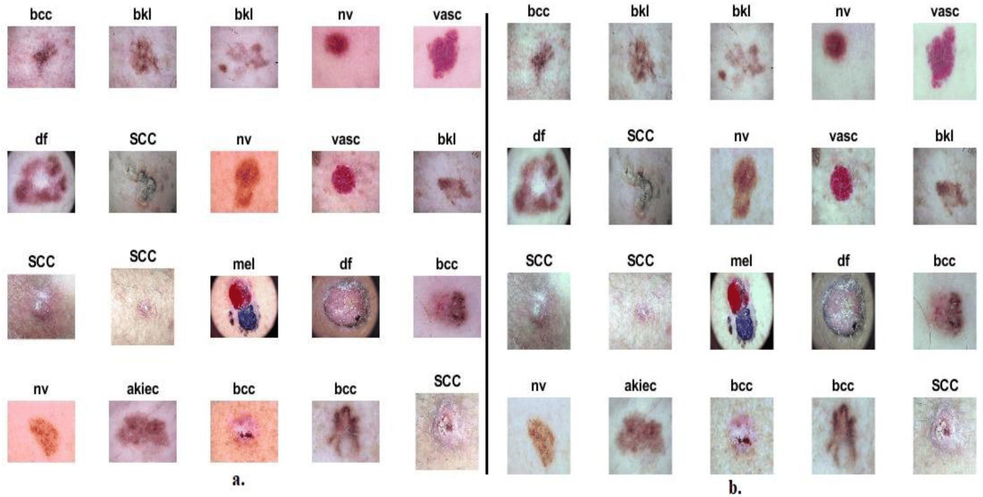
3.1. Description of ISIC 2019 Dataset
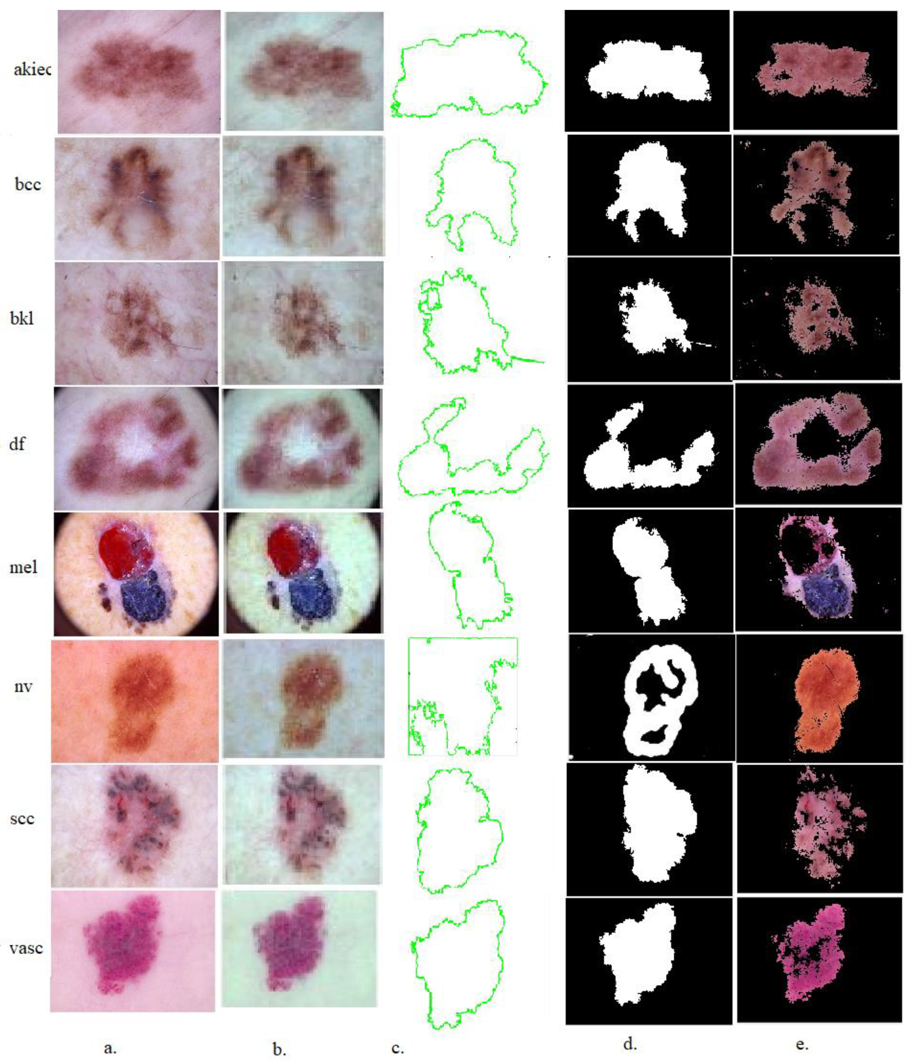
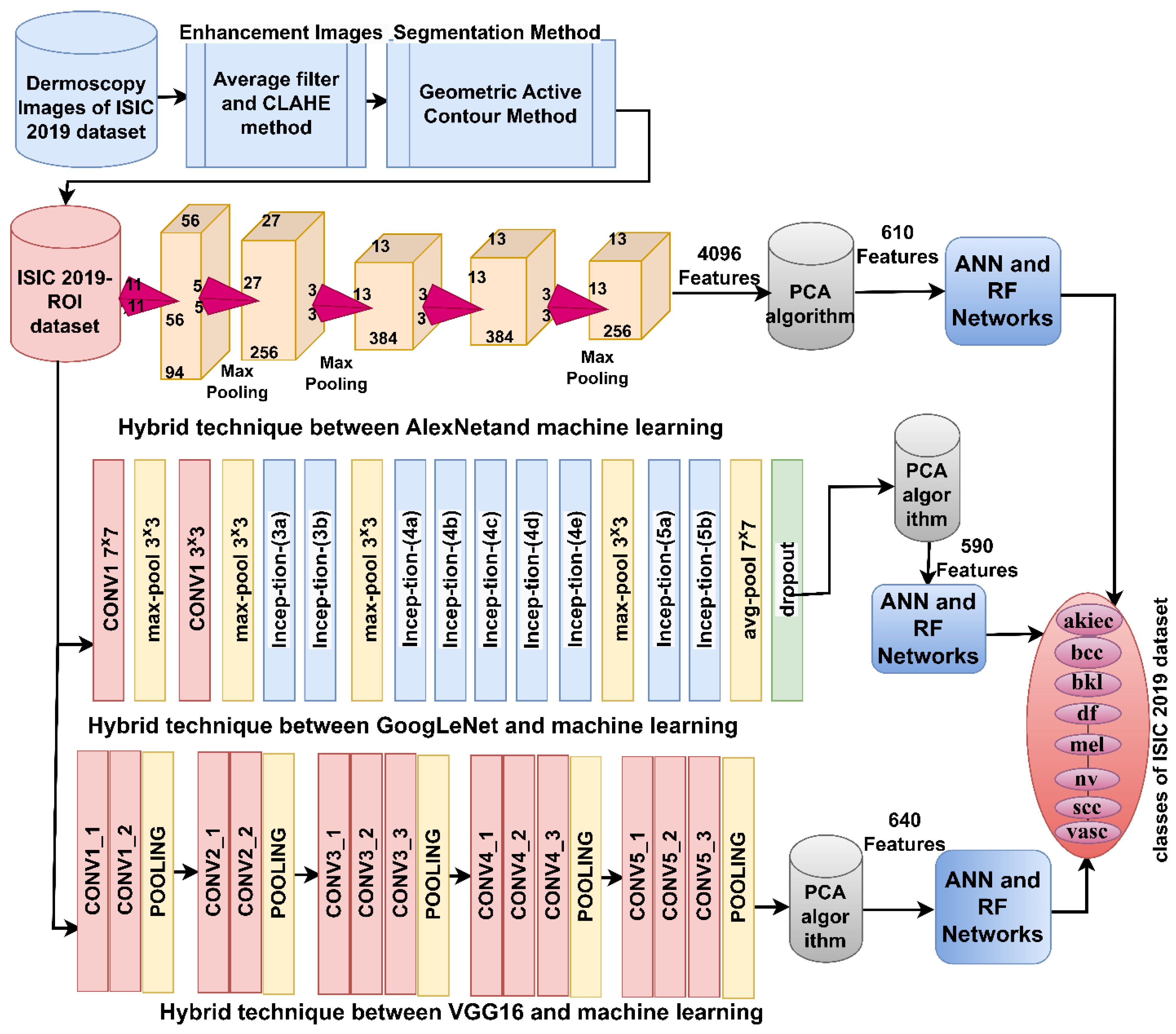
3.2. Enhancement of ISIC 2019 Dermoscopic Images
3.3. Geometric Active Contour Algorithm
3.4. Extract Deep Feature Maps
3.5. Inductive and Deductive Phase
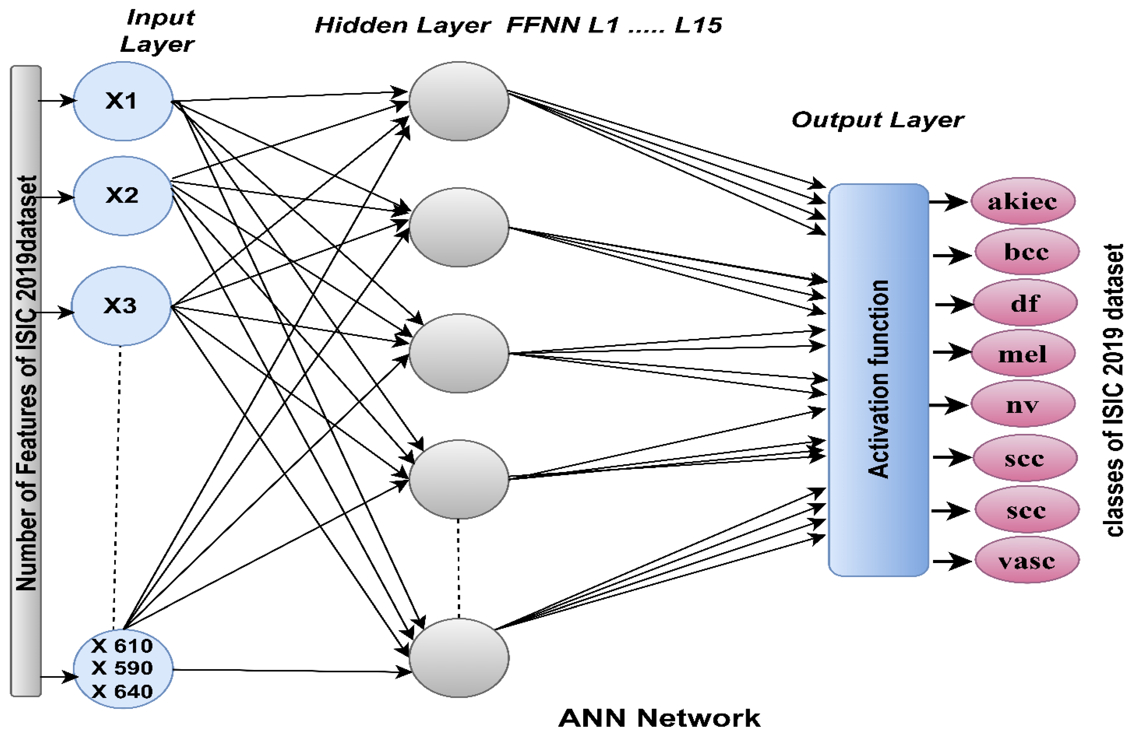
3.5.1. ANN Network
3.5.2. Random Forest Network
4. Experimental Results of the Proposed Systems
4.1. Split of ISIC 2019 Data Set
4.2. Systems Performance Measures
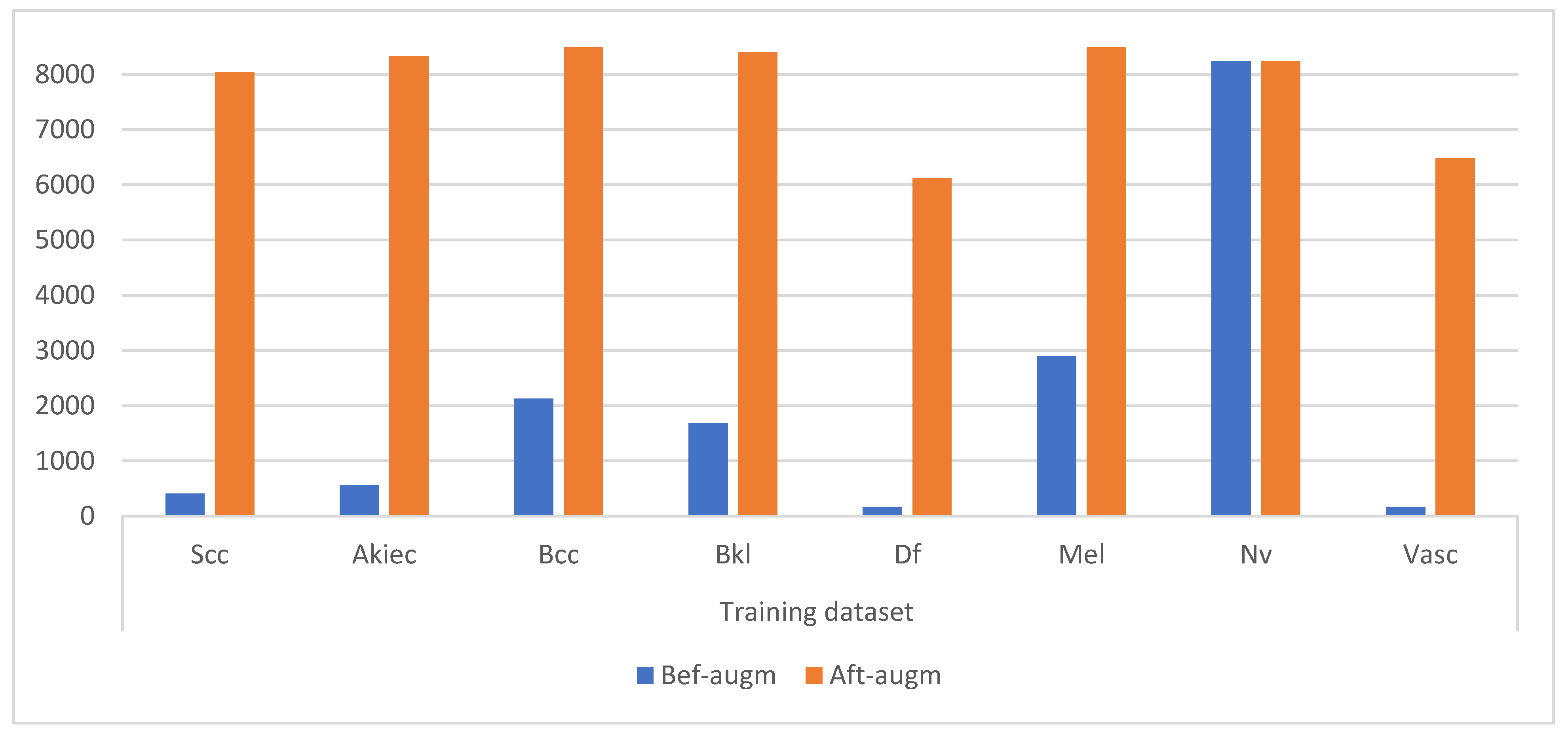
4.3. Balancing Classes of ISIC 2019 Dataset
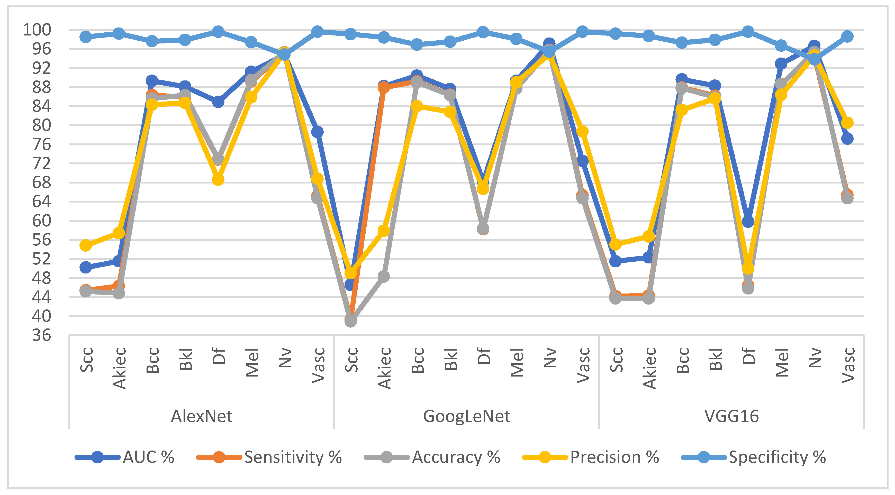
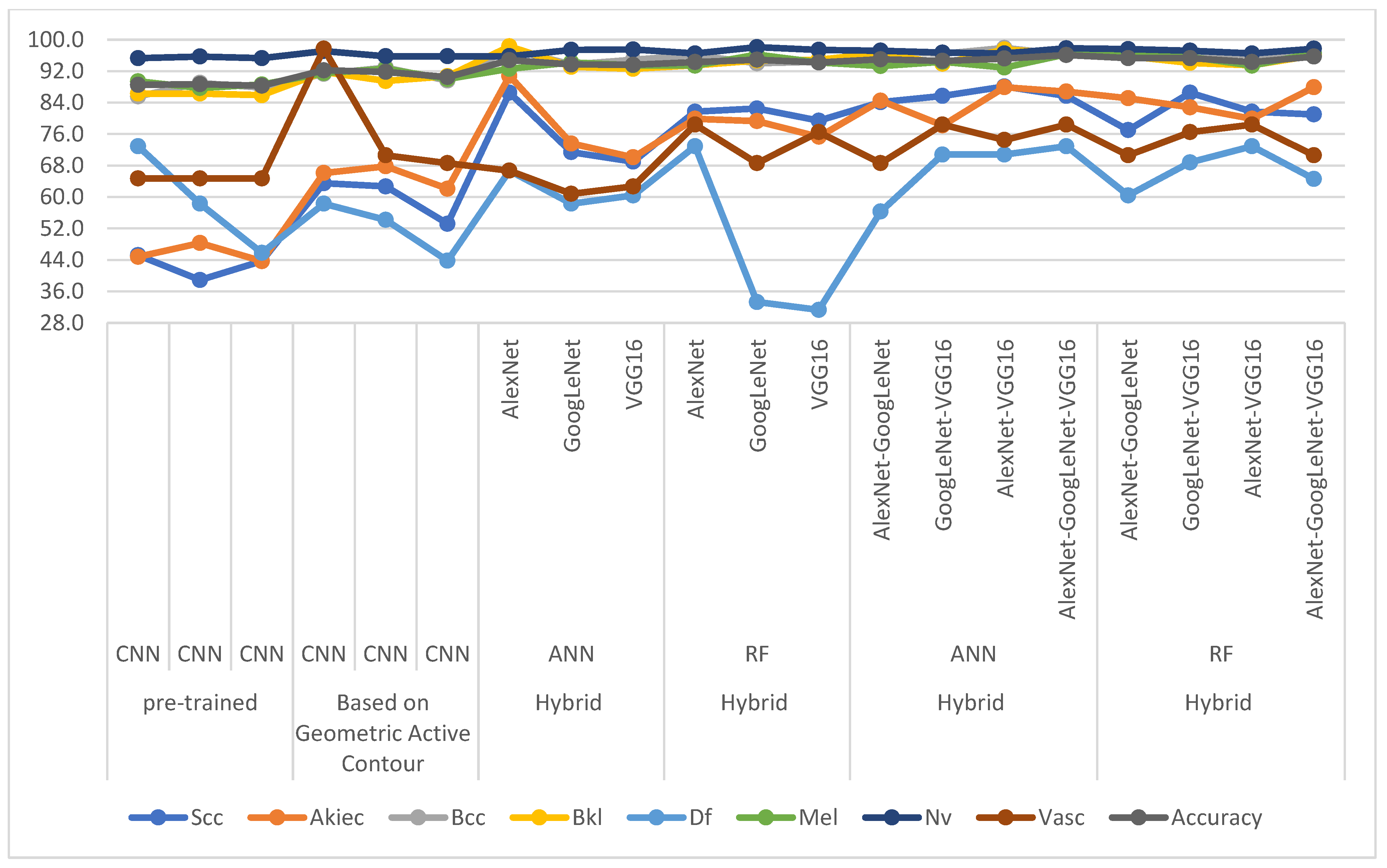
4.4. Results of Pre-Trained Deep Learning
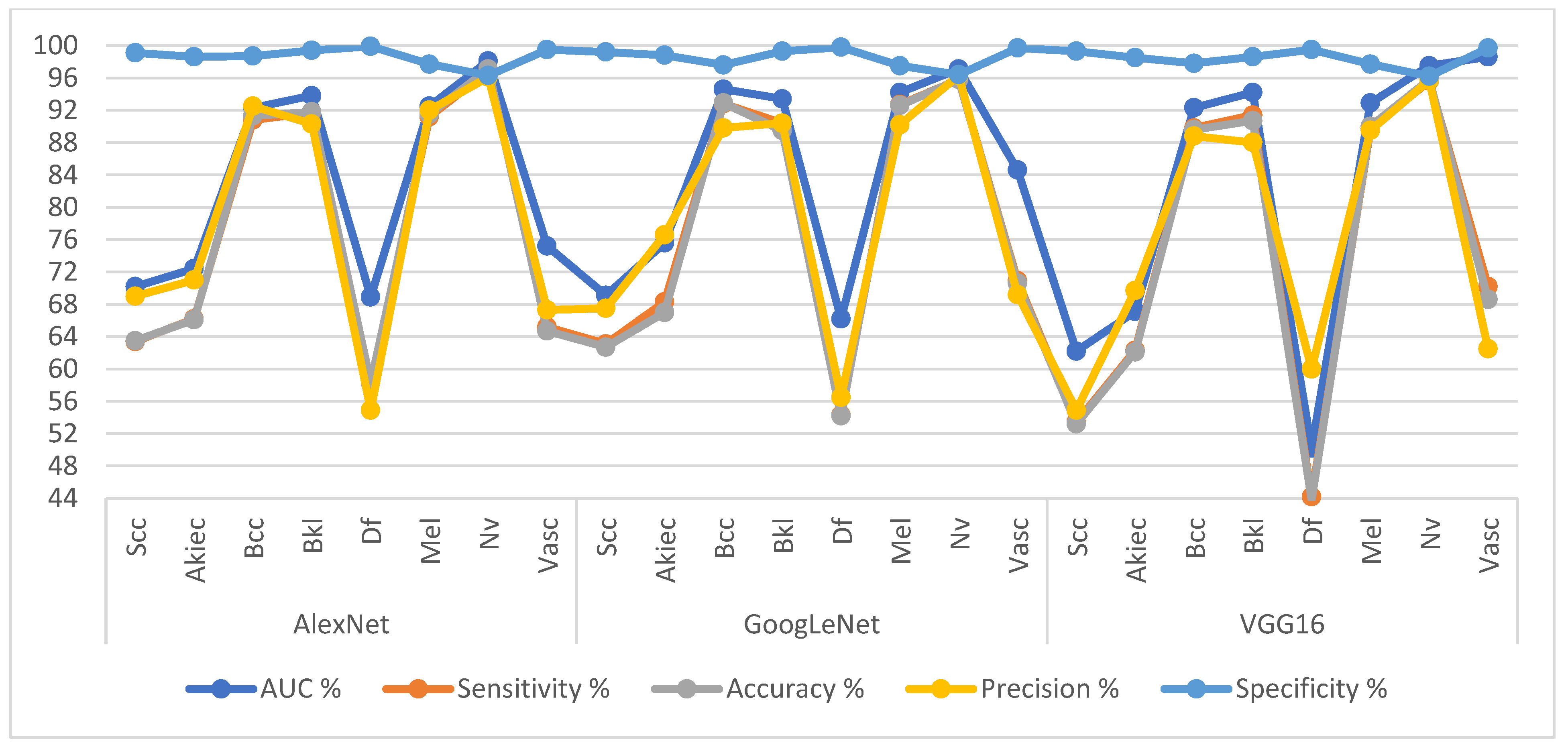
4.5. Results of Pre-Trained Deep Learning Based on GAC Algorithm
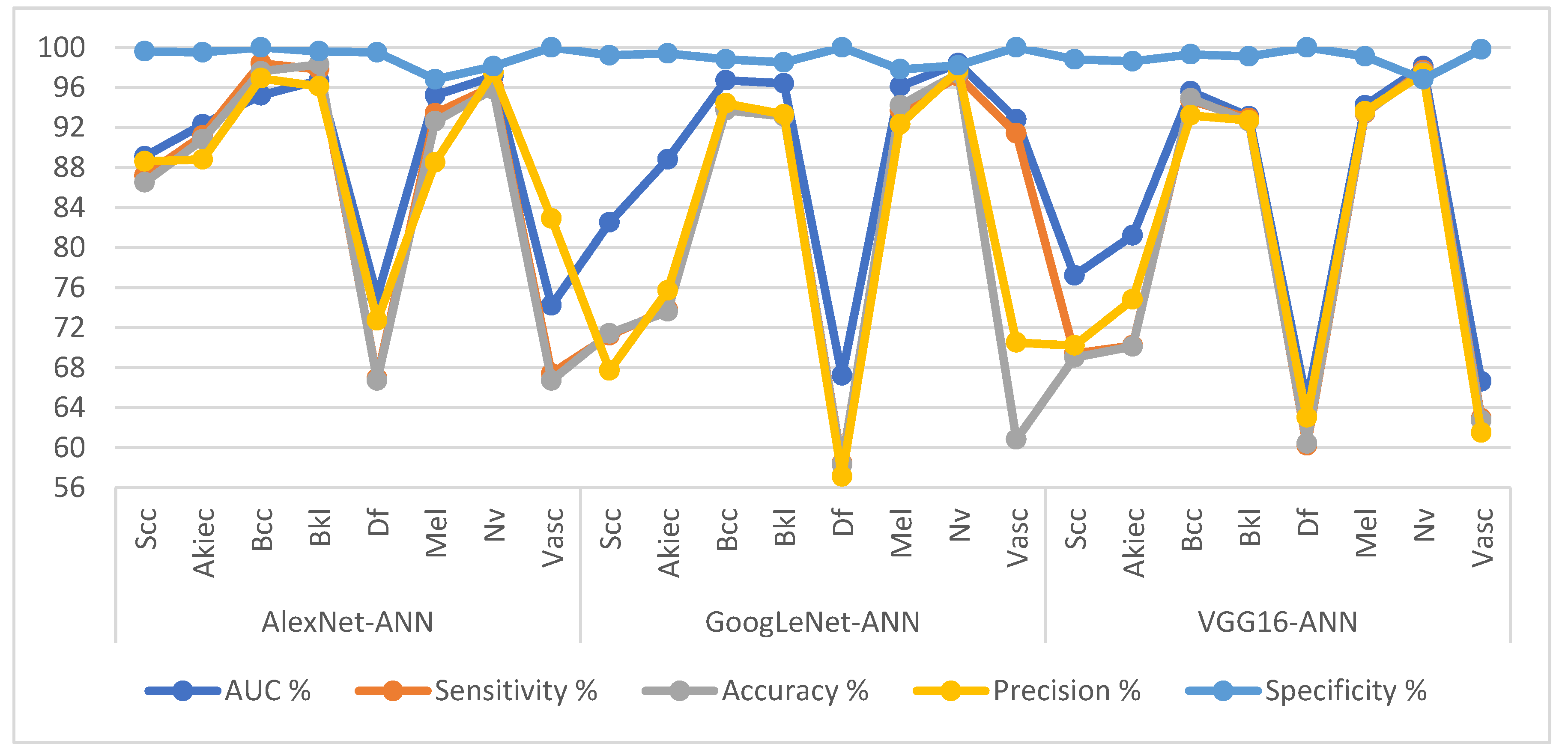
4.6. Results of Hybrid Models of CNN, ANN and RF
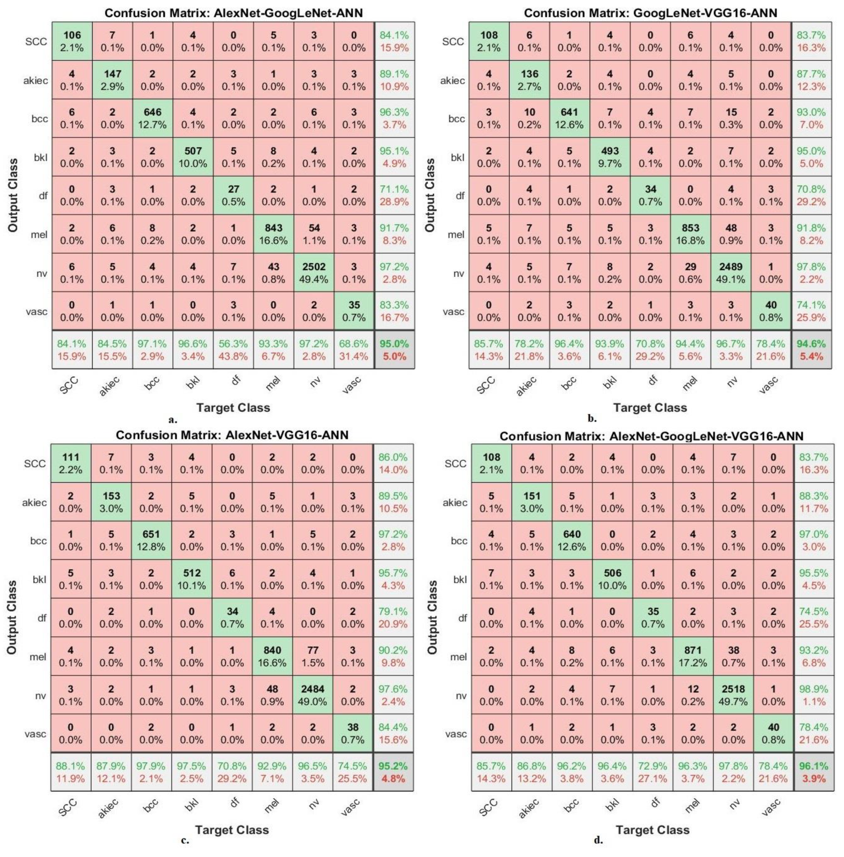
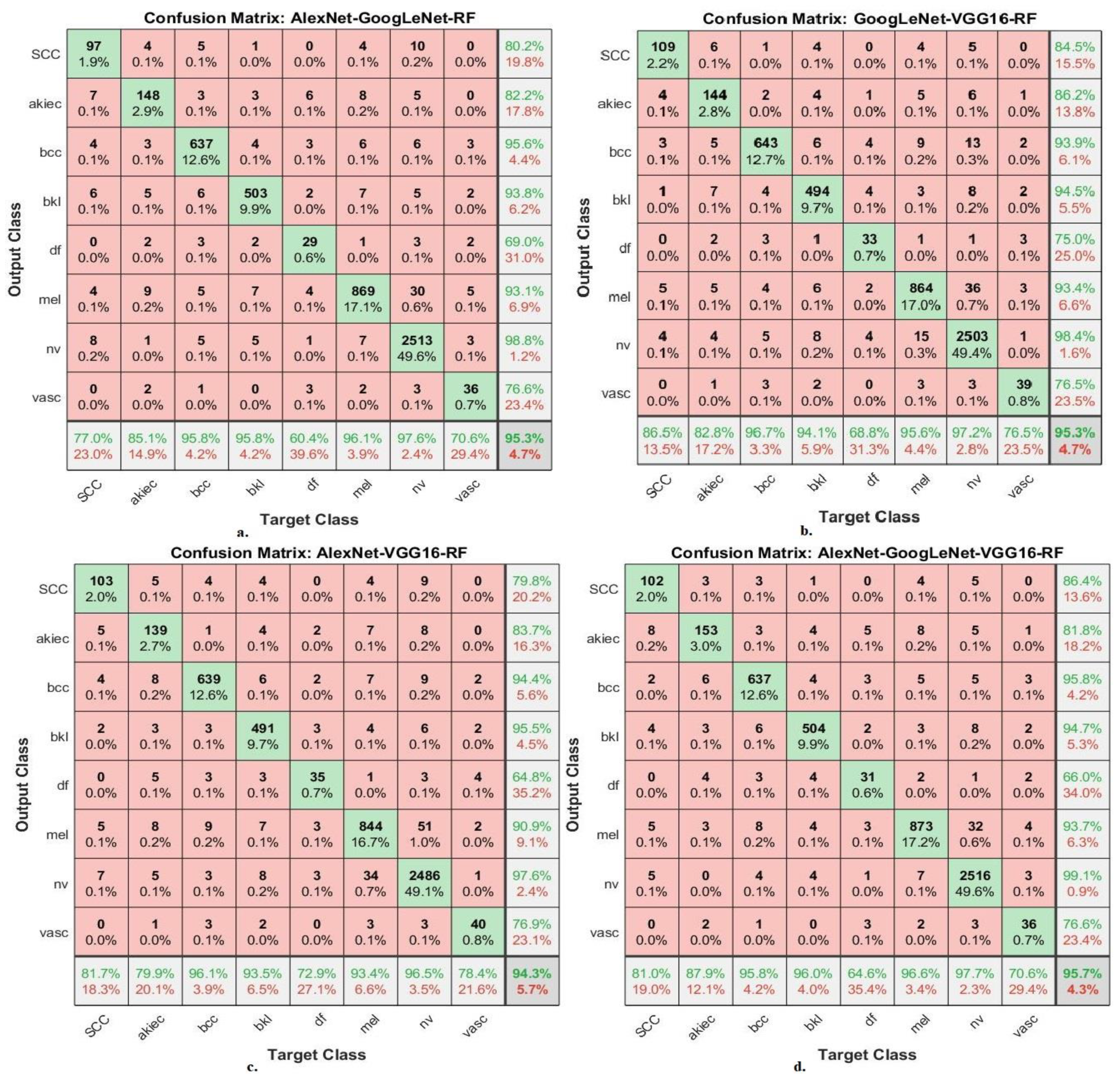
4.7. Results of Hybrid Models Based on Fused CNN Features
5. Discussion and Comparison of the Performance Results of the Systems
6. Conclusions
Author Contributions
Funding
Data Availability Statement
Acknowledgments
Conflicts of Interest
References
- Ragaa, T.M.; Sahar, S.A. Skin Managements and Diseases: A Systematic Article Review. Med. J. Cairo Univ. 2022, 90, 1773–1780. [Google Scholar] [CrossRef]
- Bortz, J.G.; Al-Shweiki, S. Free tarsal graft, and free skin graft for lower eyelid reconstruction. Ophthalmic Plast. Reconstr. Surg. 2020, 36, 605–609. [Google Scholar] [CrossRef] [PubMed]
- Holick, M.F. Sunlight, UV radiation, vitamin D, and skin cancer: How much sunlight do we need? Adv. Exp. Med. Biol. 2022, 1268, 19–36. [Google Scholar] [CrossRef]
- Saini, N.; Giacobone, C.K.; Klimczak, L.J.; Papas, B.N.; Burkholder, A.B.; Li, J.L.; Gordenin, D.A. UV-exposure, endogenous DNA damage, and DNA replication errors shape the spectra of genome changes in human skin. PLoS Genet. 2021, 17, e1009302. [Google Scholar] [CrossRef]
- Melanoma Survival Rates|Melanoma Survival Statistics. Available online: https://www.cancer.org/cancer/melanoma-skin-cancer/detection-diagnosis-staging/survival-rates-for-melanoma-skin-cancer-by-stage.html (accessed on 22 January 2023).
- Blázquez-Castro, A.; Stockert, J.C. Biomedical overview of melanin. 1. Updating melanin biology and chemistry, physico-chemical properties, melanoma tumors, and photothermal therapy. Biocell 2021, 45, 849–862. [Google Scholar] [CrossRef]
- Abd-El-Azim, H.; Tekko, I.A.; Ali, A.; Ramadan, A.; Nafee, N.; Khalafallah, N.; Donnelly, R.F. Hollow microneedle assisted intradermal delivery of hypericin lipid nanocapsules with light enabled photodynamic therapy against skin cancer. J. Control. Release 2022, 348, 849–869. [Google Scholar] [CrossRef]
- Toğaçar, M.; Cömert, Z.; Ergen, B. Intelligent skin cancer detection applying autoencoder, MobileNetV2 and spiking neural networks. Chaos Solitons Fractals 2021, 144, 110714. [Google Scholar] [CrossRef]
- Pathania, Y.S.; Apalla, Z.; Salerni, G.; Patil, A.; Grabbe, S.; Goldust, M. Non-invasive diagnostic techniques in pigmentary skin disorders and skin cancer. J. Cosmet. Dermatol. 2022, 21, 444–450. [Google Scholar] [CrossRef]
- Barhoumi, W.; Khelifa, A. Skin lesion image retrieval using transfer learning-based approach for query-driven distance recommendation. Comput. Biol. Med. 2021, 137, 104825. [Google Scholar] [CrossRef]
- Elansary, I.; Ismail, A.; Awad, W. Efficient classification model for melanoma based on convolutional neural networks. In Medical Informatics and Bioimaging Using Artificial Intelligence: Challenges, Issues, Innovations and Recent Developments; Studies in Computational Intelligence; Springer International Publishing: Cham, Switzerland, 2022; Volume 1005, pp. 15–27. [Google Scholar] [CrossRef]
- Pollastri, F.; Parreño, M.; Maroñas, J.; Bolelli, F.; Paredes, R.; Ramos, D.; Grana, C. A deep analysis on high-resolution dermoscopic image classification. IET Comput. Vis. 2021, 15, 514–526. [Google Scholar] [CrossRef]
- Sun, Q.; Huang, C.; Chen, M.; Xu, H.; Yang, Y. Skin lesion classification using additional patient information. BioMed Res. Int. 2021, 2021, 6673852. [Google Scholar] [CrossRef] [PubMed]
- Combalia, M.; Hueto, F.; Puig, S.; Malvehy, J.; Vilaplana, V. Uncertainty estimation in deep neural networks for dermoscopic image classification. In Proceedings of the IEEE/CVF Conference on Computer Vision and Pattern Recognition Workshops, New Orleans, LA, USA, 19–20 June 2022; pp. 744–745. [Google Scholar]
- Gong, A.; Yao, X.; Lin, W. Dermoscopy image classification based on StyleGANs and decision fusion. IEEE Access 2020, 8, 70640–70650. [Google Scholar] [CrossRef]
- Putra, T.A.; Rufaida, S.I.; Leu, J.S. Enhanced skin condition prediction through machine learning using dynamic training and testing augmentation. IEEE Access 2020, 8, 40536–40546. [Google Scholar] [CrossRef]
- Alizadeh, S.M.; Mahloojifar, A. Automatic skin cancer detection in dermoscopy images by combining convolutional neural networks and texture features. Int. J. Imaging Syst. Technol. 2021, 31, 695–707. [Google Scholar] [CrossRef]
- Iqbal, I.; Younus, M.; Walayat, K.; Kakar, M.U.; Ma, J. Automated multi-class classification of skin lesions through deep convolutional neural network with dermoscopic images. Comput. Med. Imaging Graph. 2021, 88, 101843. [Google Scholar] [CrossRef]
- Monika, M.K.; Vignesh, N.A.; Kumari, C.U.; Kumar, M.N.V.S.S.; Lydia, E.L. Skin cancer detection and classification using machine learning. Mater. Today Proc. 2020, 33, 4266–4270. [Google Scholar] [CrossRef]
- Pham, T.C.; Tran, C.T.; Luu, M.S.K.; Mai, D.A.; Doucet, A.; Luong, C.M. Improving binary skin cancer classification based on best model selection method combined with optimizing full connected layers of Deep CNN. In Proceedings of the IEEE.2020 International Conference on Multimedia Analysis and Pattern Recognition, Ha Noi, Vietnam, 8–9 October 2020. [Google Scholar] [CrossRef]
- Hoang, L.; Lee, S.H.; Lee, E.J.; Kwon, K.R. Multiclass skin lesion classification using a novel lightweight deep learning framework for smart healthcare. Appl. Sci. 2022, 12, 2677. [Google Scholar] [CrossRef]
- Xiao, J.; Xu, H.; Zhao, W.; Cheng, C.; Gao, H. A prior-mask-guided few-shot learning for skin lesion segmentation. Computing 2021, 105, 717–739. [Google Scholar] [CrossRef]
- Zanddizari, H.; Nguyen, N.; Zeinali, B.; Chang, J.M. A new preprocessing approach to improve the performance of CNN-based skin lesion classification. Med. Biol. Eng. Comput. 2021 59, 1123–1131. [CrossRef]
- Kassem, M.A.; Hosny, K.M.; Fouad, M.M. Skin lesions classification into eight classes for ISIC 2019 using deep convolutional neural network and transfer learning. IEEE Access 2020, 8, 114822–114832. [Google Scholar] [CrossRef]
- Villa-Pulgarin, J.P.; Ruales-Torres, A.A.; Arias-Garzon, D.; Bravo-Ortiz, M.A.; Arteaga-Arteaga, H.B.; Mora-Rubio, A.; Tabares-Soto, R. Optimized convolutional neural network models for skin lesion classification. Comput. Mater. Contin. 2022, 70, 2131–2148. [Google Scholar] [CrossRef]
- Codella, N.C.; Gutman, D.; Celebi, M.E.; Helba, B.; Marchetti, M.A.; Dusza, S.W.; Halpern, A. Skin lesion analysis toward melanoma detection: A challenge at the 2017 international symposium on biomedical imaging (ISBI), hosted by the international skin imaging collaboration (ISIC). In Proceedings of the 2018 IEEE 15th International Symposium on Biomedical Imaging (ISBI 2018), Washington, DC, USA, 4–7 April 2018; pp. 168–172. Available online: http://arxiv.org/abs/1605.01397 (accessed on 18 February 2023).
- Malik, S.; Akram, T.; Ashraf, I.; Rafiullah, M.; Ullah, M.; Tanveer, J. A Hybrid Preprocessor DE-ABC for Efficient Skin-Lesion Segmentation with Improved Contrast. Diagnostics 2022, 12, 2625. [Google Scholar] [CrossRef] [PubMed]
- Abunadi, I.; Senan, E.M. Multi-Method Diagnosis of Blood Microscopic Sample for Early Detection of Acute Lymphoblastic Leukemia Based on Deep Learning and Hybrid Techniques. Sensors 2022, 22, 1629. [Google Scholar] [CrossRef] [PubMed]
- Ahmed, I.A.; Senan, E.M.; Rassem, T.H.; Ali, M.A.; Shatnawi, H.S.A.; Alwazer, S.M.; Alshahrani, M. Eye Tracking-Based Diagnosis and Early Detection of Autism Spectrum Disorder Using Machine Learning and Deep Learning Techniques. Electronics 2022, 11, 530. [Google Scholar] [CrossRef]
- Jeyakumar, J.P.; Jude, A.; Priya, A.G.; Hemanth, J. A Survey on Computer-Aided Intelligent Methods to Identify and Classify Skin Cancer. Informatics 2022, 9, 99. [Google Scholar] [CrossRef]
- Khayretdinova, G.; Gout, C.; Chaumont-Frelet, T.; Kuksenko, S. Image Segmentation with a Priori Conditions: Applications to Medical and Geophysical Imaging. Math. Comput. Appl. 2022, 27, 26. [Google Scholar] [CrossRef]
- Zhang, H.; Liu, J.; Liu, J. Accurate Extraction of Ground Objects from Remote Sensing Image Based on Mark Clustering Point Process. ISPRS Int. J. Geo-Inf. 2022, 11, 402. [Google Scholar] [CrossRef]
- Pitchiah, M.S.; Rajamanickam, T. Efficient Feature Based Melanoma Skin Image Classification Using Machine Learning Approaches. Traitement Signal 2022, 39, 1663–1671. [Google Scholar] [CrossRef]
- Fati, S.M.; Senan, E.M.; ElHakim, N. Deep and Hybrid Learning Technique for Early Detection of Tuberculosis Based on X-ray Images Using Feature Fusion. Appl. Sci. 2022, 12, 7092. [Google Scholar] [CrossRef]
- Naeem, A.; Anees, T.; Fiza, M.; Naqvi, R.A.; Lee, S.-W. SCDNet: A Deep Learning-Based Framework for the Multiclassification of Skin Cancer Using Dermoscopy Images. Sensors 2022, 22, 5652. [Google Scholar] [CrossRef]
- Mohammed, B.A.; Senan, E.M.; Rassem, T.H.; Makbol, N.M.; Alanazi, A.A.; Al-Mekhlafi, Z.G.; Almurayziq, T.S.; Ghaleb, F.A. Multi-Method Analysis of Medical Records and MRI Images for Early Diagnosis of Dementia and Alzheimer’s Disease Based on Deep Learning and Hybrid Methods. Electronics 2021, 10, 2860. [Google Scholar] [CrossRef]
- Aljohani, K.; Turki, T. Automatic Classification of Melanoma Skin Cancer with Deep Convolutional Neural Networks. AI 2022, 3, 512–525. [Google Scholar] [CrossRef]
- Mohammed, B.A.; Senan, E.M.; Al-Mekhlafi, Z.G.; Rassem, T.H.; Makbol, N.M.; Alanazi, A.A.; Almurayziq, T.S.; Ghaleb, F.A.; Sallam, A.A. Multi-Method Diagnosis of CT Images for Rapid Detection of Intracranial Hemorrhages Based on Deep and Hybrid Learning. Electronics 2022, 11, 2460. [Google Scholar] [CrossRef]
- Vito, V.; Stefanus, L.Y. An Asymmetric Contrastive Loss for Handling Imbalanced Datasets. Entropy 2022, 24, 1303. [Google Scholar] [CrossRef]
- Silvestrini, S.; Lavagna, M. Deep Learning and Artificial Neural Networks for Spacecraft Dynamics, Navigation and Control. Drones 2022, 6, 270. [Google Scholar] [CrossRef]
- Senan, E.M.; Mohammed Jadhav, M.E.; Rassem, T.H.; Aljaloud, A.S.; Mohammed, B.A.; Al-Mekhlafi, Z.G. Early Diagnosis of Brain Tumour MRI Images Using Hybrid Techniques between Deep and Machine Learning. Comput. Math. Methods Med. 2022, 2022, 8330833. [Google Scholar] [CrossRef]
- Baig, A.R.; Abbas, Q.; Almakki, R.; Ibrahim, M.E.A.; AlSuwaidan, L.; Ahmed, A.E.S. Light-Dermo: A Lightweight Pretrained Convolution Neural Network for the Diagnosis of Multiclass Skin Lesions. Diagnostics 2023, 13, 385. [Google Scholar] [CrossRef]


| Phase | 80% (80:20) | Testing 20% | |
|---|---|---|---|
| Classes | Training (80%) | Validation (20%) | |
| Squamous cell carcinoma (Scc) | 402 | 100 | 126 |
| Actinic keratoses (Akiec) | 555 | 139 | 173 |
| Basal cell carcinoma (Bcc) | 2126 | 532 | 665 |
| Benign keratosis lesions (Bkl) | 1679 | 420 | 525 |
| Dermatofibroma (Df) | 153 | 38 | 48 |
| Melanoma (Mel) | 2894 | 724 | 904 |
| Melanocytic nevi (Nv) | 8240 | 2060 | 2575 |
| Vascular (Vasc) | 162 | 40 | 51 |
| Phase | Training Dataset | |||||||
|---|---|---|---|---|---|---|---|---|
| Classes | Scc | Akiec | Bcc | Bkl | Df | Mel | Nv | Vasc |
| Bef-augm | 402 | 555 | 2126 | 1679 | 153 | 2894 | 8240 | 162 |
| Aft-augm | 8040 | 8325 | 8504 | 8395 | 6120 | 8682 | 8240 | 6480 |
| Models | Type of Lesion | AUC % | Sensitivity % | Accuracy % | Precision % | Specificity % |
|---|---|---|---|---|---|---|
| AlexNet | Scc | 50.2 | 45.4 | 45.2 | 54.8 | 98.5 |
| Akiec | 51.5 | 46.3 | 44.8 | 57.4 | 99.2 | |
| Bcc | 89.3 | 86.3 | 85.6 | 84.3 | 97.6 | |
| Bkl | 88.1 | 85.9 | 86.3 | 84.7 | 97.9 | |
| Df | 84.9 | 72.8 | 72.9 | 68.6 | 99.6 | |
| Mel | 91.2 | 89.2 | 89.5 | 85.9 | 97.4 | |
| Nv | 94.8 | 95.2 | 95.3 | 95.2 | 94.8 | |
| Vasc | 78.6 | 65.2 | 64.7 | 68.8 | 99.6 | |
| GoogLeNet | Scc | 46.5 | 39.3 | 38.9 | 49 | 99.1 |
| Akiec | 88.2 | 87.9 | 48.3 | 57.9 | 98.4 | |
| Bcc | 90.4 | 89.2 | 89 | 84 | 96.9 | |
| Bkl | 87.6 | 86.4 | 86.3 | 82.8 | 97.5 | |
| Df | 68.2 | 58.2 | 58.3 | 66.7 | 99.5 | |
| Mel | 89.3 | 87.7 | 87.7 | 89 | 98.1 | |
| Nv | 97.1 | 95.8 | 95.7 | 94.9 | 95.4 | |
| Vasc | 72.5 | 65.3 | 64.7 | 78.7 | 99.6 | |
| VGG16 | Scc | 51.5 | 44.2 | 43.7 | 55 | 99.2 |
| Akiec | 52.3 | 44.3 | 43.7 | 56.7 | 98.7 | |
| Bcc | 89.6 | 87.9 | 87.8 | 83.2 | 97.3 | |
| Bkl | 88.3 | 86.2 | 85.9 | 85.6 | 97.9 | |
| Df | 59.8 | 46.4 | 45.8 | 50 | 99.6 | |
| Mel | 92.9 | 88.7 | 88.6 | 86.4 | 96.7 | |
| Nv | 96.6 | 94.6 | 95.3 | 94.6 | 93.8 | |
| Vasc | 77.2 | 65.4 | 64.7 | 80.5 | 98.6 |
| Models | Type of Lesion | AUC % | Sensitivity % | Accuracy % | Precision % | Specificity % |
|---|---|---|---|---|---|---|
| AlexNet | Scc | 70.2 | 63.4 | 63.5 | 69 | 99.1 |
| Akiec | 72.4 | 66.2 | 66.1 | 71 | 98.6 | |
| Bcc | 92.3 | 90.8 | 91.3 | 92.5 | 98.7 | |
| Bkl | 93.8 | 91.7 | 91.8 | 90.3 | 99.4 | |
| Df | 68.9 | 58.3 | 58.3 | 54.9 | 99.9 | |
| Mel | 92.5 | 91.2 | 91.5 | 92 | 97.7 | |
| Nv | 98.1 | 96.9 | 97.1 | 96.1 | 96.3 | |
| Vasc | 75.2 | 65.2 | 64.7 | 67.3 | 99.5 | |
| GoogLeNet | Scc | 69.1 | 63.1 | 62.7 | 67.5 | 99.2 |
| Akiec | 75.6 | 68.3 | 67 | 76.6 | 98.8 | |
| Bcc | 94.6 | 92.8 | 92.9 | 89.8 | 97.6 | |
| Bkl | 93.4 | 90.4 | 89.5 | 90.4 | 99.3 | |
| Df | 66.2 | 54.3 | 54.2 | 56.5 | 99.8 | |
| Mel | 94.2 | 92.7 | 92.6 | 90.2 | 97.5 | |
| Nv | 97.1 | 95.8 | 95.8 | 96.3 | 96.4 | |
| Vasc | 84.6 | 70.9 | 70.6 | 69.2 | 99.7 | |
| VGG16 | Scc | 62.2 | 53.4 | 53.2 | 54.9 | 99.3 |
| Akiec | 67.1 | 62.3 | 62.1 | 69.7 | 98.5 | |
| Bcc | 92.3 | 89.8 | 89.6 | 88.8 | 97.8 | |
| Bkl | 94.2 | 91.4 | 90.7 | 88 | 98.6 | |
| Df | 50.2 | 44.2 | 43.8 | 60 | 99.5 | |
| Mel | 92.9 | 89.7 | 90 | 89.5 | 97.7 | |
| Nv | 97.5 | 95.6 | 95.8 | 95.7 | 96.2 | |
| Vasc | 98.6 | 70.2 | 68.6 | 62.5 | 99.7 |
| Hybrid-Models | Type of Lesion | AUC % | Sensitivity % | Accuracy % | Precision % | Specificity % |
|---|---|---|---|---|---|---|
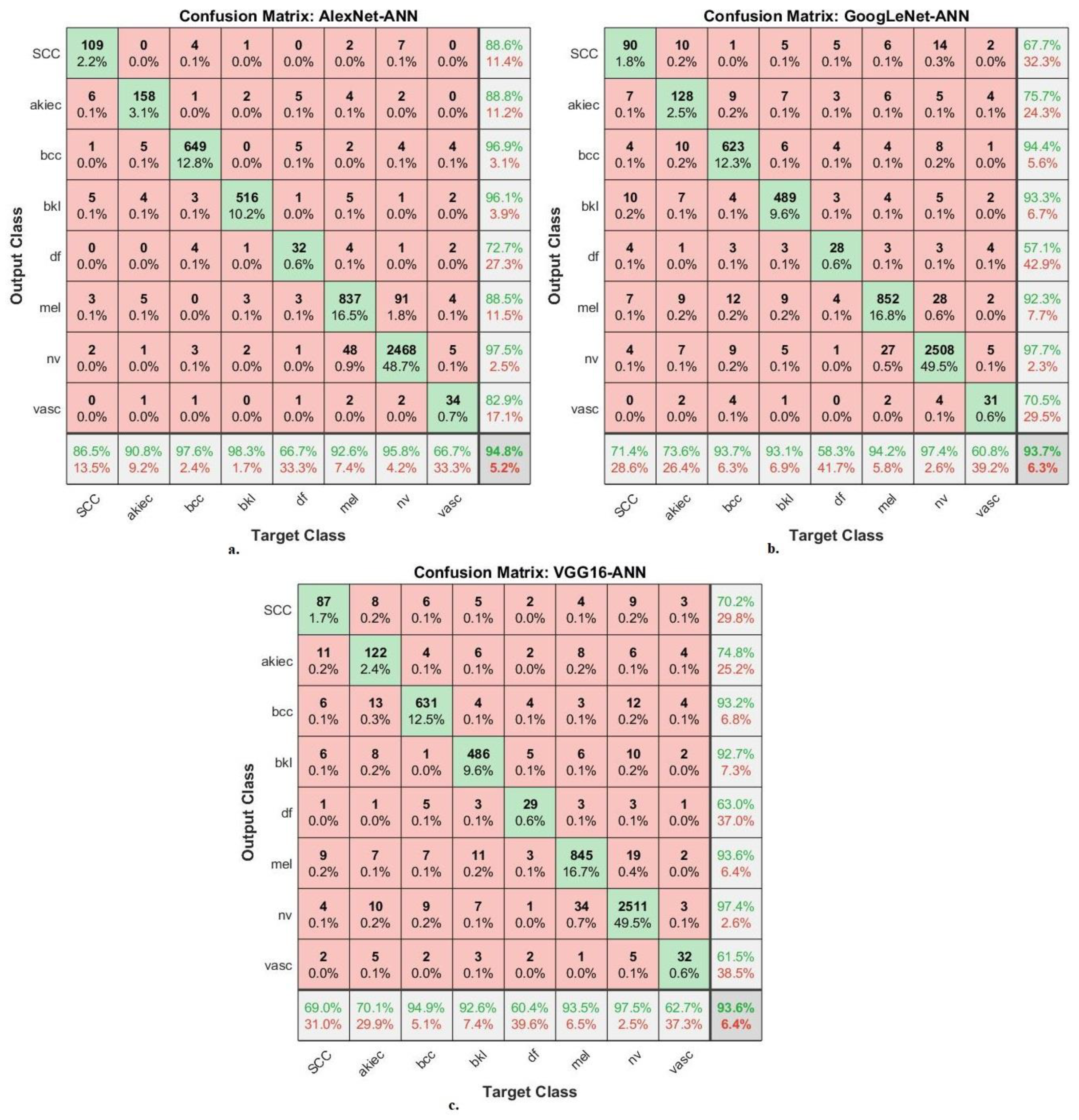
| AlexNet-ANN | Scc | 89.1 | 87.2 | 86.5 | 88.6 | 99.6 |
| Akiec | 92.3 | 91.2 | 90.8 | 88.8 | 99.5 | |
| Bcc | 95.2 | 98.4 | 97.6 | 96.9 | 100 | |
| Bkl | 96.7 | 97.8 | 98.3 | 96.1 | 99.6 | |
| Df | 75.1 | 66.9 | 66.7 | 72.7 | 99.5 | |
| Mel | 95.2 | 93.4 | 92.6 | 88.5 | 96.8 | |
| Nv | 97.1 | 96.1 | 95.8 | 97.5 | 98.1 | |
| Vasc | 74.2 | 67.4 | 66.7 | 82.9 | 100 | |
| GoogLeNet-ANN | Scc | 82.5 | 71.2 | 71.4 | 67.7 | 99.2 |
| Akiec | 88.8 | 73.8 | 73.6 | 75.7 | 99.4 | |
| Bcc | 96.7 | 93.9 | 93.7 | 94.4 | 98.8 | |
| Bkl | 96.4 | 93.2 | 93.1 | 93.3 | 98.5 | |
| Df | 67.2 | 58.4 | 58.3 | 57.1 | 100 | |
| Mel | 96.1 | 93.7 | 94.2 | 92.3 | 97.8 | |
| Nv | 98.4 | 97.1 | 97.4 | 97.7 | 98.2 | |
| Vasc | 92.8 | 91.4 | 60.8 | 70.5 | 100 | |
| VGG16-ANN | Scc | 77.2 | 69.4 | 69 | 70.2 | 98.8 |
| Akiec | 81.2 | 70.2 | 70.1 | 74.8 | 98.6 | |
| Bcc | 95.6 | 94.7 | 94.9 | 93.2 | 99.3 | |
| Bkl | 93.1 | 92.9 | 92.6 | 92.7 | 99.1 | |
| Df | 64.5 | 60.2 | 60.4 | 63 | 100 | |
| Mel | 94.2 | 93.4 | 93.5 | 93.6 | 99.1 | |
| Nv | 98.1 | 97.7 | 97.5 | 97.4 | 96.8 | |
| Vasc | 66.6 | 62.9 | 62.7 | 61.5 | 99.8 |
| Hybrid-Models | Type of Lesion | AUC % | Sensitivity % | Accuracy % | Precision % | Specificity % |
|---|---|---|---|---|---|---|
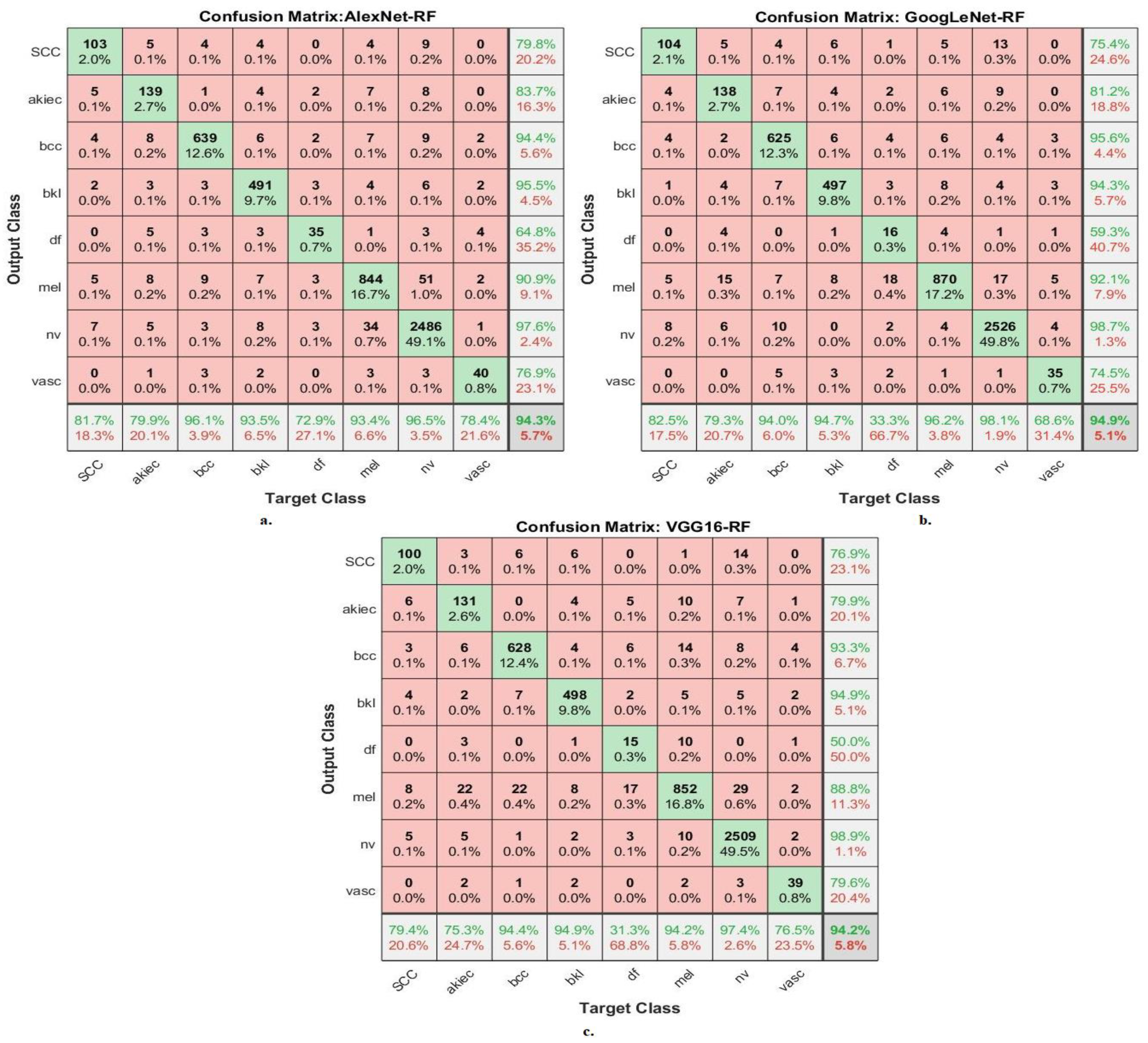
| AlexNet-RF | Scc | 89.2 | 82.3 | 81.7 | 79.8 | 99.1 |
| Akiec | 84.6 | 80.4 | 79.9 | 83.7 | 99.3 | |
| Bcc | 96.7 | 95.7 | 96.1 | 94.4 | 89.7 | |
| Bkl | 95.3 | 93.8 | 93.5 | 95.5 | 98.5 | |
| Df | 87.1 | 73.4 | 72.9 | 64.8 | 99.6 | |
| Mel | 93.1 | 92.9 | 93.4 | 90.9 | 97.8 | |
| Nv | 94.5 | 96.6 | 96.5 | 97.6 | 98.4 | |
| Vasc | 81.9 | 78.4 | 78.4 | 76.9 | 99.7 | |
| GoogLeNet-RF | Scc | 90.2 | 83.4 | 82.5 | 75.4 | 98.7 |
| Akiec | 82.1 | 79.3 | 79.3 | 81.2 | 99.2 | |
| Bcc | 94.3 | 94.3 | 94 | 95.6 | 98.6 | |
| Bkl | 91.2 | 94.8 | 94.7 | 94.3 | 99.1 | |
| Df | 79.6 | 33.4 | 33.3 | 59.3 | 99.8 | |
| Mel | 94.2 | 96.4 | 96.2 | 92.1 | 97.5 | |
| Nv | 96.8 | 97.8 | 98.1 | 98.7 | 99.5 | |
| Vasc | 85.4 | 69.3 | 68.6 | 74.5 | 100 | |
| VGG16-RF | Scc | 82.5 | 79.3 | 79.4 | 76.9 | 99.2 |
| Akiec | 86.2 | 75.2 | 75.3 | 79.9 | 98.8 | |
| Bcc | 96.1 | 94.3 | 94.4 | 93.3 | 98.6 | |
| Bkl | 94.5 | 95.4 | 94.9 | 94.9 | 98.5 | |
| Df | 67.2 | 31.4 | 31.3 | 50 | 100 | |
| Mel | 92.9 | 94.3 | 94.2 | 88.8 | 97.2 | |
| Nv | 97.2 | 96.9 | 97.4 | 98.9 | 99.3 | |
| Vasc | 87.2 | 76.4 | 76.5 | 79.6 | 99.7 |
| Fusion Features | Classifier | Type of Lesion | AUC % | Sensitivity % | Accuracy % | Precision % | Specificity % |
|---|---|---|---|---|---|---|---|
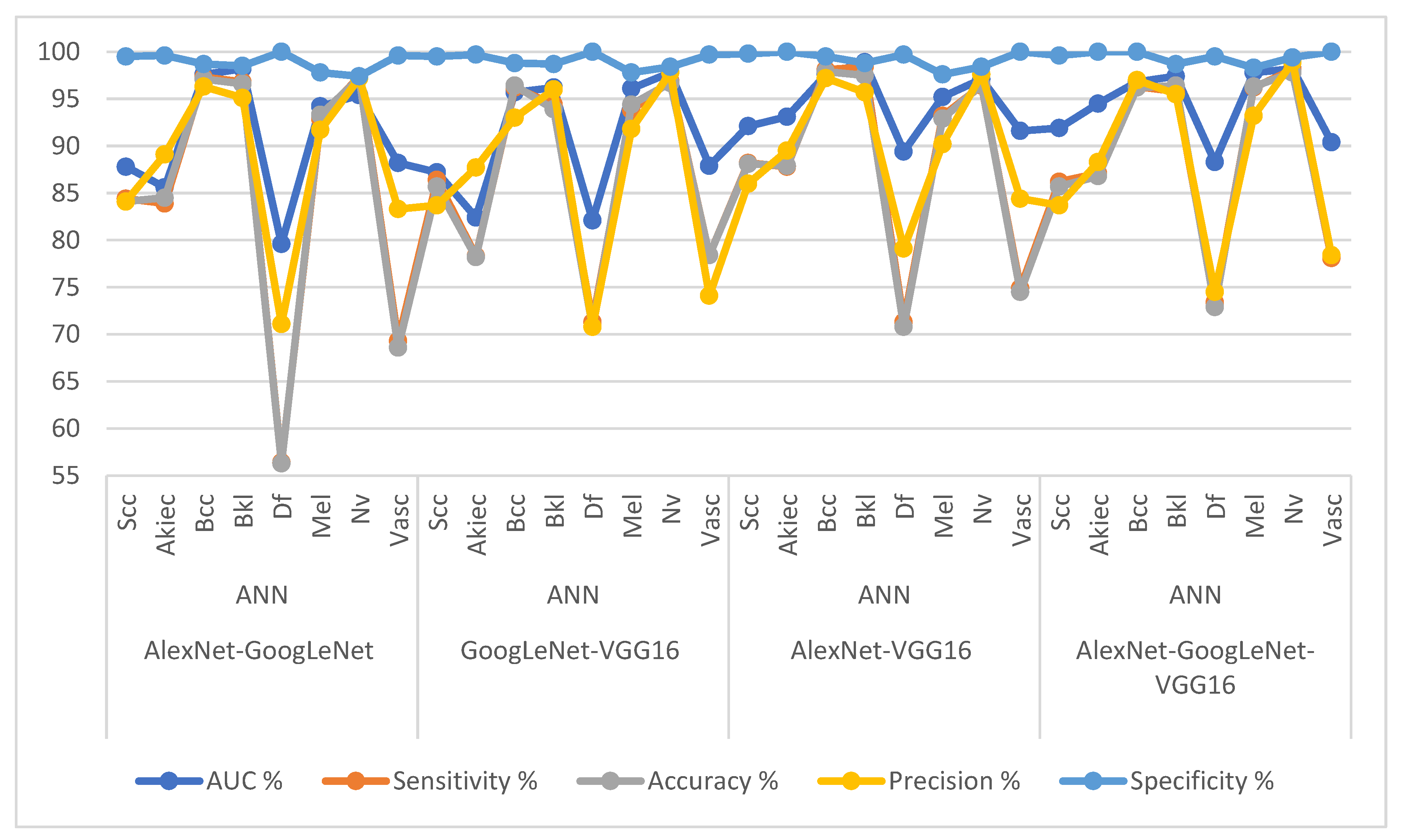
| AlexNet-GoogLeNet | ANN | Scc | 87.8 | 84.4 | 84.1 | 84.1 | 99.5 |
| Akiec | 85.6 | 83.9 | 84.5 | 89.1 | 99.6 | ||
| Bcc | 97.6 | 97.3 | 97.1 | 96.3 | 98.7 | ||
| Bkl | 98.2 | 96.8 | 96.6 | 95.1 | 98.5 | ||
| Df | 79.6 | 56.4 | 56.3 | 71.1 | 100 | ||
| Mel | 94.2 | 92.9 | 93.3 | 91.7 | 97.8 | ||
| Nv | 95.4 | 97.4 | 97.2 | 97.2 | 97.4 | ||
| Vasc | 88.2 | 69.3 | 68.6 | 83.3 | 99.6 | ||
| GoogLeNet-VGG16 | ANN | Scc | 87.2 | 86.4 | 85.7 | 83.7 | 99.5 |
| Akiec | 82.4 | 78.3 | 78.2 | 87.7 | 99.7 | ||
| Bcc | 95.7 | 96.2 | 96.4 | 93 | 98.8 | ||
| Bkl | 96.2 | 94.4 | 93.9 | 96 | 98.7 | ||
| Df | 82.1 | 71.3 | 70.8 | 70.8 | 100 | ||
| Mel | 96.1 | 93.8 | 94.4 | 91.8 | 97.8 | ||
| Nv | 97.8 | 96.9 | 96.7 | 97.8 | 98.4 | ||
| Vasc | 87.9 | 78.4 | 78.4 | 74.1 | 99.7 | ||
| AlexNet-VGG16 | ANN | Scc | 92.1 | 88.2 | 88.1 | 86 | 99.8 |
| Akiec | 93.1 | 87.8 | 87.9 | 89.5 | 100 | ||
| Bcc | 97.6 | 98.1 | 97.9 | 97.2 | 99.5 | ||
| Bkl | 98.9 | 98.4 | 97.5 | 95.7 | 98.8 | ||
| Df | 89.4 | 71.3 | 70.8 | 79.1 | 99.7 | ||
| Mel | 95.2 | 93.2 | 92.9 | 90.2 | 97.6 | ||
| Nv | 97.1 | 96.4 | 96.5 | 97.6 | 98.4 | ||
| Vasc | 91.6 | 74.9 | 74.5 | 84.4 | 100 | ||
| AlexNet-GoogLeNet-VGG16 | ANN | Scc | 91.9 | 86.2 | 85.7 | 83.7 | 99.6 |
| Akiec | 94.5 | 87.1 | 86.8 | 88.3 | 100 | ||
| Bcc | 96.8 | 96.3 | 96.2 | 97 | 100 | ||
| Bkl | 97.4 | 95.8 | 96.4 | 95.5 | 98.7 | ||
| Df | 88.3 | 73.4 | 72.9 | 74.5 | 99.5 | ||
| Mel | 97.8 | 96.2 | 96.3 | 93.2 | 98.3 | ||
| Nv | 98.2 | 98.1 | 97.8 | 98.9 | 99.4 | ||
| Vasc | 90.4 | 78.1 | 78.4 | 78.4 | 100 |
| Fusion Features | Classifier | Type of Lesion | AUC % | Sensitivity % | Accuracy % | Precision % | Specificity % |
|---|---|---|---|---|---|---|---|
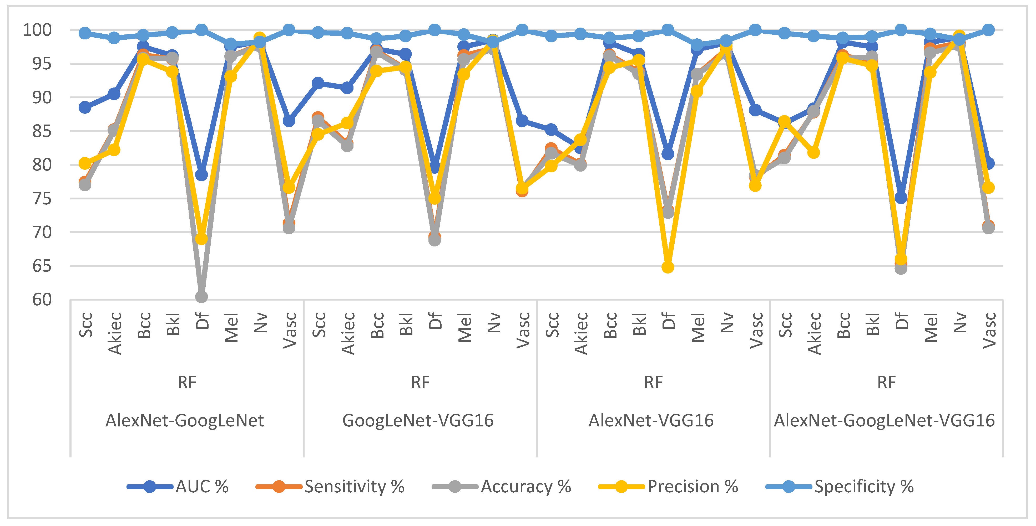
| AlexNet-GoogLeNet | RF | Scc | 88.5 | 77.4 | 77 | 80.2 | 99.5 |
| Akiec | 90.5 | 85.2 | 85.1 | 82.2 | 98.8 | ||
| Bcc | 97.5 | 96.3 | 95.8 | 95.6 | 99.2 | ||
| Bkl | 96.2 | 95.7 | 95.8 | 93.8 | 99.6 | ||
| Df | 78.5 | 60.4 | 60.4 | 69 | 100 | ||
| Mel | 97.5 | 96.1 | 96.1 | 93.1 | 97.9 | ||
| Nv | 98.3 | 97.8 | 97.6 | 98.8 | 98.2 | ||
| Vasc | 86.5 | 71.3 | 70.6 | 76.6 | 100 | ||
| GoogLeNet-VGG16 | RF | Scc | 92.1 | 87 | 86.5 | 84.5 | 99.6 |
| Akiec | 91.4 | 83.1 | 82.8 | 86.2 | 99.5 | ||
| Bcc | 97.2 | 96.9 | 96.7 | 93.9 | 98.7 | ||
| Bkl | 96.4 | 94.2 | 94.1 | 94.5 | 99.1 | ||
| Df | 79.6 | 69.3 | 68.8 | 75 | 100 | ||
| Mel | 97.5 | 96.2 | 95.6 | 93.4 | 99.3 | ||
| Nv | 98.5 | 97.3 | 97.2 | 98.4 | 98.2 | ||
| Vasc | 86.5 | 76.1 | 76.5 | 76.5 | 100 | ||
| AlexNet-VGG16 | RF | Scc | 85.2 | 82.4 | 81.7 | 79.8 | 99.1 |
| Akiec | 82.5 | 80.1 | 79.9 | 83.7 | 99.4 | ||
| Bcc | 98.1 | 96.3 | 96.1 | 94.4 | 98.8 | ||
| Bkl | 96.4 | 93.8 | 93.5 | 95.5 | 99.1 | ||
| Df | 81.6 | 73.2 | 72.9 | 64.8 | 100 | ||
| Mel | 97.1 | 93.4 | 93.4 | 90.9 | 97.8 | ||
| Nv | 98.1 | 97.1 | 96.5 | 97.6 | 98.4 | ||
| Vasc | 88.1 | 78.2 | 78.4 | 76.9 | 100 | ||
| AlexNet-GoogLeNet-VGG16 | RF | Scc | 86.2 | 81.4 | 81 | 86.4 | 99.5 |
| Akiec | 88.3 | 87.8 | 87.9 | 81.8 | 99.1 | ||
| Bcc | 98.2 | 96.2 | 95.8 | 95.8 | 98.8 | ||
| Bkl | 97.5 | 95.4 | 96 | 94.7 | 99 | ||
| Df | 75.1 | 65.3 | 64.6 | 66 | 100 | ||
| Mel | 98.2 | 97.2 | 96.6 | 93.7 | 99.4 | ||
| Nv | 98.9 | 98.1 | 97.7 | 99.1 | 98.6 | ||
| Vasc | 80.2 | 70.9 | 70.6 | 76.6 | 100 |
| Techniques | Features | Scc | Akiec | Bcc | Bkl | Df | Mel | Nv | Vasc | Accuracy | |
|---|---|---|---|---|---|---|---|---|---|---|---|
| Pre-trained | AlexNet | 45.2 | 44.8 | 85.6 | 86.3 | 72.9 | 89.4 | 95.3 | 64.7 | 88.5 | |
| GoogLeNet | 38.9 | 48.3 | 89.0 | 86.3 | 58.3 | 87.7 | 95.7 | 64.7 | 88.7 | ||
| VGG16 | 43.7 | 43.7 | 87.8 | 85.9 | 45.8 | 88.6 | 95.3 | 64.7 | 88.3 | ||
| Based on Geometric Active Contour | AlexNet | 63.5 | 66.1 | 91.3 | 91.8 | 58.3 | 91.5 | 97.1 | 97.7 | 92.2 | |
| GoogLeNet | 62.7 | 67.8 | 92.9 | 89.5 | 54.2 | 92.6 | 95.8 | 70.6 | 91.8 | ||
| VGG16 | 53.2 | 62.1 | 89.6 | 90.7 | 43.8 | 90.0 | 95.8 | 68.6 | 90.5 | ||
| Hybrid | ANN | AlexNet | 86.5 | 90.8 | 97.6 | 98.3 | 66.7 | 92.6 | 95.8 | 66.7 | 94.8 |
| GoogLeNet | 71.4 | 73.6 | 93.7 | 93.1 | 58.3 | 94.2 | 97.4 | 60.8 | 93.7 | ||
| VGG16 | 69.0 | 70.1 | 94.9 | 92.6 | 60.4 | 93.5 | 97.5 | 62.7 | 93.6 | ||
| Hybrid | RF | AlexNet | 81.7 | 79.9 | 96.1 | 93.5 | 72.9 | 93.4 | 96.5 | 78.4 | 94.3 |
| GoogLeNet | 82.5 | 79.3 | 94.0 | 94.7 | 33.3 | 96.2 | 98.1 | 68.6 | 94.9 | ||
| VGG16 | 79.4 | 75.3 | 94.4 | 94.9 | 31.3 | 94.2 | 97.4 | 76.5 | 94.2 | ||
| Hybrid | ANN | AlexNet-GoogLeNet | 84.1 | 84.5 | 97.1 | 96.6 | 56.3 | 93.3 | 97.2 | 68.6 | 95.0 |
| GoogLeNet-VGG16 | 85.7 | 78.2 | 96.4 | 93.9 | 70.8 | 94.4 | 96.7 | 78.4 | 94.6 | ||
| AlexNet-VGG16 | 88.1 | 87.9 | 97.9 | 97.5 | 70.8 | 92.9 | 96.5 | 74.5 | 95.2 | ||
| AlexNet-GoogLeNet-VGG16 | 85.7 | 86.8 | 96.2 | 96.4 | 72.9 | 96.3 | 97.8 | 78.4 | 96.1 | ||
| Hybrid | RF | AlexNet-GoogLeNet | 77.0 | 85.1 | 95.8 | 95.8 | 60.4 | 96.1 | 97.6 | 70.6 | 95.3 |
| GoogLeNet-VGG16 | 86.5 | 82.8 | 96.7 | 94.1 | 68.8 | 95.6 | 97.2 | 76.5 | 95.3 | ||
| AlexNet-VGG16 | 81.7 | 79.9 | 96.1 | 93.5 | 72.9 | 93.4 | 96.5 | 78.4 | 94.3 | ||
| AlexNet-GoogLeNet-VGG16 | 81.0 | 87.9 | 95.8 | 96.0 | 64.6 | 96.6 | 97.7 | 70.6 | 95.7 | ||
Disclaimer/Publisher’s Note: The statements, opinions and data contained in all publications are solely those of the individual author(s) and contributor(s) and not of MDPI and/or the editor(s). MDPI and/or the editor(s) disclaim responsibility for any injury to people or property resulting from any ideas, methods, instructions or products referred to in the content. |
© 2023 by the authors. Licensee MDPI, Basel, Switzerland. This article is an open access article distributed under the terms and conditions of the Creative Commons Attribution (CC BY) license (https://creativecommons.org/licenses/by/4.0/).
Share and Cite
Olayah, F.; Senan, E.M.; Ahmed, I.A.; Awaji, B. AI Techniques of Dermoscopy Image Analysis for the Early Detection of Skin Lesions Based on Combined CNN Features. Diagnostics 2023, 13, 1314. https://doi.org/10.3390/diagnostics13071314
Olayah F, Senan EM, Ahmed IA, Awaji B. AI Techniques of Dermoscopy Image Analysis for the Early Detection of Skin Lesions Based on Combined CNN Features. Diagnostics. 2023; 13(7):1314. https://doi.org/10.3390/diagnostics13071314
Chicago/Turabian StyleOlayah, Fekry, Ebrahim Mohammed Senan, Ibrahim Abdulrab Ahmed, and Bakri Awaji. 2023. "AI Techniques of Dermoscopy Image Analysis for the Early Detection of Skin Lesions Based on Combined CNN Features" Diagnostics 13, no. 7: 1314. https://doi.org/10.3390/diagnostics13071314