
Unilateral Tamoxifen-Induced Retinopathy as a Consequence of Breast Cancer Treatment—Multimodal Imaging Value
Abstract
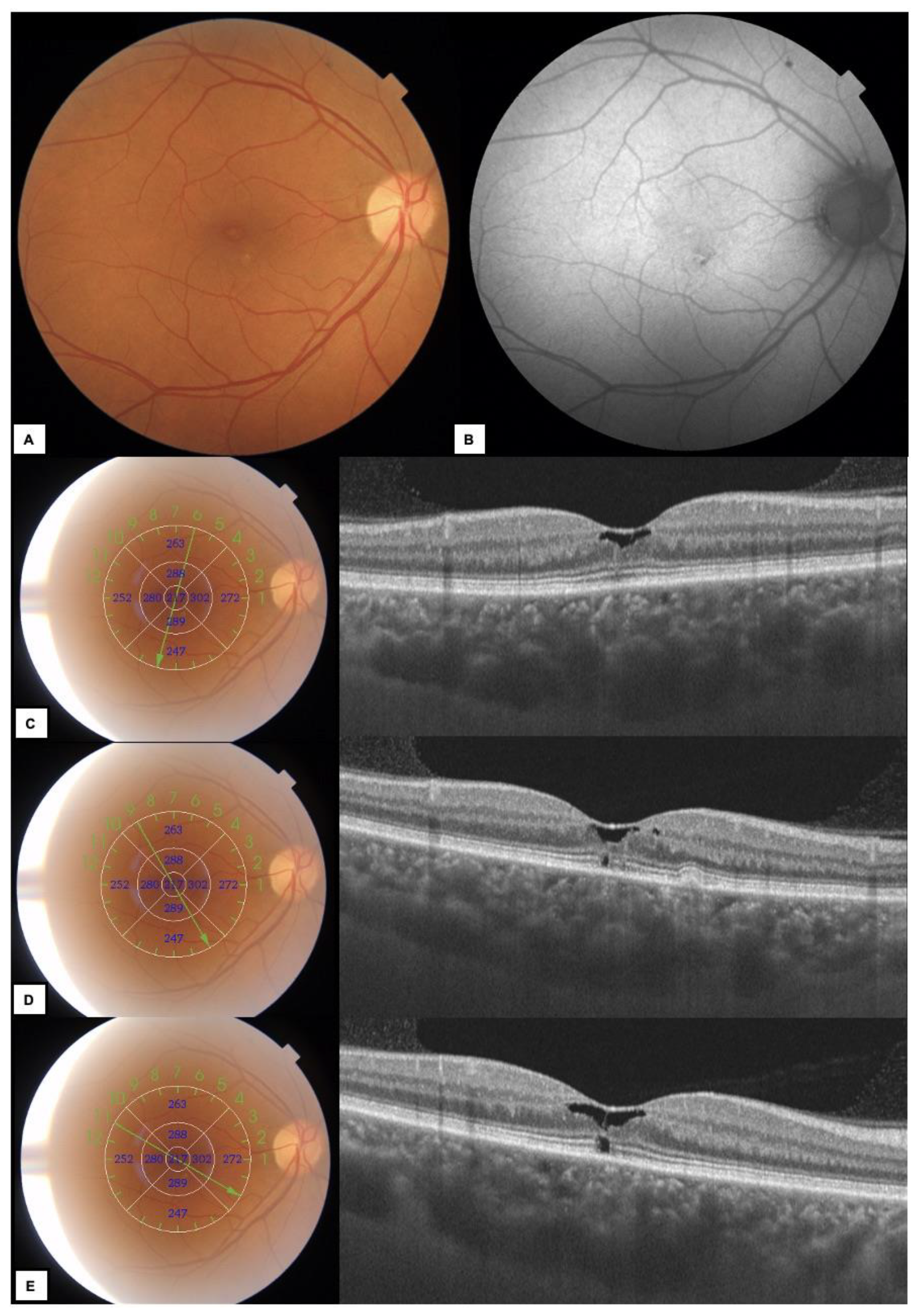
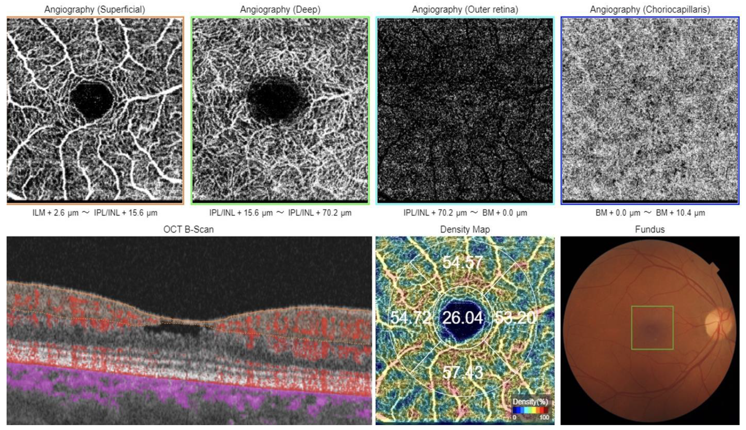
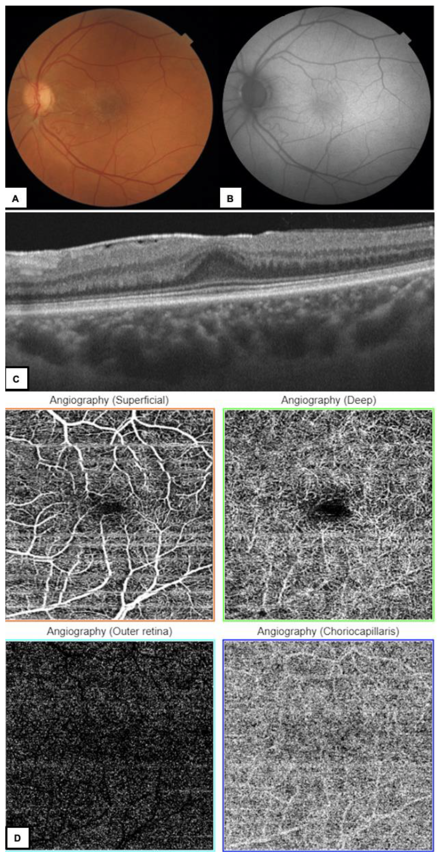
:


Supplementary Materials
Author Contributions
Funding
Institutional Review Board Statement
Informed Consent Statement
Data Availability Statement
Conflicts of Interest
References
- Eisner, A.; Luoh, S.-W. Breast Cancer Medications and Vision: Effects of Treatments for Early-stage Disease. Curr. Eye Res. 2011, 36, 867–885. [Google Scholar] [CrossRef] [Green Version]
- Crisóstomo, S.; Vieira, L.; Cardigos, J.; Fernandes, D.H.; Luís, M.E.; Nunes, S.; Morujão, I.; Anjos, R.; Flores, R.M.R.P. Tamoxifen-induced chorioretinal changes. Retina 2020, 40, 1185–1190. [Google Scholar] [CrossRef]
- Gelmann, E.P. Tamoxifen Induction of Apoptosis in Estrogen Receptor-Negative Cancers: New Tricks for an Old Dog? Gynecol. Oncol. 1996, 88, 224–226. [Google Scholar] [CrossRef] [Green Version]
- Mastronardi, L.; Puzzilli, F.; Ruggeri, A. Tamoxifen as a potential treatment of glioma. Anti-Cancer Drugs 1998, 9, 581–586. [Google Scholar] [CrossRef]
- Longstaff, S.; Sigurdsson, H.; O’Keeffe, M.; Ogston, S.; Preece, P. A controlled study of the ocular effects of tamoxifen in conventional dosage in the treatment of breast carcinoma. Eur. J. Cancer Clin. Oncol. 1989, 25, 1805–1808. [Google Scholar] [CrossRef]
- Lorizio, W.; Wu, A.H.B.; Beattie, M.S.; Rugo, H.; Tchu, S.; Kerlikowske, K.; Ziv, E. Clinical and biomarker predictors of side effects from tamoxifen. Breast Cancer Res. Treat. 2011, 132, 1107–1118. [Google Scholar] [CrossRef] [Green Version]
- Arora, S.; Surakiatchanukul, T.; Arora, T.; Errera, M.H.; Agrawal, H.; Lupidi, M.; Chhablani, J. Retinal toxicities of systemic anticancer drugs. Surv. Ophthalmol. 2021, 67, 97–148. [Google Scholar] [CrossRef]
- Bourla, D.H.; Sarraf, D.; Schwartz, S.D. Peripheral Retinopathy and Maculopathy in High-dose Tamoxifen Therapy. Am. J. Ophthalmol. 2007, 144, 126–128. [Google Scholar] [CrossRef]
- Nair, A.G.; Das, D.; Goyal, A.; Gandhi, R.A. The Eyes Have It! Tamoxifen Maculopathy Revisited: A Case Report. J. Ocul. Pharmacol. Ther. 2012, 28, 640–642. [Google Scholar] [CrossRef]
- Paganini-Hill, A.; Clark, L.J. Eye problems in breast cancer patients treated with tamoxifen. Breast Cancer Res. Treat. 2000, 60, 167–172. [Google Scholar] [CrossRef]
- Kaiser-Kupfer, M.I.; Lippman, M.E. Tamoxifen retinopathy. Cancer Treat. Rep. 1978, 62, 315–320. [Google Scholar] [CrossRef]
- Toimela, T.; Salminen, L.; Tähti, H. Effects of Tamoxifen, Toremifene and Chloroquine on the Lysosomal Enzymes in Cultured Retinal Pigment Epithelial Cells. Basic Clin. Pharmacol. Toxicol. 1998, 83, 246–251. [Google Scholar] [CrossRef]
- Mannerstrom, M.; Maenpaa, H.; Toimela, T.; Salminen, L.; Tahti, H. The Phagocytosis of Rod Outer Segments is Inhibited by Selected Drugs in Retinal Pigment Epithelial Cell Cultures. Basic Clin. Pharmacol. Toxicol. 2001, 88, 27–33. [Google Scholar] [CrossRef]
- Vinding, T.; Nielsen, N.V. Retinopathy caused by treatment with tamoxifen in low dosage. Acta Ophthalmol. 2009, 61, 45–50. [Google Scholar] [CrossRef]
- Kim, H.-A.; Lee, S.; Eah, K.S.; Yoon, Y.H. Prevalence and Risk Factors of Tamoxifen Retinopathy. Ophthalmology 2019, 127, 555–557. [Google Scholar] [CrossRef] [Green Version]
- Cai, C.X.; Choong, J.; Farsiu, S.; Chiu, S.J.; Chew, E.Y.; Jaffe, G.J. Retinal cavitations in macular telangiectasia type 2 (MacTel): Longitudinal structure–function correlations. Br. J. Ophthalmol. 2020, 105, 109–112. [Google Scholar] [CrossRef]
- Doshi, R.R.; Fortun, J.A.; Kim, B.T.; Dubovy, S.R.; Rosenfeld, P.J. Pseudocystic Foveal Cavitation in Tamoxifen Retinopathy. Am. J. Ophthalmol. 2014, 157, 1291–1298.e3. [Google Scholar] [CrossRef]
- Rijal, R.K.; Nakhwa, C.; Sindal, M. Crystalline deposits in the macula–tamoxifen maculopathy or macular telangiectasia? Nepal. J. Ophthalmol. 2014, 6, 227–229. [Google Scholar] [CrossRef] [Green Version]
- Todorich, B.; Yonekawa, Y.; Thanos, A.; Randhawa, S. OCT Angiography Findings in Tamoxifen Maculopathy. Ophthalmol. Retin. 2017, 1, 450–452. [Google Scholar] [CrossRef]
- Lee, S.; Kim, H.-A.; Yoon, Y.H. OCT Angiography Findings of Tamoxifen Retinopathy. Ophthalmol. Retin. 2019, 3, 681–689. [Google Scholar] [CrossRef]
- Hess, K.; Park, Y.J.; Kim, H.-A.; Holz, F.G.; Issa, P.C.; Yoon, Y.H.; Tzaridis, S. Tamoxifen Retinopathy and Macular Telangiectasia Type 2. Ophthalmol. Retin. 2023, 7, 101–110. [Google Scholar] [CrossRef] [PubMed]
- Lazzaroni, F.; Scorolli, L.; Pizzoleo, C.F.; Savini, G.; De Nigris, A.; Giosa, F.; Meduri, R.A. Tamoxifen retinopathy: Does it really exist? Graefe Arch. Clin. Exp. Ophthalmol. 1998, 236, 669–673. [Google Scholar] [CrossRef] [PubMed]
Disclaimer/Publisher’s Note: The statements, opinions and data contained in all publications are solely those of the individual author(s) and contributor(s) and not of MDPI and/or the editor(s). MDPI and/or the editor(s) disclaim responsibility for any injury to people or property resulting from any ideas, methods, instructions or products referred to in the content. |
© 2023 by the authors. Licensee MDPI, Basel, Switzerland. This article is an open access article distributed under the terms and conditions of the Creative Commons Attribution (CC BY) license (https://creativecommons.org/licenses/by/4.0/).
Share and Cite
Szabelska, P.; Paczwa, K.; Ciszewska, J.; Różycki, R.; Gołębiewska, J. Unilateral Tamoxifen-Induced Retinopathy as a Consequence of Breast Cancer Treatment—Multimodal Imaging Value. Diagnostics 2023, 13, 1250. https://doi.org/10.3390/diagnostics13071250
Szabelska P, Paczwa K, Ciszewska J, Różycki R, Gołębiewska J. Unilateral Tamoxifen-Induced Retinopathy as a Consequence of Breast Cancer Treatment—Multimodal Imaging Value. Diagnostics. 2023; 13(7):1250. https://doi.org/10.3390/diagnostics13071250
Chicago/Turabian StyleSzabelska, Paulina, Katarzyna Paczwa, Joanna Ciszewska, Radosław Różycki, and Joanna Gołębiewska. 2023. "Unilateral Tamoxifen-Induced Retinopathy as a Consequence of Breast Cancer Treatment—Multimodal Imaging Value" Diagnostics 13, no. 7: 1250. https://doi.org/10.3390/diagnostics13071250