

Comparison of Enamel Surface Integrity after De-Bracketing as Affected by Seven Different Orthodontic Residual Cement Removal Systems
 , and
, and
Abstract
:1. Introduction
2. Materials and Methods
2.1. Surface Roughness Initial Measurement (Pre-Bracket Bonding)
2.2. Preparation of Teeth for Bracket Bonding
2.3. Surface Roughness Final Measurement (Post-Bracket Debonding)
2.4. Statistical Analysis
3. Results
4. Discussion
5. Conclusions
Author Contributions
Funding
Institutional Review Board Statement
Informed Consent Statement
Data Availability Statement
Conflicts of Interest
References
- Chen, H.Y.; Su, M.Z.; Chang, H.F.; Chen, Y.J.; Lan, W.H.; Lin, C.P. Effects of different debonding techniques on the debonding forces and failure modes of ceramic brackets in simulated clinical set-ups. Am. J. Orthod. Dentofac. Orthop. 2007, 132, 680–686. [Google Scholar] [CrossRef]
- Joo, H.J.; Lee, Y.K.; Lee, D.Y.; Kim, Y.J.; Lim, Y.K. Influence of orthodontic adhesives and clean-up procedures on the stain susceptibility of enamel after debonding. Angle Orthod. 2011, 81, 334–340. [Google Scholar] [CrossRef]
- Janiszewska-Olszowska, J.; Szatkiewicz, T.; Tomkowski, R.; Tandecka, K.; Grocholewicz, K. Effect of orthodontic debonding and adhesive removal on the enamel—Current knowledge and future perspectives—A systematic review. Med. Sci. Monit. 2014, 20, 1991–2001. [Google Scholar] [CrossRef] [PubMed]
- Niknam, O.; Shamohammadi, M.; Ataei, Z.; Rakhshan, V. Combined Effects of Different Bracket Bonding Adhesives and Different Resin Removal Methods on Enamel Discoloration: A Preliminary Study. Int. J. Dent. 2023, 2023, 8838264. [Google Scholar] [CrossRef] [PubMed]
- Lee, H. Effect of Various Residual Adhesive Removal Methods on Enamel Surface after Bracket Debonding: A Systematic Review. 2018. Available online: https://api.semanticscholar.org/CorpusID:209473183 (accessed on 16 September 2023).
- Degrazia, F.W.; Genari, B.; Ferrazzo, V.A.; Santos-Pinto, A.D.; Grehs, R.A. Enamel Roughness Changes after Removal of Orthodontic Adhesive. Dent. J. 2018, 6, 39. [Google Scholar] [CrossRef] [PubMed]
- Boncuk, Y.; Cehreli, Z.C.; Polat-Özsoy, Ö. Effects of different orthodontic adhesives and resin removal techniques on enamel color alteration. Angle Orthod. 2014, 84, 634–641. [Google Scholar] [CrossRef]
- Pont, H.B.; Özcan, M.; Bagis, B.; Ren, Y. Loss of surface enamel after bracket debonding: An in-vivo and ex-vivo evaluation. Am. J. Orthod. Dentofac. Orthop. 2010, 138, 387.e1–387.e9. [Google Scholar] [CrossRef]
- Soares Tenório, K.C.; Neupmann Feres, M.F.; Tanaka, C.J.; Augusto, M.K.M.; Rodrigues, J.A.; Pereira da Silva, H.D.; Arana-Chavez, V.E.; Roscoe, M.G. In vitro evaluation of enamel surface roughness and morphology after orthodontic debonding: Traditional cleanup systems versus polymer bur. Int. Orthod. 2020, 18, 546–554. [Google Scholar] [CrossRef]
- Křivková, T.; Tichý, A.; Tycová, H.; Kučera, J. The Influence of Various Adhesive Systems and Polishing Methods on Enamel Surface Roughness after Debonding of Orthodontic Brackets: A Three-Dimensional In Vitro Evaluation. Materials 2023, 16, 5107. [Google Scholar] [CrossRef]
- Doddavarapu, S.K.B.; Singaraju, G.S.; Yamini Priyanka, J.S.; Vivek Reddy, G.; Mandava, P. A Comparative Evaluation of Enamel Surface Roughness of Two Different Bonding Adhesives after Debonding with Atomic Force Microscopy. Cureus 2022, 14, e31661. [Google Scholar] [CrossRef]
- Janiszewska-Olszowska, J.; Tandecka, K.; Szatkiewicz, T.; Stępień, P.; Sporniak-Tutak, K.; Grocholewicz, K. Three-dimensional analysis of enamel surface alteration resulting from orthodontic clean-up -comparison of three different tools. BMC Oral Health 2015, 15, 146. [Google Scholar] [CrossRef] [PubMed]
- Eminkahyagil, N.; Arman, A.; Cetinşahin, A.; Karabulut, E. Effect of resin-removal methods on enamel and shear bond strength of rebonded brackets. Angle Orthod. 2006, 76, 314–321. [Google Scholar] [PubMed]
- Tonetto, M.R.; Frizzera, F.; Porto, T.S.; Jordão, K.F.; de Andrade, M.F.; dos Santos, R.S.; Klug, R.J.; Bandeca, M.C. Methods for removal of resin remaining after debonding of orthodontic brackets: A literature review. J. Dent. Res. Rev. 2014, 1, 105–107. [Google Scholar]
- Hong, Y.H.; Lew, K.K. Quantitative and qualitative assessment of enamel surface following five composite removal methods after bracket debonding. Eur. J. Orthod. 1995, 17, 121–128. [Google Scholar] [CrossRef]
- Webb, B.J.; Koch, J.; Hagan, J.L.; Ballard, R.W.; Armbruster, P.C. Enamel surface roughness of preferred debonding and polishing protocols. J. Orthod. 2016, 43, 39–46. [Google Scholar] [CrossRef]
- Grazioli, G.; Hardan, L.; Bourgi, R.; Nakanishi, L.; Amm, E.; Zarow, M.; Jakubowicz, N.; Proc, P.; Cuevas-Suárez, C.E.; Lukomska-Szymanska, M. Residual Adhesive Removal Methods for Rebonding of Debonded Orthodontic Metal Brackets: Systematic Review and Meta-Analysis. Materials 2021, 14, 6120. [Google Scholar] [CrossRef] [PubMed]
- Atabek, D.; Ekçi, E.S.; Bani, M.; Öztaş, N. The effect of various polishing systems on the surface roughness of composite resins. Acta Odontol. Turc. 2016, 33, 69–74. [Google Scholar]
- Singh, S.; Mandlik, J.; Kanyal, K.; Jadhav, A.; Handa, A. An in-vitro evaluation of effect of three finishing and polishing systems on the surface of nanofilled composite resin. IJCE 2016, 1, 37–41. [Google Scholar]
- Ahrari, F.; Akbari, M.; Akbari, J.; Dabiri, G. Enamel surface roughness after debonding of orthodontic brackets and various clean-up techniques. J. Dent. 2013, 10, 82–93. [Google Scholar]
- Abzal, M.S.; Rathakrishnan, M.; Prakash, V.; Vivekanandhan, P.; Subbiya, A.; Sukumaran, V.G. Evaluation of surface roughness of three different composite resins with three different polishing systems. J. Conserv. Dent. 2016, 19, 171–174. [Google Scholar] [CrossRef]
- Livas, C.; Kuijpers-Jagtman, A.M.; Bronkhorst, E.; Derks, A.; Katsaros, C. Quantification of white spot lesions around orthodontic brackets with image analysis. Angle Orthod. 2008, 78, 585–590. [Google Scholar] [CrossRef] [PubMed]
- Banerjee, A.; Paolinelis, G.; Socker, M.; McDonald, F.; Watson, T.F. An in vitro investigation of the effectiveness of bioactive glass air-abrasion in the ‘selective’ removal of orthodontic resin adhesive. Eur. J. Oral Sci. 2008, 116, 488–492. [Google Scholar] [CrossRef]
- Shah, P.; Sharma, P.; Goje, S.K.; Kanzariya, N.; Parikh, M. Comparative evaluation of enamel surface roughness after debonding using four finishing and polishing systems for residual resin removal-an in vitro study. Prog. Orthod. 2019, 20, 18. [Google Scholar] [CrossRef]
- Erdur, E.A.; Akın, M.; Cime, L.; İleri, Z. Evaluation of Enamel Surface Roughness after Various Finishing Techniques for Debonding of Orthodontic Brackets. Turk. J. Orthod. 2016, 29, 1–5. [Google Scholar] [CrossRef] [PubMed]
- Sugsompian, K.; Tansalarak, R.; Piyapattamin, T. Comparison of the Enamel Surface Roughness from Different Polishing Methods: Scanning Electron Microscopy and Atomic Force Microscopy Investigation. Eur. J. Dent. 2020, 14, 299–305. [Google Scholar] [CrossRef] [PubMed]
- Thys, D.G.; Martins, F.R.P.; Cardinal, L.; Ribeiro, G.L.U. In vitro enamel surface roughness analysis of 4 methods for removal of remaining orthodontic adhesive after bracket debonding. Angle Orthod. 2022, 93, 213–221. [Google Scholar] [CrossRef]
- Bansal, K.; Gupta, S.; Nikhil, V.; Jaiswal, S.; Jain, A.; Aggarwal, N. Effect of Different Finishing and Polishing Systems on the Surface Roughness of Resin Composite and Enamel: An In vitro Profilometric and Scanning Electron Microscopy Study. Int. J. Appl. Basic Med. Res. 2019, 9, 154–158. [Google Scholar] [CrossRef]
- Mohebi, S.; Shafiee, H.A.; Ameli, N. Evaluation of enamel surface roughness after orthodontic bracket debonding with atomic force microscopy. Am. J. Orthod. Dentofac. Orthop. 2017, 151, 521–527. [Google Scholar] [CrossRef]
- Thawaba, A.A.; Albelasy, N.F.; Elsherbini, A.M.; Hafez, A.M. Evaluation of enamel roughness after orthodontic debonding and clean-up procedures using zirconia, tungsten carbide, and white stone burs: An in vitro study. BMC Oral Health 2023, 23, 478. [Google Scholar] [CrossRef]
- Bosco, E.; Potrubacz, M.I.; Arrizza, L.; Chimenti, C.; Tepedino, M. Enamel preservation during composite removal after orthodontic debonding comparing hydroabrasion with rotary instruments. Dent. Mater. J. 2020, 39, 367–374. [Google Scholar] [CrossRef]
- Hirasuna, K.A. Selective Removal of Residual Orthodontic Composite Using a Rapidly-Scanned CO2 Laser. UCSF. 2012. Available online: https://escholarship.org/uc/item/0dh7×01p (accessed on 16 September 2023).
- Chan, K.H.; Fried, D. Selective Removal of Dental Composite using a Rapidly Scanned Carbon Dioxide Laser. Proc. SPIE Int. Soc. Opt. Eng. 2011, 7884, 78840R1–78840R5. [Google Scholar] [CrossRef]
- Yassaei, S.; Joshan, N.; Abdolahy, S.; Abadi, A.H.R. Comparative evaluation of three methods of adhesive remnant removal after orthodontic bracket debonding. Dent. Press J. Orthod. 2023, 27, e2220352. [Google Scholar] [CrossRef]
- Atmaca, Z.; Ulusoy, M.; Ulusoy, C. Evaluation of Different Adhesive Resin Removal Methods after Debonding Ceramic Orthodontic Molar Tubes: A Scanning Electron Microscope Study. Scanning 2022, 2022, 4853035. [Google Scholar] [CrossRef]
- Ma, T.; Marangoni, R.D.; Flint, W. In vitro comparison of debonding force and intrapulpal temperature changes during ceramic orthodontic bracket removal using a carbon dioxide laser. Am. J. Orthod. Dentofac. Orthop. 1997, 111, 203–210. [Google Scholar] [CrossRef] [PubMed]
- Fan, X.C.; Chen, L.; Huang, X.F. Effects of various debonding and adhesive clearance methods on enamel surface: An in vitro study. BMC Oral Health 2017, 17, 58. [Google Scholar] [CrossRef] [PubMed]
- Ryf, S.; Flury, S.; Palaniappan, S.; Lussi, A.; van Meerbeek, B.; Zimmerli, B. Enamel loss and adhesive remnants following bracket removal and various clean-up procedures in vitro. Eur. J. Orthod. 2012, 34, 25–32. [Google Scholar] [CrossRef] [PubMed]


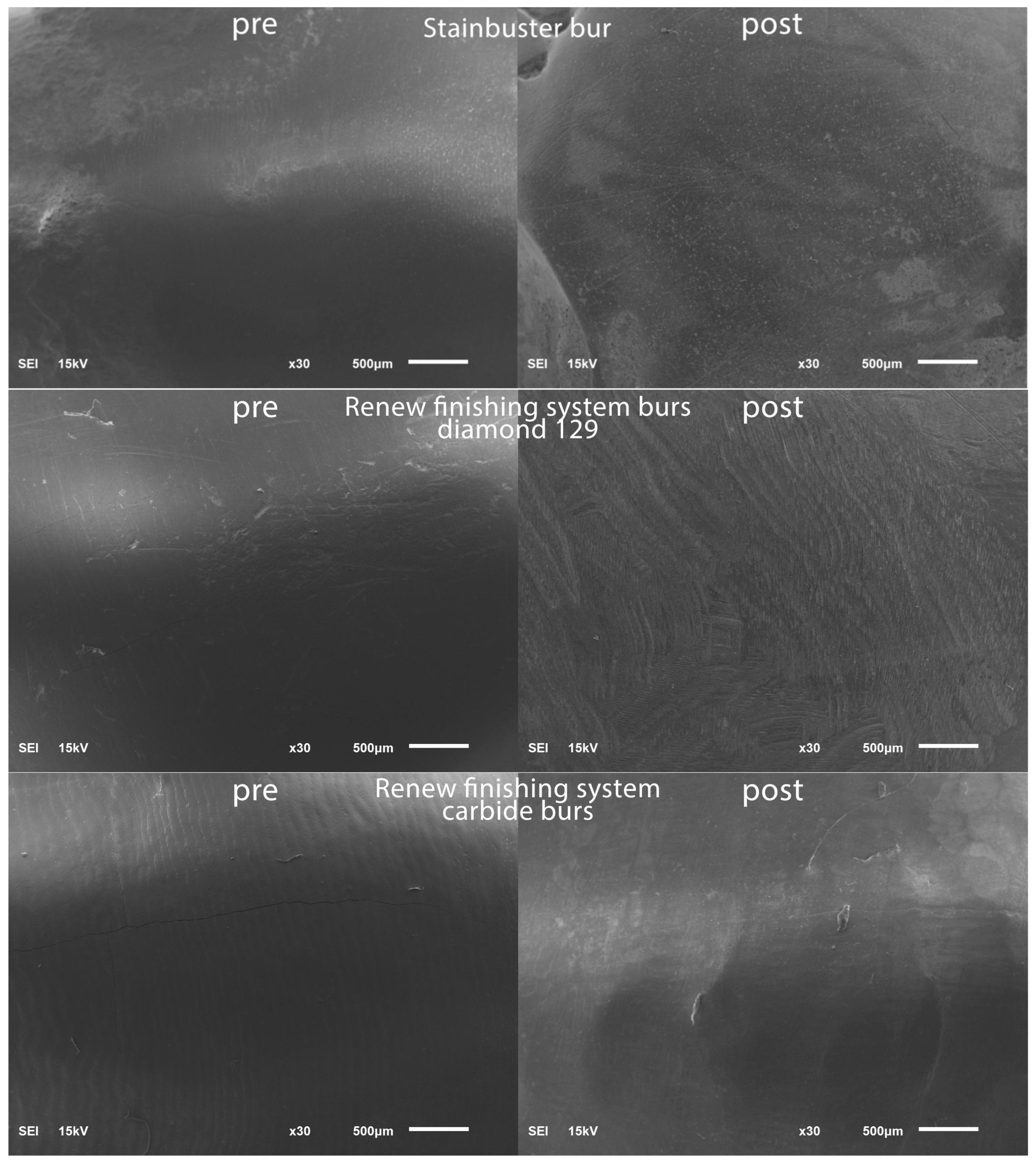
| Group | n |
|---|---|
| Group 1: Stainbuster (Abrasive Technology) | 20 |
| Group 2: Renew diamond bur #129 (Reliance Orthodontics) | 20 |
| Group 3: Renew carbide burs (Reliance Orthodontics) | 20 |
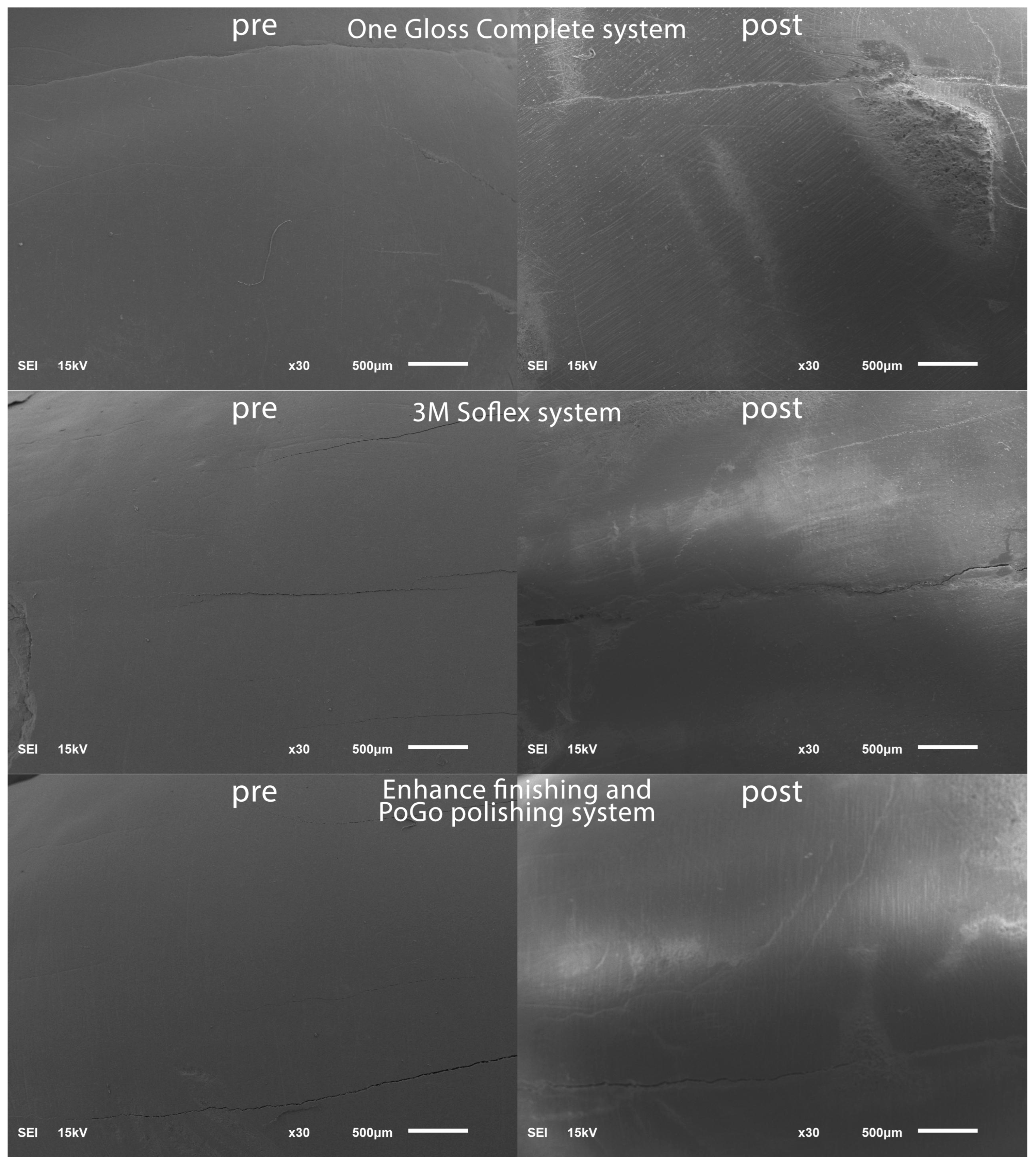
| Group 4: OneGloss Complete system (Shofu Dental) | 20 |
| Group 5: Sof-Lex system (3M ESPE), | 20 |
| Group 6: Enhance Finishing and PoGo Polishing complete kit (Dentsply Sirona) | 20 |
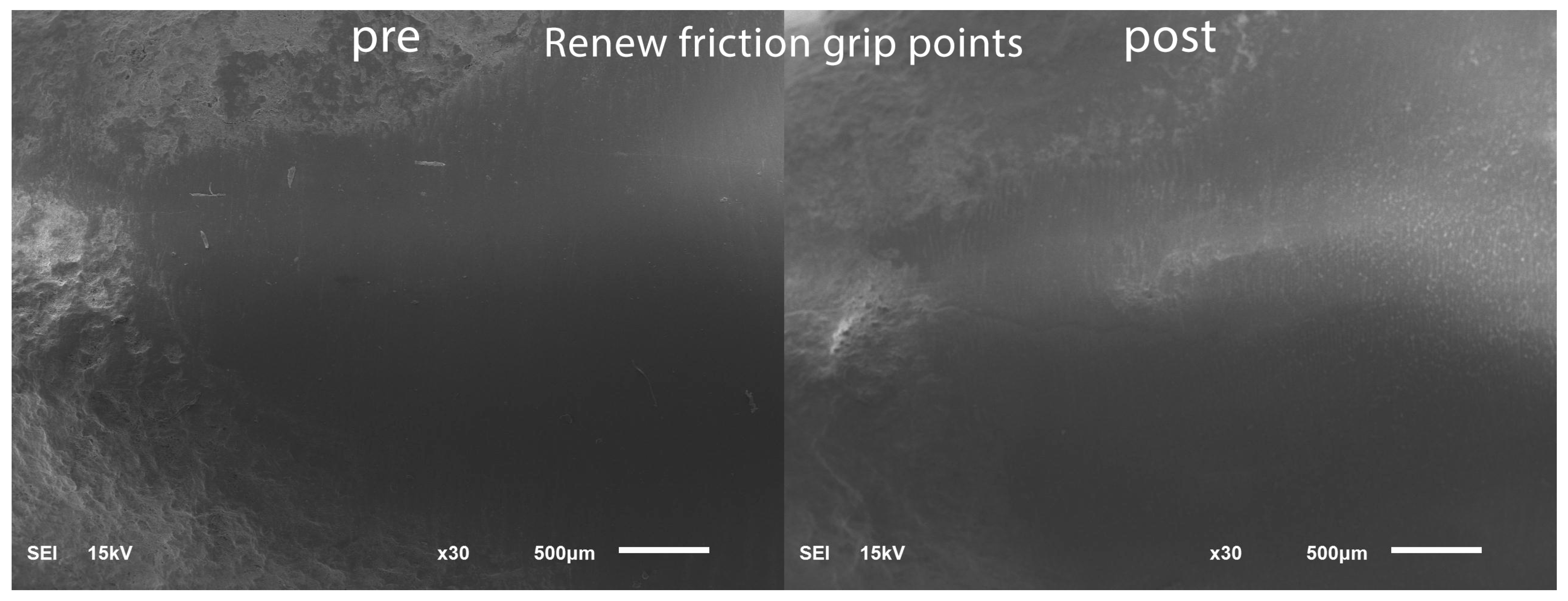
| Group 7: Renew friction grip points (Reliance Orthodontics) | 20 |
| Baseline Roughness | Sum of Squares | df | Mean Square | F | p |
|---|---|---|---|---|---|
| Between Groups | 2.112 | 6 | 0.352 | 1.447 | 0.201 |
| Within Groups | 32.358 | 133 | 0.243 |
| Roughness | Difference | Paired t-Test | p-Value Diff of Group | MCT (Tukey HSD) | |||||||||||
|---|---|---|---|---|---|---|---|---|---|---|---|---|---|---|---|
| First | Second | Group 1: Stainbuster | Group 2: Diamond | Group 3: Renew Carbide | Group 4: One Gloss | Group 5: 3M Soflex | Group 6: Enhance Finishing | Group 7: Renew Finishing System Points | |||||||
| Group | Mean | Std. Deviation | Mean | Std. Deviation | Diff | SD Dif | p-Value | ||||||||
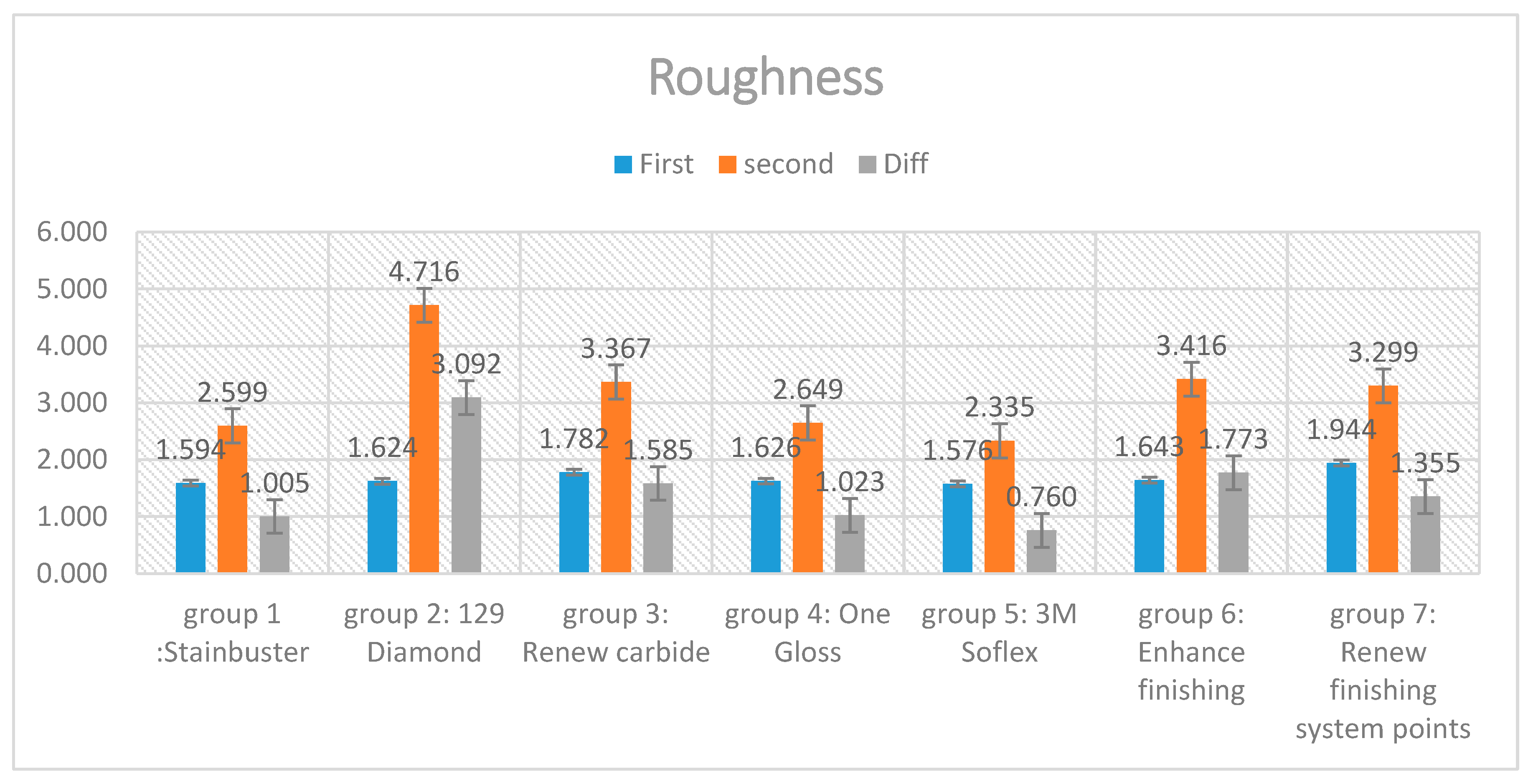
| Group 1: Stainbuster | 1.594. | 0.586 | 2.599 | 1.109 | 1.005 | 0.850 | 0.000 | 0.000 | 1 | ||||||
| Group 2: Renew #129 diamond | 1.624 | 0.443 | 4.716 | 1.074 | 3.092 | 1.156 | 0.000 | 0.000 | 1 | ||||||
| Group 3: Renew carbide | 1.782 | 0.512 | 3.367 | 1.015 | 1.585 | 1.116 | 0.000 | 0.474 | 0.000 | 1 | |||||
| Group 4: OneGloss | 1.626 | 0.530 | 2.649 | 0.706 | 1.023 | 0.730 | 0.000 | 1.000 | 0.000 | 0.513 | 1 | ||||
| Group 5: 3M Sof-Lex | 1.576 | 0.428 | 2.335 | 0.603 | 0.760 | 0.588 | 0.000 | 0.984 | 0.000 | 0.100 | 0.976 | 1 | |||
| Group 6: Enhance Finishing | 1.643 | 0.418 | 3.416 | 0.995 | 1.773 | 0.883 | 0.000 | 0.155 | 0.001 | 0.996 | 0.176 | 0.018 | 1 | ||
| Group 7: Renew Finishing System Points | 1.944 | 0.512 | 3.299 | 1.195 | 1.355 | 1.217 | 0.000 | 0.921 | 0.000 | 0.987 | 0.938 | 0.470 | 0.810 | 1 | |
| Temperature Difference | Sum of Squares | df | Mean Square | F |
|---|---|---|---|---|
| Between Groups | 217.747 | 6 | 36.291 | 6.316 |
| Within Groups | 764.268 | 133 | 5.746 |
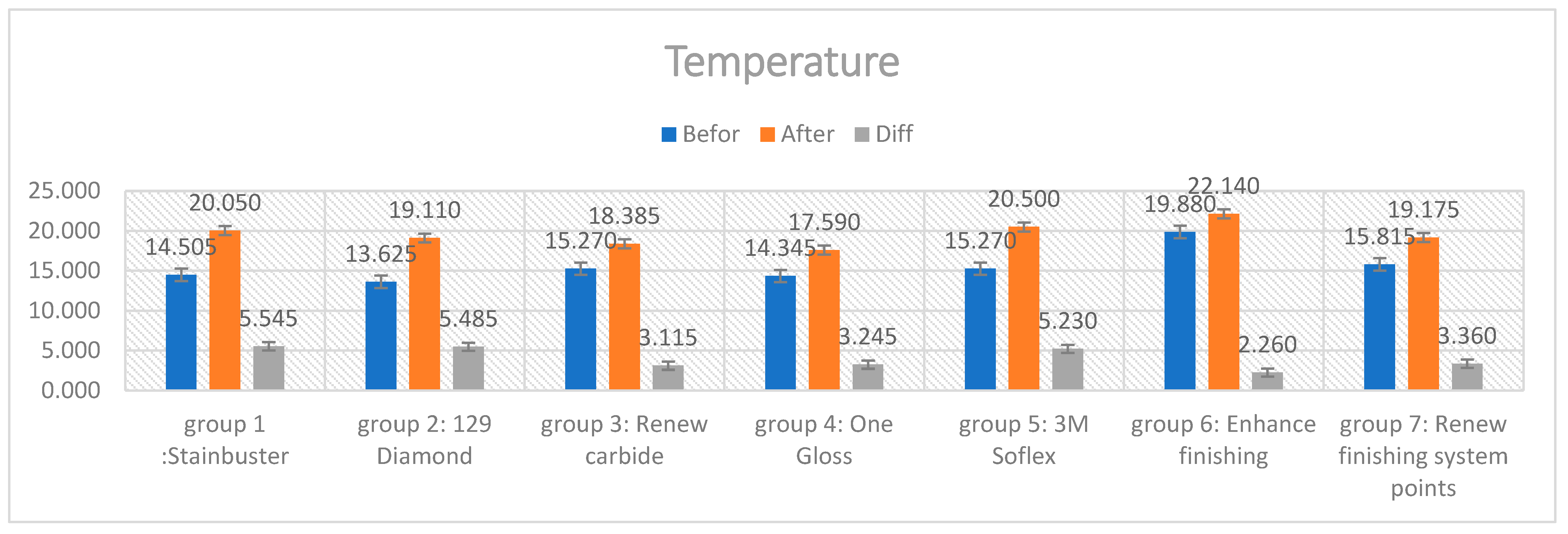
| Temperature | Difference | Paired t-Test | p-Value Diff of Group | MCT (Tukey HSD) | |||||||||||
|---|---|---|---|---|---|---|---|---|---|---|---|---|---|---|---|
| Before | After | ||||||||||||||
| Group 1: Stainbuster | Group 2: Diamond | Group 3: Renew carbide | Group 4: OneGloss | Group 5: 3M Sof-Lex | Group 6: Enhance Finishing | Group 7: Renew Finishing System Points | |||||||||
| Group | Mean | Std. Deviation | Mean | Std. Deviation | Diff | SD Dif | p-Value | ||||||||
| Group 1: Stainbuster | 14.505 | 1.209 | 20.050 | 1.963 | 5.545 | 1.862 | 0.000 | 0.000 | 1 | ||||||
| Group 2: Renew #129 diamond | 13.625 | 0.596 | 19.110 | 1.349 | 5.485 | 1.626 | 0.000 | 1.000 | 1 | ||||||
| Group 3: Renew carbide | 15.270 | 0.716 | 18.385 | 1.657 | 3.115 | 1.535 | 0.000 | 0.027 | 0.035 | 1 | |||||
| Group 4: OneGloss | 14.345 | 1.599 | 17.590 | 2.815 | 3.245 | 2.160 | 0.000 | 0.045 | 0.056 | 1.000 | 1 | ||||
| Group 5: 3M Sof-Lex | 15.270 | 0.954 | 20.500 | 1.620 | 5.230 | 2.094 | 0.000 | 1.000 | 1.000 | 0.085 | 0.129 | 1 | |||
| Group 6: Enhance Finishing | 19.880 | 3.837 | 22.140 | 2.000 | 2.260 | 3.964 | 0.000 | 0.001 | 0.001 | 0.918 | 0.851 | 0.003 | 1 | ||
| Group 7: Renew Finishing System Points | 15.815 | 1.340 | 19.175 | 2.750 | 3.360 | 2.645 | 0.000 | 0.067 | 0.082 | 1.000 | 1.000 | 0.180 | 0.770 | 1 | |
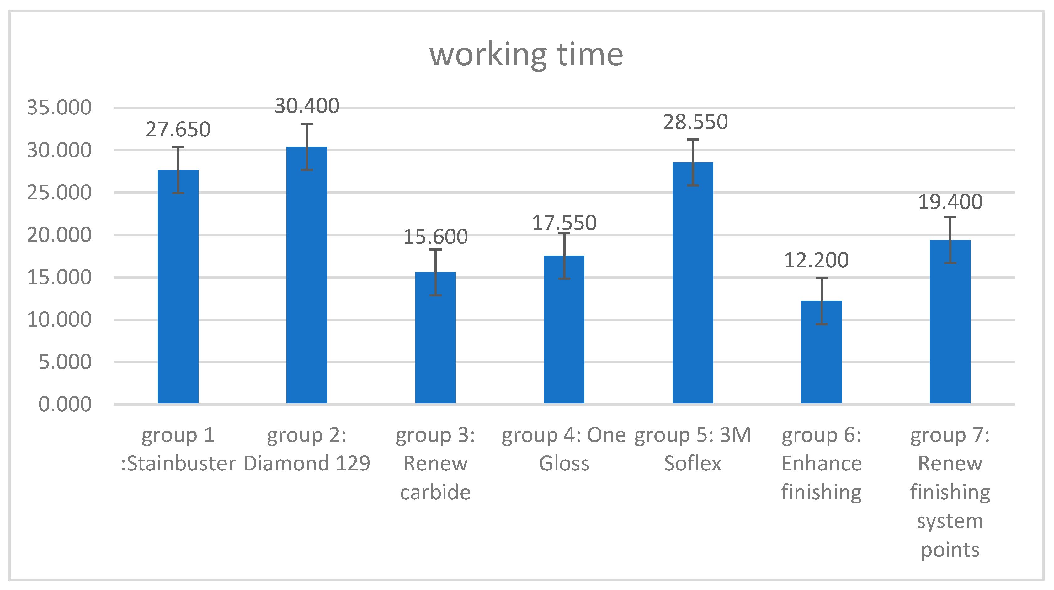
| Working Time | 95% Confidence Interval for Mean | MCT | |||||||||||
|---|---|---|---|---|---|---|---|---|---|---|---|---|---|
| Group 1: Stainbuster | Group 2: Diamond | Group 3: Renew Carbide | Group 4: One Gloss | Group 5: 3M Soflex | Group 6: Enhance Finishing | Group 7: Renew Finishing System Points | |||||||
| Group | n | Mean | Std. Deviation | p-Value | Lower Bound | Upper Bound | |||||||
| Group 1: Stainbuster | 20 | 27.650 | 15.295 | 0.000 | 5.545 | 1.862 | 1 | ||||||
| Group 2: Renew #129 Diamond | 20 | 30.400 | 6.793 | 5.485 | 1.626 | 0.934 | 1 | ||||||
| Group 3: Renew carbide | 20 | 15.600 | 3.409 | 3.115 | 1.535 | 0.000 | 0.000 | 1 | |||||
| Group 4: OneGloss | 20 | 17.550 | 6.177 | 3.245 | 2.160 | 0.002 | 0.000 | 0.988 | 1 | ||||
| Group 5: 3M Sof-Lex | 20 | 28.550 | 5.799 | 5.230 | 2.094 | 1.000 | 0.991 | 0.000 | 0.001 | 1 | |||
| Group 6: Enhance finishing | 20 | 12.200 | 4.060 | 2.260 | 3.964 | 0.000 | 0.000 | 0.835 | 0.360 | 0.000 | 1 | ||
| Group 7: Renew Finishing System Points | 20 | 19.400 | 8.696 | 3.360 | 2.645 | 0.025 | 0.001 | 0.751 | 0.991 | 0.008 | 0.079 | 1 | |
Disclaimer/Publisher’s Note: The statements, opinions and data contained in all publications are solely those of the individual author(s) and contributor(s) and not of MDPI and/or the editor(s). MDPI and/or the editor(s) disclaim responsibility for any injury to people or property resulting from any ideas, methods, instructions or products referred to in the content. |
© 2023 by the authors. Licensee MDPI, Basel, Switzerland. This article is an open access article distributed under the terms and conditions of the Creative Commons Attribution (CC BY) license (https://creativecommons.org/licenses/by/4.0/).
Share and Cite
Almudhi, A.; Aldeeri, A.; Aloraini, A.A.A.; Alomar, A.I.M.; Alqudairi, M.S.M.; Alzahrani, O.A.A.; Eldwakhly, E.; AlMugairin, S. Comparison of Enamel Surface Integrity after De-Bracketing as Affected by Seven Different Orthodontic Residual Cement Removal Systems. Diagnostics 2023, 13, 3284. https://doi.org/10.3390/diagnostics13203284
Almudhi A, Aldeeri A, Aloraini AAA, Alomar AIM, Alqudairi MSM, Alzahrani OAA, Eldwakhly E, AlMugairin S. Comparison of Enamel Surface Integrity after De-Bracketing as Affected by Seven Different Orthodontic Residual Cement Removal Systems. Diagnostics. 2023; 13(20):3284. https://doi.org/10.3390/diagnostics13203284
Chicago/Turabian StyleAlmudhi, Abdullazez, Arwa Aldeeri, Abdullah Abdulrahman A. Aloraini, Ahmed Ibrahim M. Alomar, Meshari Saad M. Alqudairi, Osama Abdullah A. Alzahrani, Elzahraa Eldwakhly, and Sarah AlMugairin. 2023. "Comparison of Enamel Surface Integrity after De-Bracketing as Affected by Seven Different Orthodontic Residual Cement Removal Systems" Diagnostics 13, no. 20: 3284. https://doi.org/10.3390/diagnostics13203284