Article

Genomic Characterization of Rare Primary Cardiac Sarcoma Entities

1
Department of Experimental, Diagnostic and Specialty Medicine (DIMES), Alma Mater Studiorum, University of Bologna, 40138 Bologna, Italy
2
Division of Oncology, IRCCS Azienda Ospedaliero, Universitaria di Bologna, 40138 Bologna, Italy
3
Department of Experimental Diagnostic and Specialty Medicine, IRCCS Azienda Ospedaliero-Universitaria, 40138 Bologna, Italy
4
Division of Pathology, Cardiovascular and Cardiac Transplant Pathology Unit, IRCCS Azienda Ospedaliero-Universitaria, 40138 Bologna, Italy
5
Department of Veterinary Medical Sciences, Alma Mater Studiorum, University of Bologna, 40138 Bologna, Italy
6
Cardiac Surgery Unit, IRCCS Azienda Ospedaliero-Universitaria, 40138 Bologna, Italy
7
Department of Pharmaceutics, Utrecht Institute for Pharmaceutical Sciences, Utrecht University, 3584 CS Utrecht, The Netherlands
8
Division of Pathology, IRCCS Azienda Ospedaliero, Universitaria di Bologna, 40138 Bologna, Italy
*
Author to whom correspondence should be addressed.
Diagnostics 2023, 13(2), 214; https://doi.org/10.3390/diagnostics13020214
Submission received: 12 November 2022 / Revised: 29 December 2022 / Accepted: 4 January 2023 / Published: 6 January 2023

Abstract

:
Primary cardiac sarcomas are considered rare malignant entities associated with poor prognosis. In fact, knowledge regarding their gene signature and possible treatments is still limited. In our study, whole-transcriptome sequencing on formalin-fixed paraffin-embedded (FFPE) samples from one cardiac osteosarcoma and one cardiac leiomyosarcoma was performed, to investigate their mutational profiles and to highlight differences and/or similarities to other cardiac histotypes. Both cases have been deeply detailed from a pathological point of view. The osteosarcoma sample presented mutations involving ATRX, ERCC5, and COL1A1, while the leiomyosarcoma case showed EXT2, DNM2, and PSIP1 alterations. Altered genes, along with the most differentially expressed genes in the leiomyosarcoma or osteosarcoma sample versus the cardiac angiosarcomas and intimal sarcomas (e.g., YAF2, PAK5, and CRABP1), appeared to be associated with cell growth, proliferation, apoptosis, and the repair of DNA damage, which are key mechanisms involved in tumorigenesis. Moreover, a distinct gene expression profile was detected in the osteosarcoma sample when compared to other cardiac sarcomas. For instance, WIF1, a marker of osteoblastic differentiation, was upregulated in our bone tumor. These findings pave the way for further studies on these entities, in order to identify targeted therapies and, therefore, improve patients’ prognoses.

Graphical Abstract

1. Introduction

Primary cardiac sarcomas are extremely rare malignant tumors of the heart, representing ~1% of all soft-tissue sarcomas and comprising a heterogeneous spectrum of histotypes [1,2]. They are classified according to their cardiac site of origin (cardiac chambers, pericardium, vena cava, and aorta) and intimal or mural localization. Angiosarcoma is the most common histotype, with an incidence of 7.3–8.5% among all variants, followed by undifferentiated high-grade pleomorphic cardiac sarcomas, with an incidence of 4.1–8.5%. Leiomyosarcomas, rhabdomyosarcomas, synovial sarcomas, liposarcomas, fibrosarcomas, myxoid fibrosarcomas, osteosarcomas, and chondrosarcomas represent rarer histological entities. Angiosarcomas are predominantly right-sided and are usually located in the right atrial cavity, while poorly differentiated cardiac sarcomas as well as other rarer histological subtypes are mostly located in the left atrial cavity.
The overall prognosis of primary cardiac sarcomas is generally poor and mainly correlates with tumor localization and with the involvement of the surrounding vital structures. These factors negatively affect surgical radicality and enhance the biological aggressiveness of most histotypes [3,4,5].
To date, clinical management of primary cardiac sarcomas has been complex and challenging, from diagnostics, often requiring a multimodality approach, to medical therapies, which still play a modest role [5]. This complexity is also due to the molecular background, which is extremely heterogeneous, though related knowledge is still very limited [6]. The molecular alterations detected in cardiac angiosarcomas often involve KDR, KIT, and the homozygous deletion of CDKN2A, while KRAS p.G13S and p.Q61K mutations have been reported in a few cases. As expected, cardiac undifferentiated pleomorphic sarcomas also have a complex genomic profile, including MDM2 and PDGFRA amplification, EGFR aneuploidy or amplification, and PDGFRB mutations. Conversely, limited molecular data on rarer histotypes are available in the literature. In the past, a primary cardiac leiomyosarcoma harboring a HRAS mutation was described [7]. Furthermore, in a single case of primary cardiac osteosarcoma, allelic losses or imbalances involving 1p36, 9p22, 10q23, 17q22, and 22q13 have been reported [8]. More recently, a whole-transcriptome sequencing analysis of a cardiac myxofibrosarcoma revealed a gene expression profile similar to that of angiosarcomas [9].
From our clinical series of adult primary cardiac sarcomas, herein we report and focus on two rare entities with transcriptome profiles that have been investigated with the aim of better describing these histotypes and, consequently, of identifying potential targets for more personalized therapies.

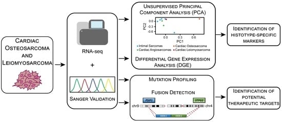
2. Materials and Methods

2.1. Patients

From 2005 to 2017, 11/358 patients with primary cardiac sarcomas were identified in our sarcoma population. Mean age was 51.8 years (range = 35–73). Main patients’ features are listed in Table 1. Histologically, the case series was extremely heterogenous, with an expected prevalence of pulmonary artery intimal sarcomas (5/11—45%) and angiosarcomas (3/11—27%). Other rare subtypes including an osteosarcoma, a myxofibrosarcoma, and a leiomyosarcoma were also identified. Almost all patients were metastatic at the time of diagnosis, presenting an aggressive clinical course.

2.2. Coding Transcriptome Sequencing

FFPE slides were reviewed by an expert pathologist and manually macrodissected to obtain an enrichment of tumor tissue of at least 70%. Total RNA was extracted using Recover. All Total Nucleic Acid Isolation Kit (Thermo Fisher Scientific, Waltham, MA, United States), and cDNA libraries were synthesized from 100 ng total RNA using TruSeq RNA Exome kit (Illumina, San Diego, CA, United States), in accordance with the instructions of the manufacturers. Briefly, cDNA libraries were synthesized from fragmented RNA, ligated to sequencing adapters, and amplified; then coding exon sequences were captured by hybridization to a pool of exonic probes. Libraries were sized with Agilent DNA 7500 chips on the Bioanalyzer 2100 (Agilent Technologies, Taiwan) and quantified with a fluorometric assay (Quant-IT Picogreen assay; Life Technologies, Carlsbad, CA, United States). Paired-end libraries (SRA Accession Number: PRJNA896891) were sequenced at 80 bp on a NextSeq500 instrument (Illumina, San Diego, CA, United States).

2.3. Bioinformatic Analysis

Our bioinformatic workflow was based on a primary quality check with FastQC and MultiQC to ensure read quality was acceptable and transcriptomes were comparable among samples [10,11]. Secondly, reads were mapped on the human genome to identify expressed genes. To determine transcript abundance, read quantification was performed obtaining raw counts. Subsequently, to ensure comparability, raw counts were normalized as counts per million (CPM) and transcripts per million (TPM), which were required for differential gene expression (DGE) and principal component analysis (PCA), respectively.
In detail, STAR was adopted to map paired reads on the reference human genome hg38 [12]; while duplicates’ removal, sorting, and indexing were performed with Samtools [13]. Gene expression was quantified and normalized as CPM and TPM, after obtaining raw gene counts with the Python package HTseq-count [14]. Starting from CPM, differentially expressed genes (q-value < 0.05, FDR-corrected) between either the leiomyosarcoma or osteosarcoma case and the group including angiosarcomas (CS6 and CS7) and intimal sarcomas (CS1, CS2, CS3, CS4 and CS5) were identified with the R-bioconductor package edgeR [15]. Using the R package prcomp [16] instead, TPM were employed to perform PCA, which also included intimal sarcomas (n = 5) and angiosarcomas (n = 2).
Additionally, gene fusion detection and small variant calling combined with variant annotation via Nirvana were performed on BaseSpace, an Illumina webtool (http://euc1.sh.basespace.illumina.com/ accessed on 4 June 2022). For gene-fusion detection, protein-coding and lncRNA genes were included to reduce false positives.
The vcf output files were filtered to include only stop gain/splice donor/splice acceptor/splice region/frameshift indels/inframe deletion/inframe insertion/initiator codon/ATG loss/missense mutations predicted as deleterious or probably damaging by SIFT predictor, with a GnomAD Frequency < 0.01, a QC metric quality ≥ 30, a total read depth > 14, an alt allele depth > 3, and a variant read frequency > 0.15. Finally, only genes belonging to Tier 1 of the Cancer Gene Census list (https://cancer.sanger.ac.uk/census accessed on 4 June 2022) were considered for further analysis. Sequencing data of both samples were analyzed using the DRAGEN RNA app (version 3.10.4).

2.4. Sanger Sequencing

To confirm the presence of alterations detected by bioinformatic analyses on genomic DNA and to ensure they were not due to RNA editing, DNA was extracted from manually macrodissected FFPE slides using the QIAmp DNA Micro KIT (Qiagen, Hilden, Germany), while cDNA was synthesized from 1 μg of total RNA using the High-Capacity cDNA Reverse Transcription Kit (Thermo Fisher Scientific, Waltham, MA, USA), in accordance with the instructions of the manufacturers.
Primers flanking the sequences of interest were designed using Primer3 Plus (https://www.bioinformatics.nl/cgi-bin/primer3plus/primer3plus.cgi accessed on 30 June 2022), either on genomic DNA or cDNA, and quality was checked by Blast alignment. Primer sequences are available upon request.
Lastly, to confirm point mutations and fusion transcripts, PCR amplification was performed on 40 ng of genomic DNA or 200 ng of cDNA using FastStart Taq Polymerase (Roche Diagnostics), with an annealing of 58 °C. Amplified fragments were sequenced on both strands using the Big Dye Terminator v1.1 Cycle Sequencing kit (Applied Biosystems) on ABI 3730 Genetic Analyzer (Applied Biosystems).

3. Results

Due to the rarity of cardiac sarcomas, we present the detailed results from the transcriptome analysis of two unusual histotypes: an osteosarcoma (CS9) and a leiomyosarcoma (CS11).

3.1. Patients

Case 1: In March 2009, a 39-year-old male presented dyspnea and tachycardia, associated with recurrent lipotimia episodes. Thoracic angio-contrast tomography (CT), cardiac catheterization, angiography, and echocardiography revealed a vascularized left atrial mass. Thus, in April 2009, the patient underwent surgical resection. Macroscopically, the mass appeared partially encapsulated with a diameter of 9 cm and was formed by solid whitish-gray tissue (Figure 1A). In multifocal areas of the chondroid and osteoid matrices, a histological examination showed mixed spindle and round cells with solid growth pattern, prominent nuclear atypia, and high mitotic activity (21/10 high-power field—HPF). Some giant osteoclastic cells were also present. According to immunohistochemical analysis (IHC), the tumor cells were diffusely positive for vimentin and had a high multifocal expression of wide-spectrum cytokeratins. Morphological aspects, along with the IHC findings, were coherent with the diagnosis of osteosarcoma (Figure 1). Subsequently, the patient underwent adjuvant chemotherapy with methotrexate, cisplatin, adriamycin, and ifosfamide until February 2010. In March 2011, due to an extended and symptomatic local relapse, the patient underwent a left pneumonectomy, dying of the disease in June 2011.
Case 2: In December 2005, a 59-year-old female presented dyspnea and pleural effusion. Trans-esophageal echocardiography detected a pedunculated left atrial mass of a gelatinous consistency. Following surgical resection, a histological examination revealed spindle cells with thickened chromatin, prominent nuclear atypia, and high mitotic activity (10/10 HPF). Rounded cells with clear cytoplasm and multinuclear giant cells were present in delimited areas (Figure 2). Additionally, immunohistochemical analysis revealed that the tumor cells were diffusely positive for smooth muscle actin (SMA) and vimentin. The morphological aspects, along with the IHC findings, were coherent with the diagnosis of pleomorphic leiomyosarcoma. The patient died of the disease in August 2006.

3.2. Mutational Profile of Cardiac Osteosarcoma and Leiomyosarcoma

An unsupervised PCA showed that the osteosarcoma sample presents a distinct gene expression profile when compared to the other histotypes included in the analysis (leiomyosarcoma, cardiac angiosarcoma, and intimal sarcoma). Indeed, it is distinctly separated from them along the first principal component (Figure 3).
Supervised gene expression analysis of the leiomyosarcoma sample and the group comprising angiosarcomas and intimal sarcomas was characterized by 55 differentially expressed genes (q-value < 0.05) (Supplementary Table S1). The same analysis applied to the osteosarcoma case versus angiosarcomas and intimal sarcomas presented 482 differentially expressed genes (q-value < 0.05), confirming the uniqueness of the osteosarcoma expression profile (Supplementary Table S2). In both analyses, among the most differentially expressed genes, the biomarkers involved in major cell processes, such as cell division, histotype-specific differentiation, and DNA repair, were upregulated in both leiomyosarcoma and osteosarcoma (Figure 4). For instance, PAK5 and YAF2, which promote cell migration and invasion, were overexpressed in the leiomyosarcoma sample; while WIF1, a marker of osteoblastic differentiation, and DLK1, which is involved in cancer stem-like cell maintenance and cancer differentiation, were upregulated in the osteosarcoma sample.
Regarding gene mutations, we identified a total of 117 and 94 mutations in the osteosarcoma and leiomyosarcoma cases, respectively. Variants were classified according to their effect on the protein (Figure 5), resulting in a prevalence of missense and splice-site mutations. However, since our focus was on cancer genes, we restricted our analysis to mutations affecting the genes belonging to Tier 1 of the Cancer Gene Census list. This led to the identification of six alterations in the osteosarcoma sample and two in the leiomyosarcoma one.
In the osteosarcoma case, the ATRX gene was identified as carrying a frameshift mutation (c.1283_1284insT) affecting exon 9 on chr23:77683973 (Table 2). This ATRX variant was also validated by Sanger sequencing and determined the loss of the reading frame, introducing a premature stop codon at position 428 (Figure 6).
The ERCC5 gene presented a missense mutation (c.2636A>G) on chr13:102868215 (exon 12), where a Serine replaced an Asparagine at position 879 (Table 2). According to SIFT, this ERCC5 variant is deleterious and probably damaging according to PolyPhen. Moreover, on COSMIC, the alteration is classified as somatic, and its FATHMM prediction is pathogenic with a score of 0.99.
Lastly, the c.1461+2T>G variant affecting the COL1A1 gene seems to cause loss of the donor splice site at intron 21-22 on chr17:50194719. According to ClinVar, this alteration is likely pathogenic (Table 2).
In the leiomyosarcoma sample, a bioinformatic analysis of the coding transcriptome data detected an EXT2 gene missense mutation (c.1186G>A) located on exon 7 (Table 2), which was validated by Sanger sequencing (Figure 6). This variant led to the substitution of a Valine at position 396 by a Methionine. Additionally, this EXT2 alteration is classified as deleterious by SIFT, as it is probably damaging with a score of 0.99 by PolyPhen-2, and reported as likely benign for its association with osteochondroma in ClinVar.
The second alteration identified in the leiomyosarcoma sample was the c.688+2T>C variant affecting the DNM2 gene, which appears to lead to the loss of the donor splice site at exon 5 on chr19:10777218 (Table 2). DNM2 is linked to GTP hydrolyzation, endocytosis, and apoptotic cell maturation. The quality score of this alteration is 33, and the ACMG classification on Varsome is pathogenic.
Another noteworthy event in the leiomyosarcoma case was the fusion between the PSIP1 gene, involved in the DNA-binding transcription factor and chromatin-binding activity, and the STPG2 gene, which was validated by Sanger sequencing (Figure 6). This fusion transcript retained the reading frame and occurred between two intact exons: exon 2 of the PSIP1 gene located on chr9:15510117 and exon 3 of the STPG2 gene located on chr4:98128592 (Figure 7).

4. Discussion

Primary cardiac sarcomas comprise a rare and heterogeneous family of several histologies, characterized by varied clinical presentations and biological behaviors. Knowledge of the molecular background of these sarcomas, especially of the rarest histotypes, is still limited. Thus, we have studied the osteosarcoma and leiomyosarcoma cases by applying transcriptome sequencing analyses to enhance the understanding of these cardiac entities and to discover useful information for their clinical management and treatment.
Among the cardiac osteosarcoma alterations, the ATRX gene is noteworthy as it encodes a protein involved in transcription and chromatin remodeling [17,18]. It also plays a role in the ALT pathway, which administers telomere maintenance [18,19]. Furthermore, ATRX is usually mutated in 30% of osteosarcomas and is in fact considered to be one of the main drivers in osteosarcoma development, along with the following genes: TP53, RB1, BRCA2, BAP1, RET, MUTYH, ATM, PTEN, WRN, RECQL4, FANCA, NUMA1, and MDC1 [20,21]. More specifically, point mutations in the ATRX gene seem to be associated with longer telomeres [21,22]. In some cases, ATRX depletion can lead to increased activity by the ALT pathway, which drives cancer cell immortalization [22].
The ERCC5 gene encodes a single-stranded structure-specific DNA endonuclease involved in the nucleotide excision repair (NER) pathway [23,24]. Thus, since both ATRX and ERCC5 are responsible for chromatin remodeling and DNA repair, mutations affecting these genes might alter DNA replication, leading to uncontrolled cell growth and proliferation.
Whilst it is established that cardiac leiomyosarcomas are rare and, therefore, lack a well-defined mutational profile, extra-cardiac leiomyosarcomas are better described in the literature. As proof, their most common signatures comprise the following genes: TP53, RB1, PTEN, BRCA1/2, ATRX, and ATM [25,26]. By contrast, germline mutations concerning the EXT2 gene, which is involved in heparan sulphate biosynthesis and in tumor suppression, appear to be associated with hereditary multiple exostosis (HME), an autosomal dominant disease [27,28,29]. HME might lead to osteochondroma development, a benign bone tumor [28,30]. Additionally, these patients have an increased risk of developing chondrosarcomas and osteosarcomas [6,28].
Remarkably, the leiomyosarcoma sample we have analyzed did not present any TP53, RB1, and ATRX mutations, which instead characterize its histological class [31]. Homozygous deletions regarding RB1, PTEN, and TP53, which are common among leiomyosarcomas [25,32], could not be detected by this sequencing approach; nonetheless, we did not observe any strong evidence of gene expression downregulation.
Regarding the PSIP1STPG2 gene fusion, the former gene appears to be involved in cell growth, proliferation, and survival [33,34,35]. When upregulated, it enhances angiogenesis and prevents apoptosis in malignant cells, leading to aggressive cancer phenotypes [33]. Moreover, since PSIP1 also plays a role in the recruitment of proteins involved in homology directed repair (HDR), when it is depleted, the number of DNA double-strand breaks (DSB) might increase [34,36]. In our case, only the first two exons of PSIP1, which consist mainly of untranslated regions, have been retained following gene fusion. Most likely, this leads to a loss of function by the PSIP1 protein and, subsequently, to genomic instability, which promotes cancer development. On the other hand, the STPG2 breakpoint was at the beginning of exon 3; thus, only the first two exons, of which the first one is mainly composed of untranslated regions, were lost. This event might explain why STPG2 was found to be upregulated among the most differentially expressed genes in the leiomyosarcoma case versus the angiosarcoma and intimal sarcoma cases.
In the leiomyosarcoma sample, the genes involved in cell growth, survival, and proliferation (MFHAS1, YAF2, SOX5, and PAK5) were also overexpressed. MFHAS1 is an oncogene with expression in tumor-associated macrophages that has been associated with colorectal cancer (CRC) progression [37]. High expression of YAF2 has been found in non-small cell lung cancer cells (NSCLCs), where it allows cell invasion and migration, preventing apoptosis [38]. SOX5 seems to enhance epithelial mesenchymal transition (EMT), leading to metastasis in breast and prostate cancers [39]. PAK5, a binding protein of GTPases, has appeared to prevent drug-induced apoptosis in CRC cells, promoting cell proliferation. Thus, it has been selected as a potential therapeutic target [40].
Interestingly, in both leiomyosarcoma and osteosarcoma, an upregulated gene was BMP5, which encodes a secret ligand of the TFG-beta (transforming growth factor-beta) protein family and is associated with bone and cartilage development [41]. Additionally, it is usually overexpressed in synovial sarcomas. This event is remarkable mainly in the leiomyosarcoma sample, since, as previously mentioned, it presented EXT2 alterations, which might contribute to osteochondroma onset. Therefore, this tumor sample has some features related to musculoskeletal malignancies.
In the osteosarcoma case versus the angiosarcomas and intimal sarcomas, the Wnt inhibitory factor 1 (WIF1) was also upregulated. WIF1 is a marker of osteoblastic differentiation that highlights the specific differentiation lineage of this rare cardiac sarcoma [42]. It is usually overexpressed in the late stages of normal osteoblast maturation and in stratified osteosarcoma tumors. Moreover, upregulation concerned genes involved in cell proliferation, survival, differentiation, and migration (DLK1 and CRABP1) [43,44]. DLK1 expression characterizes drug-resistant tumors, since this gene allows cancer cells to maintain a stem-like phenotype [43]. High levels of CRABP1 have been detected in synovial sarcomas, lymph node metastasis, and pancreatic neuroendocrine tumors (pNETs) [44]. Lastly, COL11A2 overexpression is frequently associated with poor overall and event-free survival in patients diagnosed with osteosarcoma [45]. Consequently, these genes could be considered as markers or candidates for targeted therapies for this type of sarcoma.
The alterations identified in our analysis indicate an aggressive phenotype in both samples and suggest that these cases of cardiac osteosarcoma and leiomyosarcoma do not share common features with other types of cardiac sarcomas, including angiosarcomas, undifferentiated pleomorphic sarcomas, rhabdomyosarcomas, and myxofibrosarcomas [6]. For instance, our samples do not present any POT1 and TP53 mutations, unlike most cardiac angiosarcomas, any PDGFRB variants that characterize cardiac undifferentiated pleomorphic sarcomas, and any KRAS alterations commonly detected in cardiac rhabdomyosarcomas. However, according to the PCA results, angiosarcomas, intimal sarcomas, and leiomyosarcoma seem to share at least some features regarding their gene signature, unlike the osteosarcoma case, which instead appears to have a distinct pattern.
To date, the therapeutic landscape of both cardiac osteosarcomas and leiomyosarcomas has not differed from the one applied to sarcomas arising in more common primary sites, as previously stated by consensus conference guidelines [46,47]. However, the fact that the osteosarcoma greatly differs from the other histotypes, along with the presence of novel alterations in both cases (e.g., ERCC5 and PSIP1), highlights the need to further explore these rare entities.
Since our sample size was limited, additional studies including more patients might help validating the events identified in our study, leading to multidisciplinary approaches and, perhaps, to individualized and molecular-based treatments. Another downside of our research is the lack of whole-exome sequencing (WES) analyses, where tumor samples are compared with their normal counterparts. Thus, future studies should include WES to better understand how gene amplifications and/or deletions can contribute to tumorigenesis, when combined with other mutational events. This might pave the way for more advanced combination therapies.

5. Conclusions

Ultimately, even if primary cardiac sarcomas still represent an unmet clinical need from several points of view, as we have learned from other rare diseases, sharing experiences remains the main option to improve our knowledge and patients’ treatment opportunities.

Supplementary Materials

The following supporting information can be downloaded at https://www.mdpi.com/article/10.3390/diagnostics13020214/s1. Table S1: Differentially expressed genes (q-value < 0.05) between the leiomyosarcoma sample (CS11) and the group comprising cardiac angiosarcomas (CS6 and CS7) and intimal sarcomas (CS1, CS2, CS3, CS4, and CS5) (logFC = log2 fold change; logCPM = log2 counts per million; FDR = false discovery rate); Table S2: Differentially expressed genes (q-value < 0.05) between the osteosarcoma sample (CS9) and the group comprising cardiac angiosarcomas (CS6 and CS7) and intimal sarcomas (CS1, CS2, CS3, CS4, and CS5) (logFC = log2 fold change; logCPM = log2 counts per million; FDR = false discovery rate).

Author Contributions

Conceptualization, L.G., M.N., M.A.P. and A.A.; methodology, L.G., M.N., M.A.P. and A.A.; validation, L.G.; formal analysis, L.G., M.N. and V.I.; investigation, C.P., P.P., A.F., D.P. and G.F.; resources, O.L., B.C. and C.B.; data curation, L.G., M.N., V.I. and A.S.; writing—original draft preparation, L.G., M.N. and A.A.; writing—review and editing, L.G., M.N., A.C., G.P., M.A.P. and A.A.; supervision, G.P., M.A.P. and A.A.; project administration, M.A.P. and A.A. All authors have read and agreed to the published version of the manuscript.

Funding

This study belongs to a project supported by Ministero della Salute, RC-2022-2773450.

Institutional Review Board Statement

The study was conducted in accordance with the Declaration of Helsinki and was approved by the Institutional Ethics Committee of Policlinico Sant’Orsola-Malpighi, Bologna, Italy (approval number: 95/2013/U/Tess; date: 8 October 2013).

Informed Consent Statement

Informed consent was obtained from all living subjects involved in the study.

Data Availability Statement

Publicly available datasets were analyzed in this study. The data can be found at https://www.ncbi.nlm.nih.gov/sra/PRJNA896891.

Conflicts of Interest

The authors declare no conflict of interest.

References

  1. Isambert, N.; Ray-Coquard, I.; Italiano, A.; Rios, M.; Kerbrat, P.; Gauthier, M.; Blouet, A.; Chaigneau, L.; Duffaud, F.; Piperno-Neumann, S.; et al. Primary Cardiac Sarcomas: A Retrospective Study of the French Sarcoma Group. Eur. J. Cancer 2014, 50, 128–136. [Google Scholar] [CrossRef] [PubMed]
  2. Scicchitano, P.; Chiara Sergi, M.; Cameli, M.; Miglioranza, M.H.; Ciccone, M.M.; Gentile, M.; Porta, C.; Tucci, M. Primary Soft Tissue Sarcoma of the Heart: An Emerging Chapter in Cardio-Oncology. Biomedicines 2021, 9, 774. [Google Scholar] [CrossRef] [PubMed]
  3. Siontis, B.L.; Leja, M.; Chugh, R. Current Clinical Management of Primary Cardiac Sarcoma. Expert Rev. Anticancer Ther. 2020, 20, 45–51. [Google Scholar] [CrossRef] [PubMed]
  4. Hamidi, M.; Moody, J.S.; Weigel, T.L.; Kozak, K.R. Primary Cardiac Sarcoma. Ann. Thorac. Surg. 2010, 90, 176–181. [Google Scholar] [CrossRef] [Green Version]
  5. Patel, S.D.; Peterson, A.; Bartczak, A.; Lee, S.; Chojnowski, S.; Gajewski, P.; Loukas, M. Primary Cardiac Angiosarcoma—A Review. Med. Sci. Monit. 2014, 20, 103–109. [Google Scholar] [CrossRef] [Green Version]
  6. Urbini, M.; Astolfi, A.; Indio, V.; Nannini, M.; Pizzi, C.; Paolisso, P.; Tarantino, G.; Pantaleo, M.A.; Saponara, M. Genetic Aberrations and Molecular Biology of Cardiac Sarcoma. Ther. Adv. Med. Oncol. 2020, 12, 1758835920918492. [Google Scholar] [CrossRef]
  7. Parissis, J.; Arvanitis, D.; Sourvinos, G.; Spandidos, D. A Primary Cardiac Leiomyosarcoma with Mutation at H-Ras Codon 12. Oncol. Rep. 1997, 4, 807–808. [Google Scholar] [CrossRef]
  8. Parwani, A.V.; Esposito, N.; Rao, U.N.M. Primary Cardiac Osteosarcoma with Recurrent Episodes and Unusual Patterns of Metastatic Spread. Cardiovasc. Pathol. 2008, 17, 413–417. [Google Scholar] [CrossRef]
  9. Saponara, M.; Indio, V.; Pizzi, C.; Serban, E.D.; Urbini, M.; Astolfi, A.; Paolisso, P.; Suarez, S.M.; Nannini, M.; Pacini, D.; et al. Successful Multidisciplinary Clinical Approach and Molecular Characterization by Whole Transcriptome Sequencing of a Cardiac Myxofibrosarcoma: A Case Report. World J. Clin. Cases 2019, 7, 3018–3026. [Google Scholar] [CrossRef]
  10. Andrews, S. FastQC: A Quality Control Tool for High Throughput Sequence Data. 2010. Available online: http://www.bioinformatics.babraham.ac.uk/projects/fastqc (accessed on 30 May 2022).
  11. Ewels, P.; Magnusson, M.; Lundin, S.; Käller, M. MultiQC: Summarize Analysis Results for Multiple Tools and Samples in a Single Report. Bioinformatics 2016, 32, 3047–3048. [Google Scholar] [CrossRef]
  12. Dobin, A.; Davis, C.A.; Schlesinger, F.; Drenkow, J.; Zaleski, C.; Jha, S.; Batut, P.; Chaisson, M.; Gingeras, T.R. STAR: Ultrafast Universal RNA-Seq Aligner. Bioinformatics 2013, 29, 15–21. [Google Scholar] [CrossRef] [PubMed]
  13. Danecek, P.; Bonfield, J.K.; Liddle, J.; Marshall, J.; Ohan, V.; Pollard, M.O.; Whitwham, A.; Keane, T.; McCarthy, S.A.; Davies, R.M.; et al. Twelve Years of SAMtools and BCFtools. Gigascience 2021, 10, giab008. [Google Scholar] [CrossRef]
  14. Putri, G.H.; Anders, S.; Pyl, P.T.; Pimanda, J.E.; Zanini, F. Analysing High-Throughput Sequencing Data in Python with HTSeq 2.0. Bioinformatics 2022, 38, 2943–2945. [Google Scholar] [CrossRef] [PubMed]
  15. Robinson, M.D.; McCarthy, D.J.; Smyth, G.K. EdgeR: A Bioconductor Package for Differential Expression Analysis of Digital Gene Expression Data. Bioinformatics 2009, 26, 139–140. [Google Scholar] [CrossRef] [PubMed] [Green Version]
  16. Sigg, C.D.; Buhmann, J.M. Expectation-Maximization for Sparse and Non-Negative PCA. In Proceedings of the Twenty-Fifth International Conference on Machine Learning, Helsinki, Finlandia, 5–9 June 2008. [Google Scholar] [CrossRef] [Green Version]
  17. Valenzuela, M.; Amato, R.; Sgura, A.; Antoccia, A.; Berardinelli, F. The Multiple Facets of Atrx Protein. Cancers 2021, 13, 2211. [Google Scholar] [CrossRef] [PubMed]
  18. Haase, S.; Garcia-Fabiani, M.B.; Carney, S.; Altshuler, D.; Núñez, F.J.; Méndez, F.M.; Núñez, F.; Lowenstein, P.R.; Castro, M.G. Mutant ATRX: Uncovering a New Therapeutic Target for Glioma. Expert Opin. Ther. Targets 2018, 22, 599–613. [Google Scholar] [CrossRef]
  19. Mackenzie, D.; Watters, A.K.; To, J.T.; Young, M.W.; Muratori, J.; Wilkoff, M.H.; Abraham, R.G.; Plummer, M.M.; Zhang, D. Alt Positivity in Human Cancers: Prevalence and Clinical Insights. Cancers 2021, 13, 2384. [Google Scholar] [CrossRef]
  20. Rickel, K.; Fang, F.; Tao, J. Molecular Genetics of Osteosarcoma. Bone 2017, 102, 69–79. [Google Scholar] [CrossRef]
  21. Chen, X.; Bahrami, A.; Pappo, A.; Easton, J.; Dalton, J.; Hedlund, E.; Ellison, D.; Shurtleff, S.; Wu, G.; Wei, L.; et al. Recurrent Somatic Structural Variations Contribute to Tumorigenesis in Pediatric Osteosarcoma. Cell Rep. 2014, 7, 104–112. [Google Scholar] [CrossRef]
  22. Ren, X.; Tu, C.; Tang, Z.; Ma, R.; Li, Z. Alternative Lengthening of Telomeres Phenotype and Loss of ATRX Expression in Sarcomas. Oncol. Lett. 2018, 15, 7489–7496. [Google Scholar] [CrossRef] [Green Version]
  23. Malik, S.S.; Mubarik, S.; Baig, M.; Masood, N.; Chaudhry, N. Genetic Polymorphism in ERCC5 and Breast Cancer Risk. Mol. Biol. Res. Commun. 2019, 8, 27–31. [Google Scholar] [CrossRef]
  24. Zheng, X.; Chen, K.; Liu, X.; Jiang, G.; Liu, H. High Expression of ERCC5 Predicts a Poor Prognosis in Hepatocellular Carcinoma. Int. J. Clin. Exp. Pathol. 2018, 11, 3664–3670. [Google Scholar]
  25. Astolfi, A.; Nannini, M.; Indio, V.; Schipani, A.; Rizzo, A.; Perrone, A.M.; de Iaco, P.; Pirini, M.G.; de Leo, A.; Urbini, M.; et al. Genomic Database Analysis of Uterine Leiomyosarcoma Mutational Profile. Cancers 2020, 12, 2126. [Google Scholar] [CrossRef]
  26. Lee, P.J.; Yoo, N.S.; Hagemann, I.S.; Pfeifer, J.D.; Cottrell, C.E.; Abel, H.J.; Duncavage, E.J. Spectrum of Mutations in Leiomyosarcomas Identified by Clinical Targeted Next-Generation Sequencing. Exp. Mol. Pathol. 2017, 102, 156–161. [Google Scholar] [CrossRef]
  27. Ropero, S.; Setien, F.; Espada, J.; Fraga, M.F.; Herranz, M.; Asp, J.; Benassi, M.S.; Franchi, A.; Patiño, A.; Ward, L.S.; et al. Epigenetic Loss of the Familial Tumor-Suppressor Gene Exostosin-1 (EXT1) Disrupts Heparan Sulfate Synthesis in Cancer Cells. Hum. Mol. Genet. 2004, 13, 2753–2765. [Google Scholar] [CrossRef] [Green Version]
  28. Wang, Y.; Zhong, L.; Xu, Y.; Ding, L.; Ji, Y.; Schutz, S.; Férec, C.; Cooper, D.N.; Xu, C.; Chen, J.M.; et al. EXT1 and EXT2 Variants in 22 Chinese Families with Multiple Osteochondromas: Seven New Variants and Potentiation of Preimplantation Genetic Testing and Prenatal Diagnosis. Front. Genet. 2020, 11, 607838. [Google Scholar] [CrossRef]
  29. Yang, C.; Zhang, R.; Lin, H.; Wang, H. Insights into the Molecular Regulatory Network of Pathomechanisms in Osteochondroma. J. Cell. Biochem. 2019, 120, 16362–16369. [Google Scholar] [CrossRef] [PubMed]
  30. D’arienzo, A.; Andreani, L.; Sacchetti, F.; Colangeli, S.; Capanna, R. Hereditary Multiple Exostoses: Current Insights. Orthop. Res. Rev. 2019, 11, 199–211. [Google Scholar] [CrossRef] [PubMed] [Green Version]
  31. Abeshouse, A.; Adebamowo, C.; Adebamowo, S.N.; Akbani, R.; Akeredolu, T.; Ally, A.; Anderson, M.L.; Anur, P.; Appelbaum, E.L.; Armenia, J.; et al. Comprehensive and Integrated Genomic Characterization of Adult Soft Tissue Sarcomas. Cell 2017, 171, 950–965. [Google Scholar] [CrossRef] [PubMed] [Green Version]
  32. Hensley, M.L.; Chavan, S.S.; Solit, D.B.; Murali, R.; Soslow, R.; Chiang, S.; Jungbluth, A.A.; Bandlamudi, C.; Srinivasan, P.; Tap, W.D.; et al. Genomic Landscape of Uterine Sarcomas Defined through Prospective Clinical Sequencing. Clin. Cancer Res. 2020, 26, 3881–3888. [Google Scholar] [CrossRef] [PubMed]
  33. Singh, D.K.; Gholamalamdari, O.; Jadaliha, M.; Li, X.L.; Lin, Y.C.; Zhang, Y.; Guang, S.; Hashemikhabir, S.; Tiwari, S.; Zhu, Y.J.; et al. PSIP1/P75 Promotes Tumorigenicity in Breast Cancer Cells by Promoting the Transcription of Cell Cycle Genes. Carcinogenesis 2017, 38, 966–975. [Google Scholar] [CrossRef] [PubMed] [Green Version]
  34. Liedtke, V.; Schröder, C.; Roggenbuck, D.; Weiss, R.; Stohwasser, R.; Schierack, P.; Rödiger, S.; Schenk, L. LEDGF/P75 Is Required for an Efficient Dna Damage Response. Int. J. Mol. Sci. 2021, 22, 5866. [Google Scholar] [CrossRef] [PubMed]
  35. Basu, A.; Rojas, H.; Banerjee, H.; Cabrera, I.B.; Perez, K.Y.; de León, M.; Casiano, C.A. Expression of the Stress Response Oncoprotein LEDGF/P75 in Human Cancer: A Study of 21 Tumor Types. PLoS ONE 2012, 7, e30132. [Google Scholar] [CrossRef] [PubMed] [Green Version]
  36. French, J.D.; Johnatty, S.E.; Lu, Y.; Beesley, J.; Gao, B.; Kalimutho, M.; Henderson, M.J.; Russell, A.J.; Kar, S.; Chen, X.; et al. Germline Polymorphisms in an Enhancer of PSIP1 Are Associated with Progression-Free Survival in Epithelial Ovarian Cancer. Oncotarget 2016, 7, 6353–6368. [Google Scholar] [CrossRef] [Green Version]
  37. Chen, W.; Xu, Y.; Zhong, J.; Wang, H.; Weng, M.; Cheng, Q.; Wu, Q.; Sun, Z.; Jiang, H.; Zhu, M.; et al. MFHAS1 Promotes Colorectal Cancer Progress by Regulating Polarization of Tumor-Associated Macrophages via STAT6 Signaling Pathway. Oncotarget 2016, 7, 78726–78735. [Google Scholar] [CrossRef] [Green Version]
  38. Yan, Y.; Jin, X.; Sun, H.; Pang, S.; Kong, X.; Bu, J.; Xu, S. Mir-139-5p Targetedly Regulates Yaf2 and Mediates the Akt/P38 Mapk Signaling Pathway to Alleviate the Metastasis of Non-Small Cell Lung Cancer Cells and Their Resistance against Cisplatin. Cancer Manag. Res. 2021, 13, 3639–3650. [Google Scholar] [CrossRef]
  39. Grimm, D.; Bauer, J.; Wise, P.; Krüger, M.; Simonsen, U.; Wehland, M.; Infanger, M.; Corydon, T.J. The Role of SOX Family Members in Solid Tumours and Metastasis. Semin. Cancer Biol. 2020, 67, 122–153. [Google Scholar] [CrossRef]
  40. Huang, S.; Zhu, Y.; Wang, C.; Li, X.; Cui, X.; Tu, S.; You, L.; Fu, J.W.; Chen, Z.; Hu, W.; et al. PAK5 Facilitates the Proliferation, Invasion and Migration in Colorectal Cancer Cells. Cancer Med. 2020, 9, 4777–4790. [Google Scholar] [CrossRef]
  41. Nakajima, K.; Kidani, T.; Miura, H. Molecular Profiling of Bone Remodeling Occurring in Musculoskeletal Tumors. J. Orthop. Res. 2021, 39, 1402–1410. [Google Scholar] [CrossRef]
  42. Baker, E.K.; Taylor, S.; Gupte, A.; Chalk, A.M.; Bhattacharya, S.; Green, A.C.; Martin, T.J.; Strbenac, D.; Robinson, M.D.; Purton, L.E.; et al. Wnt Inhibitory Factor 1 (WIF1) Is a Marker of Osteoblastic Differentiation Stage and Is Not Silenced by DNA Methylation in Osteosarcoma. Bone 2015, 73, 223–232. [Google Scholar] [CrossRef]
  43. Grassi, E.S.; Pietras, A. Emerging Roles of DLK1 in the Stem Cell Niche and Cancer Stemness. J. Histochem. Cytochem. 2022, 70, 17–28. [Google Scholar] [CrossRef] [PubMed]
  44. Kainov, Y.; Favorskaya, I.; Delektorskaya, V.; Chemeris, G.; Komelkov, A.; Zhuravskaya, A.; Trukhanova, L.; Zueva, E.; Tavitian, B.; Dyakova, N.; et al. CRABP1 Provides High Malignancy of Transformed Mesenchymal Cells and Contributes to the Pathogenesis of Mesenchymal and Neuroendocrine Tumors. Cell Cycle 2014, 13, 1530–1539. [Google Scholar] [CrossRef] [PubMed] [Green Version]
  45. Toledo, S.R.C.; Oliveira, I.D.; Okamoto, O.K.; Zago, M.A.; Alves, M.T.D.S.; Garcia Filho, R.J.; Macedo, C.R.P.D.; Petrilli, A.S. Bone Deposition, Bone Resorption, and Osteosarcoma. J. Orthop. Res. 2010, 28, 1142–1148. [Google Scholar] [CrossRef] [PubMed]
  46. Gronchi, A.; Miah, A.B.; Dei Tos, A.P.; Abecassis, N.; Bajpai, J.; Bauer, S.; Biagini, R.; Bielack, S.; Blay, J.Y.; Bolle, S.; et al. Soft Tissue and Visceral Sarcomas: ESMO–EURACAN–GENTURIS Clinical Practice Guidelines for Diagnosis, Treatment and Follow-Up. Ann. Oncol. 2021, 32, 1348–1365. [Google Scholar] [CrossRef] [PubMed]
  47. Strauss, S.J.; Frezza, A.M.; Abecassis, N.; Bajpai, J.; Bauer, S.; Biagini, R.; Bielack, S.; Blay, J.Y.; Bolle, S.; Bonvalot, S.; et al. Bone Sarcomas: ESMO–EURACAN–GENTURIS–ERN PaedCan Clinical Practice Guideline for Diagnosis, Treatment and Follow-Up. Ann. Oncol. 2021, 32, 1520–1536. [Google Scholar] [CrossRef]
Figure 1. Histological findings representing the osteosarcoma sample. (A) Macroscopic aspect of the neoplastic mass formed by solid whitish-gray tissue; (B,C) malignant neoplasia forming solid nests or intersecting bundles (100×) with areas of necrosis (200×), respectively; (DG) proliferation consisting of pleomorphic spindle (200×) or rounded cells (200×) with areas of chondroid (200×) and osteoid matrix (200×), respectively; (H,I) immunohistochemistry of neoplastic cells diffusely positive for vimentin (400×) and focally positive for wide-spectrum cytokeratin (400×), respectively. (BG): Hematoxylin–eosin staining.
Diagnostics 13 00214 g001
Figure 2. Histological findings representing the leiomyosarcoma case. (A,B) Malignant proliferation with extensive areas of necrosis (hematoxylin–eosin, 25×) prevalently made up of spindle cells with thickened chromatin (hematoxylin–eosin, 100×), respectively; (C) in delimited areas presence of some multinuclear giant cells and rounded cells with clear cytoplasm (hematoxylin–eosin, 200×); (D) immunohistochemistry of neoplastic cells diffusely positive for smooth muscle actin (400×).
Diagnostics 13 00214 g002
Figure 3. Unsupervised principal component analysis (PCA) performed on intimal sarcomas (n = 5), cardiac angiosarcomas (n = 2), leiomyosarcoma (n = 1), and osteosarcoma (n = 1). The osteosarcoma case (orange dot) is separated from the other samples along the first principal component, proving to have a distinct gene signature.
Diagnostics 13 00214 g003
Figure 4. Hierarchical clustering representing the 25 most differentially expressed genes in the leiomyosarcoma sample (CS11) versus the group comprising angiosarcomas (CS6 and CS7) and intimal sarcomas (CS1, CS2, CS3, CS4 and CS5) and the 25 most differentially expressed genes in the osteosarcoma case (CS9) versus the angiosarcoma and the intimal sarcoma samples (q–value < 0.05).
Diagnostics 13 00214 g004
Figure 5. Histograms showing mutations present in the osteosarcoma (A) and in the leiomyosarcoma (B) cases, classified according to their effect on the protein.
Diagnostics 13 00214 g005
Figure 6. Chromatograms from Sanger sequencing analysis showing the following events (black arrows): in the osteosarcoma case, frameshift alteration in the ATRX gene due to the insertion of a thymine, and in the leiomyosarcoma case, missense mutation caused by the substitution of an adenine with a guanine in the EXT2 gene and PSIP1-STPG2 gene fusion, respectively.
Diagnostics 13 00214 g006
Figure 7. Representation of the PSIP1-STPG2 fusion transcript supported by reads mapped on the human reference genome hg38 and overlapping the fusion breakpoint. This event was derived from a rearrangement between chromosome 9 and chromosome 4 in the leiomyosarcoma case.
Diagnostics 13 00214 g007
Table 1. Classification of our subset of patients (n = 11) diagnosed with primary cardiac sarcomas.
IDGenderAgeHistotypeDisease
Status Dia
Last
FU 1
CS1M50pulmonary artery intimal sarcomaAdvancedDOD 2
CS2M50pulmonary artery intimal sarcomaAdvancedDOD 2
CS3F45pulmonary artery intimal sarcomaAdvancedDOD 2
CS4M37pulmonary artery intimal sarcomaAdvancedDOD 2
CS5F69pulmonary artery intimal sarcomaAdvancedDOD 2
CS6M74angiosarcomaAdvancedDOD 2
CS7M39angiosarcomaAdvancedDOD 2
CS8M35angiosarcomaLocalizedDOD 2
CS9M39osteosarcomaAdvancedDOD 2
CS10F73myxofibrosarcomaLocalizedDOD 2
CS11F59leiomyosarcomaNA 3NA 3
1 FU = follow-up; 2 DOD = died of disease; 3 NA = not available.
Table 2. Mutational analysis of gene variants in cardiac osteosarcoma (CS9) and leiomyosarcoma (CS11) cases.
IDGeneChrcDNAProteinTypeCOSMICClinVarVarsomegnomAD FrequencyAlt
Allele Depth
Total Read Depth
CS9ATRX23c.1283_1284ins(T)p.(Asn428LysfsTer6)frameshiftNA 1NA 1NA 1NA 13239
CS9ERCC513c.2636A>Gp.(Asn879Ser)missensepathogenicbenignbenign0.009331938
CS9COL1A117c.1461+2T>GNA 1spl. don. 2NA 1lik. path. 3NA 1NA 11417
CS9USP617c.319G>Tp.(Gly107Cys)missenseNA 1NA 1uncertain0.00112962
CS9FH1c.172G>Ap.(Gly58Ser)missenseNA 1uncertainNA 1NA 11120
CS9SETBP118c.3562G>Ap.(Glu1188Lys)missenseNA 1uncertainuncertain0.0000794165
CS11EXT211c.1186G>Ap.(Val396Met)missensepathogenicNA 1lik. Benign 40.000441726
CS11DNM219c.688+2T>CNA 1spl. don. 2NA 1NA 1pathogenicNA 11316
1 NA = not available; 2 spl. don. = splice donor; 3 lik. path. = likely pathogenic; 4 lik. benign = likely benign.
Disclaimer/Publisher’s Note: The statements, opinions and data contained in all publications are solely those of the individual author(s) and contributor(s) and not of MDPI and/or the editor(s). MDPI and/or the editor(s) disclaim responsibility for any injury to people or property resulting from any ideas, methods, instructions or products referred to in the content.

