Review

Drug-Resistant Helicobacter pylori: Diagnosis and Evidence-Based Approach

by
Vaneet Jearth
1,
Mitali Madhumita Rath
2,
Abhirup Chatterjee
1,
Aditya Kale
3 and
Manas Kumar Panigrahi
4,*
1
Post Graduate Institute Medical Education and Research, Chandigarh 160012, India
2
Hi-Tech Medical College and Hospital, Bhubaneswar 751010, India
3
Tata Memorial Hospital, Mumbai 400012, India
4
All India Institute of Medical Sciences, Bhubaneswar 751019, India
*
Author to whom correspondence should be addressed.
Diagnostics 2023, 13(18), 2944; https://doi.org/10.3390/diagnostics13182944
Submission received: 3 July 2023 / Revised: 25 August 2023 / Accepted: 9 September 2023 / Published: 14 September 2023
(This article belongs to the Special Issue Diagnosis and Prognosis of Gastrointestinal Diseases)

Abstract

:
Helicobacter pylori (H. pylori) is the most common chronic bacterial infection, affecting approximately half of the world’s population. H. pylori is a Class I carcinogen according to the World Health Organization, and the International Agency for Research on Cancer (IARC) has linked it to 90% of stomach cancer cases worldwide. The overall pattern points to a yearly reduction in eradication rates of H. pylori with the likelihood of success further decreasing after each unsuccessful therapeutic effort. Antimicrobial resistance in Helicobacter pylori is a major public health concern and is a predominant cause attributed to eradication failure. As a result, determining H. pylori’s antibiotic susceptibility prior to the administration of eradication regimens becomes increasingly critical. Detecting H. pylori and its antimicrobial resistance has traditionally been accomplished by time-consuming culture and phenotypic drug susceptibility testing. The resistance of H. pylori to different antibiotics is caused by various molecular mechanisms, and advances in sequencing technology have greatly facilitated the testing of antibiotic susceptibility to H. pylori. This review will summarize H. pylori antibiotic resistance patterns, mechanisms, and clinical implications. We will also review the pros and cons of current antibiotic susceptibility testing methods. Along with a comparison of tailored susceptibility-guided regimens and empirical therapy based on the latest evidence, an evidence-based approach to such situations will be explored.

1. Introduction

Helicobacter pylori (H. pylori) is the most prevalent chronic bacterial infection, affecting an estimated 50 percent of the world’s population. Despite being asymptomatic in 80–90% of infected people, chronic infection can result in gastritis and peptic ulcer disease and may potentially advance to gastric adenocarcinoma and mucosa-associated lymphoid tissue (MALT) lymphoma [1,2,3]. The World Health Organization has classified H. pylori as a Class I carcinogen, and the International Agency for Research on Cancer (IARC) has implicated it in 90% of gastric cancers globally [1,4]. Gastric cancer is a leading cause of morbidity and mortality worldwide, ranking fifth among the most prevalent cancers and third in terms of mortality rate [5,6]. Several studies, including a recent updated meta-analysis, indicate that H. pylori eradication can reduce the incidence of gastric cancer in infected individuals and patients with gastric neoplasia [7,8,9]. Elimination of H. pylori at a young age or in patients with early-stage infection is the most cost-effective method for preventing gastric cancer [1,7,9]. Additionally, eradication reduces the recurrence of gastric and duodenal ulcers and resolves two-thirds of MALT lymphoma [10]. Therefore, all major society guidelines recommend H. pylori eradication in infected individuals [1].
At present, the primary obstacle to the eradication of H. pylori infection is the emergence of antibiotic resistance, which significantly compromises the effectiveness of eradication regimens [5]. The global trend indicates a gradual decline in eradication rates every year, with the probability of success diminishing even further after each unsuccessful therapeutic endeavor [11]. The ramifications of such failures include repeated exposure to high-dose acid suppression and various antibiotics, as well as the evolution of antibiotic resistance. There are also healthcare costs connected with such failures due to accompanying gastroduodenal disorders. However, it must be emphasized that, apart from the predominant reason of increasing antibiotic resistance, reasons for treatment failure may be complex, multidimensional, and associated with numerous other factors (Figure 1) [7,11,12]. Knowledge of these factors also aids in optimizing eradication rates, which have dropped to between 50 and 75 percent in some nations [12]. Currently, an acceptable H. pylori treatment regimen is one that yields at least a 90% cure rate, while it has been recommended that an optimized regimen should attain 95% cure rates on a consistent basis [13]. The current treatment regimens recommended by international guidelines are mostly empirical in nature. As a result of diminishing eradication rates, bismuth-based quadruple therapy is being increasingly advocated as a first-line treatment option, replacing clarithromycin-based triple treatments, and has been found to achieve an over 90% success rate [14].
This review will provide a summary of current H. pylori antibiotic resistance patterns, resistance mechanisms, and clinical implications. In addition, we will discuss the utilization, advantages, and limitations of different antibiotic susceptibility testing methods that are now in use. In addition, an evidence-based approach to the management of such cases will be investigated, along with a comparison of tailored susceptibility-guided regimens and empirical therapy in accordance with the most recent evidence.

2. Global Prevalence of Antibiotic Resistance among H. pylori

Several recent systematic reviews and meta-analyses have been conducted to estimate the frequency of antibiotic resistance among H. pylori strains using regional and worldwide data [15,16,17,18]. Despite significant regional heterogeneity, an increase in H. pylori’s resistance to antibiotics has been reported worldwide, accompanied by a decline in eradication rates. Regional heterogeneity can be explained by varying patterns of antibiotic consumption and disease burdens for which antibiotics are used in respective countries. The therapeutic arsenal for eradicating H. pylori consists of a limited number of antibiotics, and the widespread use of antibiotics at the population level to treat other common diseases is a significant contributor to the emergence of increased resistance to various antibiotics. Antibiotic resistance rates for the antibiotics used to treat H. pylori infection vary globally, ranging from 15% to 50% [16,19]. Figure 2 summarizes the global prevalence of antibiotic-specific resistance rates by region.
In the most recent systematic review and meta-analysis, the antibiotic resistance pattern of H. pylori in Southeast Asian nations was examined by analyzing 23 studies. The authors reported antibiotic resistance prevalence rates of 23%, 27%, 69%, 16%, 34%, and 14% for amoxicillin, clarithromycin, metronidazole, tetracycline, levofloxacin, and furazolidone, respectively. According to subgroup analysis, prevalence was high in Pakistan, India, and Bangladesh. In addition, a ten-year trend analysis revealed an increase in the prevalence of clarithromycin (from 21% to 30%), ciprofloxacin (from 3% to 16%), and tetracycline (from 5% to 20%) resistance from 2003 to 2022 (Figure 3) [15]. Recent large-scale studies from the United States, China, and Europe all agreed on the worrying rise in antibiotic resistance [20,21,22]. Rifabutin resistance appears to be rare, based on limited evidence [19].

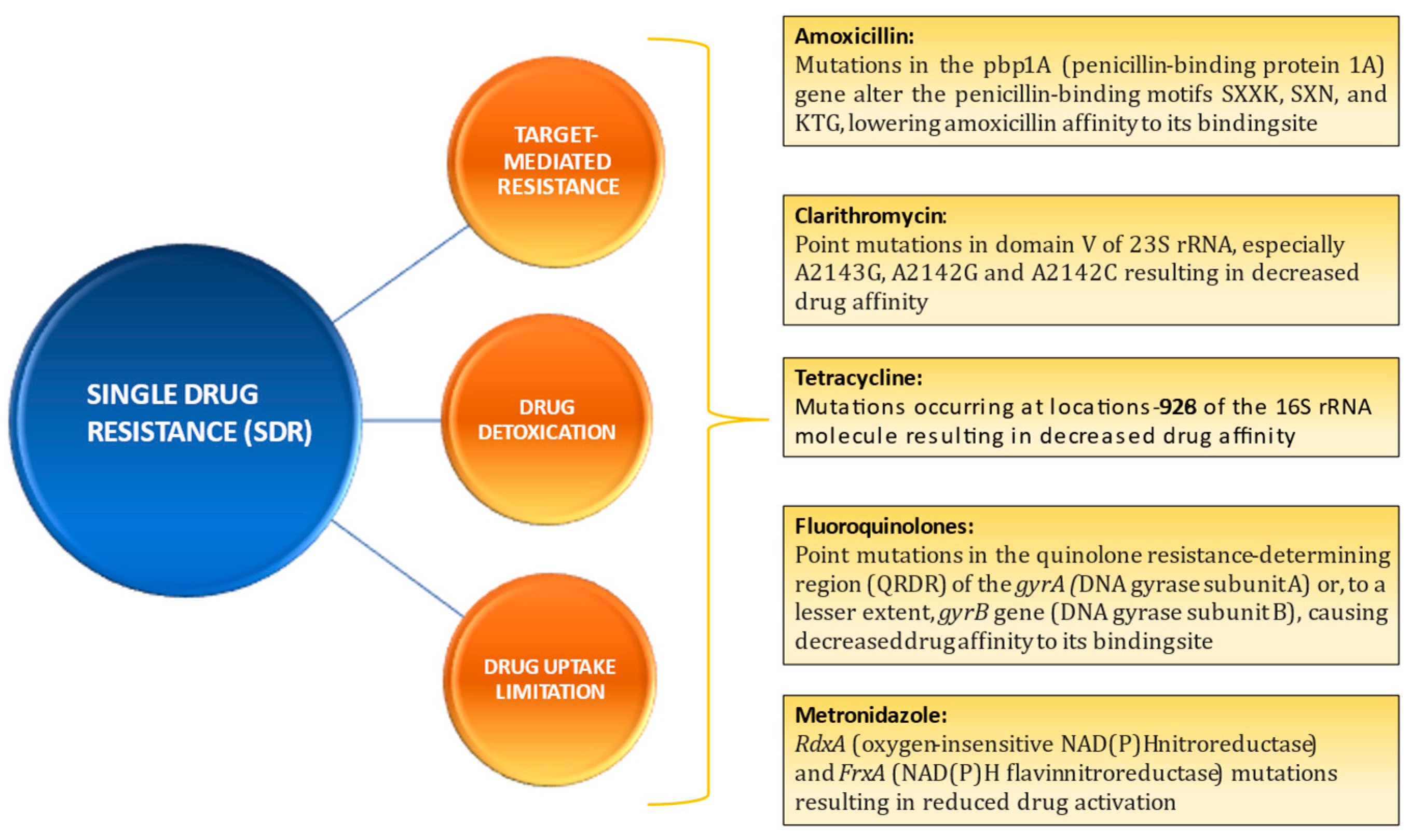
3. Mechanisms of Drug Resistance and Clinical Implications

These are broadly classified into three types of drug resistance pattern: single-drug resistance (SDR), multidrug resistance (MDR), and heteroresistance (HR).
SDR: When resistance develops against a single class of antibiotics, the mechanism is usually specific. This is known as SDR. Indeed, it is class-specific. SDR is typically defined through three mechanisms: (i) drug target-mediated resistance, (ii) drug detoxication, and (iii) drug uptake limitation [23,24]. Figure 4 demonstrates the predominant molecular mechanisms of single-drug resistance in Helicobacter pylori.
Clinical relevance of SDR in terms of efficacy of eradication regimens involving these medications has primarily been examined for clarithromycin, levofloxacin, and metronidazole. As a secondary outcome, Savoldi et al. analyzed data from 45 studies and reported a 7-fold (OR, 6.97; 95% CI, 5.23–9.01; p = 0.001) increase in the likelihood of eradication failure in patients with clarithromycin-resistant H. pylori strains treated with clarithromycin-containing regimens. A similar strong association was reported for levofloxacin (OR, 8.18; 95% CI, 3.81–17.56; p = 0.001) and combined clarithromycin and metronidazole resistance (OR, 9.40; 95% CI, 5.48–16.12; p = 0.001), though the clinical impact of metronidazole-only resistance was comparatively less significant (OR, 2.52; 95% CI, 1.82–3.48; p = 0.004) [16].
MDR: Multidrug resistance is defined as the simultaneous occurrence of resistance to three or more classes of antibiotics. Recent reports have indicated the alarming prevalence of MDR strains with triple resistance to clarithromycin, metronidazole, and quinolones as the most frequently reported MDR pattern. Primary MDR rates ranged from less than 10% in most parts of Europe to more than 20% in India and more than 40% in Peru [25,26]. Several mechanisms for MDR in H. pylori have been identified, but their clinical significance is unclear. These mechanisms include (i) increased drug efflux due to the activation of channels and efflux pumps; (ii) decreased drug uptake; (iii) biofilm formation; and (iv) transformation into an antibiotic-resistant coccoid form. Efflux pumps are upregulated or overexpressed, resulting in greater drug efflux from the cell. Biofilms are an extracellular matrix created by organisms as a long-term survival strategy that works as an effective, non-specific antibiotic barrier. They have emerged as an important MDR mechanism. These strains are also thought to have different modes of transmission. H. pylori can exist in three states: live bacillary, non-viable degenerative, and quiescent coccoid. To kill these coccoid forms, antibiotics must have a substantially higher minimum inhibitory concentration (MIC). Figure 5 depicts the predominant mechanisms attributable to H. pylori multidrug resistance [24].
HR: It is the coexistence of susceptible and resistant strains of an organism against a particular antimicrobial agent [27]. Recently, HR has been defined as the presence of a resistant strain in a subpopulation with a MIC that is at least eight times higher than the concentration required to inhibit the growth of the primary population [28]. HR in H. pylori can be intra-niche (when susceptible and resistant strains are isolated from the same gastric mucosal location) or inter-niche (when biopsies collected from separate regions of the gastric mucosa reveal H. pylori strains with different susceptibility patterns). HR can be monoclonal or polyclonal, and it can exist in both eradication-naive and previously treated patients. Polyclonal HR is characterized by the presence of genetically distinct clones exhibiting a stable antibiotic sensitivity phenotype. Monoclonal HR is characterized by the presence of genetically identical strains that can give rise to new strains with a variable pattern of susceptibility or resistance. Recent research suggests that HR in H. pylori is largely caused by co-infection with many strains in developing countries, whereas it is caused by the microevolution of a single H. pylori strain in response to antibiotic pressure in industrialized countries. HR is a serious clinical concern that is rarely addressed in major treatment guidelines and may be a precursor to future SDR or MDR [24,29]. According to a recent study by Kocsmár et al., one-fifth of clarithromycin-resistant patients can be missed with a biopsy from only the antrum, and inter-niche-only techniques can lower HR by more than 50%. To counteract this phenomenon, evidence suggests taking multiple biopsies from different gastric regions or evaluating multiple bacterial colonies from the same sample for drug susceptibility testing [30].

4. Detection of Antibiotic Resistance

Antimicrobial resistance is a ubiquitous problem requiring various solutions. The increasing prevalence of antibiotic resistance among H. pylori isolates worldwide has severely impacted the efficacy of eradication regimens, which could have significant health and economic consequences [31]. In order to personalize H. pylori therapy and formulate treatment guidelines for specific populations based on primary resistance patterns, antibiotic susceptibility testing (AST) plays a crucial role. The detection of antibiotic resistance can be assessed either by phenotypic methods employing the growth of H. pylori in culture from endoscopically retrieved gastric tissue or by genotypic or molecular methods employing pure culture isolates or biological specimens such as biopsy, stool, or saliva [23].

4.1. Phenotypic Methods

These methods involve the cultivation of H. pylori through bacterial culture. A bacterial isolate acquired by culturing is used to determine antimicrobial sensitivity. This can be accomplished using either the dilution method (agar or broth dilution method) or the diffusion method (disc or E-test). Methods such as agar dilution, broth dilution, and the E-test are utilized to determine the minimum inhibitory concentration (MIC) of antibiotics. Quantitative approaches include agar dilution and broth dilution [23].
The agar dilution method is the gold standard and is recommended by the Clinical and Laboratory Standard Institute (CLSI) [32]. First, 1 to 3 mL of a 2.0 McFarland-adjusted bacterial suspension (containing 1 × 10⁷ to 1 × 10⁸ CFU/mL) is spot inoculated on Mueller–Hinton agar supplemented with 5 to 10% sheep or horse blood and 2-fold dilutions of the antibiotics. Plates are read for bacterial growth after 72 h of incubation at 35 °C in microaerophilic conditions. The maximum dilution of antibiotics that results in no growth is referred to as the MIC. H. pylori strains are classified as sensitive, moderately resistant, or resistant based on the concentrations at which they are sensitive. According to the European Committee on Antimicrobial Susceptibility Testing (EUCAST), the MIC breakpoint for amoxicillin is >0.125 mg/L, levofloxacin’s is >1 mg/L, clarithromycin’s is >0.5 mg/L, tetracycline’s is >1 mg/L, metronidazole’s is >0.8 mg/L, and rifampicin’s is >1 mg/L [33].
The disc diffusion method offers a cut-off dependent on the diameter of the zone of inhibition. This method is widely used in epidemiological investigations, but its application in ordinary day-to-day practice has been difficult and time consuming due to tedious preparations, the length of time required, and the fact that it is not cost-effective [23,34,35]. Due to the difficulty of culturing H. pylori in broth without defibrinated blood, the broth dilution method is rarely employed [36].
The E-test employs a bacterial solution calibrated to the McFarland standard of 3.0 on Muller–Hinton agar supplemented with 5–10% sheep blood [34]. Under microaerophilic circumstances, an E-test strip impregnated with escalating quantities of antibiotics is placed on inoculation plates and incubated for 72 hours at a temperature of 37 °C. The MIC is provided by an elliptical zone of inhibition. When compared to agar dilution or broth dilution procedures, the E-test is straightforward and easy to execute, and it has been found to correlate closely with gold standard (agar dilution) results, except for metronidazole, where recent investigations have revealed some inconsistencies [37,38,39]. The disc diffusion method is a semiquantitative approach to assessing antibiotic resistance. It measures antimicrobial resistance using cut-off diameters of the zone of inhibition [23].

4.2. Molecular Methods

Although H. pylori culture followed by MIC-based AST is regarded as the gold standard, the widespread use of culture-based techniques is limited for a number of reasons. H. pylori’s finicky nature necessitates well-equipped laboratories with trained personnel to provide supplemental growth conditions, a microaerophilic atmosphere, and up to 14 days of incubation. Culture-based methodologies are also affected by sample processing delays and recent exposure to PPIs. This corresponds to a success rate of between 55 and 73% in routine clinical practice. In addition, the requirement of endoscopy for tissue acquisition restricts their use to endoscopy cohorts. Molecular-based methods have emerged as a more rapid (up to <4 h), cost-effective, and highly reproducible alternative to culture-based methods for detecting H. pylori and determining its resistance, given the challenges associated with traditional culture methods. Molecular methods can detect heteroresistant infections and are less affected by the modulation of bacterial burden caused by the recent use of PPIs [23,24,40,41]. Recent large RCTs have demonstrated that molecular testing-guided therapy is comparable to culture-based susceptibility testing-guided therapy in first-line therapy and non-inferior to the latter in third-line treatment of H. pylori infection, thereby supporting the use of molecular testing-guided therapy for H. pylori eradication [42].
Another benefit of molecular approaches is that gastric samples collected through rapid urease tests (RUTs) can be utilized for PCR-based molecular testing. The observed correlation between the utilization of RUT gastric biopsies for molecular testing after 30 days at room temperature was 93% [43]. In addition, these methods allow for the non-invasive detection of antibiotic resistance [40]. Gastric contents (i.e., gastric fluid, mucus, or mucosal biopsies), formalin-fixed, paraffin-embedded (FFPE) tissue, stools, or repurposed tissue from histology or rapid urease tests can be used for testing. A recent meta-analysis found that genotypic testing of clarithromycin resistance from stool specimens is an accurate, convenient, and rapid detection method with pooled sensitivity and specificity values of 0.93 (95% confidence interval (CI): 0.90 to 0.96) and 0.98 (95% CI: 0.93 to 1.00), respectively [44].
The alterations in genes that cause antibiotic resistance in H. pylori have been extensively investigated, particularly for the most routinely used antibiotics in eradication regimens, such as clarithromycin and levofloxacin. These are predominantly attributable to recognized localized mutations such as point mutations in 23S rRNA for clarithromycin resistance and in gyrA for levofloxacin resistance (Figure 6). Without conducting an antibiogram, molecular-based methods rely primarily on the detection of these specific H. pylori mutations encoding resistance in order to determine genotypic susceptibility to commonly employed antibiotics. The majority of assays are polymerase chain reaction (PCR)-based, and various techniques, including conventional PCR followed by Sanger sequencing, real-time (RT) PCR, droplet digital PCR, DNA microarray technology, multiplex quantitative PCR, and next-generation sequencing (NGS), are employed for this purpose. Moreover, these methodologies can be culture-based or culture-free when applied directly to biological samples [23,24,41].
Using specific primers, conventional PCR amplifies specific regions in target genes known to contain major mutations, as shown in Figure 4 for a particular antibiotic. The amplified primers are then subjected to DNA sequencing using the Sanger method in order to detect mutations. Although this method is regarded as the gold standard for identifying mutations and demonstrates a strong correlation with phenotypic antibiotic resistance [36], it is not cost-effective in routine clinical contexts [45].
The use of RT-PCR assays, which provide results for simultaneous detection of H. pylori and antimicrobial resistance in real time within hours and can be performed in any microbiological laboratory [40,41], is a more practical, rapid, and cost-effective alternative. Several commercially available kits utilize this principal for detection of clarithromycin resistance including H. pylori ClariRes (Ingenetix, Vienna, Austria) [46], Allplex H. pylori and ClariR (Seegene, Republic of Korea) [47], Lightmix H. pylori (TIBMolbiol, Germany) [48], H. pylori TaqMan real-time PCR assay (Meridian Bioscience, United States) [49], Amplidiag H. pylori + ClariR (Mobidiag, Espoo, Finland) [50], RIDA GENE H. pylori (r-Biopharm, Darmstadt, Germany) [51], Seeplex ClaR-H. pylori ACE detection system (See-gene, Eschborn, Germany), and the MutaREAL Helicobacter pylori kit (Immunodiagnostik, Benshiem, Germany) [41,52]. The GenoType HelicoDR assay (Hain Lifescience, Nehren, Germany) uses a combination PCR and DNA hybridization procedure to detect combined clarithromycin and levofloxacin resistance-causing mutations [53]. Despite the fact that these methods have demonstrated excellent performance using a biopsy or stool sample to simultaneously detect infection and antimicrobial resistance, especially for clarithromycin, with a sensitivity and specificity of up to 94% and 100%, respectively, local validation of kits in reference to culture-based AST is recommended [23,41]. The sensitivity of these approaches varies depending on the DNA extraction process and molecular assay used. False negative results can be achieved if paraffin-embedded gastric biopsy samples are used due to DNA fragmentation caused by fixatives or when using stool samples due to contamination [54,55]. High concordance with the phenotypic AST and Sanger sequencing methods, the ability to detect heteroresistant strains, and the feasibility of assessing clarithromycin resistance-causing alleles retrospectively from formalin-fixed paraffin-embedded gastric tissue are a few of the most notable features of the droplet digital PCR method [23,24,56].
As conventional methods require prior knowledge of mutations to use specific primers for detecting them in only a limited number of target genes, there is a risk of missing novel, rare, or complex antibiotic resistance mechanisms. Consequently, these techniques permit the detection of resistance mutations at a restricted number of sites. However, due to H. pylori’s vast genetic diversity and high mutation and recombination rates, the mechanisms of antimicrobial resistance are heterogeneous and complex especially for antibiotics such as metronidazole and amoxicillin. This is particularly important in areas with high clarithromycin resistance [24,57]. Using metronidazole as an example, mutations or DNA sequence alterations associated with resistance have been described in multiple genes and regions, including the rdxA, frxA, fdxB, and recA genes associated with various nucleotide alterations including nonsense, missense, frameshift, deletion, and insertion. As a result, traditional PCR-based assays based on hybridization or enzymatic analytic techniques fall short of the mark when it comes to detecting complicated and new mutations or assessing resistance to several antibiotics [58].
NGS has evolved into a potent, quick (turnaround time of 24–72 h), and cost-effective technique for predicting antibiotic resistance and evaluating several genes at the same time [19,59]. These methods could be combined with bacterial culture for whole-genome sequencing (WGS) or with other molecular techniques such as PCR for deep-amplicon sequencing. NGS-based approaches enable a comprehensive view of bacterial genotypes and can simultaneously identify DNA mutations or variants of H. pylori genes responsible for resistance in all the commonly used antibiotics for H. pylori eradication, with the possibility of identifying novel or rare resistance mechanisms. With evolving data on the molecular basis of resistance, the clinical relevance of new mutations identified by these approaches can be ascertained by retrospective analysis of WGS data, in contrast to phenotypic methods that require retesting using stored samples, albeit with reduced ability to grow in vivo, or PCR-based assays that require redesigning samples. It is also capable of detecting HR and can be performed on fresh, frozen, or archived formalin-fixed paraffin-embedded gastric tissue blocks [19,59,60,61]. Several NGS-based technologies are even affordable in low-income countries, and the high concordance between mutations and phenotypic drug resistance makes them a desirable option for monitoring genotypic markers of antibiotic resistance in H. pylori [23,24,59,62]. The combination of WGS and phenotypic methodologies, according to some experts, provides a more comprehensive approach to antimicrobial resistance surveillance [63].
In contrast to deep-amplicon sequencing, WGS approaches require prior culturing of H. pylori, which is a significant limitation. Furthermore, the presence of fragmented DNA makes WGS more challenging when applied to formalin-fixed, paraffin-embedded tissue. In comparison to WGS, the PCR-based targeted sequencing approach using NGS is more practicable and cost-effective in routine clinical practice [64]. Hulten et al. recently compared targeted NGS and culture-based H. pylori susceptibility testing using clinical isolates and paired formalin-fixed, paraffin-embedded gastric biopsies. NGS showed good agreement with the agar dilution method for clarithromycin (k = 0.90012, p = 0.0001) and levofloxacin (k = 0.78161, p = 0.0001) when applied to clinical isolates but less satisfactory agreement for metronidazole (k = 0.5588, p = 0.0001) and amoxicillin (k = 0.21400, p = 0.0051). For clarithromycin, levofloxacin, metronidazole, and amoxicillin, the accuracy of NGS in predicting resistance in clinical isolates was 97.1%, 89.5%, 77.6%, and 95.9%, respectively. When carried out on formalin-fixed, paraffin-embedded gastric biopsies, NGS showed good agreement with agar dilution for clarithromycin (k = 0.81236, p = 0.0001) and levofloxacin (k = 0.74953, p = 0.0001) but less satisfactory agreement for metronidazole (k = 0.54645, p = 0.0001) and amoxicillin (k = 0.21400, p = 0.0051). NGS was able to predict resistance in formalin-fixed, paraffin-embedded biopsies with an accuracy of 94.1% for clarithromycin, 87.7% for levofloxacin, 77.7% for metronidazole, and 95.9% for amoxicillin. These results are significant as it eliminates the need for additional specimens and cultures. However, the results for amoxicillin and metronidazole are relatively poor, most likely due to the insufficient molecular characterization of these antibiotic resistance mechanisms as explained above [58]. To obtain a perfect correlation with phenotypic data, a comprehensive WGS-based analysis may aid in identifying additional mutations of these genes or of others or other resistance mechanisms. Moss et al. were the first to report the accuracy of NGS in determining the H. pylori antimicrobial susceptibility profile in stool samples, finding that the results obtained from stool samples were concordant with those obtained from FFPE gastric biopsies in 91.4% of cases and fresh gastric specimens in 92.2% of cases. Clarithromycin (k = 0.94), levofloxacin (k = 0.88), and metronidazole (k = 0.89) had good agreement between stool and fresh stomach samples. This is a huge step forward in obtaining antibiotic susceptibility data using NGS without the discomfort, costs, and risks associated with endoscopy [65].
NGS-based techniques are incapable of determining MIC, and the contribution of each mutation to MIC is not always obvious. Moreover, as with other genomic-based methods, phenotypic and culture-based susceptibility testing and genomic prediction of resistance do not always correspond. NGS can also underestimate resistance caused by non-genetic mechanisms [19,24,59,66]. There is a need for additional research to determine which genes or gene involvement patterns correlate most strongly with outcomes [58,66]. To overcome the diminished predictive value that could be the result of DNA fragmentation in FFPE tissue samples or associated contamination in stool samples, excellent DNA extraction methods need to be developed. Consequently, studies with broader gene coverages, larger sample sizes, or multicenter designs from various geographic regions are necessary. Standardized and user-friendly computational software and tools must be created so that NGS data may be easily analyzed and implemented in routine clinical settings [24,64].

5. Empirical Therapy vs. Susceptibility-Guided Tailored Therapy

According to the principles of antibiotic stewardship, susceptibility assays (culture or molecular-based) have been proposed for any bacterial infection, and H. pylori treatment should not be an exception. Still, eradication protocols are mainly empirical in the majority of settings. The recent Maastricht VI/Florence consensus report, while recommending AST prior to prescribing first-line therapy as a plausible approach, acknowledges its limited utility in real-world settings [67]. Several meta-analyses compared cure rates for first-line therapy using an AST-based vs. an empirical method [68,69]. The evidence suggests that although the AST-based approach performs better when using triple-drug therapy, the difference is not significant when the most recent quadruple regimens are employed. The majority of international guidelines recommend avoiding clarithromycin and levofloxacin when local resistance patterns indicate rates of >15%. As a result of the fact that worldwide data reveal this to be the case in the majority of regions throughout the world, the treatment is already being shifted to bismuth-based quadruple therapy [19].
A recent AGA clinical practice update on the management of refractory H. pylori recommended susceptibility testing to guide the selection of subsequent regimens after two failed eradication attempts with confirmed patient adherence [11]. This is in accordance with Maastricht V’s recommendations [70]. However, in real-world contexts, this situation is complicated by logistical difficulties in acquiring H. pylori resistance profiles and a lack of solid evidence favoring tailored therapy over empirical therapy in patients whose H. pylori treatment has failed [1,71]. Several meta-analyses, including a recent update, have found comparable H. pylori cure rates between susceptibility-guided tailored therapies and empirical therapy for second-line treatment [72,73,74,75,76]. Recent data from the European Registry for the Management of Helicobacter pylori (Hp-EuReg) indicate that eradication rates of more than 90% can be achieved using various empirical regimens as second-line therapy [77]. Furthermore, patients who have had two or more failed H. pylori medications demonstrated equal eradication rates across customized and empirical therapies [74,75,76,78,79].
Several studies have given contradictory results about the cost-effectiveness of susceptibility-guided treatment [71]. Moreover, even when the susceptibility profile of H. pylori is known, eradication rates are not 100 percent, indicating a disparity between in vitro susceptibility and in vivo eradication [71,76]. A recent systematic review of clinical studies that investigated the efficacy of second-line treatments revealed a cure rate of only 72% in patients harboring a clarithromycin-susceptible strain after prior clarithromycin treatment [80]. Consequently, there is insufficient evidence to support the routine use of susceptibility-guided, individualized therapy after H. pylori eradication failure at this time. Recently, the Maastricht VI Consensus suggested that second-line and rescue therapies should be guided by local resistance patterns as determined by susceptibility testing and monitoring eradication rates to maximize treatment success [1]. Therefore, there is an immediate need for a well-designed trial comparing AST-directed rescue therapy with empirical rescue therapy. With the advent of molecular techniques, H. pylori management can now enter the realm of antimicrobial stewardship. In addition, the development of non-invasive molecular techniques may, in the near future, remove many rate-limiting obstacles to the pervasive use of an AST-guided approach. On the other hand, further research is needed to evaluate the efficacy of new empirical medicines, such as vonoprazan-based therapies, with susceptibility-guided therapy. Nonetheless, susceptibility tests should be performed routinely, even before prescribing first-line treatment, in specialized centers with an interest in H. pylori management.

6. Conclusions

Antimicrobial resistance is a ubiquitous problem that necessitates a variety of solutions. Drug-resistant H. pylori is becoming an increasing health problem as a result of a considerable drop in the eradication rate of empirical therapy, which could ultimately lead to clinical complications associated with persistent infection as well as increased healthcare expenses. Therefore, periodic regional drug susceptibility surveillance programs and the implementation of policies for the prudent use of antibiotics should be of the utmost importance. Due to the advancement of molecular methods for detecting antibiotic resistance, the treatment of H. pylori can now enter the domain of antimicrobial stewardship. The use of genomics to investigate antibiotic resistance in bacteria of public health concern is a burgeoning field of study; the combination of WGS and traditional phenotypic resistance data can provide powerful results in discovering new resistance mechanisms. In addition, the development of non-invasive molecular techniques may in the near future remove many rate-limiting barriers to the widespread application of an AST-guided approach. Although a susceptibility-based strategy should be used whenever possible, empirical therapy based on prior medication history and local resistance patterns is an acceptable alternative when logistical and financial factors are considered. There is a need for the development of novel non-invasive antibiotic susceptibility testing tools and the standardization of existing protocols. Future research is required to determine the relative contribution of each mutation to MIC and the correlation between gene involvement patterns and outcomes.

Author Contributions

V.J. and M.K.P.: Conceptualization, V.J.: Writing the first draft, M.M.R., A.C. and A.K.: Writing and editing the draft, M.K.P.: Supervision and final editing of the draft. All authors have read and agreed to the published version of the manuscript.

Funding

This research received no external funding.

Institutional Review Board Statement

Not applicable.

Informed Consent Statement

Not applicable.

Conflicts of Interest

The authors declare no conflict of interest.

References

  1. Malfertheiner, P.; Megraud, F.; Rokkas, T.; Gisbert, J.P.; Liou, J.M.; Schulz, C.; Gasbarrini, A.; Hunt, R.H.; Leja, M.; O’Morain, C.; et al. Management of Helicobacter pylori infection: The Maastricht VI/Florence consensus report. Gut 2022, 71, 1724–1762. [Google Scholar] [CrossRef] [PubMed]
  2. Zamani, M.; Ebrahimtabar, F.; Zamani, V.; Miller, W.H.; Alizadeh-Navaei, R.; Shokri-Shirvani, J.; Derakhshan, M.H. Systematic review with meta-analysis: The worldwide prevalence of Helicobacter pylori infection. Aliment. Pharmacol. Ther. 2018, 47, 868–876. [Google Scholar] [CrossRef]
  3. Boyanova, L.; Hadzhiyski, P.; Gergova, R.; Markovska, R. Evolution of Helicobacter pylori Resistance to Antibiotics: A Topic of Increasing Concern. Antibiotics 2023, 12, 332. [Google Scholar] [CrossRef] [PubMed]
  4. Plummer, M.; Franceschi, S.; Vignat, J.; Forman, D.; de Martel, C. Global burden of gastric cancer attributable to Helicobacter pylori. Int. J. Cancer 2015, 136, 487–490. [Google Scholar] [CrossRef] [PubMed]
  5. Al-Fakhrany, O.M.; Elekhnawy, E. Helicobacter pylori in the post-antibiotics era: From virulence factors to new drug targets and therapeutic agents. Arch. Microbiol. 2023, 205, 301. [Google Scholar] [CrossRef] [PubMed]
  6. Sung, H.; Ferlay, J.; Siegel, R.L.; Laversanne, M.; Soerjomataram, I.; Jemal, A.; Bray, F. Global Cancer Statistics 2020: GLOBOCAN Estimates of Incidence and Mortality Worldwide for 36 Cancers in 185 Countries. CA Cancer J. Clin. 2021, 71, 209–249. [Google Scholar] [CrossRef]
  7. Ford, A.C.; Yuan, Y.; Moayyedi, P. Helicobacter pylori eradication therapy to prevent gastric cancer: Systematic review and meta-analysis. Gut 2020, 69, 2113–2121. [Google Scholar] [CrossRef]
  8. Lee, Y.C.; Chiang, T.H.; Chou, C.K.; Tu, Y.K.; Liao, W.C.; Wu, M.S.; Graham, D.Y. Association between Helicobacter Pylori Eradication and Gastric Cancer Incidence: A Systematic Review and Meta-analysis. Gastroenterology 2016, 150, 1113–1124.e5. [Google Scholar] [CrossRef]
  9. Rokkas, T.; Rokka, A.; Portincasa, P. A systematic review and meta-analysis of the role of Helicobacter pylori eradication in preventing gastric cancer. Ann. Gastroenterol. 2017, 30, 414–423. [Google Scholar] [CrossRef]
  10. Liou, J.M.; Lee, Y.C.; Wu, M.S.; Taiwan Gastrointestinal Disease and Helicobacter Consortium. Treatment of Refractory Helicobacter pylori Infection-Tailored or Empirical Therapy. Gut Liver 2022, 16, 8–18. [Google Scholar] [CrossRef]
  11. Shah, S.C.; Iyer, P.G.; Moss, S.F. AGA Clinical Practice Update on the Management of Refractory Helicobacter pylori Infection: Expert Review. Gastroenterology 2021, 160, 1831–1841. [Google Scholar] [CrossRef] [PubMed]
  12. Cortés, P.; Nelson, A.D.; Bi, Y.; Stancampiano, F.F.; Murray, L.P.; Pujalte, G.G.; Gomez, V.; Harris, D.M. Treatment Approach of Refractory Helicobacter pylori Infection: A Comprehensive Review. J. Prim. Care Community Health 2021, 12, 21501327211014087. [Google Scholar] [CrossRef] [PubMed]
  13. Graham, D.Y.; Liou, J.M. Primer for Development of Guidelines for Helicobacter pylori Therapy Using Antimicrobial Stewardship. Clin. Gastroenterol. Hepatol. 2022, 20, 973–983.e1. [Google Scholar] [CrossRef] [PubMed]
  14. Hsu, P.-I.; Chen, K.-Y.; Tai, W.-C.; Yang, J.-C.; Tsay, F.-W.; Liu, Y.-H.; Chen, C.-L.; Lee, C.-L.; Yeh, H.-Z.; Kuo, C.-H.; et al. Hybrid, High-Dose Dual and Bismuth Quadruple Therapies for First-Line Treatment of Helicobacter pylori Infection in Taiwan: A Multicenter, Open-Label, Randomized Trial. Am. J. Gastroenterol. 2023, 118, 1184–1195. [Google Scholar] [CrossRef]
  15. Shrestha, A.B.; Pokharel, P.; Sapkota, U.H.; Shrestha, S.; Mohamed, S.A.; Khanal, S.; Jha, S.K.; Mohanty, A.; Padhi, B.K.; Asija, A.; et al. Drug Resistance Patterns of Commonly Used Antibiotics for the Treatment of Helicobacter pylori Infection among South Asian Countries: A Systematic Review and Meta-Analysis. Trop. Med. Infect. Dis. 2023, 8, 172. [Google Scholar] [CrossRef]
  16. Savoldi, A.; Carrara, E.; Graham, D.Y.; Conti, M.; Tacconelli, E. Prevalence of Antibiotic Resistance in Helicobacter pylori: A Systematic Review and Meta-analysis in World Health Organization Regions. Gastroenterology 2018, 155, 1372–1382.e17. [Google Scholar] [CrossRef]
  17. Kuo, Y.-T.; Liou, J.-M.; El-Omar, E.M.; Wu, J.-Y.; Leow, A.H.R.; Goh, K.L.; Das, R.; Lu, H.; Lin, J.-T.; Tu, Y.-K.; et al. Primary antibiotic resistance in Helicobacter pylori in the Asia-Pacific region: A systematic review and meta-analysis. Lancet Gastroenterol. Hepatol. 2017, 2, 707–715. [Google Scholar] [CrossRef]
  18. Ho, J.J.C.; Navarro, M.; Sawyer, K.; Elfanagely, Y.; Moss, S.F. Helicobacter pylori Antibiotic Resistance in the United States between 2011 and 2021: A Systematic Review and Meta-Analysis. Am. J. Gastroenterol. 2022, 117, 1221–1230. [Google Scholar] [CrossRef]
  19. Argueta, E.A.; Ho, J.J.C.; Elfanagely, Y.; D’Agata, E.; Moss, S.F. Clinical Implication of Drug Resistance for H. pylori Management. Antibiotics 2022, 11, 1684. [Google Scholar] [CrossRef]
  20. Megraud, F.; Bruyndonckx, R.; Coenen, S.; Wittkop, L.; Huang, T.-D.; Hoebeke, M.; Bénéjat, L.; Lehours, P.; Goossens, H.; Glupczynski, Y. Helicobacter pylori resistance to antibiotics in Europe in 2018 and its relationship to antibiotic consumption in the community. Gut 2021, 70, 1815–1822. [Google Scholar] [CrossRef]
  21. Hulten, K.G.; Lamberth, L.B.; Kalfus, I.N.; Graham, D.Y. National and Regional US Antibiotic Resistance to Helicobacter pylori: Lessons From a Clinical Trial. Gastroenterology 2021, 161, 342–344.e1. [Google Scholar] [CrossRef] [PubMed]
  22. Kuo, C.-J.; Lee, C.-H.; Chang, M.-L.; Lin, C.-Y.; Lin, W.-R.; Su, M.-Y.; Tseng, C.-N.; Wu, Y.-S.; Chiu, C.-T.; Lai, C.-H. Multidrug resistance: The clinical dilemma of refractory Helicobacter pylori infection. J. Microbiol. Immunol. Infect. 2021, 54, 1184–1187. [Google Scholar] [CrossRef]
  23. Ansari, S.; Yamaoka, Y. Helicobacter pylori Infection, Its Laboratory Diagnosis, and Antimicrobial Resistance: A Perspective of Clinical Relevance. Clin. Microbiol. Rev. 2022, 35, e0025821. [Google Scholar] [CrossRef] [PubMed]
  24. Tshibangu-Kabamba, E.; Yamaoka, Y. Helicobacter pylori infection and antibiotic resistance—From biology to clinical implications. Nat. Rev. Gastroenterol. Hepatol. 2021, 18, 613–629. [Google Scholar] [CrossRef] [PubMed]
  25. Boyanova, L.; Hadzhiyski, P.; Kandilarov, N.; Markovska, R.; Mitov, I. Multidrug resistance in Helicobacter pylori: Current state and future directions. Expert Rev. Clin. Pharmacol. 2019, 12, 909–915. [Google Scholar] [CrossRef]
  26. Phan, T.N.; Santona, A.; Tran, V.H.; Tran, T.N.H.; Le, V.A.; Cappuccinelli, P.; Rubino, S.; Paglietti, B. High rate of levofloxacin resistance in a background of clarithromycin-and metronidazole-resistant Helicobacter pylori in Vietnam. Int. J. Antimicrob. Agents 2015, 45, 244–248. [Google Scholar] [CrossRef]
  27. Mascellino, M.T.; Porowska, B.; De Angelis, M.; Oliva, A. Antibiotic susceptibility, heteroresistance, and updated treatment strategies in Helicobacter pylori infection. Drug Des. Dev. Ther. 2017, 11, 2209–2220. [Google Scholar] [CrossRef]
  28. Andersson, D.I.; Nicoloff, H.; Hjort, K. Mechanisms and clinical relevance of bacterial heteroresistance. Nat. Rev. Microbiol. 2019, 17, 479–496. [Google Scholar] [CrossRef]
  29. Keikha, M.; Karbalaei, M. Prevalence of antibiotic heteroresistance associated with Helicobacter pylori infection: A systematic review and meta-analysis. Microb. Pathog. 2022, 170, 105720. [Google Scholar] [CrossRef]
  30. Kocsmár, É.; Kocsmár, I.; Buzás, G.M.; Szirtes, I.; Wacha, J.; Takáts, A.; Hritz, I.; Schaff, Z.; Rugge, M.; Fassan, M.; et al. Helicobacter pylori heteroresistance to clarithromycin in adults-New data by in situ detection and improved concept. Helicobacter 2020, 25, e12670. [Google Scholar] [CrossRef]
  31. Dang, B.N.; Graham, D.Y. Helicobacter pylori infection and antibiotic resistance: A WHO high priority? Nat. Rev. Gastroenterol. Hepatol. 2017, 14, 383–384. [Google Scholar] [CrossRef] [PubMed]
  32. M07; Methods for Dilution Antimicrobial Susceptibility Test for Bacteria That Grow Aerobically. Clinical and Laboratory Standards Institute (CLSI): Wayne, IL, USA, 2015.
  33. The European Committee on Antimicrobial Susceptibility Testing. Breakpoint Tables for Interpretation of MICs and Zone Diameters. Version 12.0. 2022. Available online: https://www.eucast.org/fileadmin/src/media/PDFs/EUCAST_files/Breakpoint_tables/v_12.0_Breakpoint_Tables.pdf (accessed on 25 August 2023).
  34. Valdivieso-García, A.; Imgrund, R.; Deckert, A.; Varughese, B.M.; Harris, K.; Bunimov, N.; Reid-Smith, R.; McEwen, S. Cost analysis and antimicrobial susceptibility testing comparing the E test and the agar dilution method in Campylobacter jejuni and Campylobacter coli. Diagn. Microbiol. Infect. Dis. 2009, 65, 168–174. [Google Scholar] [CrossRef]
  35. Medakina, I.; Tsapkova, L.; Polyakova, V.; Nikolaev, S.; Yanova, T.; Dekhnich, N.; Khatkov, I.; Bordin, D.; Bodunova, N. Helicobacter pylori Antibiotic Resistance: Molecular Basis and Diagnostic Methods. Int. J. Mol. Sci. 2023, 24, 9433. [Google Scholar] [CrossRef] [PubMed]
  36. Lok, C.-H.; Zhu, D.; Wang, J.; Ren, Y.-T.; Jiang, X.; Li, S.-J.; Zhao, X.-Y. Phenotype and Molecular Detection of Clarithromycin and Levofloxacin Resistance in Helicobacter pylori Clinical Isolates in Beijing. Infect. Drug Resist. 2020, 13, 2145–2153. [Google Scholar] [CrossRef] [PubMed]
  37. Camorlinga-Ponce, M.; Gómez-Delgado, A.; Aguilar-Zamora, E.; Torres, R.C.; Giono-Cerezo, S.; Escobar-Ogaz, A.; Torres, J. Phenotypic and Genotypic Antibiotic Resistance Patterns in Helicobacter pylori Strains From Ethnically Diverse Population in México. Front. Cell. Infect. Microbiol. 2021, 10, 539115. [Google Scholar] [CrossRef]
  38. Huang, X.; Liu, Y.; Lin, Z.; Wu, B.; Nong, G.; Chen, Y.; Lu, Y.; Ji, X.; Zhou, X.; Suo, B.; et al. Minimum inhibitory concentrations of commonly used antibiotics against Helicobacter Pylori: A multicenter study in South China. PLoS ONE 2021, 16, e0256225. [Google Scholar] [CrossRef]
  39. Miftahussurur, M.; Fauzia, K.A.; Nusi, I.A.; Setiawan, P.B.; Syam, A.F.; Waskito, L.A.; Doohan, D.; Ratnasari, N.; Khomsan, A.; Adnyana, I.K.; et al. E-test versus agar dilution for antibiotic susceptibility testing of Helicobacter pylori: A comparison study. BMC Res. Notes 2020, 13, 22. [Google Scholar] [CrossRef]
  40. Fernandez-Caso, B.; Miqueleiz, A.; Valdez, V.B.; Alarcón, T. Are molecular methods helpful for the diagnosis of Helicobacter pylori infection and for the prediction of its antimicrobial resistance? Front. Microbiol. 2022, 13, 962063. [Google Scholar] [CrossRef]
  41. Smith, S.M.; O’Morain, C.; McNamara, D. Helicobacter pylori resistance to current therapies. Curr. Opin. Gastroenterol. 2019, 35, 6–13. [Google Scholar] [CrossRef]
  42. Chen, M.J.; Chen, P.Y.; Fang, Y.J.; Bair, M.-J.; Chen, C.-C.; Chen, C.-C.; Yang, T.-H.; Lee, J.-Y.; Yu, C.-C.; Kuo, C.-C.; et al. Molecular testing-guided therapy versus susceptibility testing-guided therapy in first-line and third-line Helicobacter pylori eradication: Two multicentre, open-label, randomised controlled, non-inferiority trials. Lancet Gastroenterol. Hepatol. 2023, 8, 623–634. [Google Scholar] [CrossRef]
  43. Li, Y.; Rimbara, E.; Thirumurthi, S.; Trespalacios, A.; Reddy, R.; Sabounchi, S.; Attumi, T.A.; Graham, D.Y. Detection of clarithromycin resistance in Helicobacter pylori following noncryogenic storage of rapid urease tests for 30 days. J. Dig. Dis. 2012, 13, 54–59. [Google Scholar] [CrossRef] [PubMed]
  44. Ren, X.; Shi, Y.; Suo, B.; Yao, X.; Lu, H.; Li, C.; Zhang, Y.; Zhou, L.; Tian, X.; Song, Z. Individualized diagnosis and eradication therapy for Helicobacter pylori infection based on gene detection of clarithromycin resistance in stool specimens: A systematic review and meta-analysis. Helicobacter 2023, 28, e12958. [Google Scholar] [CrossRef] [PubMed]
  45. Mégraud, F.; Lehours, P. Helicobacter pylori detection and antimicrobial susceptibility testing. Clin. Microbiol. Rev. 2007, 20, 280–322. [Google Scholar] [CrossRef] [PubMed]
  46. Vécsei, A.; Innerhofer, A.; Binder, C.; Gizci, H.; Hammer, K.; Bruckdorfer, A.; Riedl, S.; Gadner, H.; Hirschl, A.M.; Makristathis, A. Stool polymerase chain reaction for Helicobacter pylori detection and clarithromycin susceptibility testing in children. Clin. Gastroenterol. Hepatol. 2010, 8, 309–312. [Google Scholar] [CrossRef] [PubMed]
  47. Jehanne, Q.; Bénéjat, L.; Mégraud, F.; Bessède, E.; Lehours, P. Evaluation of the Allplex™ H. pylori and ClariR PCR Assay for Helicobacter pylori detection on gastric biopsies. Helicobacter 2020, 25, e12702. [Google Scholar]
  48. Redondo, J.J.; Keller, P.M.; Zbinden, R.; Wagner, K. A novel RT-PCR for the detection of Helicobacter pylori and identification of clarithromycin resistance mediated by mutations in the 23S rRNA gene. Diagn. Microbiol. Infect. Dis. 2018, 90, 1–6. [Google Scholar] [CrossRef]
  49. Clines, N.; Beckman, E. Development of a high throughput human stool specimen processing method for a molecular Helicobacter pylori clarithromycin resistance assay. PLoS ONE 2019, 14, e0224356. [Google Scholar] [CrossRef]
  50. Hays, C.; Delerue, T.; Lamarque, D.; Burucoa, C.; Collobert, G.; Billöet, A.; Kalach, N.; Raymond, J. Molecular diagnosis of Helicobacter pylori infection in gastric biopsies: Evaluation of the Amplidiag® H. pylori + ClariR assay. Helicobacter 2019, 24, e12560. [Google Scholar] [CrossRef]
  51. Van den Poel, B.; Gils, S.; Micalessi, I.; Carton, S.; Christiaens, P.; Cuyle, P.-J.; Moons, V.; Van Olmen, G.; Smismans, A.; Bourgain, C.; et al. Molecular detection of Helicobacter pylori and clarithromycin resistance in gastric biopsies: A prospective evaluation of RIDA®GENE Helicobacter pylori assay. Acta Clin. Belg. 2021, 76, 177–183. [Google Scholar] [CrossRef]
  52. Wang, Y.H.; Li, Z.; Wang, L.; Zhu-Ge, L.; Zhao, R.; Wu, S.; Wang, Y.; An, Y.; Xie, Y. A systematic review and meta-analysis of genotypic methods for detecting antibiotic resistance in Helicobacter pylori. Helicobacter 2018, 23, e12467. [Google Scholar] [CrossRef]
  53. Cambau, E.; Allerheiligen, V.; Coulon, C.; Corbel, C.; Lascols, C.; Deforges, L.; Soussy, C.-J.; Delchier, J.-C.; Megraud, F. Evaluation of a new test, genotype HelicoDR, for molecular detection of antibiotic resistance in Helicobacter pylori. J. Clin. Microbiol. 2009, 47, 3600–3607. [Google Scholar] [CrossRef] [PubMed]
  54. Brennan, D.E.; Omorogbe, J.; Hussey, M.; Tighe, D.; Holleran, G.; O’morain, C.; Smith, S.M.; McNamara, D. Molecular detection of Helicobacter pylori antibiotic resistance in stool vs. biopsy samples. World J. Gastroenterol. 2016, 22, 9214–9221. [Google Scholar] [CrossRef] [PubMed]
  55. Patel, S.K.; Pratap, C.B.; Jain, A.K.; Gulati, A.K.; Nath, G. Diagnosis of Helicobacter pylori: What should be the gold standard? World J. Gastroenterol. 2014, 20, 12847–12859. [Google Scholar] [CrossRef] [PubMed]
  56. Sun, L.; Talarico, S.; Yao, L.; He, L.; Self, S.; You, Y.; Zhang, H.; Zhang, Y.; Guo, Y.; Liu, G.; et al. Droplet Digital PCR-Based Detection of Clarithromycin Resistance in Helicobacter pylori Isolates Reveals Frequent Heteroresistance. J. Clin. Microbiol. 2018, 56, e00019-18. [Google Scholar] [CrossRef] [PubMed]
  57. Hu, Y.; Zhang, M.; Lu, B.; Dai, J. Helicobacter pylori and Antibiotic Resistance, A Continuing and Intractable Problem. Helicobacter 2016, 21, 349–363. [Google Scholar] [CrossRef] [PubMed]
  58. Hulten, K.G.; Genta, R.M.; Kalfus, I.N.; Zhou, Y.; Zhang, H.; Graham, D.Y. Comparison of Culture With Antibiogram to Next-Generation Sequencing Using Bacterial Isolates and Formalin-Fixed, Paraffin-Embedded Gastric Biopsies. Gastroenterology 2021, 161, 1433–1442.e2. [Google Scholar] [CrossRef]
  59. Saracino, I.M.; Pavoni, M.; Zullo, A.; Fiorini, G.; Lazzarotto, T.; Borghi, C.; Vaira, D. Next Generation Sequencing for the Prediction of the Antibiotic Resistance in Helicobacter pylori: A Literature Review. Antibiotics 2021, 10, 437. [Google Scholar] [CrossRef]
  60. Gardy, J.L.; Loman, N.J. Towards a genomics-informed, real-time, global pathogen surveillance system. Nat. Rev. Genet. 2018, 19, 9–20. [Google Scholar] [CrossRef]
  61. Hendriksen, R.S.; Bortolaia, V.; Tate, H.; Tyson, G.H.; Aarestrup, F.M.; McDermott, P.F. Using Genomics to Track Global Antimicrobial Resistance. Front. Public Health 2019, 7, 242. [Google Scholar] [CrossRef]
  62. Lauener, F.N.; Imkamp, F.; Lehours, P.; Buissonnière, A.; Benejat, L.; Zbinden, R.; Keller, P.M.; Wagner, K. Genetic determinants and prediction of antibiotic resistance phenotypes in Helicobacter pylori. J. Clin. Med. 2019, 8, 53. [Google Scholar] [CrossRef]
  63. Fauzia, K.A.; Alfaray, R.I.; Yamaoka, Y. Advantages of Whole Genome Sequencing in Mitigating the Helicobacter pylori Antimicrobial Resistance Problem. Microorganisms 2023, 11, 1239. [Google Scholar] [CrossRef] [PubMed]
  64. Ng, H.Y.; Leung, W.K.; Cheung, K.S. Antibiotic Resistance, Susceptibility Testing and Stewardship in Helicobacter pylori Infection. Int. J. Mol. Sci. 2023, 24, 11708. [Google Scholar] [CrossRef] [PubMed]
  65. Moss, S.F.; Dang, L.P.; Chua, D.; Sobrado, J.; Zhou, Y.; Graham, D.Y. Comparable Results of Helicobacter pylori Antibiotic Resistance Testing of Stools vs Gastric Biopsies Using Next-Generation Sequencing. Gastroenterology 2022, 162, 2095–2097.e2. [Google Scholar] [CrossRef]
  66. Fernández-Caso, B.; Miqueleiz, A.; Alarcón, T. Whole Genome Sequencing for Studying Helicobacter pylori Antimicrobial Resistance. Antibiotics 2023, 12, 1135. [Google Scholar] [CrossRef] [PubMed]
  67. Domanovich-Asor, T.; Motro, Y.; Khalfin, B.; Craddock, H.A.; Peretz, A.; Moran-Gilad, J. Genomic Analysis of Antimicrobial Resistance Genotype-to-Phenotype Agreement in Helicobacter pylori. Microorganisms 2020, 9, 2. [Google Scholar] [CrossRef]
  68. Wenzhen, Y.; Yumin, L.; Quanlin, G.; Kehu, Y.; Lei, J.; Donghai, W.; Lijuan, Y. Is antimicrobial susceptibility testing necessary before first-line treatment for Helicobacter pylori infection?—Meta-analysis of randomized controlled Trials. Intern. Med. 2010, 49, 1103–1109. [Google Scholar] [CrossRef]
  69. Espada, M.; Nyssen, O.P.; Gisbert, J.P. Empirical versus susceptibility-guided treatment of Helicobacter pylori infection: A meta-analysis. United Eur. Gastroenterol. J. 2020, 8, 251. [Google Scholar]
  70. Malfertheiner, P.; Megraud, F.; O’Morain, C.A.; Gisbert, J.P.; Kuipers, E.J.; Axon, A.T.; Bazzoli, F.; Gasbarrini, A.; Atherton, J.; Graham, D.Y.; et al. Management of Helicobacter pylori infection-the Maastricht V/Florence Consensus Report. Gut 2017, 66, 6–30. [Google Scholar] [CrossRef]
  71. Gisbert, J.P. Empirical or susceptibility-guided treatment for Helicobacter pylori infection? A comprehensive review. Ther. Adv. Gastroenterol. 2020, 13, 1756284820968736. [Google Scholar] [CrossRef]
  72. López-Góngora, S.; Puig, I.; Calvet, X.; Villoria, A.; Baylina, M.; Muñoz, N.; Sanchez-Delgado, J.; Suarez, D.; García-Hernando, V.; Gisbert, J.P. Systematic review and meta-analysis: Susceptibility-guided versus empirical antibiotic treatment for Helicobacter pylori infection. J. Antimicrob. Chemother. 2015, 70, 2447–2455. [Google Scholar] [CrossRef]
  73. Chen, H.; Dang, Y.; Zhou, X.; Zhang, G. Tailored therapy versus empiric chosen treatment for Helicobacter pylori eradication: A meta-analysis. Medicine 2016, 95, e2750. [Google Scholar] [CrossRef] [PubMed]
  74. Espada, M.N.O.; Gisbert, J.P. Non-bismuth quadruple concomitant treatment for H. pylori eradication: Systematic review and meta-analysis. United Eur. Gastroenterol. J. 2021, 9, 325. [Google Scholar]
  75. Gingold-Belfer, R.; Niv, Y.; Schmilovitz-Weiss, H.; Levi, Z.; Boltin, D. Susceptibility-guided versus empirical treatment for Helicobacter pylori infection: A systematic review and meta-analysis. J. Gastroenterol. Hepatol. 2021, 36, 2649–2658. [Google Scholar] [CrossRef] [PubMed]
  76. Nyssen, O.P.; Espada, M.; Gisbert, J.P. Empirical vs. Susceptibility-Guided Treatment of Helicobacter pylori Infection: A Systematic Review and Meta-Analysis. Front. Microbiol. 2022, 13, 913436. [Google Scholar] [CrossRef]
  77. Nyssen, O.P.; Vaira, D.; Pérez Aísa, Á.; Rodrigo, L.; Castro-Fernandez, M.; Jonaitis, L.; Tepes, B.; Vologzhanina, L.; Caldas, M.; Lanas, A.; et al. Empirical second-line therapy in 5000 patients of the European registry on Helicobacter pylori management (Hp-EuReg). Clin. Gastroenterol. Hepatol. 2021, 20, 2243–2257. [Google Scholar] [CrossRef]
  78. Liou, J.-M.; Chen, C.-C.; Chang, C.-Y.; Chen, M.-J.; Fang, Y.-J.; Lee, J.-Y.; Hsu, S.-J.; Hsu, Y.-C.; Tseng, C.-H.; Tseng, P.-H.; et al. Efficacy of genotypic resistance-guided sequential therapy in the third-line treatment of refractory Helicobacter pylori infection: A multicentre clinical trial. J. Antimicrob. Chemother. 2013, 68, 450–456. [Google Scholar] [CrossRef]
  79. Puig, I.; López-Góngora, S.; Calvet, X.; Villoria, A.; Baylina, M.; Sanchez-Delgado, J.; Suarez, D.; García-Hernando, V.; Gisbert, J.P. Systematic review: Third-line susceptibility guided treatment for Helicobacter pylori infection. Ther. Adv. Gastroenterol. 2016, 9, 437–448. [Google Scholar] [CrossRef]
  80. Baylina, M.; Muñoz, N.; Sánchez-Delgado, J.; López-Góngora, S.; Calvet, X.; Puig, I. Systematic review: Would susceptibility-guided treatment achieve acceptable cure rates for second-line Helicobacter pylori therapy as currently practiced? Helicobacter 2019, 24, e12584. [Google Scholar] [CrossRef]
Figure 1. Factors responsible for Helicobacter pylori eradication failure.
Diagnostics 13 02944 g001
Figure 2. Decade-wise trends of antibiotic resistance among H. pylori strains (2003–2012 vs. 2013–2022) [15].
Diagnostics 13 02944 g002
Figure 3. Region-wise data of antibiotic resistance among H. pylori strains.
Diagnostics 13 02944 g003
Figure 4. Predominant molecular mechanisms of single-drug resistance in Helicobacter pylori.
Diagnostics 13 02944 g004
Figure 5. Predominant mechanisms attributable to H. pylori multidrug resistance.
Diagnostics 13 02944 g005
Figure 6. Treatment algorithm for H pylori infection. CLR: Clarithromycin, BQT: Bismuth Quadruple Therapy, HDDT: High-dose PPI-Amoxicillin dual therapy, Levofloxacin Quadruple: Bismuth plus Levofloxacin triple therapy, Rifabutin Quadruple: Rifabutin triple plus Bismuth therapy. * High-dose PPI twice daily increases the efficacy of triple therapy [1]. # Potassium-competitive acid blocker (Vonoprazan)-based dual therapy is another option when available. $ In populations with high metronidazole resistance, a high dose of metronidazole can increase the eradication rates of H. pylori infection [10].
Diagnostics 13 02944 g006
Disclaimer/Publisher’s Note: The statements, opinions and data contained in all publications are solely those of the individual author(s) and contributor(s) and not of MDPI and/or the editor(s). MDPI and/or the editor(s) disclaim responsibility for any injury to people or property resulting from any ideas, methods, instructions or products referred to in the content.

