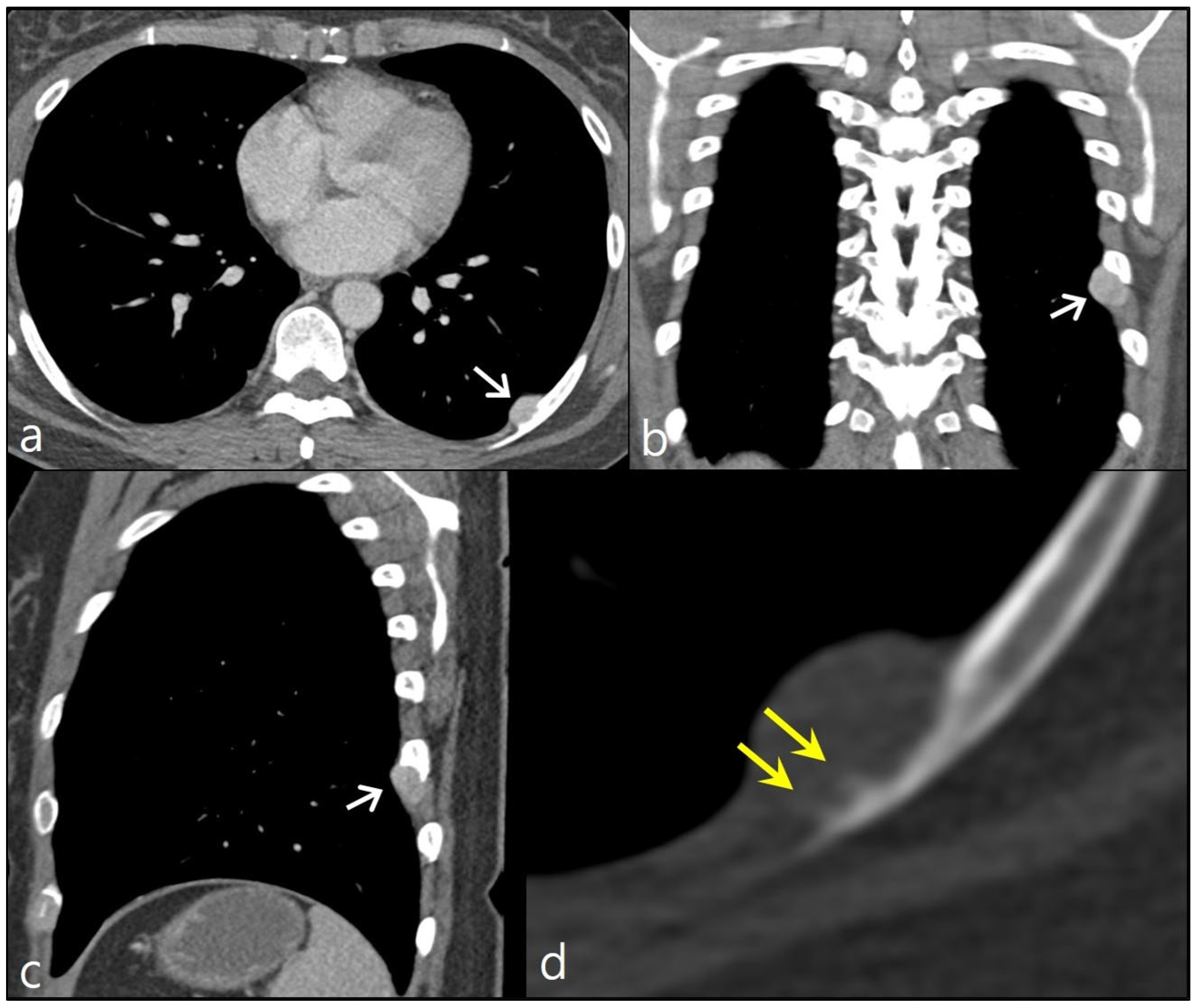
A 44-year-old woman visited an outpatient clinic due to a nodular lesion detected on a chest X-ray screening. She did not complain about any symptoms. She was never a smoker. She was previously healthy. She had no notable family history of cancer. According to her chest CT scan, the nodule was an extrapleural lesion located along the inner surface of left eighth posterior rib. It measured about 1.8 cm in its maximum diameter. The nodule showed homogeneous contrast enhancement with a smooth well-demarcated margin. Focal cortical erosion of the left eighth rib was noted. Given the imaging features and the location of the lesion, a neurogenic tumor such as schwannoma was suspected. Nevertheless, upon closer examination of a magnified CT image using a bone window setting, the presence of bony spicules oriented perpendicularly to the rib cortex was observed, indicating a potential sunburst-like periosteal reaction. This finding raises concerns about the likelihood of malignancy (
Figure 1).
We performed a video-assisted thoracoscopic excision of the nodule, along with adjacent ribs, for diagnostic and therapeutic purposes. Upon gross examination of the specimen, it was noted that the lesion abutted the eighth rib, with a very small portion of the nodule connected to the rib bone. Upon microscopic examination, the nodule was identified as a highly cellular spindle cell tumor. In a part of the lesion, woven bone with endochondral ossification was noted, suggestive of a high-grade osteosarcoma. A tiny part of the tumor was connected to the medullary cavity of the rib bone, indicating the potential presence of endosteal extension. Upon higher magnification, it was evident the malignant cells were infiltrating the medullary trabeculae (
Figure 2). Based on pathologic findings, a diagnosis of high-grade surface osteosarcoma was established.
Osteosarcoma was reported to primarily involve the rib in only 1.3% of cases [
1]. Unlike typical osteosarcomas that originate within the medullary cavity, juxtacortical osteosarcomas that constitute 4–10% of the entire spectrum of osteosarcomas have their origin on the outer surface of the bone cortex [
2,
3]. Based on their point of origin, juxtacortical osteosarcomas can be subdivided into three types, parosteal, periosteal, and high-grade surface types, corresponding to the external periosteal layer, internal periosteal layer, and any location within the periosteum, respectively. Such subtypes exhibit disparities not just in radiological and pathological characteristics but also in treatment strategies and eventual prognoses [
3]. Of these subtypes, high-grade surface osteosarcomas carry the most unfavorable prognosis. The treatment protocol involves initiating neoadjuvant chemotherapy followed by an extensive surgical resection.
We presented the radiologic and pathologic findings of a high-grade surface osteosarcoma that emerged in the intercostal space. Despite its resemblance to a neurogenic tumor, the detection of a discreet periosteal reaction in the adjacent rib cortex on CT images prompted the consideration of a potential malignancy.
Author Contributions
Conceptualization, K.N.J.; methodology, K.N.J., K.B. and J.I.M.; validation, K.N.J., H.J.A. and J.J.J.; formal analysis, K.B. and J.I.M.; investigation, K.B., K.N.J. and J.J.J.; resources, K.B. and J.J.J.; data curation, K.B. and J.I.M.; writing—original draft preparation, K.B. and J.I.M.; writing—review and editing, K.N.J., J.J.J. and H.J.A.; visualization, K.N.J. and H.J.A.; supervision, K.N.J. All authors have read and agreed to the published version of the manuscript.
Funding
This research received no external funding.
Institutional Review Board Statement
The study was conducted according to the guidelines of the Declaration of Helsinki and approved by the Institutional Review Board of Gyeongsang National University Changwon Hospital. (Approval Code: protocol code 2023-08-005; Approval Date: 2023-08-09).
Informed Consent Statement
Patient consent was waived due to study’s retrospective nature, and anonymous clinical data were used.
Data Availability Statement
Data are contained within the article. No new data were created or analyzed in this study.
Conflicts of Interest
The authors declare no conflict of interest.
References
- Nam, S.J.; Kim, S.; Lim, B.J.; Yoon, C.S.; Kim, T.H.; Suh, J.S.; Ha, D.H.; Kwon, J.W.; Yoon, Y.C.; Chung, H.W.; et al. Imaging of primary chest wall tumors with radiologic-pathologic correlation. Radiographics 2011, 31, 749–770. [Google Scholar] [CrossRef] [PubMed]
- Murphey, M.D.; Jelinek, J.S.; Temple, H.T.; Flemming, D.J.; Gannon, F.H. Imaging of periosteal osteosarcoma: Radiologic-pathologic comparison. Radiology 2004, 233, 129–138. [Google Scholar] [CrossRef] [PubMed]
- Kumar, V.S.; Barwar, N.; Khan, S.A. Surface osteosarcomas: Diagnosis, treatment and outcome. Indian J. Orthop. 2014, 48, 255–261. [Google Scholar] [CrossRef] [PubMed]
| Disclaimer/Publisher’s Note: The statements, opinions and data contained in all publications are solely those of the individual author(s) and contributor(s) and not of MDPI and/or the editor(s). MDPI and/or the editor(s) disclaim responsibility for any injury to people or property resulting from any ideas, methods, instructions or products referred to in the content. |
© 2023 by the authors. Licensee MDPI, Basel, Switzerland. This article is an open access article distributed under the terms and conditions of the Creative Commons Attribution (CC BY) license (https://creativecommons.org/licenses/by/4.0/).


