Systematic Review

Headache Attributed to Reversible Cerebral Vasoconstriction Syndrome (RCVS)

by
Vasileios Tentolouris-Piperas
,
Loukas Lymperopoulos
,
Argyro Tountopoulou
,
Sophia Vassilopoulou
and
Dimos D. Mitsikostas
*
1st Department of Neurology, Eginition Hospital, School of Medicine, National and Kapodistrian University of Athens, 11528 Athens, Greece
*
Author to whom correspondence should be addressed.
These authors contributed equally to this work.
Diagnostics 2023, 13(17), 2730; https://doi.org/10.3390/diagnostics13172730
Submission received: 10 July 2023 / Revised: 10 August 2023 / Accepted: 18 August 2023 / Published: 22 August 2023

Abstract

:
Reversible cerebral vasoconstriction syndrome (RCVS) is a condition with variable outcomes presenting a new onset thunderclap headache accompanied by focal neurological symptoms or seizures. It can be idiopathic or arise secondarily to a variety of trigger factors. The condition is increasingly recognized in clinical practice, but many facets remain poorly understood. This article aims to clarify the headache characteristics in RCVS, the temporal association of angiographic findings, the potential association of the condition with SARS-CoV-2 infection, and the clinical presentation of RCVS in children and is based on a systematic PRISMA search for published analytical or large descriptive observational studies. Data from 60 studies that fulfilled specific criteria were reviewed. Most people with RCVS exhibit a typical thunderclap, explosive, or pulsatile/throbbing headache, or a similar acute and severe headache that takes longer than 1 min to peak. Atypical presentations or absence of headaches are also reported and may be an underrecognized phenotype. In many cases, headaches may persist after resolution of RCVS. Focal deficits or seizures are attributed to associated complications including transient ischemic attacks, posterior reversible encephalopathy syndrome, ischemic stroke, cerebral edema, and intracranial hemorrhage. The peak of vasoconstriction occurs usually within two weeks after clinical onset, possibly following a pattern of centripetal propagation, and tends to resolve completely within 3 months, well after symptoms have subsided. There are a few reports of RCVS occurring in relation to SARS-CoV-2 infection, but potential underlying pathophysiologic mechanisms and etiological associations have not been confirmed. RCVS occurs in children most often in the context of an underlying disease. Overall, the available data in the literature are scattered, and large-scale prospective studies and international collaborations are needed to further characterize the clinical presentation of RCVS.

1. Introduction

Reversible cerebral vasoconstriction syndrome (RCVS) is a self-limiting condition attributed to transient cerebral vasospasm, presenting with a severe headache and/or focal neurological symptoms. As a presenting symptom, a headache usually has thunderclap characteristics. It recurs for about 1–2 weeks and it may precede an ischemic or hemorrhagic stroke. RCVS may also occur in the context of several predisposing factors, including vasoactive substances, blood products, and the postpartum state [1]. The term reversible cerebral vasoconstriction was first mentioned by Call and Flemming in 1988, who reported four cases of abrupt headache and reversible cerebral vasoconstriction. Combined with 12 more cases of patients with similar clinical characteristics, they hypothesized a new clinical entity initially termed “Call–Flemming syndrome” [2]. Following that, case reports of patients with common characteristics of headache and reversible cerebral vasoconstriction were published using various terms such as benign angiopathy of the central nervous system (CNS), postpartum angiopathy, cerebral vasculopathy, or isolated benign cerebral vasculitis. The term reversible cerebral vasoconstriction syndrome has been widely adopted after the publication of two seminal articles in 2007, reviewing and unifying the previous terminology and presenting the first large series of patients [3,4]. The proposed key diagnostic criteria have since been slightly modified [5].
RCVS is a rare syndrome with a mean age of onset in the fifth decade of life, showing female predominance in adults at a ratio ranging from 2:1 to 10:1 [3,6,7,8]. Formal epidemiological data are lacking, but incidence has been estimated to be 0.3/100.000 annually in the United States [9]. The true incidence is hypothesized to be higher, and in recent years, our understanding of the clinical course and imaging findings of RCVS has improved, making this entity more easily recognized and diagnosed. However, it can still manifest in a variety of clinical scenarios, which, in combination with its clinical and imaging heterogeneity, often makes definite diagnosis a challenge, particularly in the acute setting. Initial diagnosis requires radiographic proof of vasoconstriction, which frustratingly may not be apparent early in the disease course. Even if it is, it could be missed with the imaging modalities commonly available in an emergency department. Finally, reversibility of vasoconstriction within 3 months of symptom onset is required for definitive diagnosis, but this is by definition impossible to know early on. However, time is of the essence, as RCVS is known to harbor dangerous complications such as hemorrhage, ischemic stroke, cerebral edema, and seizures. Early diagnosis and differentiation from other possible diagnoses is imperative for appropriate management.
Headache attributed to reversible cerebral vasoconstriction syndrome (HaRCVS) is a broad classification introduced in the latest International Classification of Headaches (ICHD-3 6.7.3.1) that describes new-onset headaches in patients with confirmed RCVS, which completely resolve within 3 months of onset and fulfill one or both of the following criteria [10]:
1
The appearance of the headaches themselves led to the diagnosis of RCVS and/or;
2
Headaches possess at least one of the following characteristics:
(i)
Thunderclap onset;
(ii)
They are triggered by sexual activity, exertion, Valsalva maneuvers, emotion, and bathing/showering;
(iii)
They are present or recurrent for at most one month after onset and then subside.
In the absence of angiographical confirmation of RCVS, the criteria that allow for the diagnosis of probable HaRCVS (6.7.3.2) (hence, probable RCVS) are much stricter, and are naturally closer to what is generally known as the clinical presentation of RCVS. They require the headache to be severe and have a thunderclap onset, peaking within one minute and lasting at least 5 min, while having been triggered by at least one of the aforementioned trigger factors. The criteria further allow for persistent headaches associated with past RCVS to be diagnosed if the symptoms persist for more than 3 months (6.7.3.3). To reach a higher clinical suspicion for RCVS as early as possible, differentiate it from other conditions as accurately as possible, and inform further diagnostic and therapeutic decisions, two diagnostic scores have been devised, RCVS2 [11] and RCVS-TCH [12]. The RCVS2 score greatly incorporates the presence of thunderclap headache (TCH) in the prediction model and RCVS-TCH considers TCH as a prerequisite. However, there are many RCVS patients presenting an acute, severe headache that does not strictly adhere to the TCH definition, as well as patients with different headache presentations or no headache at all.
The aim of this review is to present the key features of HaRCVS and accompanying focal symptoms in relation to the imaging findings with various modalities via a systematic review of the published analytical observational studies (large case series, cohort, or case–control studies).

2. Materials and Methods

The review of the literature was conducted following the Preferred Reporting Items for Systematic Reviews and Meta-Analyses (PRISMA) guidelines [13]. Our search was conducted in PubMed using the search terms (reversible cerebral vasoconstriction) AND (headache) on 17 April 2023 and was repeated on 27 June 2023 to add any latest publications. No date or language restrictions were applied. Two separate members of the writing team (L.L. and V.T.-P.) reviewed the literature independently and conducted the filtering. Initial review of the titles and abstracts excluded correspondence and editorials, case reports, duplicates, and articles that were clearly irrelevant, or whose full text was in a language other than English. A second round of reviewing the full text excluded small descriptive studies (case reports and small case series of fewer than 10 patients), reviews, and other studies that were not relevant to the topic. The end result of this search was large descriptive studies (with a total cohort of at least 10 patients), analytical observational studies, and trials. The final results of this search were then read in detail to extract relevant information for the headache characteristics of RCVS patients. Articles excluded in the 2nd round were also read and incorporated in the discussion section if they contained relevant information or insights into the topic of this review. Additional unfiltered literature searches were performed for children and COVID-19. A flowchart of the literature search process and the results is shown in Figure 1.

3. Results

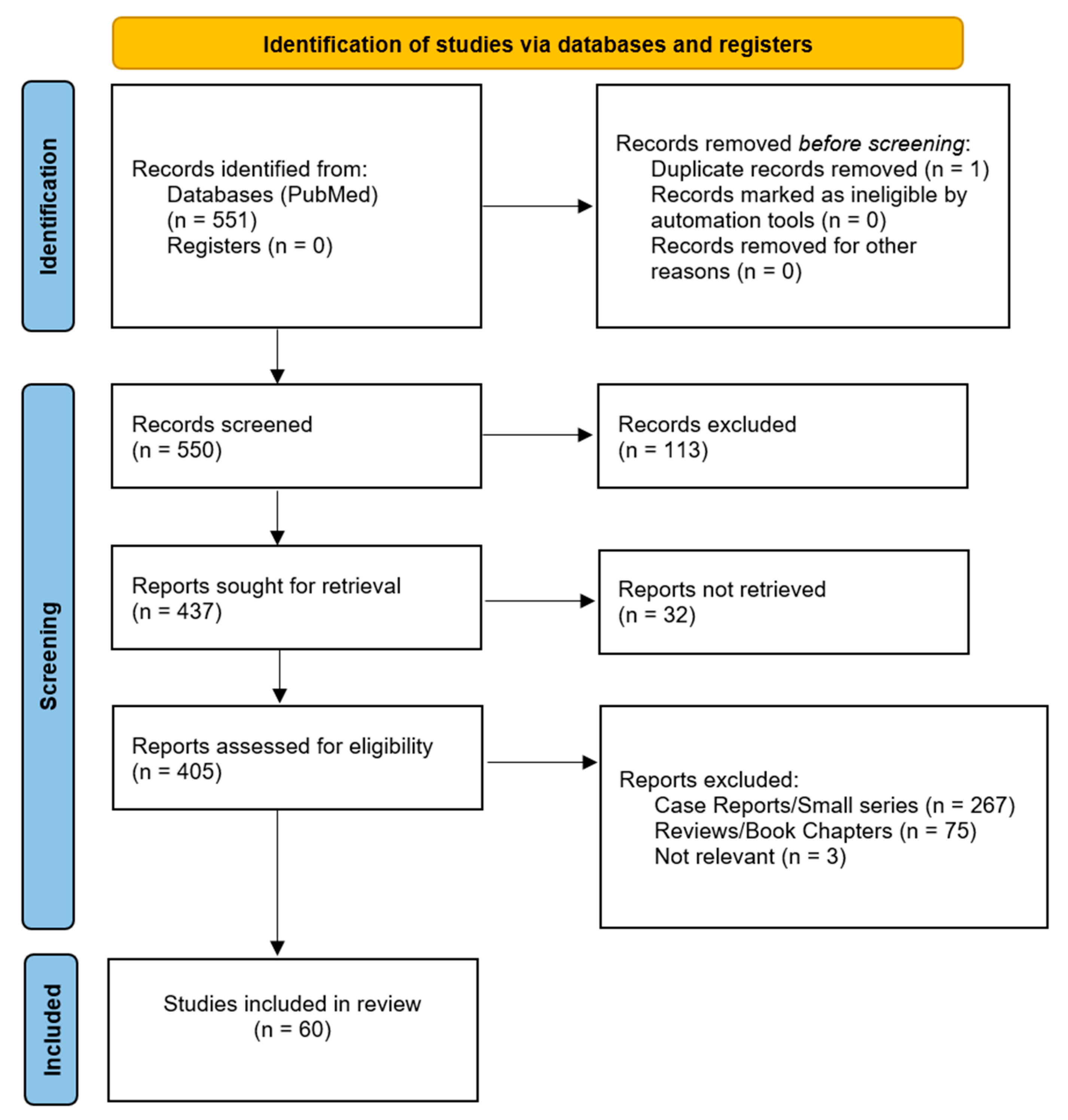
3.1. Results of the Literature Search

No published clinical trials pertaining to RCVS patients were found. Sixty observational studies fulfilled our criteria with implications on RCVS [3,6,7,8,11,12,14,15,16,17,18,19,20,21,22,23,24,25,26,27,28,29,30,31,32,33,34,35,36,37,38,39,40,41,42,43,44,45,46,47,48,49,50,51,52,53,54,55,56,57,58,59,60,61,62,63,64,65,66,67]. Of those, 16 studies contained sufficient information on particular headache characteristics of their cohorts, and they are presented and summarized in Table 1.
Results not included in Table 1 focused on postpartum patients, vascular events, white matter abnormalities, imaging, or other conditions in the differential for RCVS that did not contain enough information on headache characteristics of their RCVS cohorts to be included in the table. However, they concern other important facets of the syndrome and are further discussed in the text, where appropriate.

3.2. Headache Associated with RCVS

3.2.1. Typical Thunderclap Headache and Thunderclap-like Headache

Thunderclap headache has long been considered the hallmark symptom of RCVS since the syndrome was first described. The hallmark case series, which was published in 2007, reported a 98.5% presence of TCH headaches in RCVS patients [3] using the ICHD-2 TCH criteria [68].
Some studies in our search used the presence of thunderclap headache as a prerequisite to identify and recruit patients [6,21,37,60] and, therefore, all patients had TCH, although various definitions have been used. Studies that opted for different methods to identify RCVS patients recruited cohorts of patients with a wider variety of presentations, leading to a lower reported incidence of TCH in RCVS patients, ranging from 67 to 86.5% [7,18,25,42].
In some reports, TCH is not strictly defined [7,37,52], while in others, the definition is inadequate or is not reported at all [28,33,59,61,67]. In studies that strictly used the TCH definition, most of the non-TCH patients experienced headaches with similar characteristics: severe intensity and fast onset-to-peak, but not as fast as 1 min [3,18,25,42]. In studies not reporting the TCH definition used, most patients are still reported to have TCH or similar acute and severe headaches [7,52,61,67].
The character of the pain is usually described as explosive or pulsatile/throbbing, but it may also be dull or tearing [37,47]. Intensity tends to reach more than 7/10 on the numeric scale by definition of TCH, although many studies do not report quantitative evaluation. Smith et al. report patients who have “thunderclap” headaches of as low as 4/10 on the intensity scale [60]. Furthermore, headache intensity tends to diminish from episode to episode until full remission [7].
Location of pain has not been found to be particularly helpful in distinguishing RCVS and has not been adequately reported in all studies. For example, initial information that pain starts occipitally and then quickly spreads diffusely was reported in an early case series [3] and has since been cited in the majority of subsequent studies and reviews. However, not many corroborating data have been published since, and newer publications with larger cohorts do not report such information at all. The exception to this observation is a cohort of 174 RCVS cases triggered by sexual activity: 67% of patients had bilateral pain, with the most common locations being occipital (69%) and temporal (50.6%), but all locations, as well as diffuse pain, were reported. Pain is most commonly bilateral, but a unilateral location may be present in 20–33% of patients [3,21,47].
Headaches in RCVS patients tend to happen episodically [3,23,47], but patients with a single episode were also initially described [3]. A monophasic course may be more common, as much as 40% [18]. Episodic headaches within the first weeks are a sign that strongly suggests a diagnosis of RCVS compared to other causes of TCH [69].
The duration of each attack usually lasts for 3–5 h [3,21] and can range from several minutes to several hours or even a day and more [3,23,37,47]. A duration of less than one hour was less common in RCVS patients compared to other causes of sudden headaches [23]. The total duration of the disease until headaches remiss completely is around 2–3 weeks [3,7], but can range from 2 days to 4 months [37].
It is important to note that some patients may have thunderclap at onset, and others later in the disease course, or even never. A variety of headache types can occur in the same period of disease [42].

3.2.2. Presentation with Atypical Headache or No Headache

As discussed, RCVS can present without the typical thunderclap headache. Most commonly, this still refers to acute onset, severe headaches that peak quickly in more than 1 min; hence, this presentation was discussed above. However, there are also cases of RCVS presenting with severe but much more gradual headaches, non-severe headaches, or no headaches at all. In later publications, atypical presentations are being discussed more often, ever since a review of published case reports nicely elucidated this subset of RCVS patients [70]. In a retrospective study aiming to characterize TCH in RCVS patients, nine patients from the initial RCVS cohort were excluded from the analysis due to presenting atypical headaches. Unfortunately, no further details were reported [60]. This example highlights the bias that exists in the current literature and that atypical presentations may generally be even more prevalent. In the cohorts identified by our search, examples of other headaches include severe headaches peaking up to 120 min from onset, unusual progressive headaches, and headaches appearing upon waking with varying intensity [18,25,42]. No matter the characteristics, patients still describe these headaches as unusual.
There are also reports of patients who do not have any headaches, which are also summarized by Wolff et al. [70]. Our search provided published studies where such cases were also part of cohorts [7,23,25,33,42,59], although not much information could be gleaned about the details of the clinical presentation in this particular subgroup. Commonly, these patients are usually discovered through complications of the syndrome, such as ischemic stroke or hemorrhage. Since headache is still considered the hallmark of RCVS, cases without headaches could be missed. It is also plausible that RCVS can occur subclinically or produce such minor and insignificant symptomatology that patients never seek care for and are never evaluated.

3.2.3. Accompanying Symptoms of Headaches

The symptoms accompanying headaches of RCVS tend to resemble those of SAH and meningeal irritation; however, they have not been adequately elucidated. One reason is that RCVS can occur with transient or permanent deficits and seizures, and it is not always clear if part of the presentation is due to the headache or the complications. From our search, Caria et al. reported nausea and vomiting in 81.2% of RCVS patients with TCH [18], while Lin et al. reported nausea, vomiting, photophobia, and phonophobia in patients with RCVS that presented a headache after sexual activity [47]. Most of these patients did not have SAH, so symptoms can be more easily attributed to RCVS and the headache itself. Kim et al. reported nausea in seven of eighteen patients with RCVS and TCH, as well as rarer cases of ocular pain, vomiting, dizziness, and phonophobia. Generally, pain is often accompanied by screaming and crying (32%), photophobia (30%), nausea (57%), and vomiting (38%) [3]. Phonophobia has also been reported elsewhere [6,37,47]. Extremely intense headaches can often lead to sympathetic overactivity; therefore, crying, agitation, panic, screaming, fear of dying, confusion, or collapse are common accompanying symptoms [71]. Furthermore, about one-third of patients have a surge of blood pressure (systolic blood pressure > 160 mmHg) accompanying headache attacks [69,72]. This may be a response to the excruciating pain but may also be part of the underlying processes or vascular complications. Nevertheless, as a finding, it is an important clue to the presence of RCVS, and it has been incorporated into diagnostic scores [12]. It is not clear if any of these accompanying symptoms tend to occur in non-TCH RCVS patients as well.

3.2.4. Persistent Baseline Headaches

Although RCVS headaches typically occur in episodes, some patients experience the presence of a persistent headache between episodes. The presence of such a headache should not discourage the diagnosis of RCVS if the rest of the presentation is compatible. According to the pivotal observational study of the Ducros group, a headache of lower intensity persisted between thunderclap attacks in approximately 35% of cases [3]. As seen in Table 1, similar findings were reported in the Italian cohort [18], in cohorts of RCVS patients with TCH [21,60], and in cohorts of RCVS patients presenting sudden headaches [23], and acute ischemic stroke [67]. However, all noteworthy headaches in the vast majority of cases should be gone by 3 weeks after onset [5].

3.2.5. Trigger Factors

There is a wide variety of trigger factors that have been described in association with RCVS in the literature. The term “secondary RCVS” has been adopted by some authors for cases with a confirmed, identifiable trigger. In general, these mainly include emotional, postural, or environmental triggers, the intake of vasoactive substances, medical conditions, and pregnancy or recent childbirth. Some of these triggers have been well-described and their correlation with RCVS is undoubtful, while others seem more dubious, as they involve common, everyday situations or have a substantial delay between exposure and development of the syndrome [73]. TCH may start upon first exposure to an associated substance, or after long-term use at normal or excessive doses [71].
Commonly described precipitating factors include Valsalva maneuvers, coughing, sexual activity, physical exertion, contact with water, urinating without effort, sneezing, laughing, singing, emotional triggers (mostly negative), and sudden forward flexion [3,23,25,42].
The postpartum period has also been strongly associated with RCVS. A study by Lange et al. on 345 patients found that recent childbirth was a precipitating factor in 8% of patients [42]. On the other hand, RCVS seems to be rare among postpartum individuals, as shown in another prospective study, where out of 900 postpartum women, 34 developed TCH, but only 1 was diagnosed with RCVS [15]. According to Singhal et al., it usually emerges within1 to 3 weeks following an uncomplicated pregnancy, although presentation can occur as late as 6 weeks after delivery [74]. More recent studies have shown that RCVS emerges in a shorter time span, specifically 1–10 days after delivery, according to Topcuoglu et al. [64]. For this reason, RCVS was previously known as postpartum angiopathy [75]. Almost half of the patients have a history of proteinuria, suggesting overlap with pre-eclampsia, and there is also an association with HELLP syndrome (hemolysis, elevated liver enzymes, and low platelets), as well as posterior reversible encephalopathy syndrome (PRES) [76].
Vasoactive substances such as illicit drugs, α-sympathomimetics, and serotoninergic drugs are considered among the most common precipitating factors [42,53,77]. Drugs that may induce RCVS include selective serotonin reuptake inhibitors (SSRI) (such as escitalopram, paroxetine, sertraline) and triptans [18]. Other medications, such as nasal decongestants, have also been reported [3]. A very interesting case was described by Abruzzo et al. involving a 19-month-old female patient with retinoblastoma, who underwent selective ophthalmic artery infusion chemotherapy. Shortly after administration of a series of substances including intranasal oxymetazoline, she developed systemic hypertension and cerebral vasoconstriction, which spontaneously resolved within hours without any further complications [78]. Regarding illicit drugs, cannabis is the only one that has been associated with the development of RCVS. There is a lack of evidence for other illicit drugs (cocaine, ecstasy, amphetamine, methamphetamine, khat, heroin, LSD) as single precipitating factors [57,79].
Other reported triggers include alcohol, blood products, head trauma, neurosurgical procedures, headache disorders (exertional headache, migraine, primary thunderclap headache, benign sexual headache), tumors (in particular, pheochromocytoma and paraganglioma), vascular conditions and procedures (fibromuscular dysplasia, endovascular procedures, cervical artery dissection, unruptured intracranial aneurysm, carotid endarterectomy), and antiphospholipid syndrome [80,81,82]. RCVS in association with thrombotic thrombocytopenic purpura has also been reported [83]. Trauma/surgery/procedure as precipitating factors have been linked to poor clinical outcomes [77].
Overall, the percentage of patients with identifiable trigger factors can significantly vary between studies, as some studies suggest a lower percentage (55.8%), while others suggest a percentage up to 80–90.1% [3,6,42]. The prospective cohort study by Choi et al. identified trigger factors in only 16/104 patients with definite RCVS, with the most common being recent childbirth (5.8%) and medication use (5.8%), while 33/104 patients reported a preceding event or change in lifestyle, environment, health, or medication within one month before onset, which could not be definitively identified as a trigger [25]. In contrast, a recent review of 191 cases reported identifiable trigger factors in 88.5% of patients [77]. This extreme variability could be attributed to different trigger factors taken into account in each study or a publication bias of cases with precipitating factors, which may not represent real-world data. Secondary RCVS has been associated with atypical clinical manifestations and a higher rate of complications [18].

3.2.6. Persistent HaRCVS and Recurrency

It seems that RCVS as a phenomenon is not strictly monophasic, although most patients tend to experience it only once. In a cohort study, 6/77 patients (8%) had a recurrence during a median follow-up of 25 months (range 10–69) [6]. In a follow-up study, the same group of researchers demonstrated a recurrence of TCH in 18 of 169 RCVS patients with a long-term follow-up (mean of 37.5 months). Vasoconstriction was re-demonstrated in nine of these patients (5.4%) [22]. Another prospective study showed that 10 of 172 patients (5.6%) were found to have a recurrent episode of RCVS, though none of these recurrent cases resulted in permanent sequelae [17]. One patient in the same study had a third recurrence of RCVS. Predicting factors that were found to be associated with the recurrence of RCVS included a history of migraine and having exercise or sexual activity as a trigger for thunderclap headaches during the initial episode of RCVS [17].
There are studies suggesting that RCVS not only causes excruciating headaches in its acute phase, but also leads to chronic headaches afterward [36,84]. Rozen et al., in 2013, first reported a patient with possible RCVS (although not diagnosed as such in the study), followed by new daily persistent headaches. The authors considered this presentation as a novel phenotype [85]. Jamali et al. described three cases of RCVS followed by new daily persistent headaches [86]. A retrospective two-centered study, conducted by the Cleveland Clinic Foundation and Massachusetts General Hospital in 2016, investigated the long-term outcomes of RCVS patients. The study provided important information regarding headaches after RCVS resolution. Around half (24/45) of the patients continued to have chronic headaches after RCVS resolution [36]. In a recent study of long-term effects, 50% of patients were found to experience long-term headaches, defined as headaches beyond 6 months from onset [39]. This chronic headache, which can be triggered by RCVS, has lately begun to be recognized and described even further. It is now formally included in the third edition of the International Classification of Headache Disorders (ICHD-3, 2018) and is named “persistent headache attributed to past RCVS” [10]. Psychological distress and a prior history of migraines are regarded as predisposing factors for the occurrence of post-RCVS headaches [87].

3.2.7. Focal Neurologic Deficits, Seizures, Strokes, and Other Accompanying Conditions

Even though the majority of patients (up to 75%) describe headaches as their only symptom, fluctuating neurologic deficits and seizures are also commonly described [88]. According to various studies, focal neurologic deficits may have an estimated prevalence between 8% and 63% [69,72,89]. According to Song et al., the most common neurologic symptom of RCVS is headache (93.2%), followed by motor weakness (36.6%), visual abnormality (30.4%), mental change (20.4%), and seizures (17.3%) [77]. In addition to the aforementioned symptoms, RCVS may present encephalopathy, dysarthria, aphasia, or ataxia [89]. Patients with aphasia/neglect/apraxia as acute neurologic symptoms had a poor clinical outcome with persistent neurologic deficits [77]. There might also be an association between the severity of RCVS and atypical clinical features since these patients can often present with neurological deficits due to stroke, severe PRES, seizures, confusion, or even coma [89,90].
The focal deficits are usually a consequence of associated complications, such as transient ischemic attacks (TIAs) (up to 16%), PRES (9–14%), ischemic stroke (4–54%), and intracranial hemorrhage (34–43%), specifically cortical subarachnoid hemorrhage (cSAH) (up to 22%) or intracerebral hemorrhage (ICH) (up to 6%) [29,65,69]. In the largest RCVS series published to date [32] of 1157 patients, 18.6% had AIS, 39.5% had a hemorrhagic stroke, 11.2% had cerebral edema, and 4.3% had a new-onset seizure. This sample of patients does not include patients who died in their first admission, only patients who were discharged, so rates of complications may be even higher among the entire RCVS population. However, patients were identified using discharge diagnosis codes and not diagnostic criteria.
According to a recent review, the incidence of hemorrhagic strokes and PRES is even higher (in 44% and 38% of cases with RCVS respectively) [72]. Specifically, an observational study performed by Patel et al. showed that the total frequency of hemorrhage in patients with RCVS was 43.4%, while the frequency of SAH, ICH, or subdural hematoma was 35.9%, 13.1%, and 3.6%, respectively [53]. There can be an overlap of different types of hemorrhage in the same patient. As far as patients with SAH are concerned, 90% present with it initially [80]. Intracranial hemorrhage tends to be small (less than 10 mm3) and usually precedes SAH, although they are both early complications [80]. Both types of hemorrhage (cSAH and ICH) and PRES are early events occurring usually during the first week, while ischemic events, including TIAs and cerebral infarcts, occur later, usually during the second week, and on average 8 days later than the hemorrhagic complications [3,90]. Ischemic strokes usually involve a watershed distribution between the anterior and posterior circulation, whereas major arterial territorial infarcts are uncommon [90]. Ischemic strokes are usually bilateral. Cerebellar strokes have also been described in RCVS, although they are rare [80].
Cerebral edema is a possible complication of RCVS and it has been reported in almost 10% of cases [91]. It usually occurs in the first week after clinical onset, showing complete resolution within 1 month [80].
Dissection of the cervical arteries may also be associated with RCVS, but it is unclear whether it is a cause or a downstream effect of the syndrome [73]. A cohort study of 20 patients has demonstrated cervical artery dissection in 12% of patients with RCVS, most frequently females with a migraine history and neck pain [50]. Interestingly, a retrospective study comparing RCVS patients with the control groups of primary angiitis of CNS (PACNS) and patients with other neurological syndromes who underwent angiographic evaluations indicated that patients with RCVS were more likely to have coexisting vascular abnormalities such as cervical artery dissection (CAD), aneurysms, and cavernomas [63]. Similarly, postpartum patients with CAD were more likely to have RCVS compared to non-postpartum patients [92].

3.2.8. Diagnosis of HaRCVS and RCVS

Diagnosis of HaRCVS is simple but requires a prior confirmed diagnosis of RCVS; hence, it is not particularly useful in the acute setting. Strictly applying the ICHD-3 classification criteria means that any new type of headache in a patient that leads to a diagnosis of RCVS, regardless of the headache characteristics and if it occurs in isolation or not, is automatically termed as HaRCVS if it cannot be attributed to other causes. On the other hand, if RCVS is diagnosed in spite of the presence of a headache, any new persistent or recurring headaches that are present afterward, subside within one month, and are not otherwise explained, can also be termed as HaRCVS.
Diagnosis of RCVS itself is trickier because it requires proof of vasoconstriction and its reversal, something that occurs usually at least 1 month from disease onset. Aneurysmal subarachnoid hemorrhage should always be ruled out at symptom onset, along with vasoconstriction due to inflammatory conditions and all other potential causes of new-onset acute headaches. Imaging diagnostics will be examined in the following section. Lumbar puncture (LP) is of crucial importance for the diagnosis of RCVS in order to differentiate it from inflammatory diseases (such as PACNS) or SAH. In the majority of patients with RCVS, LP shows normal or near-normal results. Caria et al. found a slightly elevated cerebrospinal fluid (CSF) leukocyte count and slightly elevated protein level in a minority of patients [18]. CSF findings are more often normal in patients with RCVS compared to patients with PACNS [28]. In contrast to other studies, a small cohort showed that 5/6 of RCVS patients had leucocyte levels of more than 10/mm3 [38].
So far, three ways exist to suspect and diagnose RCVS early, before conducting extensive neuroimaging. These are the “Probable HaRCVS” criteria (6.7.3.2) of the ICHD-3, and the two diagnostic scores, RCVS2 and RCVS-TCH. The latter two have a primary goal of differentiating RCVS from other causes of vasoconstriction and thunderclap headaches, respectively. In a validation study, the criteria for probable HaRCVS (6.7.3.2) [45] were found to be specific for identifying RCVS in patients presenting with thunderclap headaches. The RCVS2 score and diagnostic approach can be used to promptly diagnose and distinguish RCVS from mimics, using easily available admission variables (the presence of thunderclap headache, the absence of intracranial carotid artery involvement, the presence of a vasoconstrictive trigger, gender, and the presence of subarachnoid hemorrhage) [11]. Kumar et al. supported the usefulness of the RCVS2 score in a real-world clinical context using a cohort of 55 patients and demonstrated that the RCVS2 score had a significant association with high specificity and positive predictive value for angiographic vasoconstriction [39]. Cho et al., however, developed a new prediction model, the RCVS–TCH score, which showed high specificity and sensitivity for discriminating RCVS in patients with TCH. The authors suggest that the RCVS2 score has an excellent performance to predict RCVS in patients with arteriopathies, but the RCVS-TCH score better helps distinguish RCVS from primary TCH or other secondary causes of TCH [12].
All three of these options have TCH in a central role. With recent publications suggesting a higher than previously thought prevalence of non-TCH presentations, it is not easy to suspect RCVS in the first place and request necessary diagnostic procedures. Future studies may shed light on this subgroup of patients and determine if there are definitive clues that may point to RCVS in this case. Nevertheless, new headaches or vascular events that cannot be explained otherwise, particularly in patients that report one of the known possible triggers, should evoke suspicion, even in the absence of TCH. A well-designed prospective case–control series documenting detailed headaches and other clinical characteristics could provide more information on the topic in the future.

3.2.9. Imaging in RCVS and Temporal Relation of Symptomatology and Vasoconstriction

Imaging is of vital importance in RCVS to confirm vasoconstriction, exclude other causes of TCH, such as aneurysmal SAH or cerebral vasculitis, and monitor for potential complications [93]. The first imaging exam performed in a patient with TCH due to RCVS in the emergency department is usually non-contrast computed tomography (CT). The exam may be negative in more than half of the cases [80]. Digital subtraction angiography (DSA) has, by definition, close to 100% sensitivity for detecting peripheral vasoconstriction compared to CT angiography (CTA) and MRA, as it can assess vessel size to a diameter of 0.4 mm, compared with 0.7 mm in CTA. DSA does, however, have drawbacks, particularly the risks of an invasive vascular procedure and radiation exposure. Cerebrovascular complications have been described, including TIA, ischemic strokes, arterial dissection, or vessel perforation (TIA in 9% of the patients, and iatrogenic dissections in 0.14%) [90]. Complications of DSA specific to RCVS patients have not been clearly described. DSA tends to be reserved only for patients with negative findings of non-invasive techniques or for cases that also require intraarterial treatment [93]. The sensitivity of CTA or MRA has been reported to be around 80% for both techniques in the seminal case series by Ducros [5,93]. This number has been cited in the literature ever since, but no other sensitivity data have been adequately reported so far. Sensitivity analysis is complicated by the temporal progression of the condition. Nevertheless, MRA has been shown to be a good alternative to DSA, particularly since it enables concurrent detection of other pathology and easy monitoring of vasoconstriction progression.
Magnetic resonance (MR) is frequently performed in the setting of suspected RCVS to detect complications, such as intracerebral bleeding or ischemic stroke [80]. Consequently, concurrent MRA has been increasingly used to detect segmental vasoconstriction. It can detect medium- to large-sized arteries, giving a typical “string of bead” or “sausage on a string” appearance. Arterial spin labeling (ASL) is a perfusion MR technique, which enables cerebral blood flow (CBF) evaluation, without the need for gadolinium injection [80]. According to Kano et al., ASL can infer vasoconstriction in distal vessels “hidden” from MRA by detecting areas of hypoperfusion [94]. In this study, vasoconstriction reached its peak in the second week with subsequent gradual normalization. Centripetal progression of vasoconstriction from the distal arteries to the major cerebral arteries of the circle of Willis was observed in all patients [94].
Finally, transcranial doppler (TCD) might also be a useful tool in the identification of vasospasm in patients with probable RCVS. A study of 36 patients with RCVS who underwent TCD on an average of 16 (6–26) days after headache onset showed that 29 of them had mild or moderate vasoconstriction, which completely resolved on an average of 41 days after headache onset [95]. TCD has also been used to demonstrate that vasoconstriction increases gradually and peaks between 13 and 14 days after symptom onset [35]. Another small cohort study of 15 patients also demonstrated that TCD has potential as an initial non-invasive diagnostic method and particularly as a method for monitoring patients with RCVS [46]. TCD could be the best way to look for the peak of vasoconstriction in large-sized vessels as a non-invasive, easy, and relatively fast imaging examination given an adequate acoustic window.
As far as the timing of vasoconstriction is concerned in relation to symptom onset, our search of published observational studies demonstrated that visible vasoconstriction could occur on the same day as symptom onset, as has been reported by Shimoda et al. [56]. There are also reports of vasoconstriction being visible 1–3 days afterward [25,31], but it is most commonly evident some days into the disease course [18,37,44,46]. According to two reviews focusing on the neuroimaging evaluation of the syndrome, the peak of vasoconstriction is around 16 days after clinical onset, and it resolves within 3 months [80,81]. The percentage of patients with visible vasoconstriction at onset could be underestimated due to a delay in imaging acquisition. On the other hand, there are reports of negative imaging examinations, even when they are performed early in the course of the disease [3,96]. It remains unclear whether this can be attributed to a low sensitivity of imaging methods in detecting vasoconstriction of small, distal vessels that have not propagated yet proximally, as some theories suggest, or to a true absence of vasoconstriction at that time point.
Ducros et.al theorized that the pathological process first includes distal arteries and then progresses toward the branches of the circle of Willis [5]. Vasoconstriction of small distal arteries is undetectable on angiography and may stimulate trigeminal leptomeningeal afferents to induce thunderclap headaches [97]. Due to the difficulty in detecting vasoconstriction in very distal branches, it may be impossible to detect it during the first week of the disease [93]. Chen et al. thoroughly investigated the evolution of vasoconstriction in patients with RCVS in a study of 77 patients, demonstrated that diffuse vasoconstriction peaks on day 16.2 ± 9.4 (range, 2–51) and found that different vessels can be affected at different times in the disease course [6]. In 2016, Shimoda et al. demonstrated that centripetal propagation of vasoconstriction can be depicted on magnetic resonance angiography (MRA) when performed at the time of remission of TCH [56]. Two years later, the same group performed a retrospective, case–control study and analyzed data from 48 patients with RCVS who underwent an MRI within 72 h of RCVS onset and 48 h of TCH remission. Half of these patients showed centripetal propagation of vasoconstriction (CPV). These patients had an increased risk of brain lesions and a longer interval between the first and last TCH. By repeating MRA in the period between symptom onset and TCH remission, assessing CPV can be useful for the diagnosis of RCVS. The study hypothesized that CPV gradually progresses after the onset of RCVS, peaks at the time of TCH remission, and does not progress further afterward [57]. Fukaguchi et al. also propose performing clinical and imaging follow-ups for at least 2 weeks to exclude the syndrome [31].

3.3. RCVS Headaches in Children vs. Adults

RCVS commonly affects patients in the middle decades of life, though all age groups, including children, can be affected. However, there are not many published data on the clinical characteristics of the syndrome in children. Clinical diagnosis of RCVS in this age group may be more challenging due to the higher prevalence of inflammatory vasculopathy, differences in presentation, and difficulties in conducting diagnostic and confirmatory imaging in children, as well as in history taking. The latter makes acquiring information on headache characteristics and intensity particularly difficult. In most observational studies discovered by this literature search, patients under 18 years old had not been included or were very few in number. Published case reports have been expertly summarized in a recent review [98]. Our search resulted in a few more reported cases [78,99,100,101,102,103,104,105,106]. Compared to adults, male patients are more common in children, particularly adolescents, and RCVS occurs more commonly in the setting of some underlying diseases, such as hematologic disorders. Regarding headache characteristics, TCH or severe headaches remain the most common. Common triggers in children may be contact with water [107,108,109], exertion [99,110,111,112], and various medications [78,113,114,115].

3.4. RCVS and COVID-19

Compared to other recent reviews [72,89], our search resulted in more published case reports and series of RCVS in people with COVID-19 [105,116,117,118,119,120,121,122]. The most convincing argument on the association of COVID-19 with RCVS remains a case series published in 2021 [116]. Nine patients had been diagnosed with COVID-19 in the past 30 days, while one patient was diagnosed 45 days before the diagnosis of vasospasm. However, that study as published has numerous limitations, primarily inadequate reporting of the recruitment strategy and diagnostic criteria used for RCVS. Furthermore, four out of ten patients had vasoconstriction in only one vessel, only two patients had a follow-up angiographic evaluation to confirm reversal or improvement of vasoconstriction, and only five of the patients had an RCVS2 score of 5 or more.
RCVS was reported in a single case of the analysis print of the Pfizer-BioNTech vaccine [123]. Two cases of RCVS after vaccination exist in the literature. In one case, symptom onset was 18 days after a repeat dose of the Moderna vaccine [124], and vasospasm of the right PCA was reversed seven days later after treatment with nimodipine. In the other published case [125], symptoms occurred after 12 h of the first Pfizer dose in a 30-year-old patient with a history of RCVS 3 years prior. However, in this case, no imaging whatsoever confirmed the recurrence of vasospasm, although the clinical presentation described resembles HaRCVS.
In the setting of COVID-19, vasoconstriction has been hypothesized to occur not due to the infection itself but rather the down-regulation of the ACE2-receptors by SARS-CoV2. This might lead to sympathetic hypertonia of the cerebral vessel walls and/or over-activation of the classic renin–angiotensin axis that leads to vasoconstriction, thus precipitating RCVS [117,118]. Nevertheless, with the extent of the vaccination program and the enormous numbers of infections and re-infections worldwide, one would expect this phenomenon to be more widespread and more widely reported in the literature during these past three years before one were to hypothesize a pure etiological connection between the two conditions. It is always possible, however, that many instances of this association have been missed if hemorrhagic or ischemic incidents were attributed to the widely accepted hypercoagulation phenomena associated with COVID-19 and vasospasm was never suspected and investigated angiographically.

4. Discussion

A thunderclap headache is traditionally the hallmark of RCVS since it was first described by Call and Fleming [2]. Our search demonstrates that many articles in the literature do not adhere strictly to widely accepted definitions of TCH. Studies with a strict definition show that the percentage of patients with the supposed hallmark symptom was, in fact, significantly lower. These findings come in contrast to previous reviews reporting 85–100% [72], 94–100% [69], and 95–100% [91] of patients having TCH. Foregoing the time-to-peak criterion, the majority of patients still experience acute, severe headaches that peak relatively quickly. It is not easy for a patient under excruciating pain to remember and report whether their headache reached maximal intensity within 1, 5, or 10 min, and in the setting of acute evaluation of a new headache, such differentiation does not seem practical, at least in the setting of RCVS differentiation. There is an obvious need to reevaluate the typical headache characteristics of RCVS using a more lenient term than TCH, including at least a wider time to peak intensity interval. Neurologists should be aware that the typical TCH may not emerge in over 25% of patients with RCVS according to some studies and should, therefore, proceed with angiographic evaluation when a patient describes a new-onset severe headache occurring in minutes or even a few hours, even in the absence of non-contrast CT findings, if the rest of the presentation is compatible.
Detailed characteristics of the typical headache associated with RCVS, such as duration, quantitative intensity, and accompanying symptoms, seem to be poorly reported in the large studies found with our search compared to publications on other primary and secondary headaches. Most information still relies on the early descriptions of the syndrome and older cohort publications. This could be due to a large number of retrospective studies not finding the required information and the ease of use of an umbrella term of TCH, bypassing a more detailed description of the clinical presentation. Patients with RCVS often arrive at emergency departments with alarming symptoms, and gathering relevant information on the specific characteristics of the headache may take a second seat in favor of ruling out threatening diagnoses and treating complications, particularly if there are associated focal neurologic deficits, mental changes, or seizures. Retrospective reporting of headache characteristics after the event is often sought from the patients but is prone to recall bias. More prospective studies focusing on headache characteristics (onset-to-peak, type of pain, remission with analgetic medicine, maximum intensity, location of pain, etc.) are of vital importance in order to better define the clinical spectrum of this relatively newfound syndrome.
At the same time, there is an increase in published data on patients presenting atypical features, which can include gradual severe headaches, non-severe headaches, or no headaches at all. There is a high probability that patients with non-severe headaches or no headaches will seek medical care only if they present complications from RCVS. Since the main pathophysiological theory suggests that the vasoconstriction of small distal arteries may stimulate trigeminal leptomeningeal afferents to induce thunderclap headaches [97], it is logical to assume that patients with atypical headaches or no headaches at all could have alternate pathophysiological mechanisms in play, which do not result in the vasoconstriction of peripheral cerebral arteries or which, despite vasoconstriction, do not stimulate leptomeningeal afferents. Alternatively, the progression of pathology may stop for some reason, or these patients may be less sensitive to these pain-inducing mechanisms. This might also be reflected in the fact that patients with RCVS often have comorbidities with other headache disorders compared to the general population [126].
Concerning the main proposed pathophysiological basis of RCVS, Wolff et al. have suggested that there is a variety of possible underlying mechanisms to explain the change in the adrenergic vascular tone response, which starts distally and progresses proximally on cerebral arteries. These could include a genetic predisposition like the BDNF gene polymorphism, an imbalance of proangiogenic and antiangiogenic factors, or an endothelial dysfunction with oxidative stress. Some of these changes have also been elucidated in a study investigating circulating miRNA [127], implicating the miRNA-EDN1 feedback loop and impairment of cerebral autoregulation. The variety of clinical presentations with different types of headaches, accompanying symptoms, and other clinical characteristics that different patients manifest suggest that individual factors may come into play [70].
This literature search produced a large number of review articles pertaining, among others, to the differential diagnosis of the thunderclap headache, but also RCVS specifically. This number is disanalogous to the published primary research articles on the topic, which is a common problem in modern academic publishing. However, this particular review adds to the current body of knowledge in two ways. Firstly, it primarily identifies and presents publications of a higher level in the research pyramid to exact some of the conclusions, and secondly, it focuses on particular topics pertaining to the clinical diagnosis of the syndrome, such as headaches and the detailed characteristics associated with them. One disadvantage of our approach is that it foregoes case reports and small case series in the systematic approach. Articles such as these often prove useful in describing rare conditions and commonly contain important details on the presentation, timing of events, and diagnostic findings. However, we believe the literature has now matured enough to focus mainly on larger studies since acquiring information from isolated case reports and small series always has a ceiling and is prone to report bias. Perhaps a new systematic analysis of all case reports published, such as the recent one by Song et al. [77], but with a focus on headache characteristics similar to the Wolff et al. 2016 study [70], is warranted; although efforts should now focus in large, multi-national registries.

5. Conclusions and Future Directions

RCVS is a rare condition presenting with TCH occurring either spontaneously or in a broad range of clinical situations. It is an increasingly recognized condition due to modern radiological developments and a higher degree of suspicion. Most patients with RCVS recover completely, but in some cases, the outcome is unfavorable, leaving permanent disability and, in very few cases, it might be even fatal. Surprisingly, the description of its clinical presentation regarding headache characteristics and its temporal relationship to typical angiographic findings still relies mainly on retrospective, older cohort studies and case series. More contemporary large studies have not focused on these facets of presentation. Prospective studies using validated headache questionnaires in all patients with new-onset headaches comparing cohorts based on final diagnosis (RCVS, aSAH, first episode of a primary thunderclap, etc.) would give further insight into this syndrome and its clinical presentation. An international collaboration has recently been established to further elucidate RCVS (REVERCE) [128] and is expected to provide more concrete data on many facets of this fascinating syndrome.

Author Contributions

Conceptualization, V.T.-P., L.L., A.T., S.V. and D.D.M.; methodology, V.T.-P., L.L., A.T., S.V. and D.D.M.; validation, A.T.; formal analysis, V.T.-P. and L.L.; investigation, V.T.-P. and L.L.; resources, D.D.M.; data curation, V.T.-P. and L.L.; writing—original draft preparation, V.T.-P., L.L. and A.T.; writing—review and editing, V.T.-P., L.L., A.T., S.V. and D.D.M.; visualization, V.T.-P. and L.L.; supervision, A.T., S.V. and D.D.M.; project administration, D.D.M.; V.T.-P. and L.L. contributed equally. All authors have read and agreed to the published version of the manuscript.

Funding

This research received no external funding.

Institutional Review Board Statement

Not applicable.

Informed Consent Statement

Not applicable.

Data Availability Statement

No datasets were analyzed or generated during the course of the current study. Results of the literature review process can be replicated by conducting the search as detailed in the methodology section.

Conflicts of Interest

The authors declare no conflict of interest.

References

  1. Calic, Z.; Cappelen-Smith, C.; Zagami, A.S. Reversible cerebral vasoconstriction syndrome. Intern. Med. J. 2015, 45, 599–608. [Google Scholar] [CrossRef]
  2. Call, G.K.; Fleming, M.C.; Sealfon, S.; Levine, H.; Kistler, J.P.; Fisher, C.M. Reversible cerebral segmental vasoconstriction. Stroke 1988, 19, 1159–1170. [Google Scholar] [CrossRef]
  3. Ducros, A.; Boukobza, M.; Porcher, R.; Sarov, M.; Valade, D.; Bousser, M.G. The clinical and radiological spectrum of reversible cerebral vasoconstriction syndrome. A prospective series of 67 patients. Brain 2007, 130, 3091–3101. [Google Scholar] [CrossRef] [PubMed]
  4. Calabrese, L.H.; Dodick, D.W.; Schwedt, T.J.; Singhal, A.B. Narrative review: Reversible cerebral vasoconstriction syndromes. Ann. Intern. Med. 2007, 146, 34–44. [Google Scholar] [CrossRef] [PubMed]
  5. Ducros, A. Reversible cerebral vasoconstriction syndrome. Lancet Neurol. 2012, 11, 906–917. [Google Scholar] [CrossRef] [PubMed]
  6. Chen, S.P.; Fuh, J.L.; Wang, S.J.; Chang, F.C.; Lirng, J.F.; Fang, Y.C.; Shia, B.C.; Wu, J.C. Magnetic resonance angiography in reversible cerebral vasoconstriction syndromes. Ann. Neurol. 2010, 67, 648–656. [Google Scholar] [CrossRef]
  7. Singhal, A.B.; Hajj-Ali, R.A.; Topcuoglu, M.A.; Fok, J.; Bena, J.; Yang, D.; Calabrese, L.H. Reversible cerebral vasoconstriction syndromes: Analysis of 139 cases. Arch. Neurol. 2011, 68, 1005–1012. [Google Scholar] [CrossRef]
  8. Katz, B.S.; Fugate, J.E.; Ameriso, S.F.; Pujol-Lereis, V.A.; Mandrekar, J.; Flemming, K.D.; Kallmes, D.F.; Rabinstein, A.A. Clinical worsening in reversible cerebral vasoconstriction syndrome. JAMA Neurol. 2014, 71, 68–73. [Google Scholar] [CrossRef]
  9. Magid-Bernstein, J.; Omran, S.S.; Parikh, N.S.; Merkler, A.E.; Navi, B.; Kamel, H. RCVS: Symptoms, Incidence, and Resource Utilization in a Population-Based US Cohort. Neurology 2021, 97, e248–e253. [Google Scholar] [CrossRef]
  10. Headache Classification Committee of the International Headache Society (IHS) The International Classification of Headache Disorders, 3rd edition. Cephalalgia 2018, 38, 1–211. [CrossRef]
  11. Rocha, E.A.; Topcuoglu, M.A.; Silva, G.S.; Singhal, A.B. RCVS(2) score and diagnostic approach for reversible cerebral vasoconstriction syndrome. Neurology 2019, 92, e639–e647. [Google Scholar] [CrossRef] [PubMed]
  12. Cho, S.; Lee, M.J.; Gil, Y.E.; Chung, C.S. RCVS-TCH score can predict reversible cerebral vasoconstriction syndrome in patients with thunderclap headache. Sci. Rep. 2021, 11, 7750. [Google Scholar] [CrossRef] [PubMed]
  13. Page, M.J.; McKenzie, J.E.; Bossuyt, P.M.; Boutron, I.; Hoffmann, T.C.; Mulrow, C.D.; Shamseer, L.; Tetzlaff, J.M.; Akl, E.A.; Brennan, S.E.; et al. The PRISMA 2020 statement: An updated guideline for reporting systematic reviews. BMJ 2021, 372, 105906. [Google Scholar] [CrossRef]
  14. Alons, I.M.E.; Goudsmit, B.F.J.; Jellema, K.; van Walderveen, M.A.A.; Wermer, M.J.H.; Algra, A. Prediction of vascular abnormalities on CT angiography in patients with acute headache. Brain Behav. 2018, 8, e00997. [Google Scholar] [CrossRef]
  15. Anzola, G.P.; Brighenti, R.; Cobelli, M.; Giossi, A.; Mazzucco, S.; Olivato, S.; Pari, E.; Piras, M.P.; Padovani, A.; Rinaldi, F.; et al. Reversible cerebral vasoconstriction syndrome in puerperium: A prospective study. J. Neurol. Sci. 2017, 375, 130–136. [Google Scholar] [CrossRef]
  16. Anzola, G.P.; Brighenti, R.; Cobelli, M.; Giossi, A.; Mazzucco, S.; Olivato, S.; Pari, E.; Piras, M.P.; Padovani, A.; Rinaldi, F.; et al. Postpartum Headache: A Prospective Study. J. Oral. Facial Pain. Headache 2017, 31, 346–352. [Google Scholar] [CrossRef]
  17. Boitet, R.; de Gaalon, S.; Duflos, C.; Marin, G.; Mawet, J.; Burcin, C.; Roos, C.; Fiedler, U.; Bousser, M.G.; Ducros, A. Long-Term Outcomes after Reversible Cerebral Vasoconstriction Syndrome. Stroke 2020, 51, 670–673. [Google Scholar] [CrossRef] [PubMed]
  18. Caria, F.; Zedde, M.; Gamba, M.; Bersano, A.; Rasura, M.; Adami, A.; Piantadosi, C.; Quartuccio, L.; Azzini, C.; Melis, M.; et al. The clinical spectrum of reversible cerebral vasoconstriction syndrome: The Italian Project on Stroke at Young Age (IPSYS). Cephalalgia 2019, 39, 1267–1276. [Google Scholar] [CrossRef]
  19. Chen, C.Y.; Chen, S.P.; Fuh, J.L.; Lirng, J.F.; Chang, F.C.; Wang, Y.F.; Wang, S.J. Vascular wall imaging in reversible cerebral vasoconstriction syndrome—A 3-T contrast-enhanced MRI study. J. Headache Pain 2018, 19, 74. [Google Scholar] [CrossRef]
  20. Chen, S.P.; Chou, K.H.; Fuh, J.L.; Huang, Y.H.; Huang, C.C.; Lirng, J.F.; Wang, Y.F.; Lin, C.P.; Wang, S.J. Dynamic Changes in White Matter Hyperintensities in Reversible Cerebral Vasoconstriction Syndrome. JAMA Neurol. 2018, 75, 1106–1113. [Google Scholar] [CrossRef]
  21. Chen, S.P.; Fuh, J.L.; Chang, F.C.; Lirng, J.F.; Shia, B.C.; Wang, S.J. Transcranial color doppler study for reversible cerebral vasoconstriction syndromes. Ann. Neurol. 2008, 63, 751–757. [Google Scholar] [CrossRef] [PubMed]
  22. Chen, S.P.; Fuh, J.L.; Lirng, J.F.; Wang, Y.F.; Wang, S.J. Recurrence of reversible cerebral vasoconstriction syndrome: A long-term follow-up study. Neurology 2015, 84, 1552–1558. [Google Scholar] [CrossRef]
  23. Cheng, Y.C.; Kuo, K.H.; Lai, T.H. A common cause of sudden and thunderclap headaches: Reversible cerebral vasoconstriction syndrome. J. Headache Pain 2014, 15, 13. [Google Scholar] [CrossRef]
  24. Cho, S.; Lee, M.J.; Chung, C.S. Effect of Nimodipine Treatment on the Clinical Course of Reversible Cerebral Vasoconstriction Syndrome. Front. Neurol. 2019, 10, 644. [Google Scholar] [CrossRef] [PubMed]
  25. Choi, H.A.; Lee, M.J.; Choi, H.; Chung, C.S. Characteristics and demographics of reversible cerebral vasoconstriction syndrome: A large prospective series of Korean patients. Cephalalgia 2018, 38, 765–775. [Google Scholar] [CrossRef] [PubMed]
  26. Choi, H.A.; Lee, M.J.; Chung, C.S. Cerebral endothelial dysfunction in reversible cerebral vasoconstriction syndrome: A case-control study. J. Headache Pain 2017, 18, 29. [Google Scholar] [CrossRef]
  27. Dakay, K.B.; Azher, I.; Mahta, A.; Furie, K.; Yaghi, S.; Cutting, S.M. Multifocal Atraumatic Convexity Subarachnoid Hemorrhage. Cureus 2021, 13, e16091. [Google Scholar] [CrossRef]
  28. de Boysson, H.; Parienti, J.J.; Mawet, J.; Arquizan, C.; Boulouis, G.; Burcin, C.; Naggara, O.; Zuber, M.; Touzé, E.; Aouba, A.; et al. Primary angiitis of the CNS and reversible cerebral vasoconstriction syndrome: A comparative study. Neurology 2018, 91, e1468–e1478. [Google Scholar] [CrossRef]
  29. Ducros, A.; Fiedler, U.; Porcher, R.; Boukobza, M.; Stapf, C.; Bousser, M.G. Hemorrhagic manifestations of reversible cerebral vasoconstriction syndrome: Frequency, features, and risk factors. Stroke 2010, 41, 2505–2511. [Google Scholar] [CrossRef]
  30. Forman, R.; Conners, J.J.; Song, S.Y.; John, S.; Garg, R.; Harris, J.; Lee, V.H. The Spectrum of Nontraumatic Convexity Subarachnoid Hemorrhage. J. Stroke Cerebrovasc. Dis. 2019, 28, 104473. [Google Scholar] [CrossRef]
  31. Fukaguchi, K.; Goto, T.; Fukui, H.; Sekine, I.; Yamagami, H. Reversible cerebral vasoconstriction syndrome: The importance of follow-up imaging within 2 weeks. Acute Med. Surg. 2020, 7, e559. [Google Scholar] [CrossRef] [PubMed]
  32. Garg, A.; Starr, M.; Rocha, M.; Ortega-Gutierrez, S. Early Risk of Readmission Following Hospitalization for Reversible Cerebral Vasoconstriction Syndrome. Neurology 2021, 96, e2912–e2919. [Google Scholar] [CrossRef]
  33. Graff-Radford, J.; Fugate, J.E.; Klaas, J.; Flemming, K.D.; Brown, R.D.; Rabinstein, A.A. Distinguishing clinical and radiological features of non-traumatic convexal subarachnoid hemorrhage. Eur. J. Neurol. 2016, 23, 839–846. [Google Scholar] [CrossRef] [PubMed]
  34. Grooters, G.S.; Sluzewski, M.; Tijssen, C.C. How often is thunderclap headache caused by the reversible cerebral vasoconstriction syndrome? Headache 2014, 54, 732–735. [Google Scholar] [CrossRef]
  35. Hathidara, M.; Patel, N.H.; Flores, A.; Cabrera, Y.; Cabrera, F.; Koch, S. Transcranial Doppler findings in reversible cerebral vasoconstriction syndrome. J. Neuroimaging 2022, 32, 345–351. [Google Scholar] [CrossRef]
  36. John, S.; Singhal, A.B.; Calabrese, L.; Uchino, K.; Hammad, T.; Tepper, S.; Stillman, M.; Mills, B.; Thankachan, T.; Hajj-Ali, R.A. Long-term outcomes after reversible cerebral vasoconstriction syndrome. Cephalalgia 2016, 36, 387–394. [Google Scholar] [CrossRef]
  37. Kim, T.; Ahn, S.; Sohn, C.H.; Seo, D.W.; Kim, W.Y. Reversible cerebral vasoconstriction syndrome at the emergency department. Clin. Exp. Emerg. Med. 2015, 2, 203–209. [Google Scholar] [CrossRef] [PubMed]
  38. Kraayvanger, L.; Berlit, P.; Albrecht, P.; Hartung, H.P.; Kraemer, M. Cerebrospinal fluid findings in reversible cerebral vasoconstriction syndrome: A way to differentiate from cerebral vasculitis? Clin. Exp. Immunol. 2018, 193, 341–345. [Google Scholar] [CrossRef]
  39. Kumar, N.; Kumar, S.; Rocha, E.; Lioutas, V.A. Vasoconstriction and long-term headache in reversible cerebral vasoconstriction syndrome. J. Neurol. 2023, 270, 1647–1653. [Google Scholar] [CrossRef]
  40. Kumar, S.; Goddeau, R.P., Jr.; Selim, M.H.; Thomas, A.; Schlaug, G.; Alhazzani, A.; Searls, D.E.; Caplan, L.R. Atraumatic convexal subarachnoid hemorrhage: Clinical presentation, imaging patterns, and etiologies. Neurology 2010, 74, 893–899. [Google Scholar] [CrossRef]
  41. Kunitake, K.; Ogura, A.; Iwata-Hatanaka, M.; Inagaki, R.; Furukawa, S.; Suzuki, J.; Nakai, N.; Nishida, S.; Katsuno, M.; Ito, Y. Persistent brain damage in reversible cerebral vasoconstriction syndrome on (99m)Tc-ethyl cysteinate dimer single-photon emission computed tomography: A long-term observational study. J. Neurol. Sci. 2022, 442, 120441. [Google Scholar] [CrossRef]
  42. Lange, K.S.; Forster, O.; Mawet, J.; Tuloup, G.; Burcin, C.; Corti, L.; Duflos, C.; Roos, C.; Ducros, A. Type of headache at onset and risk for complications in reversible cerebral vasoconstriction syndrome. Eur. J. Neurol. 2022, 29, 130–137. [Google Scholar] [CrossRef] [PubMed]
  43. Lange, K.S.; Tuloup, G.; Duflos, C.; Gobron, C.; Burcin, C.; Corti, L.; Roos, C.; Ducros, A.; Mawet, J. Complications of reversible cerebral vasoconstriction syndrome in relation to age. J. Neurol. 2023, 270, 3584–3594. [Google Scholar] [CrossRef] [PubMed]
  44. Lee, M.J.; Cha, J.; Choi, H.A.; Woo, S.Y.; Kim, S.; Wang, S.J.; Chung, C.S. Blood-brain barrier breakdown in reversible cerebral vasoconstriction syndrome: Implications for pathophysiology and diagnosis. Ann. Neurol. 2017, 81, 454–466. [Google Scholar] [CrossRef] [PubMed]
  45. Lee, M.J.; Choi, H.A.; Choi, H.; Chung, C.S. Serial testing of the ICHD-3 beta diagnostic criteria for probable reversible cerebral vasoconstriction syndrome: A prospective validation study. Cephalalgia 2018, 38, 1665–1671. [Google Scholar] [CrossRef] [PubMed]
  46. Levin, J.H.; Benavides, J.; Caddick, C.; Laurie, K.; Wilterdink, J.; Yaghi, S.; Silver, B.; Khan, M. Transcranial Doppler Ultrasonography As a Non-Invasive Tool for Diagnosis and Monitoring of Reversible Cerebral Vasoconstriction Syndrome. Rhode Isl. Med. J. 2016, 99, 38–41. [Google Scholar]
  47. Lin, P.T.; Wang, Y.F.; Fuh, J.L.; Lirng, J.F.; Ling, Y.H.; Chen, S.P.; Wang, S.J. Diagnosis and classification of headache associated with sexual activity using a composite algorithm: A cohort study. Cephalalgia 2021, 41, 1447–1457. [Google Scholar] [CrossRef]
  48. Ling, Y.H.; Wang, Y.F.; Lirng, J.F.; Fuh, J.L.; Wang, S.J.; Chen, S.P. Post-reversible cerebral vasoconstriction syndrome headache. J. Headache Pain 2021, 22, 14. [Google Scholar] [CrossRef]
  49. Mathon, B.; Ducros, A.; Bresson, D.; Herbrecht, A.; Mirone, G.; Houdart, E.; Saint-Maurice, J.P.; Di Emidio, P.; George, B.; Chibbaro, S. Subarachnoid and intra-cerebral hemorrhage in young adults: Rare and underdiagnosed. Rev. Neurol. 2014, 170, 110–118. [Google Scholar] [CrossRef]
  50. Mawet, J.; Boukobza, M.; Franc, J.; Sarov, M.; Arnold, M.; Bousser, M.G.; Ducros, A. Reversible cerebral vasoconstriction syndrome and cervical artery dissection in 20 patients. Neurology 2013, 81, 821–824. [Google Scholar] [CrossRef]
  51. Muehlschlegel, S.; Kursun, O.; Topcuoglu, M.A.; Fok, J.; Singhal, A.B. Differentiating reversible cerebral vasoconstriction syndrome with subarachnoid hemorrhage from other causes of subarachnoid hemorrhage. JAMA Neurol. 2013, 70, 1254–1260. [Google Scholar] [CrossRef] [PubMed]
  52. Néel, A.; Auffray-Calvier, E.; Guillon, B.; Fontenoy, A.M.; Loussouarn, D.; Pagnoux, C.; Hamidou, M.A. Challenging the diagnosis of primary angiitis of the central nervous system: A single-center retrospective study. J. Rheumatol. 2012, 39, 1026–1034. [Google Scholar] [CrossRef] [PubMed]
  53. Patel, S.D.; Topiwala, K.; Saini, V.; Patel, N.; Pervez, M.; Al-Mufti, F.; Hassan, A.E.; Khandelwal, P.; Starke, R.M. Hemorrhagic reversible cerebral vasoconstriction syndrome: A retrospective observational study. J. Neurol. 2021, 268, 632–639. [Google Scholar] [CrossRef]
  54. Rico, M.; Benavente, L.; Para, M.; Santamarta, E.; Pascual, J.; Calleja, S. Headache as a crucial symptom in the etiology of convexal subarachnoid hemorrhage. Headache 2014, 54, 545–550. [Google Scholar] [CrossRef] [PubMed]
  55. Shih, Y.C.; Chen, S.P.; Fuh, J.L.; Wang, Y.F.; Wang, S.J. Influence of Climate on the Incidence of RCVS—A Retrospective Study from Taiwan. Headache 2019, 59, 567–575. [Google Scholar] [CrossRef] [PubMed]
  56. Shimoda, M.; Oda, S.; Hirayama, A.; Imai, M.; Komatsu, F.; Hoshikawa, K.; Shigematsu, H.; Nishiyama, J.; Osada, T. Centripetal Propagation of Vasoconstriction at the Time of Headache Resolution in Patients with Reversible Cerebral Vasoconstriction Syndrome. AJNR Am. J. Neuroradiol. 2016, 37, 1594–1598. [Google Scholar] [CrossRef]
  57. Shimoda, M.; Oda, S.; Shigematsu, H.; Hoshikawa, K.; Imai, M.; Komatsu, F.; Hirayama, A.; Osada, T. Clinical significance of centripetal propagation of vasoconstriction in patients with reversible cerebral vasoconstriction syndrome: A retrospective case-control study. Cephalalgia 2018, 38, 1864–1875. [Google Scholar] [CrossRef]
  58. Singhal, A.B.; Topcuoglu, M.A. Glucocorticoid-associated worsening in reversible cerebral vasoconstriction syndrome. Neurology 2017, 88, 228–236. [Google Scholar] [CrossRef]
  59. Singhal, A.B.; Topcuoglu, M.A.; Fok, J.W.; Kursun, O.; Nogueira, R.G.; Frosch, M.P.; Caviness, V.S., Jr. Reversible cerebral vasoconstriction syndromes and primary angiitis of the central nervous system: Clinical, imaging, and angiographic comparison. Ann. Neurol. 2016, 79, 882–894. [Google Scholar] [CrossRef]
  60. Smith, J.H.; Amer, M.A.; Schwedt, T.J. Characterization of thunderclap headache in a reversible cerebral vasoconstriction syndrome case series. Headache 2021, 61, 396–398. [Google Scholar] [CrossRef]
  61. Strunk, D.; Veltkamp, R.; Meuth, S.G.; Chapot, R.; Kraemer, M. Intra-arterial application of nimodipine in reversible cerebral vasoconstriction syndrome: A neuroradiological method to help differentiate from primary central nervous system vasculitis. Neurol. Res. Pract. 2022, 4, 8. [Google Scholar] [CrossRef] [PubMed]
  62. Topcuoglu, M.A.; Chan, S.T.; Silva, G.S.; Smith, E.E.; Kwong, K.K.; Singhal, A.B. Cerebral vasomotor reactivity in reversible cerebral vasoconstriction syndrome. Cephalalgia 2017, 37, 541–547. [Google Scholar] [CrossRef] [PubMed]
  63. Topcuoglu, M.A.; Kursun, O.; Singhal, A.B. Coexisting vascular lesions in reversible cerebral vasoconstriction syndrome. Cephalalgia 2017, 37, 29–35. [Google Scholar] [CrossRef]
  64. Topcuoglu, M.A.; McKee, K.E.; Singhal, A.B. Gender and hormonal influences in reversible cerebral vasoconstriction syndrome. Eur. Stroke J. 2016, 1, 199–204. [Google Scholar] [CrossRef]
  65. Topcuoglu, M.A.; Singhal, A.B. Hemorrhagic Reversible Cerebral Vasoconstriction Syndrome: Features and Mechanisms. Stroke 2016, 47, 1742–1747. [Google Scholar] [CrossRef]
  66. Wang, S.J.; Fuh, J.L.; Wu, Z.A.; Chen, S.P.; Lirng, J.F. Bath-related thunderclap headache: A study of 21 consecutive patients. Cephalalgia 2008, 28, 524–530. [Google Scholar] [CrossRef]
  67. Wolff, V.; Armspach, J.P.; Lauer, V.; Rouyer, O.; Ducros, A.; Marescaux, C.; Gény, B. Ischaemic strokes with reversible vasoconstriction and without thunderclap headache: A variant of the reversible cerebral vasoconstriction syndrome? Cerebrovasc. Dis. 2015, 39, 31–38. [Google Scholar] [CrossRef] [PubMed]
  68. The International Classification of Headache Disorders: 2nd edition. Cephalalgia 2004, 24 (Suppl. S1), 9–160. [CrossRef]
  69. Chen, S.P.; Fuh, J.L.; Wang, S.J. Reversible cerebral vasoconstriction syndrome: An under-recognized clinical emergency. Ther. Adv. Neurol. Disord. 2010, 3, 161–171. [Google Scholar] [CrossRef]
  70. Wolff, V.; Ducros, A. Reversible Cerebral Vasoconstriction Syndrome without Typical Thunderclap Headache. Headache 2016, 56, 674–687. [Google Scholar] [CrossRef]
  71. Ducros, A.; Wolff, V. The Typical Thunderclap Headache of Reversible Cerebral Vasoconstriction Syndrome and Its Various Triggers. Headache 2016, 56, 657–673. [Google Scholar] [CrossRef] [PubMed]
  72. Erhart, D.K.; Ludolph, A.C.; Althaus, K. RCVS: By clinicians for clinicians–A narrative review. J. Neurol. 2023, 270, 673–688. [Google Scholar] [CrossRef] [PubMed]
  73. Miller, T.R.; Shivashankar, R.; Mossa-Basha, M.; Gandhi, D. Reversible Cerebral Vasoconstriction Syndrome, Part 1: Epidemiology, Pathogenesis, and Clinical Course. AJNR Am. J. Neuroradiol. 2015, 36, 1392–1399. [Google Scholar] [CrossRef] [PubMed]
  74. Singhal, A.B.; Bernstein, R.A. Postpartum angiopathy and other cerebral vasoconstriction syndromes. Neurocrit Care 2005, 3, 91–97. [Google Scholar] [CrossRef]
  75. Roth, J.; Deck, G. Neurovascular disorders in pregnancy: A review. Obstet. Med. 2019, 12, 164–167. [Google Scholar] [CrossRef] [PubMed]
  76. Fugate, J.E.; Ameriso, S.F.; Ortiz, G.; Schottlaender, L.V.; Wijdicks, E.F.; Flemming, K.D.; Rabinstein, A.A. Variable presentations of postpartum angiopathy. Stroke 2012, 43, 670–676. [Google Scholar] [CrossRef] [PubMed]
  77. Song, T.J.; Lee, K.H.; Li, H.; Kim, J.Y.; Chang, K.; Kim, S.H.; Han, K.H.; Kim, B.Y.; Kronbichler, A.; Ducros, A.; et al. Reversible cerebral vasoconstriction syndrome: A comprehensive systematic review. Eur. Rev. Med. Pharmacol. Sci. 2021, 25, 3519–3529. [Google Scholar] [CrossRef]
  78. Abruzzo, T.; Patino, M.; Leach, J.; Rahme, R.; Geller, J. Cerebral vasoconstriction triggered by sympathomimetic drugs during intra-atrerial chemotherapy. Pediatr. Neurol. 2013, 48, 139–142. [Google Scholar] [CrossRef]
  79. Short, K.; Emsley, H.C.A. Illicit Drugs and Reversible Cerebral Vasoconstriction Syndrome. Neurohospitalist 2021, 11, 40–44. [Google Scholar] [CrossRef]
  80. Perillo, T.; Paolella, C.; Perrotta, G.; Serino, A.; Caranci, F.; Manto, A. Reversible cerebral vasoconstriction syndrome: Review of neuroimaging findings. Radiol. Med. 2022, 127, 981–990. [Google Scholar] [CrossRef]
  81. Pilato, F.; Distefano, M.; Calandrelli, R. Posterior Reversible Encephalopathy Syndrome and Reversible Cerebral Vasoconstriction Syndrome: Clinical and Radiological Considerations. Front. Neurol. 2020, 11, 34. [Google Scholar] [CrossRef] [PubMed]
  82. Pacheco, K.; Ortiz, J.F.; Parwani, J.; Cruz, C.; Yépez, M.; Buj, M.; Khurana, M.; Ojeda, D.; Iturburu, A.; Aguirre, A.S.; et al. Reversible Cerebral Vasoconstriction Syndrome in the Postpartum Period: A Systematic Review and Meta-Analysis. Neurol. Int. 2022, 14, 488–496. [Google Scholar] [CrossRef]
  83. Paliwal, P.R.; Teoh, H.L.; Sharma, V.K. Association between reversible cerebral vasoconstriction syndrome and thrombotic thrombocytopenic purpura. J. Neurol. Sci. 2014, 338, 223–225. [Google Scholar] [CrossRef] [PubMed]
  84. Hajj-Ali, R.A.; Furlan, A.; Abou-Chebel, A.; Calabrese, L.H. Benign angiopathy of the central nervous system: Cohort of 16 patients with clinical course and long-term followup. Arthritis Rheum. 2002, 47, 662–669. [Google Scholar] [CrossRef] [PubMed]
  85. Rozen, T.D.; Beams, J.L. New daily persistent headache with a thunderclap headache onset and complete response to nimodipine (a new distinct subtype of NDPH). J. Headache Pain 2013, 14, 100. [Google Scholar] [CrossRef]
  86. Jamali, S.A.; Rozen, T.D. An RCVS Spectrum Disorder? New Daily Persistent Headache Starting as a Single Thunderclap Headache (3 New Cases). Headache 2019, 59, 789–794. [Google Scholar] [CrossRef]
  87. Ling, Y.H.; Chen, S.P. Narrative Review: Headaches After Reversible Cerebral Vasoconstriction Syndrome. Curr. Pain Headache Rep. 2020, 24, 74. [Google Scholar] [CrossRef]
  88. Guryildirim, M.; Kontzialis, M.; Ozen, M.; Kocak, M. Acute Headache in the Emergency Setting. Radiographics 2019, 39, 1739–1759. [Google Scholar] [CrossRef]
  89. Ribas, M.Z.; Paticcié, G.F.; de Medeiros, S.D.P.; de Oliveira Veras, A.; Noleto, F.M.; Dos Santos, J.C.C. Reversible cerebral vasoconstriction syndrome: Literature review. Egypt. J. Neurol. Psychiatr. Neurosurg. 2023, 59, 5. [Google Scholar] [CrossRef]
  90. Arrigan, M.T.; Heran, M.K.S.; Shewchuk, J.R. Reversible cerebral vasoconstriction syndrome: An important and common cause of thunderclap and recurrent headaches. Clin. Radiol. 2018, 73, 417–427. [Google Scholar] [CrossRef]
  91. Santos, L.; Azevedo, E. Reversible cerebral vasoconstriction syndrome—A narrative revision of the literature. Porto Biomed. J. 2016, 1, 65–71. [Google Scholar] [CrossRef] [PubMed]
  92. Arnold, M.; Camus-Jacqmin, M.; Stapf, C.; Ducros, A.; Viswanathan, A.; Berthet, K.; Bousser, M.G. Postpartum cervicocephalic artery dissection. Stroke 2008, 39, 2377–2379. [Google Scholar] [CrossRef] [PubMed]
  93. Burton, T.M.; Bushnell, C.D. Reversible Cerebral Vasoconstriction Syndrome. Stroke 2019, 50, 2253–2258. [Google Scholar] [CrossRef] [PubMed]
  94. Kano, Y.; Inui, S.; Uchida, Y.; Sakurai, K.; Muto, M.; Sugiyama, H.; Takeshima, T.; Yuasa, H.; Yamada, K.; Matsukawa, N. Quantitative arterial spin labeling magnetic resonance imaging analysis of reversible cerebral vasoconstriction syndrome: A case series. Headache 2021, 61, 687–693. [Google Scholar] [CrossRef]
  95. Oliveira, R.; Inácio, N.; Baptista, P.; Gil-Gouveia, R. Transcranial Doppler findings in a population with clinical probable reversible cerebral vasoconstriction syndrome. Rev. Neurol. 2022, 178, 385–390. [Google Scholar] [CrossRef]
  96. Chen, S.P.; Wang, S.J. Hyperintense vessels: An early MRI marker of reversible cerebral vasoconstriction syndrome? Cephalalgia 2014, 34, 1038–1039. [Google Scholar] [CrossRef]
  97. Levitt, A.; Zampolin, R.; Burns, J.; Bello, J.A.; Slasky, S.E. Posterior Reversible Encephalopathy Syndrome and Reversible Cerebral Vasoconstriction Syndrome: Distinct Clinical Entities with Overlapping Pathophysiology. Radiol. Clin. North. Am. 2019, 57, 1133–1146. [Google Scholar] [CrossRef]
  98. Maldonado-Soto, A.R.; Fryer, R.H. Reversible cerebral vasoconstriction syndrome in children: An update. Semin. Pediatr. Neurol. 2021, 40, 100936. [Google Scholar] [CrossRef] [PubMed]
  99. Kuga, S.; Goto, H.; Okanari, K.; Maeda, T.; Ihara, K. Reversible cerebral vasoconstriction syndrome manifesting as focal seizures without a thunderclap headache: A pediatric case report. Brain Dev. 2016, 38, 880–883. [Google Scholar] [CrossRef]
  100. Haffner, D.N.; Rollins, N.K.; Dowling, M.M. Reversible Cerebral Vasoconstriction Syndrome: A Novel Mechanism for Neurological Complications in Schimke Immuno-Osseous Dysplasia. Pediatr. Neurol. 2019, 92, 67–70. [Google Scholar] [CrossRef]
  101. Taylor, C.S.; Kelly, B.M.; Nichols, T.A. Fear the reaper: Reversible cerebrovascular vasoconstriction syndrome after hot pepper ingestion. Radiol. Case Rep. 2020, 15, 641–643. [Google Scholar] [CrossRef] [PubMed]
  102. Bliss, L.; McGowan, B.; Rahman, A. A Rare Pediatric Case of Marijuana-Induced Reversible Cerebral Vasoconstriction Syndrome. Pediatr. Neurol. 2021, 120, 33–35. [Google Scholar] [CrossRef] [PubMed]
  103. Fikse, D.J.; Grenz, P.M.; Wheatley, S.M.; Fugok, K.L. Posterior reversible encephalopathy syndrome associated with Henoch Schonlein Purpura in a pediatric patient. Am. J. Emerg. Med. 2021, 43, 291.e5–291.e7. [Google Scholar] [CrossRef] [PubMed]
  104. Manuel, A.R.; Gonçalves, C.; Silva, A.; Escobar, C.; Manaças, R.; Luís, C. Reversible Cerebral Vasoconstriction Syndrome in a Previously Healthy Child: A Case Report. Neurol. Clin. Pract. 2022, 12, e116–e120. [Google Scholar] [CrossRef]
  105. Sadeghizadeh, A.; Pourmoghaddas, Z.; Zandifar, A.; Tara, S.Z.; Rahimi, H.; Saleh, R.; Ramezani, S.; Ghazavi, M.; Yaghini, O.; Hosseini, N.; et al. Reversible Cerebral Vasoconstriction Syndrome and Multisystem Inflammatory Syndrome in Children with COVID-19. Pediatr. Neurol. 2022, 129, 1–6. [Google Scholar] [CrossRef]
  106. Rizzati, F.; Marie, G.; Chanez, V.; Ferry, T.; Natterer, J.; Longchamp, D.; Saliou, G.; Perez, M.H. Intra-arterial vasodilators infusion for management of reversible cerebral vasoconstriction syndrome in a 12-year-old girl: A case report. Front. Pediatr. 2023, 11, 1042509. [Google Scholar] [CrossRef] [PubMed]
  107. Kirton, A.; Diggle, J.; Hu, W.; Wirrell, E. A pediatric case of reversible segmental cerebral vasoconstriction. Can J. Neurol. Sci. 2006, 33, 250–253. [Google Scholar] [CrossRef]
  108. Liu, H.Y.; Fuh, J.L.; Lirng, J.F.; Chen, S.P.; Wang, S.J. Three paediatric patients with reversible cerebral vasoconstriction syndromes. Cephalalgia 2010, 30, 354–359. [Google Scholar] [CrossRef]
  109. Probert, R.; Saunders, D.E.; Ganesan, V. Reversible cerebral vasoconstriction syndrome: Rare or underrecognized in children? Dev. Med. Child. Neurol. 2013, 55, 385–389. [Google Scholar] [CrossRef]
  110. Guerriero, R.M.; Rivkin, M.J. Reversible vasoconstriction syndrome involving the basilar artery in an adolescent: Imaging and clinical features. Pediatr. Neurol. 2015, 52, 635–637. [Google Scholar] [CrossRef]
  111. Coffino, S.W.; Fryer, R.H. Reversible Cerebral Vasoconstriction Syndrome in Pediatrics: A Case Series and Review. J. Child Neurol. 2017, 32, 614–623. [Google Scholar] [CrossRef] [PubMed]
  112. Kamide, T.; Tsutsui, T.; Misaki, K.; Sano, H.; Mohri, M.; Uchiyama, N.; Nakada, M. A Pediatric Case of Reversible Cerebral Vasoconstriction Syndrome with Similar Radiographic Findings to Posterior Reversible Encephalopathy Syndrome. Pediatr. Neurol. 2017, 71, 73–76. [Google Scholar] [CrossRef] [PubMed]
  113. Kazato, Y.; Fujii, K.; Oba, H.; Hino, M.; Ochiai, H.; Uchikawa, H.; Kohno, Y. Reversible cerebral vasoconstriction syndrome associated with brain parenchymal hemorrhage. Brain Dev. 2012, 34, 696–699. [Google Scholar] [CrossRef] [PubMed]
  114. Ueki, H.; Sanayama, Y.; Miyajima, A.; Tsuchimochi, T.; Igarashi, S.; Sunami, S. Reversible Cerebral Vasoconstriction Syndrome Promptly Diagnosed with Magnetic Resonance Imaging Including Magnetic Resonance Angiography During Immunosuppressive Therapy in a 16-Year-Old Girl with Refractory Cytopenia of Childhood. Hematol. Rep. 2016, 8, 6673. [Google Scholar] [CrossRef]
  115. Oikawa, Y.; Okubo, Y.; Numata-Uematsu, Y.; Aihara, Y.; Kitamura, T.; Takayanagi, M.; Takahashi, Y.; Kure, S.; Uematsu, M. Initial vasodilatation in a child with reversible cerebral vasoconstriction syndrome. J. Clin. Neurosci. 2017, 39, 108–110. [Google Scholar] [CrossRef]
  116. Arandela, K.; Samudrala, S.; Abdalkader, M.; Anand, P.; Daneshmand, A.; Dasenbrock, H.; Nguyen, T.; Ong, C.; Takahashi, C.; Shulman, J.; et al. Reversible Cerebral Vasoconstriction Syndrome in Patients with Coronavirus Disease: A Multicenter Case Series. J. Stroke Cerebrovasc. Dis. 2021, 30, 106118. [Google Scholar] [CrossRef]
  117. Dakay, K.; Kaur, G.; Gulko, E.; Santarelli, J.; Bowers, C.; Mayer, S.A.; Gandhi, C.D.; Al-Mufti, F. Reversible cerebral vasoconstriction syndrome and dissection in the setting of COVID-19 infection. J. Stroke Cerebrovasc. Dis. 2020, 29, 105011. [Google Scholar] [CrossRef]
  118. Mansoor, T.; Alsarah, A.A.; Mousavi, H.; Khader Eliyas, J.; Girotra, T.; Hussein, O. COVID-19 Associated Reversible Cerebral Vasoconstriction Syndrome Successfully Treated with Nimodipine and Aspirin. J. Stroke Cerebrovasc. Dis. 2021, 30, 105822. [Google Scholar] [CrossRef]
  119. Srinivasan, A.; Wilson, B.C.; Bear, M.; Hasan, A.; Ezzeldin, O.; Alim, S.; Elfallal, S.; Fang, X.; Ezzeldin, M. Intracerebral Hemorrhage and Reversible Cerebral Vasoconstriction Syndrome in a Patient with COVID-19. Cureus 2021, 13, e17408. [Google Scholar] [CrossRef]
  120. Harahsheh, E.; Gritsch, D.; Mbonde, A.; Apolinario, M.; Hoxworth, J.M.; Demaerschalk, B.M. Reversible Cerebral Vasoconstriction Syndrome in the Setting of COVID-19 and Pleomorphic Sarcoma: A Case Report. Neurologist 2022, 27, 135–138. [Google Scholar] [CrossRef]
  121. Ray, S.; Kamath, V.V.; Raju, A.; Rajesh, K.N.; Shalini, N. Fulminant Reversible Cerebral Vasoconstriction Syndrome in Breakthrough COVID-19 Infection. J. Stroke Cerebrovasc. Dis. 2022, 31, 106238. [Google Scholar] [CrossRef] [PubMed]
  122. Scheer, M.; Harder, A.; Wagner, S.; Ibe, R.; Prell, J.; Scheller, C.; Strauss, C.; Simmermacher, S. Case report of a fulminant non-aneurysmal convexity subarachnoid hemorrhage after COVID-19. Interdiscip. Neurosurg. 2022, 27, 101437. [Google Scholar] [CrossRef] [PubMed]
  123. COVID-19 mRNA Pfizer-BioNTech Vaccine Analysis Print. Available online: https://assets.publishing.service.gov.uk/government/uploads/system/uploads/attachment_data/file/1072043/COVID-19_mRNA_Pfizer-_BioNTech_vaccine_analysis_print.pdf (accessed on 25 June 2023).
  124. Finsterer, J. First Reported Case of Reversible Cerebral Vasoconstriction Syndrome after a SARS-CoV-2 Vaccine. Cureus 2021, 13, e19987. [Google Scholar] [CrossRef] [PubMed]
  125. Lund, A.M.; Al-Karagholi, M.A. COVID-19 Vaccination Might Induce Reversible Cerebral Vasoconstriction Syndrome Attacks: A Case Report. Vaccines 2022, 10, 823. [Google Scholar] [CrossRef]
  126. Mawet, J.; Debette, S.; Bousser, M.G.; Ducros, A. The Link Between Migraine, Reversible Cerebral Vasoconstriction Syndrome and Cervical Artery Dissection. Headache 2016, 56, 645–656. [Google Scholar] [CrossRef]
  127. Chen, S.P.; Chang, Y.A.; Chou, C.H.; Juan, C.C.; Lee, H.C.; Chen, L.K.; Wu, P.C.; Wang, Y.F.; Fuh, J.L.; Lirng, J.F.; et al. Circulating microRNAs Associated with Reversible Cerebral Vasoconstriction Syndrome. Ann. Neurol. 2021, 89, 459–473. [Google Scholar] [CrossRef]
  128. Lange, K.S.; Choi, S.Y.; Ling, Y.H.; Chen, S.P.; Mawet, J.; Duflos, C.; Lee, M.J.; Ducros, A.; Wang, S.J.; Pezzini, A. Reversible cerebral Vasoconstriction syndrome intERnational CollaborativE (REVERCE) network: Study protocol and rationale of a multicentre research collaboration. Eur. Stroke J. 2023. [Google Scholar] [CrossRef]
Figure 1. Flowchart depicting the process of the literature search (PRISMA chart).
Diagnostics 13 02730 g001
Table 1. Large series and cohort RCVS studies or other analytical studies with RCVS cohorts with information on headache characteristics.
Type of Study, Population, and AimRCVS Cohort SizeTCH n (%)TCH Definition UsedBaseline Headaches n (%)Non-TCH
Headaches n (%)
Non-TCH CharacteristicsAccompanying SymptomsAbsence of Headaches n (%)
Studies with general RCVS Cohorts 1
Ducros 2007 [3]Prospective description of RCVS patients6766 (98.5)HIS 2004(73)1 (1.5%)Acute, severe, >1 min to reach peakNausea (57%), vomiting (38%), agitation (32%), photophobia (30%)0
Chen 2008 [21]Prospective analysis of TCD in RCVS cohort vs. controls3232 (100) (by design)BACNS ICHD-217 (53)NRNRNausea (63%), photophobia (41%), vomiting (38%), phonophobia (34%)0 (by design)
Chen 2010 [6]Prospective analysis of MRA findings in RCVS7777 (100) (by design)BACNS ICHD-2NR0 (by design)NRNR0 (by design)
Singhal 2011 [7]Retrospective analysis of MRA findings in RCVS139(85)Severe, acute headacheNR(10)NRNR(5)
Choi 2018 [25]Prospective description of a Korean RCVS cohort10490 (86.5)Onset-to-peak <1 minNR11 (10.6)Severe headache of gradual onset (peak from 2 to 120 min). Two wake-upNR3 (2.9)
Caria 2019 [18]Retrospective description of an Italian RCVS population10269 (67.6)Onset-to-peak <1 minYes, in most patients of mild severity33 (32.4)severe headaches of gradual onset, 14.7%. Throbbing headaches, 7.4%. Persistent headaches of variable severity, acute headaches peaking >1 minNausea and vomiting in 81.2% of TCH patients0
Strunk 2022 [61]Retrospective RCVS population, angiographic nimodipine test138 (61.5)NRNR5 (38.5)NRNR0
Lange 2022 [42]RCVS TCH/Non-TCH345281 (81.4) Reaching a maximum intensity above 7/10 in <1 minNR55 (15.9) 2 27 (7.8) 3Excruciating reaching max <10 min n = 13, >1 h n = 2, unusual progressive n = 22, mild n = 2, wake-up n = 13NR9 (2.6)
Studies with an RCVS sub-cohort 1
Neel 2012 [52]Retrospective, RCVS vs. PACNS1310 (76.9)“Similar to thunderclap”NR3 (23.1)NRNR0
Cheng 2014 [23]Prospective, patients with sudden headache2511 (45.8)ICHD-211 (44)14 (45.2)Sudden headacheNR5 (26%)
Wolff 2015 [67]Prospective, young patients with AIS and RCVS210NR11 (44%)14 (74)Unusual and severeNR0
Kim 2015 [37]Retrospective, RCVS in TCH patients that did imaging1818 (100) (by design)Sudden severe headaches that reached maximum intensity within minutesAll (unclear)0 (by design)NRNausea (7/18), ocular pain (2/18), vomiting (2/18), dizziness (1/18), phonophobia (1/18)0 (by design)
Singhal 2016 [59]Retrospective RCVS vs. PACNS159(89) 1NRNR(7) 1NRNR(3) 1
Graff-Radford 2016 [33]Retrospective, convex SAH patients2621 (80.7)NRNR2NRNR3 (unclear)
de Boysson 2018 [28]Retro-prospective, RCVS vs. PACNS173163 (94)NRNRNRNRNRNR
Smith 2021 [60]Retrospective RCVS cohort with acute headaches5656 (100) (by design)Any increase in headache intensity occurring within a 1 min timespan.10 (17.8)0 (by design)Peak pain intensity as neither severe nor 8/10 or greater (in some episodes)NE0 (by design)
Lin 2021 [47]Prospective cohort study, headache after sexual activity174151 (86.8)Onset-to-peak <1 minNRNRNRNausea 31%, vomiting 14.9%, photophobia 14.4%, phonophobia 27%0 (by design)
1 Some of these studies come from the same research groups and probably share patients between cohorts. 2 At disease onset. 3 During entire disease course. AIS: acute ischemic stroke. MRA: magnetic resonance angiography. NR: not adequately reported. PACNS: primary angiitis of the central nervous system. RCVS: reversible cerebral vasoconstriction syndrome. SAH: subarachnoid hemorrhage. TCH: thunderclap headache.
Disclaimer/Publisher’s Note: The statements, opinions and data contained in all publications are solely those of the individual author(s) and contributor(s) and not of MDPI and/or the editor(s). MDPI and/or the editor(s) disclaim responsibility for any injury to people or property resulting from any ideas, methods, instructions or products referred to in the content.

