
Deep Learning in Diagnosis of Dental Anomalies and Diseases: A Systematic Review

 ,
,  , , and
Abstract
:1. Introduction

- The essential contributions of this article can be listed as follows:
- This study is the first systematic review of dental anomalies and deep learning.
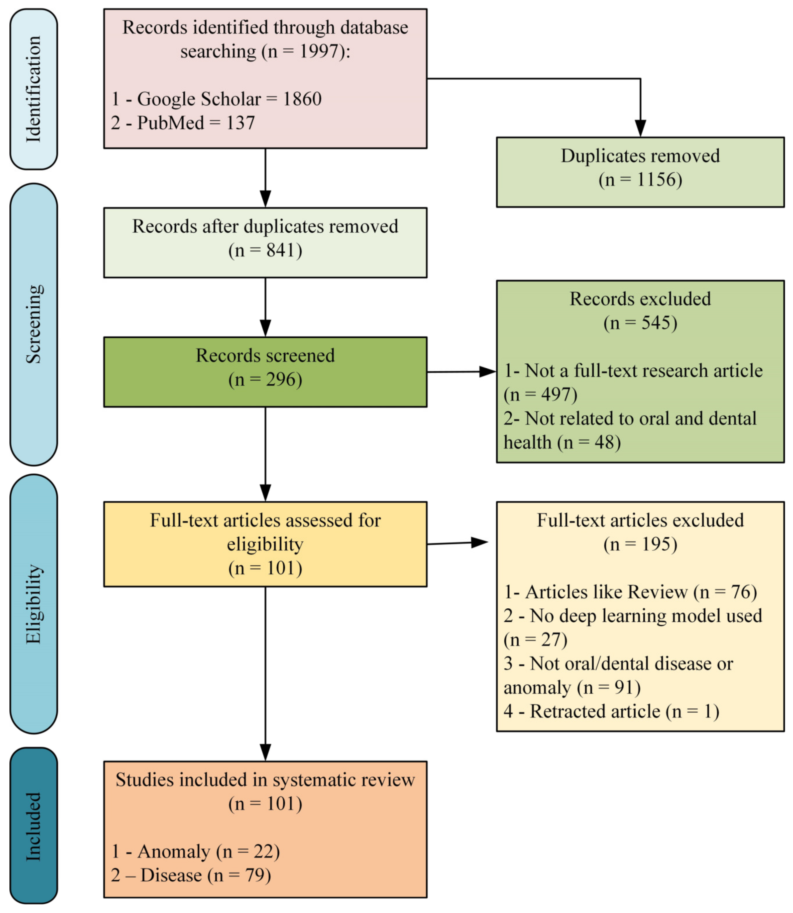
- This study includes 101 shortlisted research articles from Scholar and PubMed that apply deep learning methods for diagnosing dental anomalies and diseases.
- This review included variables such as the size of the dataset, the dental imaging method, the deep learning architecture used for performance evaluation criteria, and the explainable AI method.
- Unlike other reviews in the literature, in this review, studies comparing human-AI performance among shortlisted research articles are discussed in detail, especially statistical tests.
2. Material and Methods
2.1. Information Sources and Eligibility Criteria
- Articles published between January 2019–May 2023.
- Articles on the diagnosis of dental anomalies or diseases.
- Articles suggesting deep learning methods.
- Articles created using a reference dataset on dental imaging techniques.
- Full-text research articles.
- Articles written in English.
- The article must contain detailed information about the dataset, methods, results, and tests applied.
- Articles on topics such as healthy tooth detection, tooth labeling/numbering, dental implants, and endodontic treatment.
- Articles that have applied other AI methods that do not include deep learning methodologies, such as classical machine learning.
- Review articles and other types such as conferences, article abstracts, book chapters, preprints, or non-full-text articles, even if it is a research article.
2.2. Search Strategy and Selection Process
2.3. Data Extraction and Analysis
3. Results
4. Discussion
5. Conclusions
Author Contributions
Funding
Institutional Review Board Statement
Informed Consent Statement
Data Availability Statement
Conflicts of Interest
Abbreviations
References
- WHO. Global Oral Health Status Report: Towards Universal Health Coverage for Oral Health by 2030; WHO: Geneva, Switzerland, 2022. [Google Scholar]
- Pauwels, R. A brief introduction to concepts and applications of artificial intelligence in dental imaging. Oral Radiol. 2021, 37, 153–160. [Google Scholar] [CrossRef] [PubMed]
- Folly, P. Imaging Techniques in Dental Radiology: Acquisition, Anatomic Analysis and Interpretation of Radiographic Images. BDJ Stud. 2021, 28, 11. [Google Scholar] [CrossRef]
- Mazhar, T.; Haq, I.; Ditta, A.; Mohsan, S.A.H.; Rehman, F.; Zafar, I.; Gansau, J.A.; Goh, L.P.W. The Role of Machine Learning and Deep Learning Approaches for the Detection of Skin Cancer. Healthcare 2023, 11, 415. [Google Scholar] [CrossRef] [PubMed]
- Haq, I.; Mazhar, T.; Malik, M.A.; Kamal, M.M.; Ullah, I.; Kim, T.; Hamdi, M.; Hamam, H. Lung Nodules Localization and Report Analysis from Computerized Tomography (CT) Scan Using a Novel Machine Learning Approach. Appl. Sci. 2022, 12, 12614. [Google Scholar] [CrossRef]
- Naqvi, R.A.; Hussain, D.; Loh, W.K. Artificial Intelligence-Based Semantic Segmentation of Ocular Regions for Biometrics and Healthcare Applications. Comput. Mater. Contin. 2020, 66, 715–732. [Google Scholar] [CrossRef]
- Prados-Privado, M.; Villalón, J.G.; Martínez-Martínez, C.H.; Ivorra, C. Dental images recognition technology and applications: A literature review. Appl. Sci. 2020, 10, 2856. [Google Scholar] [CrossRef] [Green Version]
- Naqvi, R.A.; Arsalan, M.; Qaiser, T.; Khan, T.M.; Razzak, I. Sensor Data Fusion Based on Deep Learning for Computer Vision Applications and Medical Applications. Sensors 2022, 22, 8058. [Google Scholar] [CrossRef]
- Hinton, G.E.; Osindero, S.; Teh, Y.W. A Fast Learning Algorithm for Deep Belief Nets. Neural Comput. 2006, 18, 1527–1554. [Google Scholar] [CrossRef]
- Miki, Y.; Muramatsu, C.; Hayashi, T.; Zhou, X.; Hara, T.; Katsumata, A.; Fujita, H. Classification of teeth in cone-beam CT using deep convolutional neural network. Comput. Biol. Med. 2017, 80, 24–29. [Google Scholar] [CrossRef]
- Estai, M.; Tennant, M.; Gebauer, D.; Brostek, A.; Vignarajan, J.; Mehdizadeh, M.; Saha, S. Deep learning for automated detection and numbering of permanent teeth on panoramic images. Dentomaxillofacial Radiol. 2022, 51, 20210296. [Google Scholar] [CrossRef]
- Awari, H.; Subramani, N.; Janagaraj, A.; Balasubramaniapillai Thanammal, G.; Thangarasu, J.; Kohar, R. Three-dimensional dental image segmentation and classification using deep learning with tunicate swarm algorithm. Expert Syst. 2022, e13198. [Google Scholar] [CrossRef]
- Khanagar, S.B.; Alfadley, A.; Alfouzan, K.; Awawdeh, M.; Alaqla, A.; Jamleh, A. Developments and Performance of Artificial Intelligence Models Designed for Application in Endodontics: A Systematic Review. Diagnostics 2023, 13, 414. [Google Scholar]
- Sadr, S.; Mohammad-Rahimi, H.; Motamedian, S.R.; Zahedrozegar, S.; Motie, P.; Vinayahalingam, S.; Dianat, O.; Nosrat, A. Deep Learning for Detection of Periapical Radiolucent Lesions: A Systematic Review and Meta-analysis of Diagnostic Test Accuracy. J. Endod. 2023, 49, 248–261.e3. [Google Scholar]
- Sultan, A.S.; Elgharib, M.A.; Tavares, T.; Jessri, M.; Basile, J.R. The use of artificial intelligence, machine learning and deep learning in oncologic histopathology. J. Oral Pathol. Med. 2020, 49, 849–856. [Google Scholar] [CrossRef]
- Alhazmi, A.; Alhazmi, Y.; Makrami, A.; Masmali, A.; Salawi, N.; Masmali, K.; Patil, S. Application of artificial intelligence and machine learning for prediction of oral cancer risk. J. Oral Pathol. Med. 2021, 50, 444–450. [Google Scholar] [CrossRef]
- Rajee, M.V.; Mythili, C. Gender classification on digital dental x-ray images using deep convolutional neural network. Biomed. Signal Process. Control 2021, 69, 102939. [Google Scholar] [CrossRef]
- Guo, Y.-C.; Han, M.; Chi, Y.; Long, H.; Zhang, D.; Yang, J.; Yang, Y.; Chen, T.; Du, S. Accurate age classification using manual method and deep convolutional neural network based on orthopantomogram images. Int. J. Legal Med. 2021, 135, 1589–1597. [Google Scholar] [CrossRef]
- Takahashi, T.; Nozaki, K.; Gonda, T.; Mameno, T.; Wada, M.; Ikebe, K. Identification of dental implants using deep learning—Pilot study. Int. J. Implant Dent. 2020, 6, 53. [Google Scholar] [CrossRef]
- Lee, D.W.; Kim, S.Y.; Jeong, S.N.; Lee, J.H. Artificial intelligence in fractured dental implant detection and classification: Evaluation using dataset from two dental hospitals. Diagnostics 2021, 11, 233. [Google Scholar] [CrossRef]
- Ahn, Y.; Hwang, J.J.; Jung, Y.-H.; Jeong, T.; Shin, J. Automated Mesiodens Classification System Using Deep Learning on Panoramic Radiographs of Children. Diagnostics 2021, 11, 1477. [Google Scholar] [CrossRef]
- Li, C.W.; Lin, S.Y.; Chou, H.S.; Chen, T.Y.; Chen, Y.A.; Liu, S.Y.; Liu, Y.L.; Chen, C.A.; Huang, Y.C.; Chen, S.L.; et al. Detection of Dental Apical Lesions Using CNNs on Periapical Radiograph. Sensors 2021, 21, 7049. [Google Scholar] [CrossRef] [PubMed]
- Choi, E.; Kim, D.; Lee, J.Y.; Park, H.K. Artificial intelligence in detecting temporomandibular joint osteoarthritis on orthopantomogram. Sci. Rep. 2021, 11, 10246. [Google Scholar] [CrossRef] [PubMed]
- Bayrakdar, S.K.; Orhan, K.; Bayrakdar, I.S.; Bilgir, E.; Ezhov, M.; Gusarev, M.; Shumilov, E. A deep learning approach for dental implant planning in cone-beam computed tomography images. BMC Med. Imaging 2021, 21, 86. [Google Scholar] [CrossRef]
- Casalegno, F.; Newton, T.; Daher, R.; Abdelaziz, M.; Lodi-Rizzini, A.; Schürmann, F.; Krejci, I.; Markram, H. Caries Detection with Near-Infrared Transillumination Using Deep Learning. J. Dent. Res. 2019, 98, 1227–1233. [Google Scholar] [CrossRef] [PubMed] [Green Version]
- Vimalarani, G.; Ramachandraiah, U. Automatic diagnosis and detection of dental caries in bitewing radiographs using pervasive deep gradient based LeNet classifier model. Microprocess. Microsyst. 2022, 94, 104654. [Google Scholar] [CrossRef]
- Park, S.; Erkinov, H.; Hasan, M.A.M.; Nam, S.H.; Kim, Y.R.; Shin, J.; Chang, W. Du Periodontal Disease Classification with Color Teeth Images Using Convolutional Neural Networks. Electronics 2023, 12, 1518. [Google Scholar] [CrossRef]
- Alalharith, D.M.; Alharthi, H.M.; Alghamdi, W.M.; Alsenbel, Y.M.; Aslam, N.; Khan, I.U.; Shahin, S.Y.; Dianišková, S.; Alhareky, M.S.; Barouch, K.K. A Deep Learning-Based Approach for the Detection of Early Signs of Gingivitis in Orthodontic Patients Using Faster Region-Based Convolutional Neural Networks. Int. J. Environ. Res. Public Health 2020, 17, 8447. [Google Scholar] [CrossRef]
- Mohammad-Rahimi, H.; Motamedian, S.R.; Rohban, M.H.; Krois, J.; Uribe, S.E.; Mahmoudinia, E.; Rokhshad, R.; Nadimi, M.; Schwendicke, F. Deep learning for caries detection: A systematic review. J. Dent. 2022, 122, 104115. [Google Scholar] [CrossRef]
- Benakatti, V.; Nayakar, R.; Anandhalli, M.; Lagali-Jirge, V. Accuracy of machine learning in identification of dental implant systems in radiographs-A systematic review and meta-analysis. J. Indian Acad. Oral Med. Radiol. 2022, 34, 354–358. [Google Scholar] [CrossRef]
- Alwohaibi, R.N.; Almaimoni, R.A.; Alshrefy, A.J.; AlMusailet, L.I.; AlHazzaa, S.A.; Menezes, R.G. Dental implants and forensic identification: A systematic review. J. Forensic Leg. Med. 2023, 96, 102508. [Google Scholar] [CrossRef]
- Khanagar, S.B.; Vishwanathaiah, S.; Naik, S.; Al-Kheraif, A.A.; Devang Divakar, D.; Sarode, S.C.; Bhandi, S.; Patil, S. Application and performance of artificial intelligence technology in forensic odontology—A systematic review. Leg. Med. 2021, 48, 101826. [Google Scholar]
- Farook, T.H.; Dudley, J. Automation and deep (machine) learning in temporomandibular joint disorder radiomics: A systematic review. J. Oral Rehabil. 2023, 50, 501–521. [Google Scholar] [CrossRef]
- Revilla-León, M.; Gómez-Polo, M.; Barmak, A.B.; Inam, W.; Kan, J.Y.K.; Kois, J.C.; Akal, O. Artificial intelligence models for diagnosing gingivitis and periodontal disease: A systematic review. J. Prosthet. Dent. 2022. [Google Scholar] [CrossRef]
- AbuSalim, S.; Zakaria, N.; Islam, M.R.; Kumar, G.; Mokhtar, N.; Abdulkadir, S.J. Analysis of Deep Learning Techniques for Dental Informatics: A Systematic Literature Review. Healthcare 2022, 10, 1892. [Google Scholar]
- Corbella, S.; Srinivas, S.; Cabitza, F. Applications of deep learning in dentistry. Oral Surg. Oral Med. Oral Pathol. Oral Radiol. 2021, 132, 225–238. [Google Scholar] [CrossRef]
- Carrillo-Perez, F.; Pecho, O.E.; Morales, J.C.; Paravina, R.D.; Della Bona, A.; Ghinea, R.; Pulgar, R.; del Mar Pérez, M.; Herrera, L.J. Applications of artificial intelligence in dentistry: A comprehensive review. J. Esthet. Restor. Dent. 2022, 34, 259–280. [Google Scholar]
- Tandon, D.; Rajawat, J. Present and future of artificial intelligence in dentistry. J. Oral Biol. Craniofacial Res. 2020, 10, 391–396. [Google Scholar]
- Khanagar, S.B.; Al-ehaideb, A.; Maganur, P.C.; Vishwanathaiah, S.; Patil, S.; Baeshen, H.A.; Sarode, S.C.; Bhandi, S. Developments, application, and performance of artificial intelligence in dentistry—A systematic review. J. Dent. Sci. 2021, 16, 508–522. [Google Scholar]
- Mohammad-Rahimi, H.; Rokhshad, R.; Bencharit, S.; Krois, J.; Schwendicke, F. Deep learning: A primer for dentists and dental researchers. J. Dent. 2023, 130, 104430. [Google Scholar]
- Shah, N. Recent advances in imaging technologies in dentistry. World J. Radiol. 2014, 6, 794. [Google Scholar] [CrossRef]
- Singh, N.K.; Raza, K. Progress in deep learning-based dental and maxillofacial image analysis: A systematic review. Expert Syst. Appl. 2022, 199, 116968. [Google Scholar]
- Page, M.J.; McKenzie, J.E.; Bossuyt, P.M.; Boutron, I.; Hoffmann, T.C.; Mulrow, C.D.; Shamseer, L.; Tetzlaff, J.M.; Akl, E.A.; Brennan, S.E.; et al. The PRISMA 2020 statement: An updated guideline for reporting systematic reviews. BMJ 2021, 372, n71. [Google Scholar] [CrossRef] [PubMed]
- Al Kheraif, A.A.; Wahba, A.A.; Fouad, H. Detection of dental diseases from radiographic 2d dental image using hybrid graph-cut technique and convolutional neural network. Meas. J. Int. Meas. Confed. 2019, 146, 333–342. [Google Scholar] [CrossRef]
- Mine, Y.; Iwamoto, Y.; Okazaki, S.; Nakamura, K.; Takeda, S.; Peng, T.; Mitsuhata, C.; Kakimoto, N.; Kozai, K.; Murayama, T. Detecting the presence of supernumerary teeth during the early mixed dentition stage using deep learning algorithms: A pilot study. Int. J. Paediatr. Dent. 2022, 32, 678–685. [Google Scholar] [CrossRef]
- Okazaki, S.; Mine, Y.; Iwamoto, Y.; Urabe, S.; Mitsuhata, C.; Nomura, R.; Kakimoto, N.; Murayama, T. Analysis of the feasibility of using deep learning for multiclass classification of dental anomalies on panoramic radiographs. Dent. Mater. J. 2022, 41, 889–895. [Google Scholar] [CrossRef]
- Ragodos, R.; Wang, T.; Padilla, C.; Hecht, J.T.; Poletta, F.A.; Orioli, I.M.; Buxó, C.J.; Butali, A.; Valencia-Ramirez, C.; Restrepo Muñeton, C.; et al. Dental anomaly detection using intraoral photos via deep learning. Sci. Rep. 2022, 12, 11577. [Google Scholar] [CrossRef]
- Aljabri, M.; Aljameel, S.S.; Min-Allah, N.; Alhuthayfi, J.; Alghamdi, L.; Alduhailan, N.; Alfehaid, R.; Alqarawi, R.; Alhareky, M.; Shahin, S.Y.; et al. Canine impaction classification from panoramic dental radiographic images using deep learning models. Inform. Med. Unlocked 2022, 30, 100918. [Google Scholar] [CrossRef]
- Liu, J.; Liu, Y.; Li, S.; Ying, S.; Zheng, L.; Zhao, Z. Artificial intelligence-aided detection of ectopic eruption of maxillary first molars based on panoramic radiographs. J. Dent. 2022, 125, 104239. [Google Scholar] [CrossRef]
- Askar, H.; Krois, J.; Rohrer, C.; Mertens, S.; Elhennawy, K.; Ottolenghi, L.; Mazur, M.; Paris, S.; Schwendicke, F. Detecting white spot lesions on dental photography using deep learning: A pilot study. J. Dent. 2021, 107, 103615. [Google Scholar] [CrossRef]
- Schönewolf, J.; Meyer, O.; Engels, P.; Schlickenrieder, A.; Hickel, R.; Gruhn, V.; Hesenius, M.; Kühnisch, J. Artificial intelligence-based diagnostics of molar-incisor-hypomineralization (MIH) on intraoral photographs. Clin. Oral Investig. 2022, 26, 5923–5930. [Google Scholar] [CrossRef]
- Alevizakos, V.; Bekes, K.; Steffen, R.; von See, C. Artificial intelligence system for training diagnosis and differentiation with molar incisor hypomineralization (MIH) and similar pathologies. Clin. Oral Investig. 2022, 26, 6917–6923. [Google Scholar] [CrossRef]
- Ha, E.G.; Jeon, K.J.; Kim, Y.H.; Kim, J.Y.; Han, S.S. Automatic detection of mesiodens on panoramic radiographs using artificial intelligence. Sci. Rep. 2021, 11, 23061. [Google Scholar] [CrossRef]
- Jeon, K.J.; Ha, E.G.; Choi, H.; Lee, C.; Han, S.S. Performance comparison of three deep learning models for impacted mesiodens detection on periapical radiographs. Sci. Rep. 2022, 12, 15402. [Google Scholar] [CrossRef]
- Dai, X.; Jiang, X.; Jing, Q.; Zheng, J.; Zhu, S.; Mao, T.; Wang, D. A one-stage deep learning method for fully automated mesiodens localization on panoramic radiographs. Biomed. Signal Process. Control 2023, 80, 104315. [Google Scholar] [CrossRef]
- Kuwada, C.; Ariji, Y.; Fukuda, M.; Kise, Y.; Fujita, H.; Katsumata, A.; Ariji, E. Deep learning systems for detecting and classifying the presence of impacted supernumerary teeth in the maxillary incisor region on panoramic radiographs. Oral Surg. Oral Med. Oral Pathol. Oral Radiol. 2020, 130, 464–469. [Google Scholar] [CrossRef]
- Celik, M.E. Deep Learning Based Detection Tool for Impacted Mandibular Third Molar Teeth. Diagnostics 2022, 12, 942. [Google Scholar] [CrossRef]
- Başaran, M.; Çelik, Ö.; Bayrakdar, I.S.; Bilgir, E.; Orhan, K.; Odabaş, A.; Aslan, A.F.; Jagtap, R. Diagnostic charting of panoramic radiography using deep-learning artificial intelligence system. Oral Radiol. 2022, 38, 363–369. [Google Scholar] [CrossRef]
- Lee, S.; Kim, D.; Jeong, H.G. Detecting 17 fine-grained dental anomalies from panoramic dental radiography using artificial intelligence. Sci. Rep. 2022, 12, 5172. [Google Scholar] [CrossRef]
- Kim, J.; Hwang, J.J.; Jeong, T.; Cho, B.-H.H.; Shin, J. Deep learning-based identification of mesiodens using automatic maxillary anterior region estimation in panoramic radiography of children. Dentomaxillofacial Radiol. 2022, 51, 20210528. [Google Scholar]
- Ariji, Y.; Mori, M.; Fukuda, M.; Katsumata, A.; Ariji, E. Automatic visualization of the mandibular canal in relation to an impacted mandibular third molar on panoramic radiographs using deep learning segmentation and transfer learning techniques. Oral Surg. Oral Med. Oral Pathol. Oral Radiol. 2022, 134, 749–757. [Google Scholar] [CrossRef]
- Imak, A.; Çelebi, A.; Polat, O.; Türkoğlu, M.; Şengür, A. ResMIBCU-Net: An encoder–decoder network with residual blocks, modified inverted residual block, and bi-directional ConvLSTM for impacted tooth segmentation in panoramic X-ray images. Oral Radiol. 2023, 1, 1–15. [Google Scholar] [CrossRef]
- Zhu, H.; Yu, H.; Zhang, F.; Cao, Z.; Wu, F.; Zhu, F. Automatic segmentation and detection of ectopic eruption of first permanent molars on panoramic radiographs based on nnU-Net. Int. J. Paediatr. Dent. 2022, 32, 785–792. [Google Scholar] [CrossRef] [PubMed]
- Duman, S.; Yılmaz, E.F.; Eşer, G.; Çelik, Ö.; Bayrakdar, I.S.; Bilgir, E.; Costa, A.L.F.; Jagtap, R.; Orhan, K. Detecting the presence of taurodont teeth on panoramic radiographs using a deep learning-based convolutional neural network algorithm. Oral Radiol. 2023, 39, 207–214. [Google Scholar] [CrossRef] [PubMed]
- Megalan Leo, L.; Kalpalatha Reddy, T. Dental Caries Classification System Using Deep Learning Based Convolutional Neural Network. J. Comput. Theor. Nanosci. 2020, 17, 4660–4665. [Google Scholar] [CrossRef]
- Wang, C.; Qin, H.; Lai, G.; Zheng, G.; Xiang, H.; Wang, J.; Zhang, D. Automated classification of dual channel dental imaging of auto-fluorescence and white lightby convolutional neural networks. J. Innov. Opt. Health Sci. 2020, 13, 2050014. [Google Scholar] [CrossRef]
- Schwendicke, F.; Elhennawy, K.; Paris, S.; Friebertshäuser, P.; Krois, J. Deep learning for caries lesion detection in near-infrared light transillumination images: A pilot study. J. Dent. 2020, 92, 103260. [Google Scholar] [CrossRef]
- Megalan Leo, L.; Kalapalatha Reddy, T. Learning compact and discriminative hybrid neural network for dental caries classification. Microprocess. Microsyst. 2021, 82, 103836. [Google Scholar] [CrossRef]
- Vinayahalingam, S.; Kempers, S.; Limon, L.; Deibel, D.; Maal, T.; Hanisch, M.; Bergé, S.; Xi, T. Classification of caries in third molars on panoramic radiographs using deep learning. Sci. Rep. 2021, 11, 12609. [Google Scholar] [CrossRef]
- Singh, P.; Sehgal, P.G. V Black dental caries classification and preparation technique using optimal CNN-LSTM classifier. Multimed. Tools Appl. 2021, 80, 5255–5272. [Google Scholar] [CrossRef]
- Bui, T.H.; Hamamoto, K.; Paing, M.P. Automated Caries Screening Using Ensemble Deep Learning on Panoramic Radiographs. Entropy 2022, 24, 1358. [Google Scholar] [CrossRef]
- Panyarak, W.; Wantanajittikul, K.; Suttapak, W.; Charuakkra, A.; Prapayasatok, S. Feasibility of deep learning for dental caries classification in bitewing radiographs based on the ICCMSTM radiographic scoring system. Oral Surg. Oral Med. Oral Pathol. Oral Radiol. 2023, 135, 272–281. [Google Scholar] [CrossRef]
- Haghanifar, A.; Majdabadi, M.M.; Haghanifar, S.; Choi, Y.; Ko, S.B. PaXNet: Tooth segmentation and dental caries detection in panoramic X-ray using ensemble transfer learning and capsule classifier. Multimed. Tools Appl. 2023, 82, 27659–27679. [Google Scholar] [CrossRef]
- Zhou, X.; Yu, G.; Yin, Q.; Yang, J.; Sun, J.; Lv, S.; Shi, Q. Tooth Type Enhanced Transformer for Children Caries Diagnosis on Dental Panoramic Radiographs. Diagnostics 2023, 13, 689. [Google Scholar] [CrossRef]
- Ezhov, M.; Gusarev, M.; Golitsyna, M.; Yates, J.M.; Kushnerev, E.; Tamimi, D.; Aksoy, S.; Shumilov, E.; Sanders, A.; Orhan, K. Clinically applicable artificial intelligence system for dental diagnosis with CBCT. Sci. Rep. 2021, 11, 15006. [Google Scholar] [CrossRef]
- Rajee, M.V.; Mythili, C. Dental Image Segmentation and Classification Using Inception Resnetv2. IETE J. Res. 2021, 1–17. [Google Scholar] [CrossRef]
- Pauwels, R.; Brasil, D.M.; Yamasaki, M.C.; Jacobs, R.; Bosmans, H.; Freitas, D.Q.; Haiter-Neto, F. Artificial intelligence for detection of periapical lesions on intraoral radiographs: Comparison between convolutional neural networks and human observers. Oral Surg. Oral Med. Oral Pathol. Oral Radiol. 2021, 131, 610–616. [Google Scholar] [CrossRef]
- Calazans, M.A.A.; Ferreira, F.A.B.S.; de Lourdes Melo Guedes Alcoforado, M.; dos Santos, A.; dos Anjos Pontual, A.; Madeiro, F. Automatic Classification System for Periapical Lesions in Cone-Beam Computed Tomography. Sensors 2022, 22, 6481. [Google Scholar] [CrossRef]
- Sankaran, K.S. An improved multipath residual CNN-based classification approach for periapical disease prediction and diagnosis in dental radiography. Neural Comput. Appl. 2022, 34, 20067–20082. [Google Scholar] [CrossRef]
- Chuo, Y.; Lin, W.M.; Chen, T.Y.; Chan, M.L.; Chang, Y.S.; Lin, Y.R.; Lin, Y.J.; Shao, Y.H.; Chen, C.A.; Chen, S.L.; et al. A High-Accuracy Detection System: Based on Transfer Learning for Apical Lesions on Periapical Radiograph. Bioengineering 2022, 9, 777. [Google Scholar] [CrossRef]
- Li, S.; Liu, J.; Zhou, Z.; Zhou, Z.; Wu, X.; Li, Y.; Wang, S.; Liao, W.; Ying, S.; Zhao, Z. Artificial intelligence for caries and periapical periodontitis detection. J. Dent. 2022, 122, 104107. [Google Scholar] [CrossRef]
- Liu, F.; Gao, L.; Wan, J.; Lyu, Z.L.; Huang, Y.Y.; Liu, C.; Han, M. Recognition of Digital Dental X-ray Images Using a Convolutional Neural Network. J. Digit. Imaging 2022, 36, 73–79. [Google Scholar] [CrossRef] [PubMed]
- Jaiswal, P.; Bhirud, D.S. An intelligent deep network for dental medical image processing system. Biomed. Signal Process. Control 2023, 84, 104708. [Google Scholar] [CrossRef]
- Chauhan, R.B.; Shah, T.; Shah, D.; Gohil, T. A novel convolutional neural network–Fuzzy-based diagnosis in the classification of dental pulpitis. Adv. Hum. Biol. 2023, 13, 79–86. [Google Scholar] [CrossRef]
- Chang, H.J.; Lee, S.J.; Yong, T.H.; Shin, N.Y.; Jang, B.G.; Kim, J.E.; Huh, K.H.; Lee, S.S.; Heo, M.S.; Choi, S.C.; et al. Deep Learning Hybrid Method to Automatically Diagnose Periodontal Bone Loss and Stage Periodontitis. Sci. Rep. 2020, 10, 7531. [Google Scholar] [CrossRef]
- Krois, J.; Ekert, T.; Meinhold, L.; Golla, T.; Kharbot, B.; Wittemeier, A.; Dörfer, C.; Schwendicke, F. Deep Learning for the Radiographic Detection of Periodontal Bone Loss. Sci. Rep. 2019, 9, 8495. [Google Scholar] [CrossRef] [Green Version]
- Kim, J.; Lee, H.S.; Song, I.S.; Jung, K.H. DeNTNet: Deep Neural Transfer Network for the detection of periodontal bone loss using panoramic dental radiographs. Sci. Rep. 2019, 9, 17615. [Google Scholar] [CrossRef] [Green Version]
- Lee, J.H.; Kim, D.H.; Jeong, S.N. Diagnosis of cystic lesions using panoramic and cone beam computed tomographic images based on deep learning neural network. Oral Dis. 2020, 26, 152–158. [Google Scholar] [CrossRef]
- Rao, R.S.; Shivanna, D.B.; Mahadevpur, K.S.; Shivaramegowda, S.G.; Prakash, S.; Lakshminarayana, S.; Patil, S. Deep Learning-Based Microscopic Diagnosis of Odontogenic Keratocysts and Non-Keratocysts in Haematoxylin and Eosin-Stained Incisional Biopsies. Diagnostics 2021, 11, 2184. [Google Scholar] [CrossRef]
- Sivasundaram, S.; Pandian, C. Performance analysis of classification and segmentation of cysts in panoramic dental images using convolutional neural network architecture. Int. J. Imaging Syst. Technol. 2021, 31, 2214–2225. [Google Scholar] [CrossRef]
- Lee, J.S.; Adhikari, S.; Liu, L.; Jeong, H.G.; Kim, H.; Yoon, S.J. Osteoporosis detection in panoramic radiographs using a deep convolutional neural network-based computer-assisted diagnosis system: A preliminary study. Dentomaxillofacial Radiol. 2019, 48, 20170344. [Google Scholar] [CrossRef]
- Lee, K.S.; Jung, S.K.; Ryu, J.J.; Shin, S.W.; Choi, J. Evaluation of Transfer Learning with Deep Convolutional Neural Networks for Screening Osteoporosis in Dental Panoramic Radiographs. J. Clin. Med. 2020, 9, 392. [Google Scholar] [CrossRef] [Green Version]
- Sukegawa, S.; Fujimura, A.; Taguchi, A.; Yamamoto, N.; Kitamura, A.; Goto, R.; Nakano, K.; Takabatake, K.; Kawai, H.; Nagatsuka, H.; et al. Identification of osteoporosis using ensemble deep learning model with panoramic radiographs and clinical covariates. Sci. Rep. 2022, 12, 6088. [Google Scholar] [CrossRef]
- Tassoker, M.; Öziç, M.Ü.; Yuce, F. Comparison of five convolutional neural networks for predicting osteoporosis based on mandibular cortical index on panoramic radiographs. Dentomaxillofacial Radiol. 2022, 51, 20220108. [Google Scholar] [CrossRef]
- Nishiyama, M.; Ishibashi, K.; Ariji, Y.; Fukuda, M.; Nishiyama, W.; Umemura, M.; Katsumata, A.; Fujita, H.; Ariji, E. Performance of deep learning models constructed using panoramic radiographs from two hospitals to diagnose fractures of the mandibular condyle. Dentomaxillofacial Radiol. 2021, 50, 50. [Google Scholar] [CrossRef]
- Yang, P.; Guo, X.; Mu, C.; Qi, S.; Li, G. Detection of vertical root fractures by cone-beam computed tomography based on deep learning. Dentomaxillofacial Radiol. 2023, 52, 20220345. [Google Scholar] [CrossRef]
- Murata, M.; Ariji, Y.; Ohashi, Y.; Kawai, T.; Fukuda, M.; Funakoshi, T.; Kise, Y.; Nozawa, M.; Katsumata, A.; Fujita, H.; et al. Deep-learning classification using convolutional neural network for evaluation of maxillary sinusitis on panoramic radiography. Oral Radiol. 2019, 35, 301–307. [Google Scholar] [CrossRef]
- Li, W.; Liang, Y.; Zhang, X.; Liu, C.; He, L.; Miao, L.; Sun, W. A deep learning approach to automatic gingivitis screening based on classification and localization in RGB photos. Sci. Rep. 2021, 11, 16831. [Google Scholar] [CrossRef]
- Jung, W.; Lee, K.E.; Suh, B.J.; Seok, H.; Lee, D.W. Deep learning for osteoarthritis classification in temporomandibular joint. Oral Dis. 2023, 29, 1050–1059. [Google Scholar] [CrossRef]
- Kuwada, C.; Ariji, Y.; Kise, Y.; Fukuda, M.; Nishiyama, M.; Funakoshi, T.; Takeuchi, R.; Sana, A.; Kojima, N.; Ariji, E. Deep-learning systems for diagnosing cleft palate on panoramic radiographs in patients with cleft alveolus. Oral Radiol. 2022, 39, 349–354. [Google Scholar] [CrossRef]
- Al-Sarem, M.; Al-Asali, M.; Alqutaibi, A.Y.; Saeed, F. Enhanced Tooth Region Detection Using Pretrained Deep Learning Models. Int. J. Environ. Res. Public Health 2022, 19, 15414. [Google Scholar] [CrossRef]
- Zhang, X.; Liang, Y.; Li, W.; Liu, C.; Gu, D.; Sun, W.; Miao, L. Development and evaluation of deep learning for screening dental caries from oral photographs. Oral Dis. 2022, 28, 173–181. [Google Scholar] [CrossRef] [PubMed]
- Chen, H.; Li, H.; Zhao, Y.; Zhao, J.; Wang, Y. Dental disease detection on periapical radiographs based on deep convolutional neural networks. Int. J. Comput. Assist. Radiol. Surg. 2021, 16, 649–661. [Google Scholar] [CrossRef] [PubMed]
- Kim, C.; Jeong, H.; Park, W.; Kim, D. Tooth-Related Disease Detection System Based on Panoramic Images and Optimization Through Automation: Development Study. JMIR Med. Inform. 2022, 10, e38640. [Google Scholar] [CrossRef]
- Chen, X.; Guo, J.; Ye, J.; Zhang, M.; Liang, Y. Detection of Proximal Caries Lesions on Bitewing Radiographs Using Deep Learning Method. Caries Res. 2022, 56, 455–463. [Google Scholar] [CrossRef] [PubMed]
- Park, E.Y.; Cho, H.; Kang, S.; Jeong, S.; Kim, E.K. Caries detection with tooth surface segmentation on intraoral photographic images using deep learning. BMC Oral Health 2022, 22, 573. [Google Scholar] [CrossRef]
- Fatima, A.; Shafi, I.; Afzal, H.; Mahmood, K.; de la Torre Díez, I.; Lipari, V.; Ballester, J.B.; Ashraf, I. Deep Learning-Based Multiclass Instance Segmentation for Dental Lesion Detection. Healthcare 2023, 11, 347. [Google Scholar] [CrossRef]
- Jiang, L.; Chen, D.; Cao, Z.; Wu, F.; Zhu, H.; Zhu, F. A two-stage deep learning architecture for radiographic staging of periodontal bone loss. BMC Oral Health 2022, 22, 106. [Google Scholar] [CrossRef]
- Thanathornwong, B.; Suebnukarn, S. Automatic detection of periodontal compromised teeth in digital panoramic radiographs using faster regional convolutional neural networks. Imaging Sci. Dent. 2020, 50, 169. [Google Scholar] [CrossRef]
- Kwon, O.; Yong, T.H.; Kang, S.R.; Kim, J.E.; Huh, K.H.; Heo, M.S.; Lee, S.S.; Choi, S.C.; Yi, W.J. Automatic diagnosis for cysts and tumors of both jaws on panoramic radiographs using a deep convolution neural network. Dentomaxillofacial Radiol. 2020, 49, 20200185. [Google Scholar] [CrossRef]
- Yang, H.; Jo, E.; Kim, H.J.; Cha, I.H.; Jung, Y.S.; Nam, W.; Kim, J.Y.; Kim, J.K.; Kim, Y.H.; Oh, T.G.; et al. Deep Learning for Automated Detection of Cyst and Tumors of the Jaw in Panoramic Radiographs. J. Clin. Med. 2020, 9, 1839. [Google Scholar] [CrossRef]
- Ariji, Y.; Yanashita, Y.; Kutsuna, S.; Muramatsu, C.; Fukuda, M.; Kise, Y.; Nozawa, M.; Kuwada, C.; Fujita, H.; Katsumata, A.; et al. Automatic detection and classification of radiolucent lesions in the mandible on panoramic radiographs using a deep learning object detection technique. Oral Surg. Oral Med. Oral Pathol. Oral Radiol. 2019, 128, 424–430. [Google Scholar] [CrossRef]
- Kise, Y.; Ariji, Y.; Kuwada, C.; Fukuda, M.; Ariji, E. Effect of deep transfer learning with a different kind of lesion on classification performance of pre-trained model: Verification with radiolucent lesions on panoramic radiographs. Imaging Sci. Dent. 2023, 53, 27–34. [Google Scholar] [CrossRef]
- Kuwana, R.; Ariji, Y.; Fukuda, M.; Kise, Y.; Nozawa, M.; Kuwada, C.; Muramatsu, C.; Katsumata, A.; Fujita, H.; Ariji, E. Performance of deep learning object detection technology in the detection and diagnosis of maxillary sinus lesions on panoramic radiographs. Dentomaxillofacial Radiol. 2020, 50, 20200171. [Google Scholar] [CrossRef]
- Watanabe, H.; Ariji, Y.; Fukuda, M.; Kuwada, C.; Kise, Y.; Nozawa, M.; Sugita, Y.; Ariji, E. Deep learning object detection of maxillary cyst-like lesions on panoramic radiographs: Preliminary study. Oral Radiol. 2021, 37, 487–493. [Google Scholar] [CrossRef]
- Fukuda, M.; Inamoto, K.; Shibata, N.; Ariji, Y.; Yanashita, Y.; Kutsuna, S.; Nakata, K.; Katsumata, A.; Fujita, H.; Ariji, E. Evaluation of an artificial intelligence system for detecting vertical root fracture on panoramic radiography. Oral Radiol. 2020, 36, 337–343. [Google Scholar] [CrossRef]
- Son, D.M.; Yoon, Y.A.; Kwon, H.J.; An, C.H.; Lee, S.H. Automatic Detection of Mandibular Fractures in Panoramic Radiographs Using Deep Learning. Diagnostics 2021, 11, 933. [Google Scholar] [CrossRef]
- Lee, K.S.; Kwak, H.J.; Oh, J.M.; Jha, N.; Kim, Y.J.; Kim, W.; Baik, U.B.; Ryu, J.J. Automated Detection of TMJ Osteoarthritis Based on Artificial Intelligence. J. Dent. Res. 2020, 99, 1363–1367. [Google Scholar] [CrossRef]
- Park, J.; Lee, J.; Moon, S.; Lee, K. Deep Learning Based Detection of Missing Tooth Regions for Dental Implant Planning in Panoramic Radiographic Images. Appl. Sci. 2022, 12, 1595. [Google Scholar] [CrossRef]
- Khan, H.A.; Haider, M.A.; Ansari, H.A.; Ishaq, H.; Kiyani, A.; Sohail, K.; Muhammad, M.; Khurram, S.A. Automated feature detection in dental periapical radiographs by using deep learning. Oral Surg. Oral Med. Oral Pathol. Oral Radiol. 2021, 131, 711–720. [Google Scholar] [CrossRef]
- Cantu, A.G.; Gehrung, S.; Krois, J.; Chaurasia, A.; Rossi, J.G.; Gaudin, R.; Elhennawy, K.; Schwendicke, F. Detecting caries lesions of different radiographic extension on bitewings using deep learning. J. Dent. 2020, 100, 103425. [Google Scholar] [CrossRef]
- Bayrakdar, I.S.; Orhan, K.; Akarsu, S.; Çelik, Ö.; Atasoy, S.; Pekince, A.; Yasa, Y.; Bilgir, E.; Sağlam, H.; Aslan, A.F.; et al. Deep-learning approach for caries detection and segmentation on dental bitewing radiographs. Oral Radiol. 2022, 38, 468–479. [Google Scholar] [CrossRef] [PubMed]
- You, W.; Hao, A.; Li, S.; Wang, Y.; Xia, B. Deep learning-based dental plaque detection on primary teeth: A comparison with clinical assessments. BMC Oral Health 2020, 20, 141. [Google Scholar] [CrossRef]
- Lee, S.; Oh, S.-I.; Jo, J.; Kang, S.; Shin, Y.; Park, J.-W. Deep learning for early dental caries detection in bitewing radiographs. Sci. Rep. 2021, 11, 16807. [Google Scholar] [CrossRef] [PubMed]
- Lian, L.; Zhu, T.; Zhu, F.; Zhu, H. Deep Learning for Caries Detection and Classification. Diagnostics 2021, 11, 1672. [Google Scholar] [CrossRef] [PubMed]
- Zhu, H.; Cao, Z.; Lian, L.; Ye, G.; Gao, H.; Wu, J. CariesNet: A deep learning approach for segmentation of multi-stage caries lesion from oral panoramic X-ray image. Neural Comput. Appl. 2022, 35, 16051–16059. [Google Scholar] [CrossRef]
- Ari, T.; Sağlam, H.; Öksüzoğlu, H.; Kazan, O.; Bayrakdar, İ.Ş.; Duman, S.B.; Çelik, Ö.; Jagtap, R.; Futyma-Gąbka, K.; Różyło-Kalinowska, I.; et al. Automatic Feature Segmentation in Dental Periapical Radiographs. Diagnostics 2022, 12, 3081. [Google Scholar] [CrossRef]
- Dayı, B.; Üzen, H.; Çiçek, İ.B.; Duman, Ş.B. A Novel Deep Learning-Based Approach for Segmentation of Different Type Caries Lesions on Panoramic Radiographs. Diagnostics 2023, 13, 202. [Google Scholar] [CrossRef]
- Rajee, M.V.; Mythili, C. Novel technique for caries detection using curvilinear semantic deep convolutional neural network. Multimed. Tools Appl. 2023, 82, 10745–10762. [Google Scholar] [CrossRef]
- Kirnbauer, B.; Hadzic, A.; Jakse, N.; Bischof, H.; Stern, D. Automatic Detection of Periapical Osteolytic Lesions on Cone-beam Computed Tomography Using Deep Convolutional Neuronal Networks. J. Endod. 2022, 48, 1434–1440. [Google Scholar] [CrossRef]
- Song, I.S.; Shin, H.K.; Kang, J.H.; Kim, J.E.; Huh, K.H.; Yi, W.J.; Lee, S.S.; Heo, M.S. Deep learning-based apical lesion segmentation from panoramic radiographs. Imaging Sci. Dent. 2022, 52, 351–357. [Google Scholar] [CrossRef]
- Chen, C.C.; Wu, Y.F.; Aung, L.M.; Lin, J.C.Y.; Ngo, S.T.; Su, J.N.; Lin, Y.M.; Chang, W.J. Automatic recognition of teeth and periodontal bone loss measurement in digital radiographs using deep-learning artificial intelligence. J. Dent. Sci. 2023, 18, 1301–1309. [Google Scholar] [CrossRef]
- Endres, M.G.; Hillen, F.; Salloumis, M.; Sedaghat, A.R.; Niehues, S.M.; Quatela, O.; Hanken, H.; Smeets, R.; Beck-Broichsitter, B.; Rendenbach, C.; et al. Development of a Deep Learning Algorithm for Periapical Disease Detection in Dental Radiographs. Diagnostics 2020, 10, 430. [Google Scholar] [CrossRef]
- Yu, D.; Hu, J.; Feng, Z.; Song, M.; Zhu, H. Deep learning based diagnosis for cysts and tumors of jaw with massive healthy samples. Sci. Rep. 2022, 12, 1855. [Google Scholar] [CrossRef]
- Chau, R.C.W.; Li, G.-H.; Tew, I.M.; Thu, K.M.; McGrath, C.; Lo, W.-L.; Ling, W.-K.; Hsung, R.T.-C.; Lam, W.Y.H. Accuracy of Artificial Intelligence-Based Photographic Detection of Gingivitis. Int. Dent. J. 2023. [Google Scholar] [CrossRef]
- Wang, X.; Pastewait, M.; Wu, T.H.; Lian, C.; Tejera, B.; Lee, Y.T.; Lin, F.C.; Wang, L.; Shen, D.; Li, S.; et al. 3D morphometric quantification of maxillae and defects for patients with unilateral cleft palate via deep learning-based CBCT image auto-segmentation. Orthod. Craniofac. Res. 2021, 24, 108–116. [Google Scholar] [CrossRef]
- Laganà, G.; Venza, N.; Borzabadi-Farahani, A.; Fabi, F.; Danesi, C.; Cozza, P. Dental anomalies: Prevalence and associations between them in a large sample of non-orthodontic subjects, a cross-sectional study. BMC Oral Health 2017, 17, 62. [Google Scholar] [CrossRef] [Green Version]
- Sella Tunis, T.; Sarne, O.; Hershkovitz, I.; Finkelstein, T.; Pavlidi, A.M.; Shapira, Y.; Davidovitch, M.; Shpack, N. Dental Anomalies’ Characteristics. Diagnostics 2021, 11, 1161. [Google Scholar] [CrossRef]


| Research Question | What Are the Applications and Performance of Deep Learning for Diagnosing Dental Anomalies and Diseases? |
|---|---|
| Population | Diagnostic medical images of patients with dental anomalies or disease (radiographs, CBCT, intraoral images, near-infrared-light transillumination (NILT) images, optical color Images, microscopic histopathology) |
| Intervention | Deep learning-based models for diagnosis and clinical decision making |
| Comparison | Expert diagnosis |
| Outcome | Predicted results that can be measured with performance metrics (accuracy (ACC), sensitivity (SEN), specificity (SPEC), Area Under the Curve (AUC), Matthews Correlation Coefficient (MCC), Intersection over Union (IoU), Positive/Negative Predictive Values (PPV/NPV), etc.) |
| Database | Search Strategy | Search Date |
|---|---|---|
| Google Scholar | all: (“deep learning” OR “CNN” OR “convolutional neural network”) AND (“oral” OR “dental” OR “tooth” OR “teeth”) AND (“anomalies” OR “diseases”) | 26 May 2023 |
| Medline/PubMed | (“deep learning”[All Fields] OR “CNN”[All Fields] OR “convolutional neural network”[All Fields]) AND (“oral”[All Fields] OR “dental”[All Fields] OR “tooth” [All Fields] OR “teeth” [All Fields]) AND (“anomalies” [All Fields] OR “diseases” [All Fields]) | 25 May 2023 |
| Author, Year, Reference | Anomaly | Image Type | Dataset Size | Method | Primary Performance Metrics and Values (%) | Other Performance Metrics | Explainable AI Method |
|---|---|---|---|---|---|---|---|
| Classification | |||||||
| Ahn et al., 2021, [21] | Mesiodens | Panoramic | 1100 | InceptionResNetV2 | ACC: 92.40 | Precision, Recall, F1 score, AUC | Grad-CAM |
| Kheraif et al., 2019, [44] | Supernumerary, Number teeth, Jaws position, Structure, Restoration, Implants, Cavities | Panoramic | 1500 | Hybrid Graph Cut Segmentation + CNN | ACC: 97.07 | Precision, Recall, F1 score, SPEC | - |
| Mine et al., 2022, [45] | Supernumerary | Panoramic | 220 | VGG16 | ACC: 84.00 | SEN, SPEC, AUC | - |
| Okazaki et al., 2022, [46] | Supernumerary, Odontomas | Panoramic | 150 | AlexNet | ACC: 70.00 | Precision, SEN, F1 score | - |
| Ragodos et al., 2022, [47] | Supernumerary, Rotation, Agenesis, Mammalons, Microdontia, Impacted, Hypoplasia, Incisal Fissure, Hypocalcification, Displaced | Intraoral photos | 38,486 | ResNet18 | AUC for supernumerary class: 57.10 | Precision, Recall, F1 score | Grad-CAM |
| Aljabri et al., 2022, [48] | Maxillary canine impaction | Panoramic | 416 | InceptionV3 | ACC: 92.59 | Precision, Recall, F1 score, SPEC | Grad-CAM |
| Liu et al., 2022, [49] | Ectopic eruption of maxillary first molars | Panoramic | 1580 | CNN-based Fusion Model | SPEC: 86 | SEN, F1 score, PPV, NPV | Grad-CAM |
| Askar et al., 2021, [50] | White spot lesions, Hypomineralized lesions | Intraoral photos | 434 | SqueezeNet | ACC: 84.00 | SEN, SPEC, F1 score, AUC, PPV, NPV | Grad-CAM |
| Schönewolf et al., 2022, [51] | Molar-incisor-hypomineralization, Enamel breakdown | Intraoral photos | 3241 | ResNeXt-101 | ACC: 95.20 | SEN, SPEC, AUC, PPV, NPV | Grad-CAM |
| Alevizakos et al., 2022, [52] | Molar-incisor-hypomineralization, Amelogenesis imperfecta, Dental fluorosis, White spot lesions | Intraoral photos | 462 | DenseNet121 | ACC: 92.86 | Loss | - |
| Detection | |||||||
| Ha et al., 2021, [53] | Mesiodens | Panoramic | 612 | YOLOv3 | ACC: 96.20 | SEN, SPEC | - |
| Jeon et al., 2022, [54] | Mesiodens | Periapical | 720 | EfficientDetD3 | ACC: 99.20 | SEN, SPEC | - |
| Dai et al., 2023, [55] | Mesiodens | Panoramic | 850 | Authors Specific CNN: DMLnet | ACC: 94.00 | SEN, SPEC, mAP | - |
| Kuwada et al., 2020, [56] | Supernumerary | Panoramic | 550 | DetectNet | AUC: 96.00 | Precision, Recall, F1 score, ACC | - |
| Celik, 2022, [57] | Third molar impacted teeth | Panoramic | 440 | YOLOv3 | mAP: 96.00 | IoU, ACC, Precision, Recall | - |
| Başaran et al., 2022, [58] | Impacted tooth, Residual root, and eight fine-grained dental anomalies | Panoramic | 1084 | Faster R-CNN InceptionV2 (COCO) | SEN for Impacted class: 96.58 | TP, FP, FN, Precision, F1 score | - |
| Lee et al., 2022, [59] | Supernumerary, Impacted, Residual root, and 14 fine-grained dental anomalies | Panoramic | 23,000 | Faster R-CNN | SEN: 99.00 | Precision, SPEC | - |
| Segmentation | |||||||
| Kim et al., 2022, [60] | Mesiodens | Panoramic | 988 | DeepLabV3plus + InceptionResNetV2 | ACC: DeepLabV3plus +: 83.90, InceptionResNetV2: 97.10 | IoU, MeanBF score, Precision, Recall, F1 score | Grad-CAM |
| Ariji et al., 2022, [61] | Third molar impacted teeth | Panoramic | 3200 | U-Net | DSC: 83.10 | JSC, SEN | - |
| Imak et al., 2023, [62] | Impacted tooth | Panoramic | 304 | Authors Specific CNN: ResMIBCU-Net: an encoder–decoder network with residual blocks, modified inverted residual block, and bi-directional ConvLSTM | ACC: 99.82 | IoU, Recall, F1 score | - |
| Zhu et al., 2022, [63] | Ectopic eruption of first permanent molars | Panoramic | 285 | nnU-Net | ACC: 99.00 | DSC, IoU, Precision, SEN, SPEC, F1 score | - |
| Duman et al., 2023, [64] | Taurodont | Panoramic | 434 | U-Net | SEN: 86.50 | TP, FP, FN, Precision, F1 score | - |
| Author, Year, Reference | Disease | Image Type | Dataset Size | Method | Primary Performance Metrics and Values (%) | Other Performance Metrics | Explainable AI Method |
|---|---|---|---|---|---|---|---|
| Classification | |||||||
| Megalan Leo and Kalpalatha Reddy, 2020, [65] | Dental caries | Bite viewing | 480 | InceptionV3 | ACC: 86.70 | - | - |
| Wang et al., 2020, [66] | Dental caries, Dental plaque | Intraoral photos | 7200 | Authors Specific CNN | ACC: Dental caries: 95.30, Dental plaque: 95.90 | SEN, SPEC | - |
| Schwendicke et al., 2020, [67] | Dental caries | NILT | 226 | ResNet18 | ACC: 69.00 | SEN, SPEC, AUC, PPV, NPV | CAM |
| Megalan Leo and Kalpalatha Reddy, 2021, [68] | Dental caries: Enamel, Dentin, Pulp, Root lesions | Bite viewing | 480 | Hybrid Neural Network (HNN) | ACC: 96.00 | - | - |
| Vinayahalingam et al., 2021, [69] | Dental caries | Panoramic | 400 | MobileNetV2 | ACC: 87.00 | SEN, SPEC, AUC | CAM |
| Singh and Sehgal, 2021, [70] | G.V Black dental caries | Periapical | 1500 | CNN-LSTM | ACC: 96.00 | Precision, SEN, SPEC, F1 score, G-mean, AUC | - |
| Bui et al., 2022, [71] | Dental caries | Panoramic | 95 | Pretrained CNNs-SVM | ACC: 93.58 | SEN, SPEC, F1 score, PPV, NPV | - |
| Vimalarani and Ramachandraiah, 2022, [26] | Dental caries | Bite viewing | 1000 | Pervasive deep gradient-based LeNet | ACC: 98.74 | SEN, SPEC, ER, PPV, NPV | - |
| Panyarak et al., 2023, [72] | Dental caries | Bite viewing | 2758 | ResNet152 | ACC: 71.11 | SEN, SPEC, CR, AUC | CAM |
| Haghanifar et al., 2023, [73] | Dental caries | Panoramic | 470 | Authors Specific CNN: PaXNet: Ensemble transfer learning and capsule classifier | ACC: 86.05 | Loss, Precision, Recall, F0.5 score | Grad-CAM |
| Zhou et al., 2023, [74] | Dental caries | Panoramic | 304 | Swin Transformer | ACC: 85.57 | Precision, Recall, F1 score | - |
| Ezhov et al., 2021, [75] | Dental caries, Periapical lesion, Periodontal bone loss | CBCT | 1346 | U-Net + DenseNet | SEN: 92.39 | SPEC | - |
| Rajee and Mythili, 2021, [76] | Dental caries, Periapical infection, Periodontal, and Pericoronal diseases | Periapical, Panoramic | 2000 | Curvilinear Semantic DCNN+ InceptionResNetV2 | ACC: 94.51 | MCC, DSC, JSC, ER, Precision, Recall, SPEC | - |
| Pauwels et al., 2021, [77] | Periapical lesion | Periapical | 280 | Authors Specific CNN | SEN: 87.00 | SPEC, AUC | - |
| Calazans et al., 2022, [78] | Periapical lesion | CBCT | 1000 | Siamese Network + DenseNet121 | ACC: 70.00 | SPEC, Precision, Recall, F1 score | - |
| Sankaran, 2022, [79] | Periapical lesion | Panoramic | 1500 | Improved Multipath Residual CNN (IMRCNN) | ACC: 98.90 | SEN, SPEC, Precision, F1 score | - |
| Li et al., 2021, [22] | Dental apical lesions | Periapical | 476 | Authors Specific CNN | ACC: 92.50 | Loss | - |
| Chuo et al., 2022, [80] | Dental apical lesions | Periapical | 760 | AlexNet | ACC: 96.21 | - | - |
| Li et al., 2022, [81] | Dental caries, Periapical periodontitis | Periapical | 4129 | Modified ResNet18 | F1 score: Dental caries: 82.90, Periapical periodontitis: 82.80 | SEN, SPEC, AUC, PPV, NPV | Grad-CAM |
| Liu et al., 2022, [82] | Dental caries, Periapical periodontitis, Periapical cysts | Periapical | 1880 | DenseNet121 | ACC: 99.50 | SEN, SPEC, PPV, NPV | CAM |
| Park et al., 2023, [27] | Calculus and Inflammation | Optical Color Images | 220 | YOLOv5 + Parallel 1D CNN | ACC: 74.54 | - | - |
| Jaiswal and Bhirud, 2023, [83] | Erosive wear, Periodontitis | OPG | 500 | CNN with Antlion Optimization | ACC: 77.00 | Precision, Recall, F1 score | - |
| Chauhan et al., 2023, [84] | Dental pulpitis | Periapical | 428 | CNN-Fuzzy logic | ACC: 94.00 | SEN, SPEC, Precision, F1 score, MCC | Grad-CAM |
| Chang et al., 2020, [85] | Periodontal bone loss, Periodontitis | Panoramic | 330 | Mask R-CNN + CNN | Pixel ACC: 92.00 | DSC, JSC | - |
| Krois et al., 2019, [86] | Periodontal bone loss | Panoramic | 85 | Authors Specific CNN | ACC: 81.00 | SEN, SPEC, F1 score, AUC, PPV, NPV | - |
| Kim et al., 2019, [87] | Periodontal bone loss | Panoramic | 12,179 | Authors Specific CNN: DeNTNet: Deep Neural Transfer Network | F1 score: 75.00 | Precision, Recall, AUC, NPV | Grad-CAM |
| Lee et al., 2020, [88] | Odontogenic cyst | Panoramic, CBCT | Panoramic 1140, CBCT 986 | InceptionV3 | AUC: Panoramic: 84.70, CBCT: 91.40 | SEN, SPEC | - |
| Rao et al., 2021, [89] | Odontogenic cysts | Microscopic histopathology | 2657 | DenseNet169 | ACC: 93.00 | Loss, Precision, Recall, F1 score | - |
| Sivasundaram and Pandian, 2021, [90] | Dental cyst | Panoramic | 1171 | Morphology-based Segmentation + Modified LeNet | ACC: 98.50 | CR, Precision, F1 score, DSC, SEN, SPEC, PPV, NPV | - |
| Lee et al., 2019, [91] | Osteoporosis | Panoramic | 1268 | Multicolumn DCNN | AUC: 99.87 | ACC, Precision, Recall, F1 score | - |
| Lee et al., 2020, [92] | Osteoporosis | Panoramic | 680 | VGG16 | AUC: 85.80 | SEN, SPEC, ACC | Grad-CAM |
| Sukegawa et al., 2022, [93] | Osteoporosis | Panoramic | 778 | EfcientNet Ensemble Model | ACC: 84.50 | Precision, Recall, F1 score, AUC | Grad-CAM |
| Tassoker et al., 2022, [94] | Osteoporosis | Panoramic | 1488 | AlexNet | ACC: 81.14 | SEN, SPEC, F1 score, AUC | Grad-CAM |
| Nishiyama et al., 2021, [95] | Mandibular condyle fractures | Panoramic | 400 | AlexNet | ACC: 84.50 | SEN, SPEC, AUC | - |
| Yang et al., 2023, [96] | Vertical root fractures | CBCT | 1641 | ResNet50 | AUC: 92.90 | SEN, SPEC, ACC, PPV, NPV | CAM |
| Murata et al., 2019, [97] | Maxillary sinusitis | Panoramic | 920 | AlexNet | ACC: 87.50 | SEN, SPEC, AUC, | - |
| Li et al., 2021, [98] | Gingivitis | Intraoral photos | 625 | CNN with Multi-task Learning | AUC: 87.11 | SEN, SPEC, FPR, | Grad-CAM |
| Choi et al., 2021, [23] | TMJOA | OPG | 1189 | ResNet | ACC: 80.00 | SEN, SPEC, Cohen’s Kappa | - |
| Jung et al., 2023, [99] | TMJOA | Panoramic | 858 | EfficientNetB7 | ACC: 88.37 | SEN, SPEC, AUC | Grad-CAM |
| Kuwada et al., 2022, [100] | Cleft palate | Panoramic | 491 | DetectNet, VGG16 | AUC: DetectNe: 95.00, VGG16: 93.00 | SEN, SPEC, ACC | - |
| Al-Sarem et al., 2022, [101] | Missing tooth | CBCT | 500 | U-Net + DenseNet169 | Precision: 94.00 | ACC, Recall, F1 score, Loss, MCC | - |
| Detection | |||||||
| Zhang et al., 2022, [102] | Dental caries | Intraoral photos | 3932 | Single-Shot Detector | AUC: 95.00 | TPR | - |
| Chen et al., 2021, [103] | Dental caries, Periapical periodontitis, Periodontitis | Periapical | 2900 | Faster R-CNN | IoU: Dental caries: 71.59, Periapical periodontitis: 69.42, Periodontitis: 68.35 | AP, AUC, Recall | - |
| Kim et al., 2022, [104] | Dental caries, Periapical radiolucency, Residual root | Panoramic | 10,000 | Fast R-CNN | ACC: 90.00 | SEN, SPEC, Precision | - |
| Chen et al., 2022, [105] | Dental caries | Bite viewing | 978 | Faster R-CNN | ACC: 87.00 | SEN, SPEC, PPV, NPV | - |
| Park et al., 2022, [106] | Dental caries | Intraoral photos | 2348 | Faster R-CNN | ACC: 81.30 | AUC, SEN, AP | - |
| Fatima et al., 2023, [107] | Periapical lesions | Periapical | 534 | Lightweight Mask R-CNN | ACC: 94.00 | IoU, mAP | - |
| Jiang et al., 2022, [108] | Periodontal bone loss | Panoramic | 640 | U-Net + YOLOv4 | ACC: 77.00 | AP, Recall, F1 score | - |
| Thanathornwong and Suebnukarn, 2020, [109] | Periodontally compromised teeth | Panoramic | 100 | Faster R-CNN | Precision: 81.00 | SEN, SPEC, F1 score | - |
| Kwon et al., 2020, [110] | Odontogenic cysts | Panoramic | 1282 | YOLOv3 | ACC: 91.30 | SEN, SPEC, AUC | - |
| Yang et al., 2020, [111] | Odontogenic cysts | Panoramic | 1603 | YOLOv2 | Precision: 70.70 | Recall, ACC, F1 score | - |
| Ariji et al., 2019, [112] | Radiolucent lesions in the mandible (Ameloblastomas, Odontogenic keratocysts, Dentigerous cysts, Radicular cysts, Simple bone cysts) | Panoramic | 285 | DetectNet | SEN: 88.00 | IoU, FPR | - |
| Kise et al., 2023, [113] | Mandibular radiolucent cyst-like lesions (Radicular cyst, Dentigerous cyst, Odontogenic keratocyst, Ameloblastoma) | Panoramic | 310 | DetectNet | ACC: 89.00 | SEN, SPEC | - |
| Kuwana et al., 2020, [114] | Inflamed maxillary sinuses, Maxillary sinus cysts | Panoramic | 611 | DetectNet | ACC: 92.00 | SEN, SPEC, FPR | - |
| Watanabe et al., 2021, [115] | Maxillary cyst-like lesions | Panoramic | 410 | DetectNet | Precision: 90.00 | Recall, F1 score | - |
| Fukuda et al., 2020, [116] | Vertical root fractures | Panoramic | 300 | DetectNet | Precision: 93.00 | Recall, F1 score | - |
| Son et al., 2021, [117] | Mandibular Fractures | Panoramic | 420 | YOLOv4 | Precision: 98.50 | Recall, F1 score, | - |
| Alalharith et al., 2020, [28] | Gingivitis | Intraoral photos | 134 | Faster R-CNN | ACC: 100 | Recall, mAP | - |
| Lee et al., 2020, [118] | TMJOA | CBCT | 3514 | Single-Shot Detector | ACC: 86.00 | Precision, Recall, F1 score | - |
| Park et al., 2022, [119] | Missing tooth | Panoramic | 455 | Faster R-CNN | mAP: 59.09 | AP, IoU | - |
| Segmentation | |||||||
| Casalegno et al., 2019, [25] | Dental caries | NILT | 217 | U-Net | mIoU: 72.70 | AUC | - |
| Khan et al., 2021, [120] | Dental caries, Alveolar bone recession, Interradicular radiolucencies | Periapical | 206 | U-Net | mIoU: 40.20 | DSC, Precision, Recall, NPV, F1 score | - |
| Cantu et al., 2020, [121] | Dental caries | Bite viewing | 3686 | U-Net | ACC: 80.00 | SEN, SPEC, PPV, NPV, MCC, F1 Score | - |
| Bayrakdar et al., 2022, [122] | Dental caries | Bite viewing | 621 | U-Net | SEN: 81.00 | Precision, F1 score | - |
| You et al., 2020, [123] | Dental plaque | Intraoral photos | 886 | DeepLabV3+ | mIOU: 72.60 | - | - |
| Lee et al., 2021, [124] | Dental caries | Bite viewing | 304 | U-Net | Precision: 63.29 | Recall, F1 score, PPV | - |
| Lian et al., 2021, [125] | Dental caries | Panoramic | 1160 | Caries detection: nnU-Net, Caries severity detection: DenseNet121 | IoU: nnU-Net: 78.50, ACC: DenseNet121: 95.70 | DSC, Precision, Recall, NPV, F1 score | - |
| Zhu et al., 2022, [126] | Dental caries | Panoramic | 1159 | Authors Specific CNN: CariesNet | DSC: 93.64 | ACC, Precision, Recall, F1 score | - |
| Ari et al., 2022, [127] | Dental caries, Periapical lesion | Periapical | 1169 | U-Net | SEN: Dental caries: 82.00, Periapical lesion: 92.00 | Precision, F1 Score | - |
| Dayı et al., 2023, [128] | Dental caries | Panoramic | 504 | Authors Specific CNN: DCDNet | F1 score: 61.86 | Precision, Recall, c | - |
| Rajee and Mythili, 2023, [129] | Dental caries | Panoramic | 2000 | Curvilinear Semantic DCNN | ACC: 93.7 | DSC, JSC, TPR, FPR | - |
| Kirnbauer et al., 2022, [130] | Periapical lesion | CBCT | 144 | U-Net | ACC: 97.30 | SEN, SPEC, FNR, DSC | - |
| Song et al., 2022, [131] | Dental apical lesions | Panoramic | 1000 | U-Net | IoU: 82.80 | Precision, Recall, F1 score | - |
| Chen et al., 2023, [132] | Periodontal bone loss | Periapical | 8000 | U-Net-based Ensemble Model | ACC: 97.00 | AP | - |
| Endres et al., 2020, [133] | Periapical inflammation, Granuloma, Cysts, Osteomyelitis, Tumor | Panoramic | 2902 | U-Net | PPV: 67.00 | TPR, AP, F1 score | - |
| Yu et al., 2022, [134] | Odontogenic cysts | Panoramic | 10,872 | MoCoV2 + U-Net | ACC: MoCoV2: 88.72, IoU: U-Net: 70.84 | Precision, F1 score, SEN, SPEC | Grad-CAM |
| Chau et al., 2023, [135] | Gingivitis | Intraoral photos | 567 | DeepLabV3plus | SEN: 92.00 | SPEC, IoU | - |
| Wang et al., 2021, [136] | Cleft lip and palate | CBCT | 60 | 3D U-Net | DSC: 77.00 | - | - |
| Bayrakdar et al., 2021, [24] | Missing tooth | CBCT | 75 | 3D U-Net | Right percentages: 95.30 | False percentages | - |
| Author, Year, Reference | Test Dataset | Reference Dataset Annotators | Comparator Dentists | Statistical Significance Test | Diagnostic Performance (%) | Diagnostic Time | AI Performance (+) Effective, (−) Noneffective |
|---|---|---|---|---|---|---|---|
| Ahn et al., 2021, [21] | Panoramic, 100 | 1 PS | 6 GP, 6 PS | Kruskal–Wallis test, p < 0.05 | (ACC) GP: 95.00, PS: 99.00, AI Model: 88.00 | GP: 811.8 s, PS: 375.5 s, AI Model: 1.5 s | Performance: −, Time: + |
| Ragodos et al., 2022, [47] | Intraoral photos, Reference test size 7.697, Comparative test size 30 | 1 SD | 1 SD | Pre-calibration performance measurements | (F1 score for mammalons class) SD: 85.70, AI Model: 50.60 | SD: 1 year, AI Model: 16 min for the entire dataset | Performance: −, Time: + |
| Liu et al., 2022, [49] | Panoramic, 100 | 3 PS | 3 PS | Cochran test, p = 0.114 | (SPEC) PS1: 88.00, PS2: 83.00, PS3: 87.00, AI Model: 86.00 | PS: -, AI Model: 1 s | Performance: −, Time: + |
| Zhu et al., 2022, [63] | Panoramic, 65 | 1 OMFR, 2 PS | 2 GP, 1 PS | McNemar’s χ2 test, p < 0.001 | (ACC) GP1: 82.50, GP2: 83.30, PS: 77.50, AI Model: 99.00 | - | Performance: + |
| Zhou et al., 2023, [74] | Panoramic, 30 | An experienced data annotation worker trained by dentists | 2 SD | Kappa statistic | (ACC) SD: 88.42, AI Model: 85.57 | SD: 64.5 s, AI Model: 68.97 s | Performance: − Time: − |
| Ezhov et al., 2021, [75] | CBCT, 600 | OMFR | 4 OMFR | Student’s t-test, p < 0.05 | (SEN) OMFR1: 94.11, OMFR2: 94.38, OMFR3: 93.18, OMFR4: 93.37, AI Model: 92.39 | - | Performance: − |
| CBCT, 40 | OMFR | 12 AI-aided group, 12 AI-unaided group | Mann–Whitney-u test, p < 0.05 | (SEN) AI-unaided group: 76.72, AI-aided group: 85.37 | AI-unaided group: 18.74 min, AI-aided group: 17.55 min | Performance: +, Time: + | |
| Pauwels et al., 2021, [77] | Periapical, 112 (Val. dataset) | 3 OMFR | 3 OMFR | Quadratic weighted kappa | (SEN) OMFR: 58.00, AI Model: 87.00 | - | Performance: + |
| Li et al., 2022, [81] | Periapical, 300 | 3 SD | 3 JD | Kappa statistic | (F1 score) JD1: 61.29, JD2: 61.87, JD3: 65.39, AI Model: 82.85 | - | Performance: + |
| Chang et al., 2020, [85] | Panoramic, 34 | OMFR | 3 OMFR (1 Professor, 1 Fellow, 1 Resident) | Intraclass Correlation Coefficient (ICC), p < 0.01 | (ICC) AI Model-Professor: 86.00, AI Model-Fellow: 84.00, AI Model-Resident: 82.00 | - | Performance: + |
| Krois et al., 2019, [86] | Panoramic, 25 | 3 SD | 6 SD (1 PS, 1 ES, 4 GP) | Welch’s t-test, p = 0.067 | (ACC) SD average: 76.00, AI Model: 81.00 | - | Performance: + |
| Kim et al., 2019, [87] | Panoramic, 800 | 5 SD | Same 5 SD | - | (F1 score) SD average: 69.00, AI Model: 75.00 | - | Performance: + |
| Murata et al., 2019, [97] | Panoramic, 120 | CBCT | 2 OMFR, 2 JD | McNemar’s χ2 test, p < 0.05 | (ACC) OMFR: 89.60, JD: 76.7, AI Model: 87.50 | OMFR, JD: -, AI Model: 9 s | Performance: +, Time: + |
| Choi et al., 2021, [23] | OPG, 450 | CBCT | 1 SD | McNemar’s test, p < 0.05 | (ACC) SD: 81.00, AI Model: 80.00 | - | Performance: − |
| Kuwada et al., 2022, [100] | Panoramic, 60 | - | 2 OMFR | McNemar’s χ2 test, p < 0.05 | (AUC) OMFR1: 70.00, OMFR2: 63.00, AI Models: 95.00, 93.00 | - | Performance: + |
| Chen et al., 2022, [105] | Bite viewing, 160 | 2 ES, 1 OMFR | 2 JD | McNemar’s χ2 test, p < 0.05 | (ACC) JD: 82.00, AI Model: 87.00 | - | Performance: + |
| Yang et al., 2020, [111] | Panoramic, 181 | - | 3 OMFS, 2 GP | Kruskal–Wallis test, p = 0.77 | (Precision) OMFS: 67.10, GP: 65.80, AI Model: 70.70 | OMFS and GP average time: 33.8 min, AI Model: - | Performance: +, Time: + |
| Cantu et al., 2020, [121] | Bite viewing, 141 | 3 SD | 7 SD | Two-sided paired t-test, p < 0.05 | (ACC) SD average: 71.00, Model: 80.00 | - | Performance: + |
| You et al., 2020, [123] | Intraoral photos, 98 | A researcher | 1 PS | Paired t-test, p > 0.05 | (mIOU) PS: 69.50, AI Model: 73.60 | - | Performance: + |
| Intraoral photos, 102 | A researcher | 1 PS | Paired t-test, p > 0.05 | (mIOU) PS: 65.20, AI Model: 72.40 | - | Performance: + | |
| Lee et al., 2021, [124] | Bite viewing, 50 | 2 SD | 3 SD | Generalized estimating equations, p < 0.05 | (SEN) AI-unaided group: SD1: 85.34, SD2: 85.86, SD3: 69.11, AI-aided group: SD1: 92.15, SD2: 93.72, SD3: 79.06, AI Model: 83.25 | - | Performance: + |
| Lian et al., 2021, [125] | Panoramic, 89 | 4 SD | 6 SD | McNemar’s χ2 test, p < 0.05 | Segmentation (IoU) SD average: 69.60, AI Model: 78.50; Classification (ACC) SD average: 91.50, AI Model: 95.70 | - | Performance: + |
| Endres et al., 2020, [133] | Panoramic, 102 | 1 OMFS | 24 OMFS | Wilcoxon signed-rank test, p < 0.05 | (PPV) OMFS average: 69.00, AI Model: 67.00 | - | Performance: + (The AI model outperformed 14 of the 24 OMFS) |
Disclaimer/Publisher’s Note: The statements, opinions and data contained in all publications are solely those of the individual author(s) and contributor(s) and not of MDPI and/or the editor(s). MDPI and/or the editor(s) disclaim responsibility for any injury to people or property resulting from any ideas, methods, instructions or products referred to in the content. |
© 2023 by the authors. Licensee MDPI, Basel, Switzerland. This article is an open access article distributed under the terms and conditions of the Creative Commons Attribution (CC BY) license (https://creativecommons.org/licenses/by/4.0/).
Share and Cite
Sivari, E.; Senirkentli, G.B.; Bostanci, E.; Guzel, M.S.; Acici, K.; Asuroglu, T. Deep Learning in Diagnosis of Dental Anomalies and Diseases: A Systematic Review. Diagnostics 2023, 13, 2512. https://doi.org/10.3390/diagnostics13152512
Sivari E, Senirkentli GB, Bostanci E, Guzel MS, Acici K, Asuroglu T. Deep Learning in Diagnosis of Dental Anomalies and Diseases: A Systematic Review. Diagnostics. 2023; 13(15):2512. https://doi.org/10.3390/diagnostics13152512
Chicago/Turabian StyleSivari, Esra, Guler Burcu Senirkentli, Erkan Bostanci, Mehmet Serdar Guzel, Koray Acici, and Tunc Asuroglu. 2023. "Deep Learning in Diagnosis of Dental Anomalies and Diseases: A Systematic Review" Diagnostics 13, no. 15: 2512. https://doi.org/10.3390/diagnostics13152512