
Thoracic Diseases: Technique and Applications of Dual-Energy CT
 , ,
, ,
Abstract
:1. Introduction
2. Relevant Sections
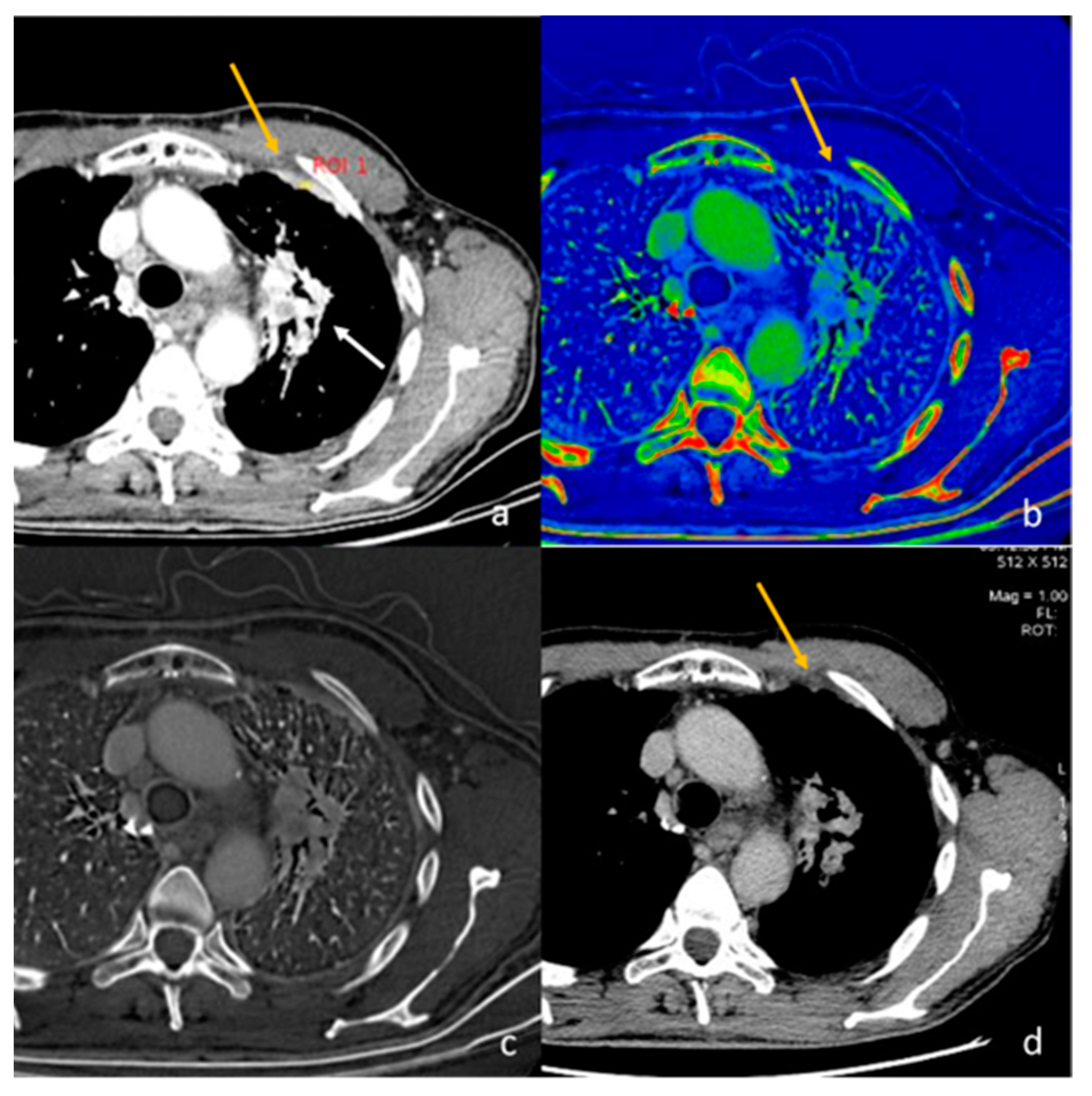
2.1. Pleura
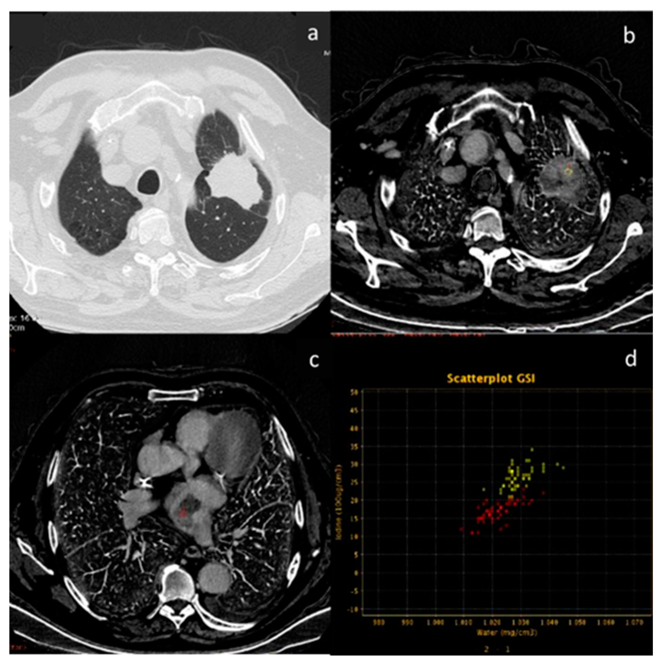
2.2. Lung
2.3. Breast
2.4. Lymph Nodes
2.5. Mediastinal Neoplasms
2.6. Esophagus
2.7. Vascular Emergency
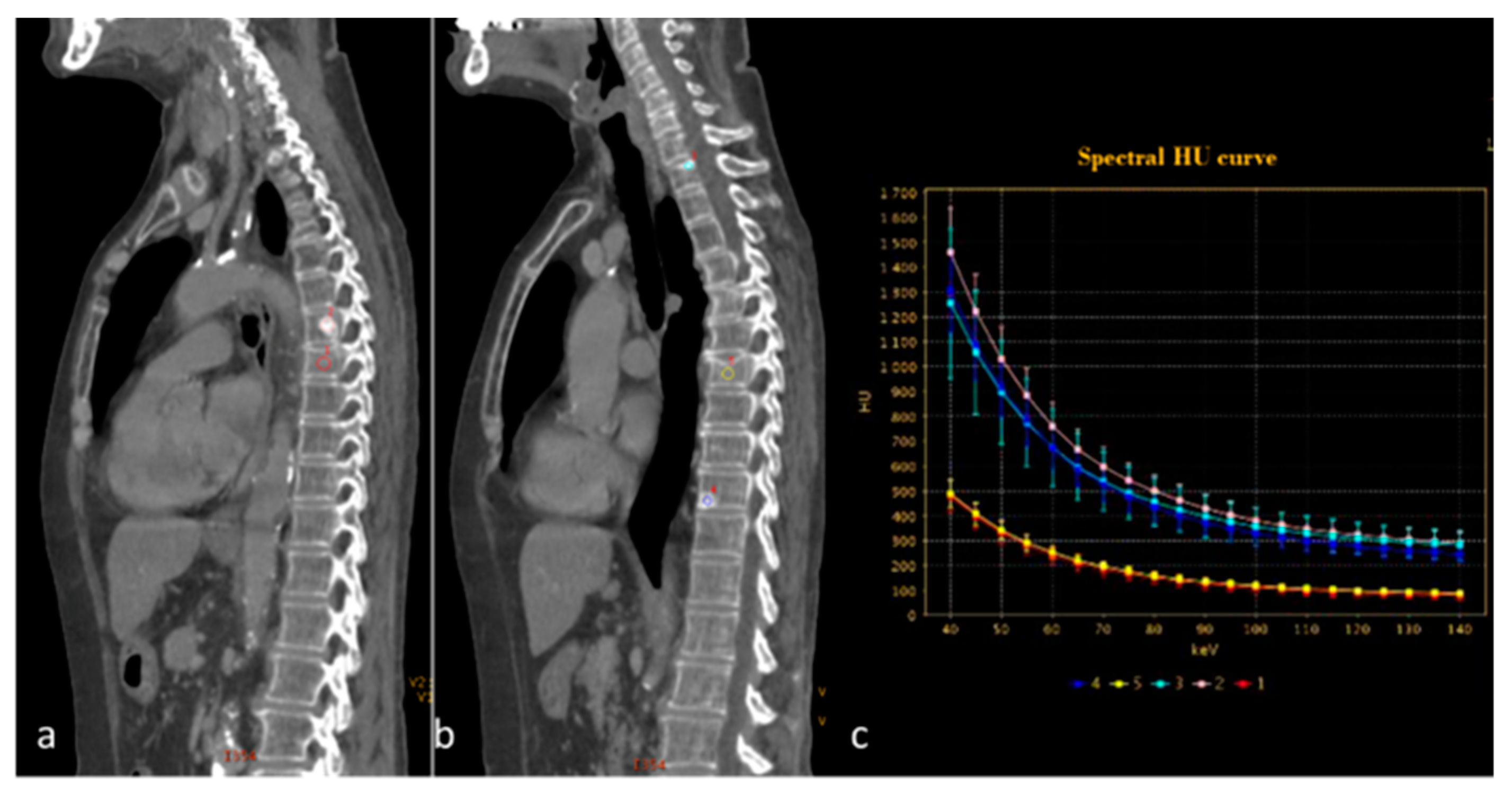
2.8. Bone
3. Conclusions
Author Contributions
Funding
Institutional Review Board Statement
Informed Consent Statement
Data Availability Statement
Conflicts of Interest
Abbreviations
| AUC | area under the receiver operating characteristic curve |
| CT | computed tomography |
| DECT | dual-energy CT |
| HU | Hounsfield unit |
| IC | iodine concentration |
| ILD | interstitial lung disease |
| keV | Kiloelettronvolt |
| λHU | slope |
| VMI | virtual monoenergetic images |
| VNC | virtual non-contrast |
| TNC | true non-contrast |
| Zeff | effective atomic number |
References
- Johnson, T.R.C.; Krauss, B.; Sedlmair, M.; Grasruck, M.; Bruder, H.; Morhard, D.; Fink, C.; Weckbach, S.; Lenhard, M.; Schmidt, B.; et al. Material differentiation by dual energy CT: Initial experience. Eur. Radiol. 2007, 17, 1510–1517. [Google Scholar] [CrossRef]
- Agostini, A.; Borgheresi, A.; Mari, A.; Floridi, C.; Bruno, F.; Carotti, M.; Schicchi, N.; Barile, A.; Maggi, S.; Giovagnoni, A. Dual-energy CT: Theoretical principles and clinical applications. Radiol. Med. 2019, 124, 1281–1295. [Google Scholar] [CrossRef]
- Carotti, M.; Salaffi, F.; Beci, G.; Giovagnoni, A. The application of dual-energy computed tomography in the diagnosis of musculoskeletal disorders: A review of current concepts and applications. Radiol. Med. 2019, 124, 1175–1183. [Google Scholar] [CrossRef]
- Biondi, M.; Vanzi, E.; De Otto, G.; Buonamici, F.B.; Belmonte, G.; Mazzoni, L.; Guasti, A.; Carbone, S.F.; Mazzei, M.A.; La Penna, A.; et al. Water/cortical bone decomposition: A new approach in dual energy CT imaging for bone marrow oedema detection. A feasibility study. Phys. Medica 2016, 32, 1712–1716. [Google Scholar] [CrossRef] [PubMed]
- Foley, W.D.; Shuman, W.P.; Siegel, M.J. White Paper of the Society of Computed Body Tomography and Magnetic Resonance on Dual-Energy CT, Part 2: Radiation Dose and Iodine Sensitivity. J. Comput. Assist Tomogr. 2016, 40, 846–850. [Google Scholar] [CrossRef] [PubMed]
- Graser, A.; Johnson, T.R.C.; Chandarana, H.; Macari, M. Dual energy CT: Preliminary observations and potential clinical applications in the abdomen. Eur. Radiol. 2008, 19, 13–23. [Google Scholar] [CrossRef] [PubMed]
- Brown, C.L.; Hartman, R.P.; Dzyubak, O.P.; Takahashi, N.; Kawashima, A.; McCollough, C.H.; Bruesewitz, M.R.; Primak, A.M.; Fletcher, J.G. Dual-energy CT iodine overlay technique for characterization of renal masses as cyst or solid: A phantom feasibility study. Eur. Radiol. 2009, 19, 1289–1295. [Google Scholar] [CrossRef] [PubMed]
- Agrawal, M.D.; Pinho, D.F.; Kulkarni, N.M.; Hahn, P.F.; Guimaraes, A.R.; Sahani, D.V. Oncologic Applications of Dual-Energy CT in the Abdomen. Radiographics 2014, 34, 589–612. [Google Scholar] [CrossRef]
- Volterrani, L.; Perrella, A.; Bagnacci, G.; Di Meglio, N.; Di Martino, V.; Bertelli, P.; Bellan, C.; Mazzei, M.A.; Luzzi, L. Washout-Computed Tomography Discriminates Pulmonary “Fat-poor” Hamartomas from Neuroendocrine Neoplasms: A Simple Method in the Radiomics Era. J. Thorac. Imaging, 2023; ahead of print. [Google Scholar] [CrossRef]
- Ueguchi, T.; Ogihara, R.; Yamada, S. Accuracy of Dual-Energy Virtual Monochromatic CT Numbers: Comparison between the single-source projection-based and dual-source image-based methods. Acad. Radiol. 2018, 25, 1632–1639. [Google Scholar] [CrossRef]
- Di Chiro, G.; Brooks, R.A.; Kessler, R.M.; Johnston, G.S.; Jones, A.E.; Herdt, J.R.; Sheridan, W.T. Tissue Signatures with Dual-Energy Computed Tomography. Radiology 1979, 131, 521–523. [Google Scholar] [CrossRef]
- Siegel, M.J.; Kaza, R.K.; Bolus, D.N. White Paper of the Society of Computed Body Tomography and Magnetic Resonance on Dual-Energy CT, Part 1: Technology and Terminology. J. Comput. Assist Tomogr. 2016, 40, 841–845. [Google Scholar] [CrossRef] [PubMed]
- Schenzle, J.C.; Sommer, W.H.; Neumaier, K.; Michalski, G.; Lechel, U.; Nikolaou, K.; Becker, C.R.; Reiser, M.F.; Johnson, T.R.C. Dual Energy CT of the Chest: How about the Dose? Investig. Radiol. 2010, 45, 347–353. [Google Scholar] [CrossRef] [PubMed]
- Hickethier, T.; Kroeger, J.R.; Lennartz, S. Venous-phase chest CT with reduced contrast medium dose: Utilization of spectral low KeV monoenergetic images improves image quality. Eur. J. Radiol. 2020, 122, 108756. [Google Scholar] [CrossRef] [Green Version]
- Jamali, S.; Michoux, N.; Coche, E. Virtual unenhanced phase with spectral dual-energy CT: Is it an alternative to conventional true unenhanced phase for abdominal tissues? Diagn. Interv. Imaging 2019, 100, 503–511. [Google Scholar] [CrossRef] [PubMed]
- Sugawara, H.; Takayanagi, T.; Ishikawa, T.; Katada, Y.; Fukui, R.; Yamamoto, Y.; Suzuki, S. New Fast kVp Switching Dual-Energy CT: Reduced Severity of Beam Hardening Artifacts and Improved Image Quality in Reduced-Iodine Virtual Monochromatic Imaging. Acad. Radiol. 2020, 27, 1586–1593. [Google Scholar] [CrossRef] [PubMed]
- Mellander, H.; Fransson, V.; Ydström, K.; Lätt, J.; Ullberg, T.; Wassélius, J.; Ramgren, B. Metal artifact reduction by virtual monoenergetic reconstructions from spectral brain CT. Eur. J. Radiol. Open 2023, 10, 100479. [Google Scholar] [CrossRef]
- McComiskey, D.M.; Erdenebold, U.-E.M.; McInnes, M.D.M.; Salameh, J.-P.M.; Chatelain, R.R.; Torres, C.M.; Chakraborty, S.M.; Zakhari, N.M. The Impact of Virtual Monoenergetic Imaging on Visualization of the Cervical Spinal Canal. J. Comput. Assist. Tomogr. 2023, 47, 160–164. [Google Scholar] [CrossRef]
- Parakh, A.; Lennartz, S.; An, C.; Rajiah, P.; Yeh, B.M.; Simeone, F.J.; Sahani, D.V.; Kambadakone, A.R. Dual-Energy CT Images: Pearls and Pitfalls. Radiographics 2021, 41, 98–119. [Google Scholar] [CrossRef]
- Cicero, G.; Ascenti, G.; Albrecht, M.H.; Blandino, A.; Cavallaro, M.; D’angelo, T.; Carerj, M.L.; Vogl, T.J.; Mazziotti, S. Extra-abdominal dual-energy CT applications: A comprehensive overview. Radiol. Med. 2020, 125, 384–397. [Google Scholar] [CrossRef]
- Bottari, A.; Silipigni, S.; Carerj, M.L.; Cattafi, A.; Maimone, S.; Marino, M.A.; Mazziotti, S.; Pitrone, A.; Squadrito, G.; Ascenti, G. Dual-source dual-energy CT in the evaluation of hepatic fractional extracellular space in cirrhosis. Radiol. Med. 2019, 125, 7–14. [Google Scholar] [CrossRef]
- Mazzei, M.A.; Gentili, F.; Volterrani, L. Dual-Energy CT Iodine Mapping and 40-keV Monoenergetic Applications in the Diagnosis of Acute Bowel Ischemia: A Necessary Clarification. AJR Am. J. Roentgenol. 2019, 212, W93–W94. [Google Scholar] [CrossRef]
- Mileto, A.; Marin, D.; Alfaro-Cordoba, M. Iodine quantification to distinguish clear cell from papillary renal cell carcinoma at dual-energy multidetector CT: A multireader diagnostic performance study. Radiology 2014, 273, 813–820. [Google Scholar] [CrossRef] [PubMed]
- Agostini, A.; Mari, A.; Lanza, C.; Schicchi, N.; Borgheresi, A.; Maggi, S.; Giovagnoni, A. Trends in radiation dose and image quality for pediatric patients with a multidetector CT and a third-generation dual-source dual-energy CT. Radiol. Med. 2019, 124, 745–752. [Google Scholar] [CrossRef] [PubMed]
- Yoshida, R.; Usui, K.; Katsunuma, Y.; Honda, H.; Hatakeyama, K. Reducing contrast dose using virtual monoenergetic imaging for aortic CTA. J. Appl. Clin. Med. Phys. 2020, 21, 272–277. [Google Scholar] [CrossRef] [PubMed]
- Foti, G.; Beltramello, A.; Minerva, G.; Catania, M.; Guerriero, M.; Albanese, S.; Carbognin, G. Identification of residual-recurrent cholesteatoma in operated ears: Diagnostic accuracy of dual-energy CT and MRI. Radiol. Med. 2019, 124, 478–486. [Google Scholar] [CrossRef]
- Chawla, A.; Srinivasan, S.; Lim, T.C.; Pulickal, G.G.; Shenoy, J.; Peh, W.C.G. Dual-energy CT applications in salivary gland lesions. Br. J. Radiol. 2017, 90, 20160859. [Google Scholar] [CrossRef]
- Helck, A.; Hummel, N.; Meinel, F.G. Can single-phase dual-energy CT reliably identify adrenal adenomas? Eur. Radiol. 2014, 24, 1636–1642. [Google Scholar] [CrossRef]
- Botsikas, D.; Triponez, F.; Boudabbous, S. Incidental adrenal lesions detected on enhanced abdominal dual-energy CT: Can the diagnostic workup be shortened by the implementation of virtual unenhanced images? Eur. J. Radiol. 2014, 83, 1746–1751. [Google Scholar] [CrossRef]
- Gupta, R.T.; Ho, L.M.; Marin, D.; Boll, D.T.; Barnhart, H.X.; Nelson, R.C. Dual-Energy CT for Characterization of Adrenal Nodules: Initial Experience. Am. J. Roentgenol. 2010, 194, 1479–1483. [Google Scholar] [CrossRef] [PubMed]
- El Kayal, N.; Lennartz, S.; Ekdawi, S.; Holz, J.; Slebocki, K.; Haneder, S.; Wybranski, C.; Mohallel, A.; Eid, M.; Grüll, H.; et al. Value of spectral detector computed tomography for assessment of pancreatic lesions. Eur. J. Radiol. 2019, 118, 215–222. [Google Scholar] [CrossRef]
- Bao, J.; Liu, A.; Zhao, C. Correlation Between Dual-Energy Computed Tomography Single Scan and Computed Tomography Perfusion for Pancreatic Cancer Patients: Initial Experience. J. Comput. Assist Tomogr. 2019, 43, 599–604. [Google Scholar] [CrossRef] [PubMed]
- Beer, L.; Toepker, M.; Ba-Ssalamah, A.; Schestak, C.; Dutschke, A.; Schindl, M.; Wressnegger, A.; Ringl, H.; Apfaltrer, P. Objective and subjective comparison of virtual monoenergetic vs. polychromatic images in patients with pancreatic ductal adenocarcinoma. Eur. Radiol. 2019, 29, 3617–3625. [Google Scholar] [CrossRef] [PubMed] [Green Version]
- Di Maso, L.D.; Huang, J.; Bassetti, M.F.; DeWerd, L.A.; Miller, J.R. Investigating a novel split-filter dual-energy CT technique for improving pancreas tumor visibility for radiation therapy. J. Appl. Clin. Med. Phys. 2018, 19, 676–683. [Google Scholar] [CrossRef] [PubMed] [Green Version]
- Guerrini, S.; Bagnacci, G.; Perrella, A.; Di Meglio, N.; Sica, C.; Mazzei, M.A. Dual Energy CT in Oncology: Benefits for Both Patients and Radiologists From an Emerging Quantitative and Functional Diagnostic Technique. Semin. Ultrasound CT MRI 2023, 44, 205–213. [Google Scholar] [CrossRef]
- Morgan, D.E. Dual-energy CT of the abdomen. Abdom. Imaging 2014, 39, 108–134. [Google Scholar] [CrossRef]
- Yang, F.; Dong, J.; Wang, X.; Fu, X.; Zhang, T. Non-small cell lung cancer: Spectral computed tomography quantitative parameters for preoperative diagnosis of metastatic lymph nodes. Eur. J. Radiol. 2017, 89, 129–135. [Google Scholar] [CrossRef]
- Deniffel, D.; Sauter, A.; Dangelmaier, J.; Fingerle, A.; Rummeny, E.J.; Pfeiffer, D. Differentiating intrapulmonary metastases from different primary tumors via quantitative dual-energy CT based iodine concentration and conventional CT attenuation. Eur. J. Radiol. 2019, 111, 6–13. [Google Scholar] [CrossRef]
- Lee, J.-A.; Jeong, W.K.; Kim, Y.; Song, S.-Y.; Kim, J.; Heo, J.N.; Park, C.K. Dual-energy CT to detect recurrent HCC after TACE: Initial experience of color-coded iodine CT imaging. Eur. J. Radiol. 2013, 82, 569–576. [Google Scholar] [CrossRef]
- Apfaltrer, P.; Meyer, M.B.; Meier, C.B.; Henzler, T.; Barraza, J.M.J.B.; Dinter, D.J.; Hohenberger, P.; Schoepf, U.J.; Schoenberg, S.O.; Fink, C. Contrast-enhanced dual-energy CT of gastrointestinal stromal tumors: Is iodine-related attenuation a potential indicator of tumor response? Investig. Radiol. 2012, 47, 65–70. [Google Scholar] [CrossRef]
- Lv, P.; Liu, J.; Yan, X.; Chai, Y.; Chen, Y.; Gao, J.; Pan, Y.; Li, S.; Guo, H.; Zhou, Y. CT spectral imaging for monitoring the therapeutic efficacy of VEGF receptor kinase inhibitor AG-013736 in rabbit VX2 liver tumours. Eur. Radiol. 2017, 27, 918–926. [Google Scholar] [CrossRef]
- Vlahos, I.; Jacobsen, M.C.; Godoy, M.C.; Stefanidis, K.; Layman, R.R. Dual-energy CT in pulmonary vascular disease. Br. J. Radiol. 2022, 95, 20210699. [Google Scholar] [CrossRef]
- Rajiah, P.; Tanabe, Y.; Partovi, S.; Moore, A. State of the art: Utility of multi-energy CT in the evaluation of pulmonary vasculature. Int. J. Cardiovasc. Imaging 2019, 35, 1509–1524. [Google Scholar] [CrossRef]
- Lefebvre, B.; Kyheng, M.; Giordano, J.; Lamblin, N.; de Groote, P.; Fertin, M.; Delobelle, M.; Perez, T.; Faivre, J.-B.; Remy, J.; et al. Dual-energy CT lung perfusion characteristics in pulmonary arterial hypertension (PAH) and pulmonary veno-occlusive disease and/or pulmonary capillary hemangiomatosis (PVOD/PCH): Preliminary experience in 63 patients. Eur. Radiol. 2022, 32, 4574–4586. [Google Scholar] [CrossRef] [PubMed]
- Masy, M.; Giordano, J.; Petyt, G.; Hossein-Foucher, C.; Duhamel, A.; Kyheng, M.; De Groote, P.; Fertin, M.; Lamblin, N.; Bervar, J.-F.; et al. Dual-energy CT (DECT) lung perfusion in pulmonary hypertension: Concordance rate with V/Q scintigraphy in diagnosing chronic thromboembolic pulmonary hypertension (CTEPH). Eur. Radiol. 2018, 28, 5100–5110. [Google Scholar] [CrossRef] [PubMed]
- Lennartz, S.; Le Blanc, M.; Zopfs, D.; Hokamp, N.G.; Abdullayev, N.; Laukamp, K.R.; Haneder, S.; Borggrefe, J.; Maintz, D.; Persigehl, T. Dual-Energy CT-derived Iodine Maps: Use in Assessing Pleural Carcinomatosis. Radiology 2019, 290, 796–804. [Google Scholar] [CrossRef]
- Zhang, X.; Duan, H.; Yu, Y.; Ma, C.; Ren, Z.; Lei, Y.; He, T.; Zhang, M. Differential diagnosis between benign and malignant pleural effusion with dual-energy spectral CT. PLoS ONE 2018, 13, e0193714. [Google Scholar] [CrossRef] [PubMed] [Green Version]
- Sato, Y.; Ishiyama, M.; Nakano, S.; Nakao, M.; Mun, M.; Ninomiya, H.; Terauchi, T.; Oikado, K. Ringlike Peripheral Increased Iodine Concentration for the Differentiation of Primary Lung Cancer and Pulmonary Metastases on Contrast-Enhanced Dual-Energy CT. AJR Am. J. Roentgenol. 2023, 220, 828–837. [Google Scholar] [CrossRef] [PubMed]
- He, C.; Liu, J.; Li, Y.; Lin, L.; Qing, H.; Guo, L.; Hu, S.; Zhou, P. Quantitative parameters of enhanced dual-energy computed tomography for differentiating lung cancers from benign lesions in solid pulmonary nodules. Front. Oncol. 2022, 12, 1027985. [Google Scholar] [CrossRef]
- Yan, G.; Li, H.; Fan, X.; Deng, J.; Yan, J.; Qiao, F.; Yan, G.; Liu, T.; Chen, J.; Wang, L.; et al. Multimodality CT imaging contributes to improving the diagnostic accuracy of solitary pulmonary nodules: A multi-institutional and prospective study. Radiol. Oncol. 2023, 57, 20–34. [Google Scholar] [CrossRef]
- Ha, T.; Kim, W.; Cha, J.; Lee, Y.H.; Seo, H.S.; Park, S.Y.; Kim, N.H.; Hwang, S.H.; Yong, H.S.; Oh, Y.-W.; et al. Differentiating pulmonary metastasis from benign lung nodules in thyroid cancer patients using dual-energy CT parameters. Eur. Radiol. 2022, 32, 1902–1911. [Google Scholar] [CrossRef]
- Choe, J.; Lee, S.M.; Do, K.-H.; Lee, J.B.; Lee, J.-G.; Seo, J.B. Prognostic value of radiomic analysis of iodine overlay maps from dual-energy computed tomography in patients with resectable lung cancer. Eur. Radiol. 2019, 29, 915–923. [Google Scholar] [CrossRef]
- Hagen, F.; Walder, L.; Fritz, J.; Gutjahr, R.; Schmidt, B.; Faby, S.; Bamberg, F.; Schoenberg, S.; Nikolaou, K.; Horger, M. Image Quality and Radiation Dose of Contrast-Enhanced Chest-CT Acquired on a Clinical Photon-Counting Detector CT vs. Second-Generation Dual-Source CT in an Oncologic Cohort: Preliminary Results. Tomography 2022, 8, 1466–1476. [Google Scholar] [CrossRef]
- Chen, L.; Zhu, M.; Lu, H.; Yang, T.; Li, W.; Zhang, Y.; Xie, Q.; Li, Z.; Wan, H.; Luo, F. Quantitative evaluation of disease severity in connective tissue disease-associated interstitial lung disease by dual-energy computed tomography. Respir. Res. 2022, 23, 47. [Google Scholar] [CrossRef] [PubMed]
- Scharm, S.C.; Vogel-Claussen, J.; Schaefer-Prokop, C.; Dettmer, S.; Knudsen, L.; Jonigk, D.; Fuge, J.; Apel, R.-M.; Welte, T.; Wacker, F.; et al. Quantification of dual-energy CT-derived functional parameters as potential imaging markers for progression of idiopathic pulmonary fibrosis. Eur. Radiol. 2021, 31, 6640–6651. [Google Scholar] [CrossRef]
- Wang, X.; Liu, D.; Jiang, S.; Zeng, X.; Li, L.; Yu, T.; Zhang, J. Subjective and Objective Assessment of Monoenergetic and Polyenergetic Images Acquired by Dual-Energy CT in Breast Cancer. Korean J. Radiol. 2021, 22, 502–512. [Google Scholar] [CrossRef]
- Volterrani, L.; Gentili, F.; Fausto, A.; Pelini, V.; Megha, T.; Sardanelli, F.; Mazzei, M.A. Dual-Energy CT for Locoregional Staging of Breast Cancer: Preliminary Results. Am. J. Roentgenol. 2020, 214, 707–714. [Google Scholar] [CrossRef] [PubMed]
- Moon, J.I.; Choi, B.H.; Baek, H.J.; Ryu, K.H.; Park, S.E.; Ha, J.Y.; Jung, E.J.; Lee, H.S.; An, H.J. Comprehensive analyses with radiological and biological markers of breast cancer on contrast-enhanced chest CT: A single center experience using dual-layer spectral detector CT. Eur. Radiol. 2020, 30, 2782–2790. [Google Scholar] [CrossRef]
- Lenga, L.; Bernatz, S.; Martin, S.S.; Booz, C.; Solbach, C.; Mulert-Ernst, R.; Vogl, T.J.; Leithner, D. Iodine Map Radiomics in Breast Cancer: Prediction of Metastatic Status. Cancers 2021, 13, 2431. [Google Scholar] [CrossRef]
- Zhang, X.; Zheng, C.; Yang, Z.; Cheng, Z.; Deng, H.; Chen, M.; Duan, X.; Mao, J.; Shen, J. Axillary Sentinel Lymph Nodes in Breast Cancer: Quantitative Evaluation at Dual-Energy CT. Radiology 2018, 289, 337–346. [Google Scholar] [CrossRef] [PubMed]
- Terada, K.; Kawashima, H.; Yoneda, N.; Toshima, F.; Hirata, M.; Kobayashi, S.; Gabata, T. Predicting axillary lymph node metastasis in breast cancer using the similarity of quantitative dual-energy CT parameters between the primary lesion and axillary lymph node. Jpn. J. Radiol. 2022, 40, 1272–1281. [Google Scholar] [CrossRef] [PubMed]
- Nagano, H.; Takumi, K.; Nakajo, M.; Fukukura, Y.; Kumagae, Y.; Jinguji, M.; Tani, A.; Yoshiura, T. Dual-Energy CT-Derived Electron Density for Diagnosing Metastatic Mediastinal Lymph Nodes in Non-Small Cell Lung Cancer: Comparison with Conventional CT and FDG PET/CT Findings. AJR Am. J. Roentgenol. 2022, 218, 66–74. [Google Scholar] [CrossRef] [PubMed]
- Chang, S.; Hur, J.; Im, D.J.; Suh, Y.J.; Hong, Y.J.; Lee, H.-J.; Kim, Y.J.; Han, K.; Kim, D.J.; Lee, C.Y.; et al. Volume-based quantification using dual-energy computed tomography in the differentiation of thymic epithelial tumours: An initial experience. Eur. Radiol. 2017, 27, 1992–2001. [Google Scholar] [CrossRef]
- Zhou, Q.; Huang, X.; Xie, Y.; Liu, X.; Li, S.; Zhou, J. Role of quantitative energy spectrum CT parameters in differentiating thymic epithelial tumours and thymic cysts. Clin. Radiol. 2022, 77, 136–141. [Google Scholar] [CrossRef] [PubMed]
- Cheng, F.; Liu, Y.; Du, L.; Wang, L.; Li, L.; Shi, J.; Wang, X.; Zhang, J. Evaluation of optimal monoenergetic images acquired by dual-energy CT in the diagnosis of T staging of thoracic esophageal cancer. Insights into Imaging 2023, 14, 33. [Google Scholar] [CrossRef]
- Zopfs, D.; Hokamp, N.G.; Reimer, R.; Bratke, G.; Maintz, D.; Bruns, C.; Mallmann, C.; Persigehl, T.; Haneder, S.; Lennartz, S. Value of spectral detector CT for pretherapeutic, locoregional assessment of esophageal cancer. Eur. J. Radiol. 2021, 134, 109423. [Google Scholar] [CrossRef] [PubMed]
- Si-Mohamed, S.; Dupuis, N.; Tatard-Leitman, V.; Rotzinger, D.; Boccalini, S.; Dion, M.; Vlassenbroek, A.; Coulon, P.; Yagil, Y.; Shapira, N.; et al. Virtual versus true non-contrast dual-energy CT imaging for the diagnosis of aortic intramural hematoma. Eur. Radiol. 2019, 29, 6762–6771. [Google Scholar] [CrossRef] [PubMed]
- Perez-Johnston, R.; Plodkowski, A.J.; Halpenny, D.F.; Hayes, S.A.; Capanu, M.; Araujo-Filho, J.A.B.; Weinsaft, J.W.; Ginsberg, M.S. Perfusion defects on dual-energy CTA in patients with suspected pulmonary embolism correlate with right heart strain and lower survival. Eur. Radiol. 2021, 31, 2013–2021. [Google Scholar] [CrossRef] [PubMed]
- Abdellatif, W.; Ebada, M.A.; Alkanj, S.; Negida, A.; Murray, N.; Khosa, F.; Nicolaou, S. Diagnostic Accuracy of Dual-Energy CT in Detection of Acute Pulmonary Embolism: A Systematic Review and Meta-Analysis. Can. Assoc. Radiol. J. 2021, 72, 285–292. [Google Scholar] [CrossRef] [Green Version]
- Monti, C.B.; Zanardo, M.; Cozzi, A.; Schiaffino, S.; Spagnolo, P.; Secchi, F.; De Cecco, C.N.; Sardanelli, F. Dual-energy CT performance in acute pulmonary embolism: A meta-analysis. Eur. Radiol. 2021, 31, 6248–6258. [Google Scholar] [CrossRef]
- Flors, L.; Leiva-Salinas, C.; Norton, P.T.; Patrie, J.T.; Hagspiel, K.D. Endoleak Detection After Endovascular Repair of Thoracic Aortic Aneurysm Using Dual-Source Dual-Energy CT: Suitable Scanning Protocols and Potential Radiation Dose Reduction. AJR Am. J. Roentgenol. 2013, 200, 451–460. [Google Scholar] [CrossRef]
- Shuman, W.P.; O’Malley, R.B.; Busey, J.M. Prospective comparison of dual-energy CT aortography using 70% reduced iodine dose versus single-energy CT aortography using standard iodine dose in the same patient. Abdom. Radiol. 2017, 42, 759–765. [Google Scholar] [CrossRef] [PubMed]
- Kosmala, A.; Weng, A.; Heidemeier, A.; Krauss, B.; Knop, S.; Bley, T.A.; Petritsch, B. Multiple Myeloma and Dual-Energy CT: Diagnostic Accuracy of Virtual Noncalcium Technique for Detection of Bone Marrow Infiltration of the Spine and Pelvis. Radiology 2018, 286, 205–213. [Google Scholar] [CrossRef] [PubMed]
- Kosmala, A.; Weng, A.M.; Krauss, B.; Knop, S.; Bley, T.A.; Petritsch, B. Dual-energy CT of the bone marrow in multiple myeloma: Diagnostic accuracy for quantitative differentiation of infiltration patterns. Eur. Radiol. 2018, 28, 5083–5090. [Google Scholar] [CrossRef] [PubMed]
- Ishiwata, Y.; Hieda, Y.; Kaki, S.; Aso, S.; Horie, K.; Kobayashi, Y.; Nakamura, M.; Yamada, K.; Yamashiro, T.; Utsunomiya, D. Improved Diagnostic Accuracy of Bone Metastasis Detection by Water-HAP Associated to Non-contrast CT. Diagnostics 2020, 10, 853. [Google Scholar] [CrossRef] [PubMed]
- Borggrefe, J.; Neuhaus, V.-F.; Le Blanc, M.; Hokamp, N.G.; Maus, V.; Mpotsaris, A.; Lennartz, S.; dos Santos, D.P.; Maintz, D.; Abdullayev, N. Accuracy of iodine density thresholds for the separation of vertebral bone metastases from healthy-appearing trabecular bone in spectral detector computed tomography. Eur. Radiol. 2018, 29, 3253–3261. [Google Scholar] [CrossRef] [PubMed]
- Huang, H.-C.; Srinivasan, R.; Sun, Y.; Kazakia, G.J.; Lin, P.-C.; Yeh, B.M. Detection of Lumbar Spine Osseous Metastases Using Dual-Energy CT: Phantom Results and Preliminary Clinical Validation. AJR Am. J. Roentgenol. 2019, 212, 402–410. [Google Scholar] [CrossRef]
- Siegel, M.J.; Ramirez-Giraldo, J.C. Dual-Energy CT in Children: Imaging Algorithms and Clinical Applications. Radiology 2019, 291, 286–297. [Google Scholar] [CrossRef]
- Ren, Y.; Jiao, Y.; Ge, W.; Zhang, L.; Hua, Y.; Li, C.; Zhai, W.; Tang, X.; He, W.; Fang, M.; et al. Dual-Energy Computed Tomography-Based Iodine Quantitation for Response Evaluation of Lung Cancers to Chemoradiotherapy/Radiotherapy: A Comparison with Fluorine-18 Fluorodeoxyglucose Positron Emission Tomography/Computed Tomography-Based Positron Emission Tomography/Computed Tomography Response Evaluation Criterion in Solid Tumors. J. Comput. Assist. Tomogr. 2018, 42, 614–622. [Google Scholar] [CrossRef]
- Hou, W.S.; Wu, H.W.; Yin, Y.; Cheng, J.J.; Zhang, Q.; Xu, J.R. Differentiation of Lung Cancers From Inflammatory Masses with Dual-Energy Spectral CT Imaging. Acad. Radiol. 2015, 22, 337–344. [Google Scholar] [CrossRef]
- Zhang, Y.; Cheng, J.; Hua, X.; Yu, M.; Xu, C.; Zhang, F.; Xu, J.; Wu, H. Can Spectral CT Imaging Improve the Differentiation between Malignant and Benign Solitary Pulmonary Nodules? PLoS ONE 2016, 11, e0147537. [Google Scholar] [CrossRef]
- Baxa, J.; Vondráková, A.; Matoušková, T.; Růžičková, O.; Schmidt, B.; Flohr, T.; Sedlmair, M.; Ferda, J. Dual-phase dual-energy CT in patients with lung cancer: Assessment of the additional value of iodine quantification in lymph node therapy response. Eur. Radiol. 2014, 24, 1981–1988. [Google Scholar] [CrossRef]
- Siegel, M.J.; Bhalla, S.; Cullinane, M. Dual-Energy CT Material Decomposition in Pediatric Thoracic Oncology. Radiol. Imaging Cancer 2021, 3, e200097. [Google Scholar] [CrossRef] [PubMed]
- Dewaguet, J.; Copin, M.-C.; Duhamel, A.; Faivre, J.-B.; Deken, V.; Sedlmair, M.; Flohr, T.; Schmidt, B.; Cortot, A.; Wasielewski, E.; et al. Dual-Energy CT Perfusion of Invasive Tumor Front in Non-Small Cell Lung Cancers. Radiology 2022, 302, 448–456. [Google Scholar] [CrossRef] [PubMed]
- Yuan, X.; Zhang, J.; Quan, C.; Cao, J.; Ao, G.; Tian, Y.; Li, H. Differentiation of malignant and benign pulmonary nodules with first-pass dual-input perfusion CT. Eur. Radiol. 2013, 23, 2469–2474. [Google Scholar] [CrossRef] [PubMed]
- Inoue, T.; Nakaura, T.; Iyama, A.; Kidoh, M.; Nagayama, Y.; Uetani, H.; Oda, S.; Utsunomiya, D.; Yamashita, Y. Usefulness of Virtual Monochromatic Dual-Layer Computed Tomographic Imaging for Breast Carcinoma. J. Comput. Assist. Tomogr. 2020, 44, 78–82. [Google Scholar] [CrossRef]
- Metin, Y.; Metin, N.O.; Özdemir, O.; Taşçı, F.; Kul, S. The role of low keV virtual monochromatic imaging in increasing the conspicuity of primary breast cancer in dual-energy spectral thoracic CT examination for staging purposes. Acta Radiol. 2020, 61, 168–174. [Google Scholar] [CrossRef]
- Li, J.-X.; Xie, F.-J.; Chen, C.-H.; Chen, K.-M.; Tsai, C.-J. Dual-Energy Computed Tomography for Evaluation of Breast Cancer Follow-Ups: Comparison of Virtual Monoenergetic Images and Iodine-Map. Diagnostics 2022, 12, 946. [Google Scholar] [CrossRef]
- Li, H.; Wang, H.; Chen, F.; Gao, L.; Zhou, Y.; Zhou, Z.; Huang, J.; Xu, L. Detection of axillary lymph node metastasis in breast cancer using dual-layer spectral computed tomography. Front. Oncol. 2022, 12, 967655. [Google Scholar] [CrossRef]
- Volterrani, L.; Mazzei, M.A.; Banchi, B.; Voltolini, L.; La Sala, F.; Carbone, S.F.; Ricci, V.; Gotti, G.; Zompatori, M. MSCT multi-criteria: A novel approach in assessment of mediastinal lymph node metastases in non-small cell lung cancer. Eur. J. Radiol. 2011, 79, 459–466. [Google Scholar] [CrossRef]
- Sekiguchi, T.; Ozawa, Y.; Hara, M.; Nakagawa, M.; Goto, T.; Shibamoto, Y. Visibility of the hilar lymph nodes using advanced virtual monoenergetic low-KeV images for preoperative evaluation of lung cancer. Br. J. Radiol. 2019, 92, 20180734. [Google Scholar] [CrossRef]
- Khandelwal, A.; Sholl, L.; Araki, T.; Ramaiya, N.; Hatabu, H.; Nishino, M. Patterns of metastasis and recurrence in thymic epithelial tumours: Longitudinal imaging review in correlation with histological subtypes. Clin. Radiol. 2016, 71, 1010–1017. [Google Scholar] [CrossRef] [Green Version]
- Gentili, F.; Pelini, V.; Lucii, G.; Luzzi, L.; Mazzei, F.G.; Fausto, A.; Volterrani, L.; Mazzei, M.A. Update in diagnostic imaging of the thymus and anterior mediastinal masses. Gland. Surg. 2019, 8 (Suppl. 3), S188–S207. [Google Scholar] [CrossRef]
- Gentili, F.; Monteleone, I.; Mazzei, F.G.; Luzzi, L.; Del Roscio, D.; Guerrini, S.; Volterrani, L.; Mazzei, M.A. Advancement in Diagnostic Imaging of Thymic Tumors. Cancers 2021, 13, 3599. [Google Scholar] [CrossRef] [PubMed]
- Gentili, F.; Guerrini, S.; Mazzei, F.G.; Monteleone, I.; Di Meglio, N.; Sansotta, L.; Perrella, A.; Puglisi, S.; De Filippo, M.; Gennaro, P.; et al. Dual energy CT in gland tumors: A comprehensive narrative review and differential diagnosis. Gland. Surg. 2020, 9, 2269–2282. [Google Scholar] [CrossRef]
- Yan, W.-Q.; Xin, Y.-K.; Jing, Y.; Li, G.-F.; Wang, S.-M.; Rong, W.-C.; Xiao, G.; Lei, X.-B.; Li, B.; Hu, Y.-C.; et al. Iodine Quantification Using Dual-Energy Computed Tomography for Differentiating Thymic Tumors. J. Comput. Assist. Tomogr. 2018, 42, 873–880. [Google Scholar] [CrossRef]
- Yu, C.; Li, T.; Zhang, R.; Yang, X.; Yang, Z.; Xin, L.; Zhao, Z. Dual-energy CT perfusion imaging for differentiating WHO subtypes of thymic epithelial tumors. Sci. Rep. 2020, 10, 5511, Erratum in 2020, 10, 7493. [Google Scholar] [CrossRef] [Green Version]
- Rajamohan, N.; Goyal, A.; Kandasamy, D.; Bhalla, A.S.; Parshad, R.; Jain, D.; Sharma, R. CT texture analysis in evaluation of thymic tumors and thymic hyperplasia: Correlation with the international thymic malignancy interest group (ITMIG) stage and WHO grade. Br. J. Radiol. 2021, 94, 20210583. [Google Scholar] [CrossRef]
- Vulasala, S.S.R.; Wynn, G.C.; Hernandez, M.; Kadambi, I.; Gopireddy, D.R.; Bhosale, P.; Virarkar, M.K. Dual-Energy Imaging of the Chest. Semin. Ultrasound CT MRI 2022, 43, 311–319. [Google Scholar] [CrossRef]
- Hong, Y.J.; Shim, J.; Lee, S.M.; Im, D.J.; Hur, J. Dual-Energy CT for Pulmonary Embolism: Current and Evolving Clinical Applications. Korean J. Radiol. 2021, 22, 1555–1568. [Google Scholar] [CrossRef] [PubMed]
- Okamura-Kawasaki, M.; Uesugi, Y.; Yabusaki, S. Dual-energy CT for gastrointestinal bleeding. BJR Open 2023, 5, 20220054. [Google Scholar] [CrossRef] [PubMed]
- Poschenrieder, F.; Meiler, S.; Lubnow, M.; Zeman, F.; Rennert, J.; Scharf, G.; Schaible, J.; Stroszczynski, C.; Pfeifer, M.; Hamer, O.W. Severe COVID-19 pneumonia: Perfusion analysis in correlation with pulmonary embolism and vessel enlargement using dual-energy CT data. PLoS ONE 2021, 16, e0252478. [Google Scholar] [CrossRef] [PubMed]
- Idilman, I.S.; Dizman, G.T.; Duzgun, S.A.; Irmak, I.; Karcaaltincaba, M.; Inkaya, A.C.; Demirkazik, F.; Durhan, G.; Akpinar, M.G.; Ariyurek, O.M.; et al. Lung and kidney perfusion deficits diagnosed by dual-energy computed tomography in patients with COVID-19-related systemic microangiopathy. Eur. Radiol. 2021, 31, 1090–1099. [Google Scholar] [CrossRef]
- Huang, F.; Wu, H.; Lai, Q.-Q.; Ke, X.-T. Application value of preoperative dual-source computed tomography in assessing the rupture site of thoracic aortic dissection. J. Cardiothorac. Surg. 2021, 16, 346. [Google Scholar] [CrossRef] [PubMed]
- Behrendt, F.F.; Schmidt, B.; Plumhans, C.; Keil, S.; Woodruff, S.G.; Ackermann, D.; Mühlenbruch, G.; Flohr, T.; Günther, R.W.; Mahnken, A.H. Image fusion in dual energy computed tomography: Effect on contrast enhancement, signal-to-noise ratio and image quality in computed tomography angiography. Investig. Radiol. 2009, 44, 1–6. [Google Scholar] [CrossRef] [PubMed]
- Li, W.; Liu, M.; Yu, F.; Zhu, W.; Yu, X.; Guo, X.; Yang, Q. Detection of left atrial appendage thrombus by dual-energy computed tomography-derived imaging biomarkers in patients with atrial fibrillation. Front. Cardiovasc. Med. 2022, 9, 809688. [Google Scholar] [CrossRef] [PubMed]
- Ascenti, G.; Sofia, C.; Mazziotti, S.; Silipigni, S.; D’Angelo, T.; Pergolizzi, S.; Scribano, E. Dual-energy CT with iodine quantification in distinguishing between bland and neoplastic portal vein thrombosis in patients with hepatocellular carcinoma. Clin. Radiol. 2016, 71, 938.e1–938.e9. [Google Scholar] [CrossRef]
- Yue, N.; Xin, W.R.; Jing, C.; Rong, C.F.; Fei, S.L.; Lian, L.A.; Hong, L.Y. Virtual monochromatic spectral imaging for the evaluation of vertebral inconspicuous osteoblastic metastases from lung. Acta Radiol. 2017, 58, 1485–1492. [Google Scholar] [CrossRef]
- Tan, M.T.; Lloyd, T.B. Utility of dual energy computed tomography in the evaluation of infiltrative skeletal lesions and metastasis: A literature review. Skelet. Radiol. 2022, 51, 1731–1741. [Google Scholar] [CrossRef]
- Issa, G.; Davis, D.; Mulligan, M.E. The Ability of Dual-Energy Computed Tomography to Distinguish Normal Bone Marrow From Metastases Using Bone Marrow Color Maps. J. Comput. Assist. Tomogr. 2018, 42, 552–558. [Google Scholar] [CrossRef]


| Items | |
|---|---|
| Date of search | 15/01/2023 |
| Sources | Pubmed, Cochrane |
| Search terms | (dual energy CT OR dual-energy CT OR dual-energy computed tomography OR dual-energy computed tomography OR DECT) and (pleura OR lung OR breast OR mediastinum OR thymus OR embolism OR bone metastasis OR esophagus OR esophageal cancer) |
| Timeframe | No restrictions |
| Inclusion criteria | Articles published in English |
| Selection process | Two authors selected articles according to the following criteria: priority has been given to meta-analysis, systematic reviews, and original articles. Only original articles with diagnostic accuracy or specific dual energy measures (e.g., iodine concentration) reported in the results were included. Narrative reviews were reported only if a few original articles and no systematic reviews or meta-analysis were available about oncologic thoracic diseases. |
| Authors and Study Design | Country | Aim/Rationale | No. of Patients | DECT Scan and Conflict of Interests | Main Conclusion |
|---|---|---|---|---|---|
| Lennartz et al. [46] Retrospective study | Germany, USA | To evaluate the use of spectral CT for differentiation between noncalcified benign pleural lesions and pleural carcinomatosis | 84 | IQon; Philips Healthcare, Best, the Netherlands No C.I. | Iodine overlay images and quantitative iodine maps improve the differentiation of noncalcified benign pleural lesions from pleural carcinomatosis compared with conventional CT. The benefit of spectral CT for diagnosis of pleural lesions was greater for less-experienced radiologists compared with experienced radiologists. |
| Zhang et al. [47] Retrospective study | China | To investigate the value of spectral CT in the differential diagnosis of benign from malignant pleural effusion | 29 | Discovery CT750 HD; GE Healthcare No C.I. | The CT value measurement at both high and low energy levels and the effective atomic number obtained in a single spectral CT scan can assist the differential diagnosis of benign from malignant pleural effusion. Combining them with patient age and disease history can further improve diagnostic accuracy. |
| Sato et al. [48] Retrospective study | Japan | To compare the utility of ringlike peripheral increased IC and conventional findings for differentiating primary lung cancers from pulmonary metastases on DECT | 93 | Discovery CT750 HD or Revolution HD, GE Healthcare. No C.I. | Ringlike peripheral high IC had excellent interobserver agreement and high specificity (but poor sensitivity) for differentiating pulmonary metastasis from primary lung cancer and was independently predictive of pulmonary metastasis. |
| He et al. [49] Retrospective study | China | To investigate the ability of quantitative parameters of dual-energy computed tomography (DECT) and nodule size for differentiation between lung cancers and benign lesions in solid pulmonary nodules | 147 | Somatom Definition Flash, Siemens Healthcare, Germany. No C.I. | The DECT-derived IC V and NIC V may be useful in differentiating lung cancers from benign lesions in solid pulmonary nodules. |
| Yan et al. [50] Multi-institutional and prospective study | China | To investigate the value of non-contrast-enhanced CT, contrast-enhanced CT, CT perfusion imaging, and dual- energy CT used for differentiating benign and malignant SPNs with a multi-institutional and prospective study | 285 | Revolution CT, GE Healthcare, Milwaukee WI, USA). No C.I. | SPNs evaluated with multimodality CT imaging contribute to improving the diagnostic accuracy of benign and malignant SPNs. DECT using the parameter of NIC at the venous phase is helpful for improving the diagnostic performance. |
| Ha et al. [51] Retrospective study | South Korea | To explore the importance of quantitative characteristics of DECT between pulmonary metastasis and benign lung nodules in thyroid cancer | 63 | Philips IQon 128-slice dual-layer detector spectral CT scanner. No C.I. | DECT parameters can help to differentiate metastatic and benign lung nodules in thyroid cancer. The highest diagnostic accuracy was achieved with the NIC and IC, followed by the NIC PA and λHU, and their cutoff values were 0.29, 3.10, 0.28, and 3.57, respectively. |
| Choe et al. [52] Retrospective study | Korea | To investigate whether radiomics on iodine overlay maps from DECT can predict survival outcomes in patients with resectable lung cancer | 93 | Somatom Definition, Siemens Healthcare, Germany. No C.I. | Radiomic features extracted from iodine overlay map reflecting heterogeneity of tumor perfusion could add prognostic information for patients with resectable lung cancer. |
| Hagen et al. [53] Prospective study | Germany | To compare the image quality and the patient dose of contrast-enhanced oncologic chest-CT of a first-generation photon-counting CT and a second-generation dual-source dual-energy CT using comparable exam protocol settings | 100 | Somatom Definition Flash, Siemens Healthcare, Germany. NAEOTOM Alpha, Siemens Healthineers, Forchheim, Germany. Some Authors received institutional research support from Siemens | Photon-counting CT enables oncologic chest-CT with a significantly reduced dose while retaining image quality similar to a second-generation dual-source DECT. |
| Chen et al. [54] Cross-sectional study of the “ICE study” | China | To investigate whether DECT, a novel quantitative technique, can be used for quantitative severity assessment in connective tissue disease-associated interstitial lung disease | 147 | Revolution CT, GE Healthcare, Milwaukee WI, USA No C.I. | DECT could be applied to evaluate the severity of connective tissue disease-associated interstitial lung disease. |
| Scharm et al. [55] Retrospective study | Germany, The Netherlands | To evaluate whether regional ventilation, lung perfusion, and late enhancement can serve as early imaging markers for disease progression in patients with idiopathic pulmonary fibrosis | 32 | Somatom Force®, Siemens Healthineers No C.I. | CT-derived functional parameters of regional ventilation and parenchymal late enhancement are potential early imaging markers for idiopathic pulmonary fibrosis progression. |
| Wang et al. [56] Retrospective study | China | To objectively and subjectively assess and compare the characteristics of mono-energetic images and poly-energetic images acquired by DECT of patients with breast cancer | 42 | SOMATOM Drive, Siemens Healthineers No C.I. | Reconstructions at low keV in the venous phase acquired by DECT improved the objective and subjective assessment of lesion conspicuity in patients with malignant breast lesions. |
| Volterrani et al. [57] Retrospective study | Italy | To demonstrate the feasibility of DECT for locoregional staging of breast cancer and differentiation of tumor histotypes | 31 | Discovery CT 750 HD, GE Healthcare No C.I. | DECT is feasible and seems to be a reliable tool for locoregional staging of breast cancer. |
| Moon et al. [58] Retrospective study | South Korea | To evaluate the predictive value of VMI by assessing tumor conspicuity on dual-layer spectral detector CT and correlate tumor conspicuity on VMI with prognostic biomarkers in patients with breast cancer | 64 | IQon Spectral CT, Philips Health System No C.I. | VMI40DEL may be useful in the diagnosis of breast cancers due to higher tumor conspicuity and better enhancement than VMI40ART. VMI40ART may be beneficial for the prediction of poor breast cancer prognoses. |
| Lenga et al. [59] Retrospective study | Germany | To evaluate whether breast cancer spread can be predicted by radiomic features derived from iodine maps, an application on a new generation of CT scanners visualizing tissue blood flow | 77 | Somatom Force, Siemens Healthineers, Forchheim, Germany. No C.I. | DECT iodine map-derived radiomic signatures have the potential to predict metastatic status in breast cancer patients. In addition, microstructural differences between primary and metastatic breast cancer tissue are also reflected by differences in the respective DECT radiomic features. |
| Zhang et al. [60] Prospective study | China | To evaluate the diagnostic performance of quantitative parameters derived from DECT for the preoperative diagnosis of metastatic sentinel lymph nodes, in participants with breast cancer | 193 | Discovery CT 750 HD, GE Healthcare No C.I. | The accuracy of the venous phase slope HU for detecting metastatic sentinel lymph nodes was 90.5% on a per-lymph node basis and 87.0% on a per-patient basis. The accuracy and specificity at venous phase slope HU were higher than their counterparts in the morphologic parameters (p < 0.001). |
| Terada et al. [61] Retrospective study | Japan | To evaluate the similarity of quantitative DECT parameters between the primary breast cancer lesion and axillary LN for predicting LN metastasis. | 137 | Revolution CT; GE Healthcare, Chicago, IL, USA No C.I. | The quantitative DECT parameters, including the slope HU, IC, and attenuation values at 40 keV and 70 keV, were useful for predicting LN metastasis, as previously reported. However, these DECT parameters may be influenced by differences in the CT scanner, scanning protocols, and injection protocols of the contrast medium. |
| Nagano et al. [62] Retrospective study | Japan | To assess the utility of ED from DECT in diagnosing metastatic mediastinal lymph nodes in patients with non-small-cell lung cancer in comparison with conventional CT and FDG PET/CT | 57 | IQon Spectral CT, Philips Healthcare No C.I. | ED may complement conventional CT findings and FDG uptake on PET/CT in diagnosing metastatic nodes. |
| Chang et al. [63] Prospective study | Korea | To investigate the diagnostic value of DECT in differentiating between low- and high-risk thymomas and thymic carcinomas. | 37 | Discovery CT750 HD; GE Healthcare, Wauwatosa, WI, USA, No C.I. | DECT using a quantitative analytical method based on IC measurement can be used to differentiate among thymic epithelial tumors using single-phase scanning. IHU and IC were lower in high-risk thymomas/carcinomas than in low-risk thymomas. |
| Zhou et al. [64] Retrospective study | China | To explore the utility of DECT parameters in distinguishing thymic epithelial tumours from thymic cysts among lesions <5 cm in diameter. | 56 | Discovery 750HD CT system (GE Healthcare, Madison, WI, USA No C.I. | DECT could distinguish thymic epithelial tumours from thymic cysts (d. < 5 cm). The CT value under 60 keV in the arterial phase has better diagnostic performance. |
| Cheng et al. [65] Retrospective study | China | To objectively and subjectively assess optimal VMI characteristics from DECT and the diagnostic performance for the T-staging in patients with thoracic esophageal cancer | 68 | SOMATOM Drive, Siemens Healthineers No C.I. | DECT has great advantages in evaluating T-staging in patients with EC. The venous phase VMI40 keV can improve the accuracy of evaluating T- staging, and quantitative parameters derived from DECT also can help to identify T1-2 from T3-4. |
| Zopfs et al. [66] Retrospective study | Germany | To investigate the diagnostic value of spectral detector DECT-derived low-keV VMI and iodine overlays for locoregional, pretherapeutic assessment of esophageal cancer | 74 | IQon, Philips Healthcare, Best, The Netherlands) No C.I. | Virtual monoenergetic images at 40–60 keV improve qualitative assessment of the esophageal cancer lesion and depiction of lymph nodes and vessels at pretherapeutic. |
| Si-Mohamed et al. [67] Retrospective study | France | To assess whether VNC images derived from contrast dual-layer DECT images could replace TNC images for aortic intramural hematoma diagnosis in acute aortic syndrome imaging protocols by performing quantitative as well as qualitative phantom and clinical studies. | 21 | IQon, Philips Healthcare No C.I. | Dual-layer -DECT offers similar performances with VNC and TNC images for intramural hematoma diagnosis without compromise in diagnostic image quality. VNC imaging with dual-layer DECT reduces the number of acquisitions and radiation exposure in acute aortic syndrome imaging protocol. |
| Perez-Johnston et al. [68] Retrospective study | USA | To evaluate the utility of perfusion defects on dual-energy CT angiograms in assessing the clinical severity of pulmonary embolism | 1136 | Discovery CT750 HD, GE Healthcare No C.I. | The presence of a perfusion defect correlates with several parameters evaluating pulmonary embolism severity. A perfusion defect and higher perfusion defect score were associated with a lower survival. |
| Abdellatif et al. [69] Meta-Analysis | Canada | To investigate the accuracy of DECT in the detection of acute | 7 | No C.I. | DECT shows high sensitivity, specificity, and diagnostic accuracy in the detection of acute pulmonary embolism. The high positive likelihood ratio highlights the high clinical importance of DECT as a prevalence-independent, rule-in test. Studies with a larger sample size with standardized reference tests are still needed to increase the statistical power of the study and support these findings. |
| Monti et al. [70] Meta-Analysis | Italy | To evaluate the diagnostic performance of DECT with regard to its post-processing techniques, namely, linear blending, iodine maps, and VMI reconstructions, in diagnosing acute pulmonary embolism | 17 studies | No C.I. | DECT displayed pooled sensitivity and specificity of 0.87 and 0.93 for linear blending alone, 0.89 and 0.90 for linear blending and iodine maps, and 0.90 and 0.90 for linear blending iodine maps, and VMI reconstructions. The performance of DECT for patient management is not superior to that reported in the literature for single-energy CT (0.83 sensitivity and 0.96 specificity). DECT did not yield substantial advantages in the identification of patients with acute pulmonary embolism compared to single-energy techniques. |
| Flors et al. [71] Retrospective study | USA | To evaluate the diagnostic performance of dual-source DECT in the detection of endoleaks after thoracic endovascular aortic repair for thoracic aortic aneurysm and to investigate if a double-phase (arterial and dual-energy late delayed phase) or a single-phase (dual-energy late delayed phase) acquisition can replace the standard triphasic protocol | 48 | Somatom Definition, Siemens Healthcare No C.I. | VNC and late delayed phase images reconstructed from a single DECT acquisition can replace the standard triphasic protocol in follow-up examinations after thoracic endovascular aortic repair, thereby providing a significant dose reduction. |
| Shuman et al. [72] Prospective study | USA | To compare DECT aortography using a 70% reduced iodine dose to single-energy CT aortography using a standard iodine dose in the same patient | 21 | Discovery CT750 HD; GE Healthcare, Waukesha, WI) One author received research grants from GE Healthcare No C.I. | 70% reduced iodine DECT aortography may result in similar aortic attenuation, CNR, SNR, and lower although acceptable subjective image scores when compared to standard iodine single-energy aortography in the same patient. |
| Kosmala et al. [73] Prospective study | Germany | To determine the diagnostic performance of DECT for the detection of bone marrow infiltration in patients with multiple myeloma by using a VNCa technique | 34 | Somatom Force, Siemens Healthineers,, Germany. No C.I. related to this article | Visual and ROI-based analyses of dual-energy VNCa images had excellent diagnostic performance for assessing bone marrow infiltration in patients with multiple myeloma with precision comparable to that of MR imaging. |
| Kosmala et al. [74] Retrospective study | Germany | To evaluate whether different MRI patterns also result in different bone marrow DECT VNCa attenuation values | 53 | Somatom Force; Siemens Healthineers t One author (BK) is an employee of Siemens Healthcare | Bone marrow VNCa attenuation numbers of various imaging patterns in patients with plasma cell disorders differ significantly and a diffuse imaging pattern can be determined confidently using DECT, when ROIs are carefully selected on the basis of MRI findings. |
| Ishiwata et al. [75] Retrospective study | Japan | To examine whether water-hydroxyapatite images improve the diagnostic accuracy of bone metastasis compared with non-contrast CT alone | 83 | Revolution CT, GE Healthcare, Waukesha, USA No C.I. | CT with water-hydroxyapatite images reduces the need for additional radiographic imaging, potentially reducing costs and radiation exposure. |
| Borggrefe et al. [76] Retrospective study | Germany | To evaluate quantitative iodine density mapping as a quantitative biomarker for the separation of vertebral trabecular bone metastases from healthy-appearing trabecular bone | 43 | IQon Spectral Detector CT, Philips Healthcare D.M. and J.B. received honorarium from Philips for scientific lectures | Iodine density measured yielded highest sensitivity and specificity for the statistical differentiation of vertebral trabecular metastases and healthy trabecular bone using an iodine density threshold of 4.5 mg/mL |
| Huang et al. [77] Retrospective study | USA | To evaluate the sensitivity, tumor conspicuity, and image quality of different material decomposition images of phantoms and patients with nearly isodense bone metastases using rapid-kilovoltage-switching DECT. | 6 | Discovery CT750 HD scanner, GE Healthcare B.Y. is a consultant for GE Healthcare | Dual-energy CT with hydroxyapatite–water material decomposition may improve the detection of bone marrow metastases, especially for subtle isodense tumors. |
Disclaimer/Publisher’s Note: The statements, opinions and data contained in all publications are solely those of the individual author(s) and contributor(s) and not of MDPI and/or the editor(s). MDPI and/or the editor(s) disclaim responsibility for any injury to people or property resulting from any ideas, methods, instructions or products referred to in the content. |
© 2023 by the authors. Licensee MDPI, Basel, Switzerland. This article is an open access article distributed under the terms and conditions of the Creative Commons Attribution (CC BY) license (https://creativecommons.org/licenses/by/4.0/).
Share and Cite
Perrella, A.; Bagnacci, G.; Di Meglio, N.; Di Martino, V.; Mazzei, M.A. Thoracic Diseases: Technique and Applications of Dual-Energy CT. Diagnostics 2023, 13, 2440. https://doi.org/10.3390/diagnostics13142440
Perrella A, Bagnacci G, Di Meglio N, Di Martino V, Mazzei MA. Thoracic Diseases: Technique and Applications of Dual-Energy CT. Diagnostics. 2023; 13(14):2440. https://doi.org/10.3390/diagnostics13142440
Chicago/Turabian StylePerrella, Armando, Giulio Bagnacci, Nunzia Di Meglio, Vito Di Martino, and Maria Antonietta Mazzei. 2023. "Thoracic Diseases: Technique and Applications of Dual-Energy CT" Diagnostics 13, no. 14: 2440. https://doi.org/10.3390/diagnostics13142440