
Total Pancreatectomy for Multicentric Cystic Neuroendocrine Tumor of the Pancreas: A Case Report

 , , ,
, , , 
Abstract
:1. Introduction
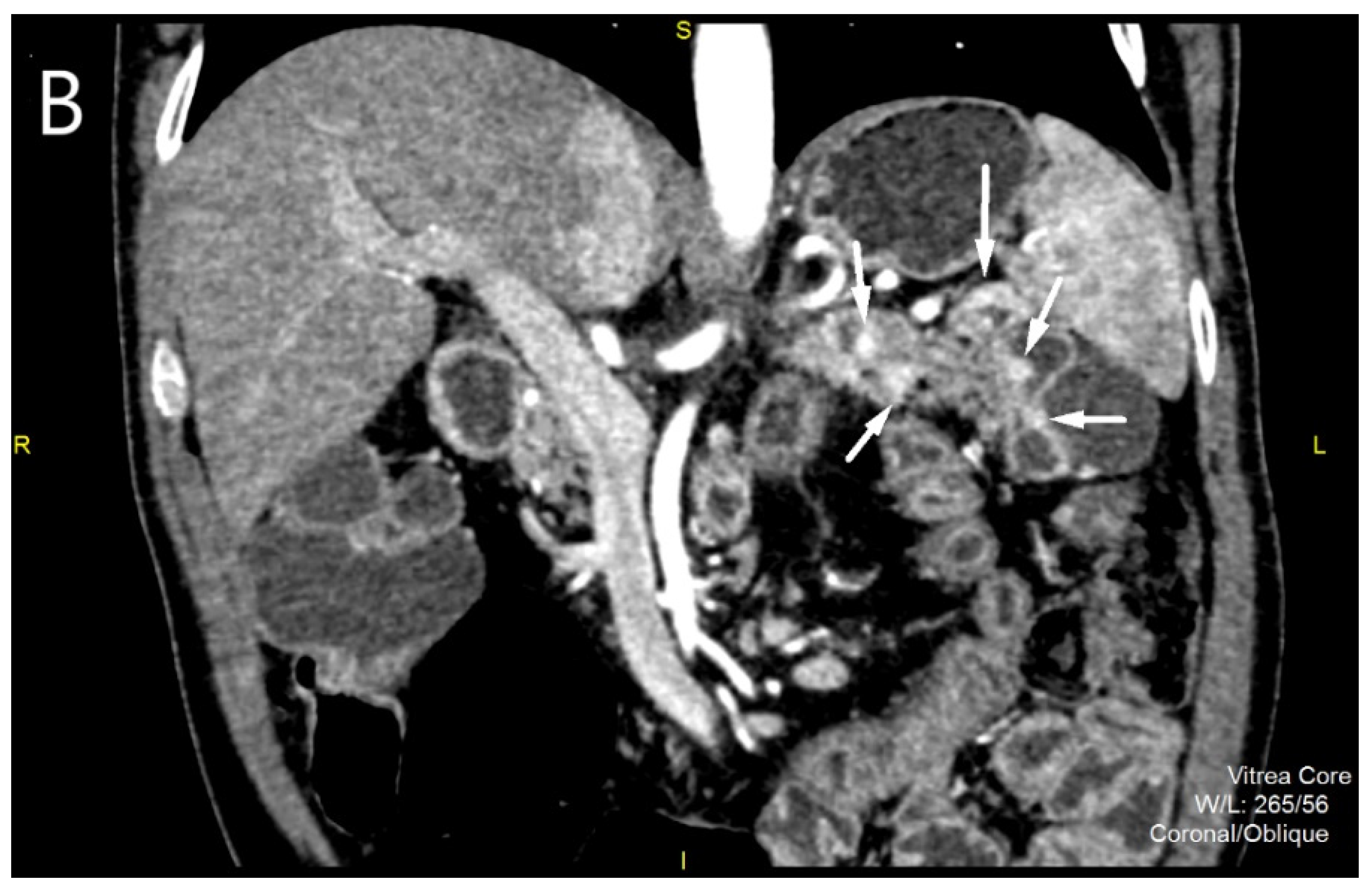
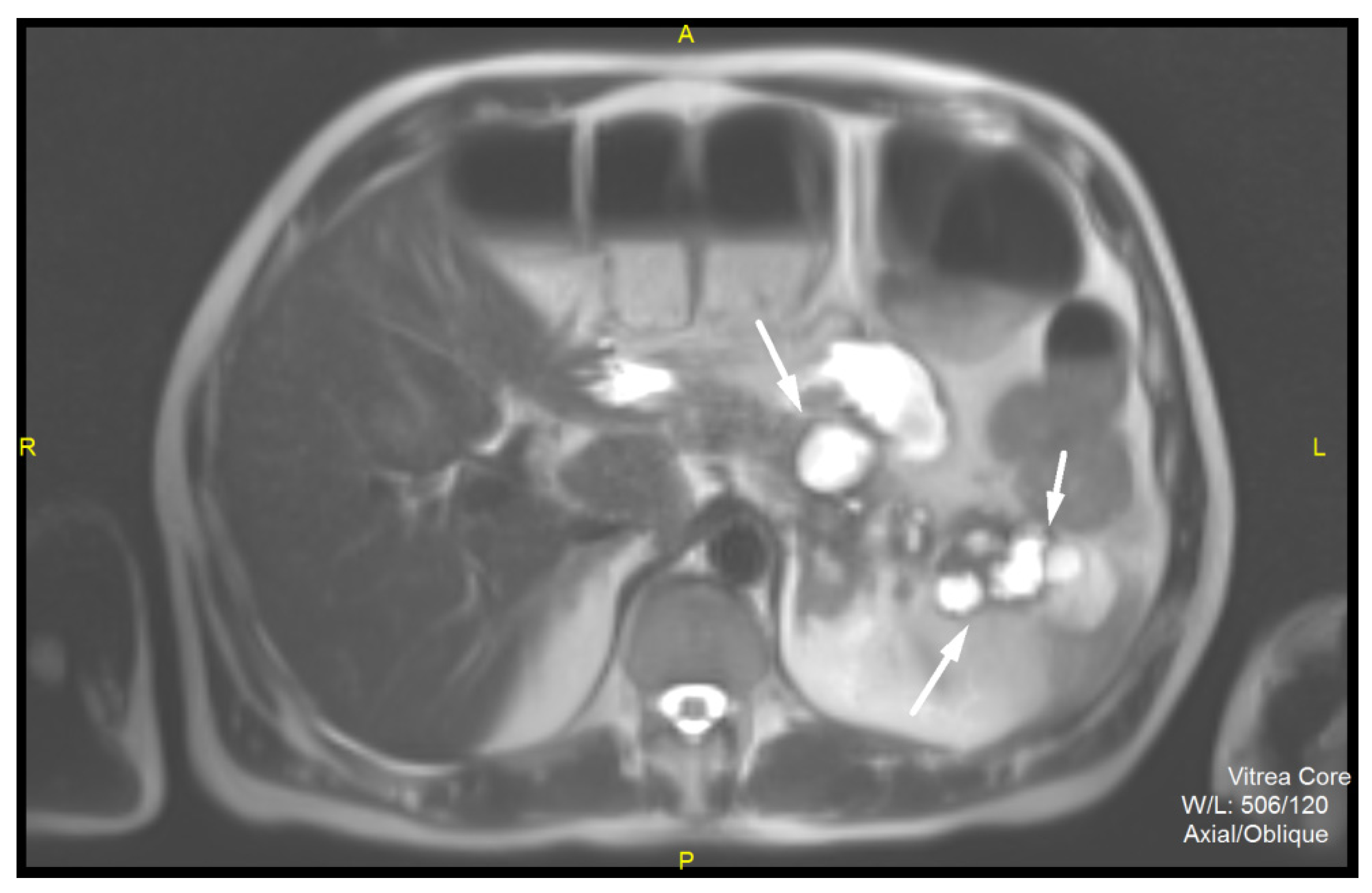
2. Case Report
3. Discussion
4. Conclusions
Author Contributions
Funding
Institutional Review Board Statement
Informed Consent Statement
Data Availability Statement
Conflicts of Interest
References
- Lee, N.J.; Hruban, R.H.; Fishman, E.K. Pancreatic neuroendocrine tumor: Review of heterogeneous spectrum of CT appearance. Abdom. Radiol. 2018, 43, 3025–3034. [Google Scholar] [CrossRef] [PubMed]
- Choe, J.; Kim, K.W.; Kim, H.J.; Kim, D.W.; Kim, K.P.; Hong, S.M.; Ryu, J.S.; Tirumani, S.H.; Krajewski, K.; Ramaiya, N. What Is New in the 2017 World Health Organization Classification and 8th American Joint Committee on Cancer Staging System for Pancreatic Neuroendocrine Neoplasms? Korean J. Radiol. 2019, 20, 5–17. [Google Scholar] [CrossRef]
- Carr, R.A.; Bletsis, P.; Roch, A.M.; House, M.G.; Zyromski, N.J.; Nakeeb, A.; Schmidt, C.M.; Ceppa, E.P. Cystic pancreatic neuroendocrine tumors: A more favorable lesion? Pancreatology 2019, 19, 372–376. [Google Scholar] [CrossRef] [PubMed]
- Caglià, P.; Cannizzaro, M.T.; Tracia, A.; Amodeo, L.; Tracia, L.; Buffone, A.; Amodeo, C.; Cannizzaro, M.A. Cystic pancreatic neuroendocrine tumors: To date a diagnostic challenge. Int. J. Surg. 2015, 21, 44–49. [Google Scholar] [CrossRef] [PubMed]
- Salahshour, F.; Taslimi, R.; Moosavi, N.S.; Yazdi, N.A.; Esfandbod, M. Pancreatic Neuroendocrine Tumor presenting as a diffuse pancreatic enlargement, case report and review of literature. J. Radiol. Case Rep. 2021, 15, 11–20. [Google Scholar] [PubMed]
- Bosman, F.T.; Carneiro, F.; Hruban, R.H.; Theise, N.D. WHO Classification of Tumors of the Digestive System, 4th ed.; IRAC Press: Lyon, France, 2010; pp. 322–326. [Google Scholar]
- Chiruvella, A.; Kooby, D.A. Surgical Management of Pancreatic Neuroendocrine Tumors. Surg. Oncol. Clin. N. Am. 2016, 25, 401–421. [Google Scholar] [CrossRef] [PubMed]
- McKenna, L.R.; Edil, B.H. Update on pancreatic neuroendocrine tumors. Gland Surg. 2014, 3, 258–275. [Google Scholar] [PubMed]
- Cloyd, J.M.; Kopecky, K.E.; Norton, J.A.; Kunz, P.L.; Fisher, G.A.; Visser, B.C.; Dua, M.M.; Park, W.G.; Poultsides, G.A. Neuroendocrine tumors of the pancreas: Degree of cystic component predicts prognosis. Surgery 2016, 160, 708–713. [Google Scholar] [CrossRef]
- Mortaji, P.; Morris, K.T.; Samedi, V.; Eberhardt, S.; Ryan, S. Pancreatic neuroendocrine tumor in a patient with a TSC1 variant: Case report and review of the literature. Fam. Cancer 2018, 17, 275–280. [Google Scholar] [CrossRef]
- Hassan, M.M.; Phan, A.; Li, D.; Dagohoy, C.G.; Leary, C.; Yao, J.C. Family history of cancer and associated risk of developing neuroendocrine tumors: A case-control study. Cancer Epidemiol. Biomark. Prev. 2008, 17, 959–965. [Google Scholar] [CrossRef] [Green Version]
- Gut, P.; Komarowska, H.; Czarnywojtek, A.; Waligórska-Stachura, J.; Bączyk, M.; Ziemnicka, K.; Fischbach, J.; Wrotkowska, E.; Ruchała, M. Familial syndromes associated with neuroendocrine tumours. Contemp. Oncol. Pozn. 2015, 19, 176–183. [Google Scholar] [CrossRef] [PubMed] [Green Version]
- Malagò, R.; D’Onofrio, M.; Zamboni, G.A.; Faccioli, N.; Falconi, M.; Boninsegna, L.; Mucelli, R.P. Contrast-enhanced sonography of nonfunctioning pancreatic neuroendocrine tumors. AJR Am. J. Roentgenol. 2009, 192, 424–430. [Google Scholar] [CrossRef] [PubMed]
- Bocchini, M.; Nicolini, F.; Severi, S.; Bongiovanni, A.; Ibrahim, T.; Simonetti, G.; Grassi, I.; Mazza, M. Biomarkers for Pancreatic Neuroendocrine Neoplasms (PanNENs) Management-An Updated Review. Front. Oncol. 2020, 27, 831. [Google Scholar] [CrossRef] [PubMed]
- Atiq, M.; Bhutani, M.S.; Bektas, M.; Lee, J.E.; Gong, Y.; Tamm, E.P.; Shah, C.P.; Ross, W.A.; Yao, J.; Raju, G.S.; et al. EUS-FNA for pancreatic neuroendocrine tumors: A tertiary cancer center experience. Dig. Dis. Sci. 2012, 57, 791–800. [Google Scholar] [CrossRef] [PubMed]
- Kawamoto, S.; Johnson, P.T.; Shi, C.; Singhi, A.D.; Hruban, R.H.; Wolfgang, C.L.; Edil, B.H.; Fishman, E.K. Pancreatic neuroendocrine tumor with cystlike changes: Evaluation with MDCT. AJR Am. J. Roentgenol. 2013, 200, 283–290. [Google Scholar] [CrossRef] [Green Version]
- Naringrekar, H.; Vogel, A.; Prestipino, A.; Shahid, H. Imitators of chronic pancreatitis: Diffuse neuroendocrine tumour of the pancreas. BJR Case Rep. 2017, 3, 20170015. [Google Scholar] [CrossRef]
- Kartalis, N.; Mucelli, R.M.; Sundin, A. Recent developments in imaging of pancreatic neuro-endocrine tumors. Ann. Gastroenterol. 2015, 28, 193–202. [Google Scholar]
- Mojtahedi, A.; Thamake, S.; Tworowska, I.; Ranganathan, D.; Delpassand, E. The value of 68Ga-DOTATATE PET/CT in diagnosis and management of neuroendocrine tumors compared to current FDA approved imaging modalities: A review of literature. Am. J. Nucl. Med. Mol. Imaging 2014, 4, 426–434. [Google Scholar]
- Singh, R.; Calhoun, S.; Shin, M.; Katz, R. Pancreatic neuroendocrine tumor with atypical radiologic presentation. Radiol. Case Rep. 2015, 3, 162. [Google Scholar] [CrossRef] [Green Version]
- Santes, O.; Morales-Maza, J.; Domínguez-Rosado, I. Diffuse Enlargement of the Pancreas: An Unusual Radiologic Presentation of a Pancreatic Neuroendocrine Tumor. Clin. Gastroenterol. Hepatol. 2017, 15, 165–166. [Google Scholar] [CrossRef] [Green Version]
- Humphrey, P.E.; Alessandrino, F.; Bellizzi, A.M.; Mortele, K.J. Non-hyperfunctioning pancreatic endocrine tumors: Multimodality imaging features with histopathological correlation. Abdom. Imaging 2015, 40, 2398–2410. [Google Scholar] [CrossRef] [PubMed]
- Kaosombatwattana, U.; Hirooka, Y.; Kawashima, H.; Ohno, E.; Ishikawa, T.; Suhara, H.; Goto, H. Neuroendocrine neoplasm of pancreas with cystic degeneration mimicking mucinous cystic neoplasm. Clin. J. Gastroenterol. 2018, 11, 333–337. [Google Scholar] [CrossRef] [PubMed]
- Lamarca, A.; Clouston, H.; Barriuso, J.; McNamara, M.G.; Frizziero, M.; Mansoor, W.; Hubner, R.A.; Manoharan, P.; O’Dwyer, S.; Valle, J.W. Follow-Up Recommendations after Curative Resection of Well-Differentiated Neuroendocrine Tumours: Review of Current Evidence and Clinical Practice. J. Clin. Med. 2019, 8, 1630. [Google Scholar] [CrossRef] [PubMed] [Green Version]
- Singh, S.; Chan, D.L.; Moody, L.; Liu, N.; Fischer, H.D.; Austin, P.C.; Segelov, E. Recurrence in Resected Gastroenteropancreatic Neuroendocrine Tumors. JAMA Oncol. 2018, 4, 583–585. [Google Scholar] [CrossRef]
- Li, D.; Rock, A.; Kessler, J.; Ballena, R.; Hyder, S.; Mo, C.; Chang, S.; Singh, G. Understanding the Management and Treatment of Well-Differentiated Pancreatic Neuroendocrine Tumors: A Clinician’s Guide to a Complex Illness. JCO Oncol. Pract. 2020, 16, 720–728. [Google Scholar] [CrossRef]


Publisher’s Note: MDPI stays neutral with regard to jurisdictional claims in published maps and institutional affiliations. |
© 2022 by the authors. Licensee MDPI, Basel, Switzerland. This article is an open access article distributed under the terms and conditions of the Creative Commons Attribution (CC BY) license (https://creativecommons.org/licenses/by/4.0/).
Share and Cite
Mitrovic-Jovanovic, M.; Grubor, N.; Milosevic, S.; Jankovic, A.; Stosic, K.; Ostojic, S.; Ninic, A.; Micev, M.; Djokic Kovac, J. Total Pancreatectomy for Multicentric Cystic Neuroendocrine Tumor of the Pancreas: A Case Report. Diagnostics 2022, 12, 1003. https://doi.org/10.3390/diagnostics12041003
Mitrovic-Jovanovic M, Grubor N, Milosevic S, Jankovic A, Stosic K, Ostojic S, Ninic A, Micev M, Djokic Kovac J. Total Pancreatectomy for Multicentric Cystic Neuroendocrine Tumor of the Pancreas: A Case Report. Diagnostics. 2022; 12(4):1003. https://doi.org/10.3390/diagnostics12041003
Chicago/Turabian StyleMitrovic-Jovanovic, Milica, Nikica Grubor, Stefan Milosevic, Aleksandra Jankovic, Katarina Stosic, Slavenko Ostojic, Aleksandar Ninic, Marjan Micev, and Jelena Djokic Kovac. 2022. "Total Pancreatectomy for Multicentric Cystic Neuroendocrine Tumor of the Pancreas: A Case Report" Diagnostics 12, no. 4: 1003. https://doi.org/10.3390/diagnostics12041003