
Machine Learning and Deep Learning Methods for Skin Lesion Classification and Diagnosis: A Systematic Review

 , , and
Abstract
:1. Introduction
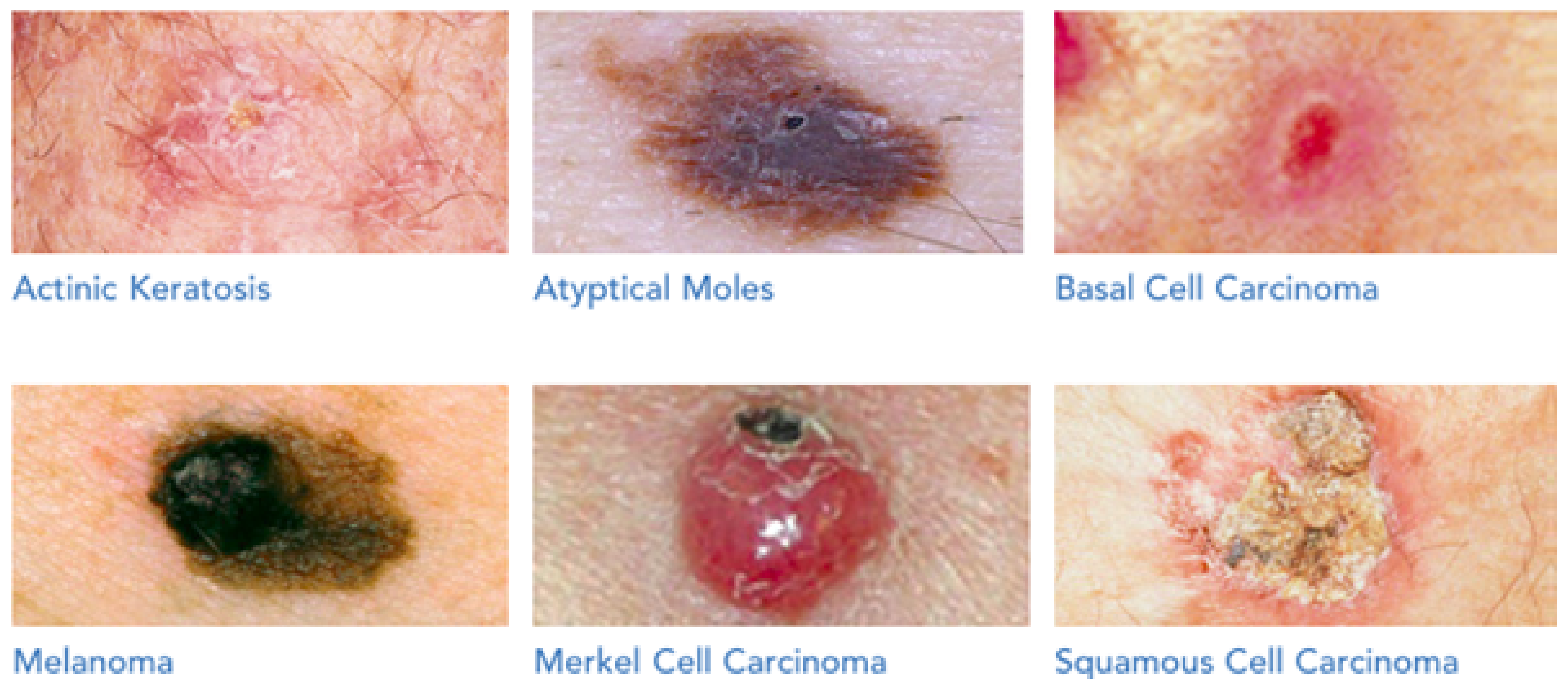
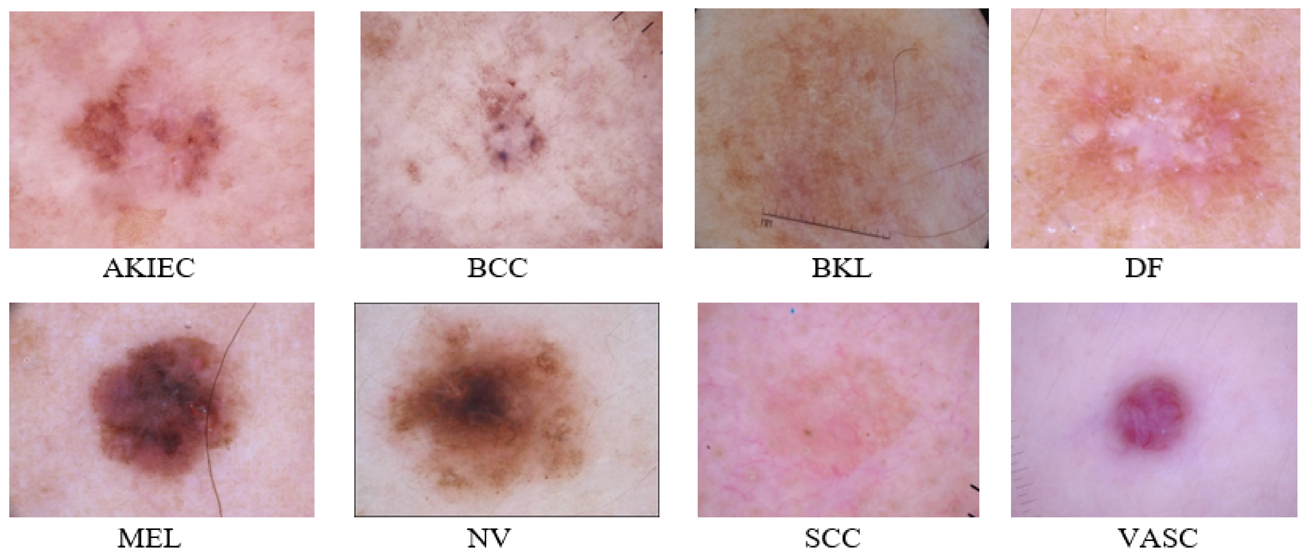
- Actinic Keratosis (AKIEC) or solar keratosis: This is a form of keratosis that occurs on the skin. It is a crusty, scaly growth on the skin. It is classified as pre-cancer because it has the potential to turn into skin cancer if left untreated.
- Atypical moles: Also known as dysplastic nevi, they are benign moles that have an irregular appearance. They may look like melanoma, and the ones that have them are more likely to develop melanoma in a mole or anywhere on the body. They have a greater chance of having melanom;
- Basal Cell Carcinoma (BCC): This is the most common form of skin cancer. This form of skin cancer spreads rarely. Its common symptoms are open sores, shiny bumps, red spots, pink growths, or scars;
- Melanoma: This is the most lethal kind of skin cancer. It is often black or brown, although it can also be pink, red, purple, blue, or white. UV radiation from the sun or tanning beds causes cancerous tumors. Melanoma is frequently treatable if detected and treated early, but, if not, the disease can spread to other places of the body and the therapy would be complicated and deadly;
- Merkel cell carcinoma: This is an uncommon and aggressive kind of skin cancer that has a high chance of metastasizing. However, it is 40 times less prevalent than melanoma;
- Squamous Cell Carcinoma (SCC): This is the second most frequent kind of skin cancer. Scaly red spots, open sores, raised growths with a central depression, or warts are frequent signs.
- Image enhancement: This phase aims to eliminate all noise and artifacts such as hair and blood vessels in dermoscopic images;
- Segmentation: Segmenting the Region of Interest (ROI) is a crucial step in CAD systems. The process of segmenting skin cancer images is made more complex by a large number of different skin lesions. It quickly became one of the most complex and tedious tasks in the CAD system;
- Feature extraction: After defining the ROI, the goal of the feature extraction step is to identify the best set of features that have high discrimination capability to classify the dataset into two or more classes;
- Classification and detection: The proposed system is evaluated according to its capability to classify the dataset into different classes. Hence, the choice of classifier is critical for a better performance. However, it depends on the set of extracted feature and the required number of classes. The classification performance measures are accuracy, specificity, sensitivity, precision, and Receiver Operating Characteristic (ROC).
2. Methods
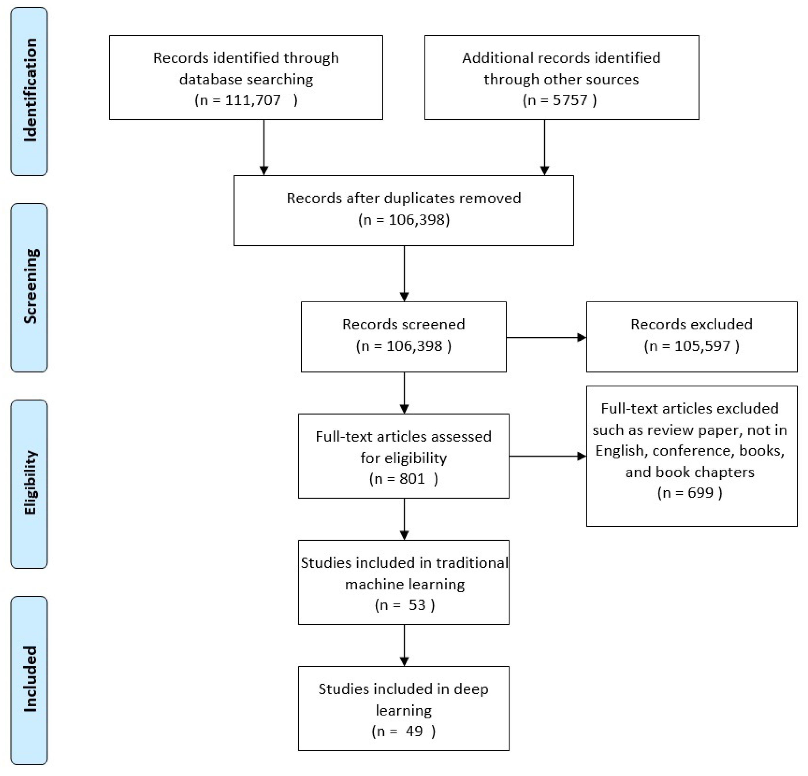
2.1. Systematic Review
2.2. Datasets
2.3. Traditional Machine Learning
2.4. Deep Learning
3. Discussion and Conclusions
Author Contributions
Funding
Institutional Review Board Statement
Informed Consent Statement
Data Availability Statement
Conflicts of Interest
References
- Capdehourat, G.; Corez, A.; Bazzano, A.; Alonso, R.; Musé, P. Toward a combined tool to assist dermatologists in melanoma detection from dermoscopic images of pigmented skin lesions. Pattern Recognit. Lett. 2011, 32, 2187–2196. [Google Scholar] [CrossRef]
- American Cancer Society. Statistics 2013. Available online: https://www.cancer.org/research/cancer-facts-statistics/all-cancer-facts-figures/cancer-facts-figures-2013.html?fbclid=IwAR2gMmnaky1m3LdETjBwoTiRkaxDiaKvWss9UlSVx6YqWmR-rrehUjBMpvs (accessed on 10 May 2021).
- Korotkov, K.; Garcia, R. Computerized analysis of pigmented skin lesions: A review. Artif. Intell. Med. 2012, 56, 69–90. [Google Scholar] [CrossRef] [PubMed]
- Skin Cancer Foundation. Skin Cancer Information. 2016. Available online: http://www.skincancer.org/skin-cancer-information (accessed on 20 August 2016).
- AlZubi, S.; Islam, N.; Abbod, M. Multiresolution analysis using wavelet, ridgelet, and curvelet transforms for medical image segmentation. J. Biomed. Imaging 2011, 4, 2011. Available online: http://www.dermquest.com (accessed on 20 July 2015). [CrossRef] [Green Version]
- Arroyo, J.L.G.; Zapirain, B.G. Detection of pigment 131 network in dermoscopy images using supervised machine learning and structural analysis. Comput. Biol. Med. 2014, 44, 144–157. [Google Scholar] [CrossRef]
- Lee, H.Y.; Chay, W.Y.; Tang, M.B.; Chio, M.T.; Tan, S.H. Melanoma: Differences between asian and caucasian patients. Ann. Acad. Med. Singap. 2012, 41, 17–20. [Google Scholar] [PubMed]
- Rigel, D.S.; Russak, J.; Friedman, R. The evolution of melanoma diagnosis: 25 years beyond the abcds. CA Cancer J. Clin. 2010, 60, 301–316. [Google Scholar] [CrossRef]
- Laikova, K.V.; Oberemok, V.V.; Krasnodubets, A.M.; Gal’chinsky, N.V.; Useinov, R.Z.; Novikov, I.A.; Temirova, Z.Z.; Gorlov, M.V.; Shved, N.A.; Kumeiko, V.V.; et al. Advances in the Understanding of Skin Cancer: Ultraviolet Radiation, Mutations, and Antisense Oligonucleotides as Anticancer Drugs. Molecules 2019, 24, 1516. [Google Scholar] [CrossRef] [PubMed] [Green Version]
- Apalla, Z.; Lallas, A.; Sotiriou, E.; Lazaridou, E.; Ioannides, D. Epidemiological trends in skin cancer. Dermatol. Pract. Concept. 2017, 7, 1–6. [Google Scholar] [CrossRef] [Green Version]
- Karimkhani, C.; Green, A.C.; Nijsten, T.; Weinstock, M.A.; Dellavalle, R.P.; Naghavi, M.; Fitzmaurice, C. The global burden of melanoma: Results from the Global Burden of Disease Study 2015. Br. J. Dermatol. 2017, 177, 134–140. [Google Scholar] [CrossRef] [Green Version]
- Schadendorf, D.; Lebbé, C.; Hausenzur, A.; Avril, M.F.; Hariharan, S.; Bharmal, M.; Becker, J.C. Merkel cell carcinoma: Epidemiology, prognosis, therapy and unmet medical needs. Eur. J. Cancer 2017, 71, 53–69. [Google Scholar] [CrossRef] [PubMed] [Green Version]
- Timerman, D.; McEnery-Stonelake, M.; Joyce, C.J.; Nambudiri, V.E.; Hodi, F.S.; Claus, E.B.; Ibrahim, N.; Lin, J.Y. Vitamin D deficiency is associated with a worse prognosis in metastatic melanoma. Oncotarget 2017, 8, 6873–6882. [Google Scholar] [CrossRef] [PubMed] [Green Version]
- Feller, L.; Khammissa, R.A.; Kramer, B.; Altini, M.; Lemmer, J. Basal cell carcinoma, squamous cell carcinoma and melanoma of the head and face. Head Face Med. 2016, 12, 11. [Google Scholar] [CrossRef] [Green Version]
- Becker, J.C.; Stang, A.; DeCaprio, J.A.; Cerroni, L.; Lebbé, C.; Veness, M.; Nghiem, P. Merkel cell carcinoma. Nat. Rev. Dis. Primers 2017, 3, 170–177. [Google Scholar] [CrossRef] [PubMed]
- Glazer, A.M.; Winkelmann, R.R.; Farberg, A.S.; Rigel, D.S. Analysis of trends in US melanoma incidence and mortality. JAMA Dermatol. 2016, 153, 225–226. [Google Scholar] [CrossRef] [Green Version]
- Lv, R.; Sun, Q. A Network Meta-Analysis of Non-Melanoma Skin Cancer (NMSC) Treatments: Efficacy and Safety Assessment. J. Cell. Biochem. 2017, 118, 3686–3695. [Google Scholar] [CrossRef]
- Lindelof, B.; Hedblad, M.-A. Accuracy in the clinical diagnosis and pattern of malignant melanoma at a dermatological clinic. J. Dermatol. 1994, 21, 461–464. [Google Scholar] [CrossRef] [PubMed]
- Morton, C.A.; Mackie, R.M. Clinical accuracy of the diagnosis of cutaneous malignant melanoma. Br. J. Dermatol. 1998, 138, 283–287. [Google Scholar] [CrossRef]
- Menzies, S.W.; Bischof, L.; Talbot, H.; Gutenev, A.; Avramidis, M.; Wong, L.; Lo, S.K.; Mackellar, G.; Skladnev, V.; McCarthy, W.; et al. The performance of SolarScan: An automated dermoscopy image analysis instrument for the diagnosis of primary melanoma. Arch. Dermatol. 2005, 141, 1388–1396. [Google Scholar] [CrossRef] [PubMed] [Green Version]
- Binder, M.; Schwarz, M.; Winkler, A.; Steiner, A.; Kaider, A.; Wolff, K.; Pehamberger, H. Epiluminescence microscopy: A useful tool for the diagnosis of pigmented skin lesions for formally trained dermatologists. Arch. Dermatol. 1995, 131, 286–291. [Google Scholar] [CrossRef]
- Pehamberger, H.; Binder, M.; Steiner, A.; Wolff, K. In vivo epiluminescence microscopy: Improvement of early diagnosis of melanoma. J. Investig. Dermatol. 1993, 100, 3. [Google Scholar] [CrossRef] [Green Version]
- Dhawan, A.P.; Gordon, R.; Rangayyan, R.M. Nevoscopy: Three-dimensional computed tomography of nevi and melanomas in situ by transillumination. IEEE Trans. Onmedical Imaging 1984, 3, 54–61. [Google Scholar] [CrossRef]
- Zouridakis, G.; Duvic, M.D.M.; Mullani, N.A. Transillumination Imaging for Early Skin Cancer Detection; Technol Report 2005; Biomedical Imaging Lab., Department of Computer Science, University of Houston: Houston, TX, USA, 2005. [Google Scholar]
- Vestergaard, M.E.; Macaskill, P.; Holt, P.E.; Menzies, S.W. Dermoscopy compared with naked eye examination for the diagnosis of primary melanoma: A meta-analysis of studies performed in a clinical setting. Br. J. Dermatol. 2008, 159, 669–676. [Google Scholar] [CrossRef]
- Carli, P.; Giorgi, V.D.; Crocetti, E.; Mannone, F.; Massi, D.; Chiarugi, A.; Giannotti, B. Improvement of malignant/benign ratio in excised melanocytic lesions in the “dermoscopy era”: A retrospective study 1997–2001. Br. J. Dermatol. 2004, 150, 687–692. [Google Scholar] [CrossRef]
- Carli, P.; Giorgi, V.D.; Chiarugi, A.; Nardini, P.; Weinstock, M.A.; Crocetti, E.; Stante, M.; Giannotti, B. Addition of dermoscopy to conventional naked-eye examination in melanoma screening: A randomized study. J. Am. Acad. Dermatol. 2004, 50, 683–689. [Google Scholar] [CrossRef] [PubMed]
- Mayer, J. Systematic review of the diagnostic accuracy of dermatoscopy in detecting malignant melanoma. Med. J. Aust. 1997, 167, 206–210. [Google Scholar] [CrossRef] [PubMed]
- Piccolo, D.; Ferrari, A.; Peris, K.; Daidone, R.; Ruggeri, B.; Chimenti, S. Dermoscopic diagnosis by a trained clinician vs. a clinician with minimal dermoscopy training vs. computeraided diagnosis of 341 pigmented skin lesions: A comparative study. Br. J. Dermatol. 2002, 147, 481–486. [Google Scholar] [CrossRef]
- Braun, R.P.; Rabinovitz, H.S.; Oliviero, M.; Kopf, A.W.; Saurat, J.-H. Dermoscopy of pigmented skin lesions. J. Am. Acad. Dermatol. 2005, 52, 109–121. [Google Scholar] [CrossRef]
- Kittler, H.; Pehamberger, H.; Wolff, K.; Binder, M. Diagnostic accuracy of dermoscopy. Lancet Oncol. 2002, 3, 159–165. [Google Scholar] [CrossRef]
- Whited, J.D.; Grichnik, J.M. Does this patient have a mole or a melanoma? J. Am. Med. Assoc. 1998, 279, 696–701. [Google Scholar] [CrossRef]
- Burroni, M.; Corona, R.; Dell’Eva, G.; Sera, F.; Bono, R.; Puddu, P.; Perotti, R.; Nobile, F.; Andreassi, L.; Rubegni, P. Melanoma computer-aided diagnosis: Reliability and feasibility study. Clin. Cancer Res. 2004, 10, 1881–1886. [Google Scholar] [CrossRef] [Green Version]
- Nami, N.; Giannini, E.; Burroni, M.; Fimiani, M.; Rubegni, P. Teledermatology: State-of-the-art and future perspectives. Expert Rev. Dermatol. 2014, 7, 1–3. [Google Scholar] [CrossRef] [Green Version]
- Fabbrocini, G.; Triassi, M.; Mauriello, M.C.; Torre, G.; Annunziata, M.C.; De Vita, V.; Pastore, F.; D’Arco, V.; Monfrecola, G. Epidemiology of skin cancer: Role of some environmental factors. Cancers 2010, 2, 1980–1989. [Google Scholar] [CrossRef] [Green Version]
- Haenssle, H.; Fink, C.; Schneiderbauer, R.; Toberer, F.; Buhl, T.; Blum, A.; Reader Study Level-I and Level-II Groups. Man against machine: Diagnostic performance of a deep learning convolutional neural network for dermoscopic melanoma recognition in comparison to 58 dermatologists. Ann. Oncol. 2018, 29, 1836–1842. [Google Scholar] [CrossRef]
- Argenziano, G.; Soyer, H.P. Dermoscopy of pigmented skin lesions: A valuable tool for early diagnosis of melanoma. Lancet Oncol. 2001, 2, 443–449. [Google Scholar] [CrossRef]
- Ali, A.R.A.; Deserno, T.M. A systematic review of automated melanoma detection in dermatoscopic images and its ground truth data. Proc. SPIE Int. Soc. Opt. Eng. 2012, 8318, 1–6. [Google Scholar] [CrossRef]
- Fabbrocini, G.; De Vita, V.; Pastore, F.; D’Arco, V.; Mazzella, C.; Annunziata, M.C.; Cacciapuoti, S.; Mauriello, M.C.; Monfrecola, A. Teledermatology: From prevention to diagnosis of nonmelanoma and melanoma skin cancer. Int. J. Telemed. Appl. 2011, 2011, 125762. [Google Scholar] [CrossRef]
- Foraker, R.; Kite, B.; Kelley, M.M.; Lai, A.M.; Roth, C.; Lopetegui, M.A.; Shoben, A.B.; Langan, M.; Rutledge, N.; Payne, P.R.O. EHR-based visualization tool: Adoption rates, satisfaction, and patient outcomes. EGEMS 2015, 3, 1159. [Google Scholar] [CrossRef] [Green Version]
- Fabbrocini, G.; Betta, G.; Di Leo, G.; Liguori, C.; Paolillo, A.; Pietrosanto, A.; Sommella, P.; Rescigno, O.; Cacciapuoti, S.; Pastore, F.; et al. Epiluminescence image processing for melanocytic skin lesion diagnosis based on 7-point check-list: A preliminary discussion on three parameters. Open Dermatol. J. 2010, 4, 110–115. [Google Scholar] [CrossRef] [Green Version]
- Hart, P.E.; Stork, D.G.; Duda, R.O. Pattern Classification, 2nd ed.; John Wiley & Sons: Hoboken, NJ, USA, 2000. [Google Scholar]
- Oliveira, R.B.; Papa, J.P.; Pereira, A.S.; Tavares, J.M.R.S. Computational methods for pigmented skin lesion classification in images: Review and future trends. Neural Comput. Appl. 2016, 29, 613–636. [Google Scholar] [CrossRef] [Green Version]
- Cascinelli, N.; Ferrario, M.; Tonelli, T.; Leo, E. Apossible new tool for clinical diagnosis of melanoma: The computer. J. Am. Acad. Dermatol. 1987, 16, 361–367. [Google Scholar] [CrossRef]
- Hall, P.N.; Claridge, E.; Smith, J.D.M. Computer screening for early detection of melanoma—Is there a future? Br. J. Dermatol. 1995, 132, 325–338. [Google Scholar] [CrossRef]
- Cristofolini, M.; Bauer, P.; Boi, S.; Cristofolini, P.; Micciolo, R.; Sicher, M.C. Diagnosis of cutaneous melanoma: Accuracy of a computerized image analysis system(SkinView). Ski. Res. Technol. 1997, 3, 23–27. [Google Scholar] [CrossRef] [PubMed]
- Umbaugh, S.E. Computer Vision in Medicine: Color Metrics and Image Segmentation Methods for Skin Cancer Diagnosis; Electrical Engineering Department, University of Missouri: Rolla, MO, USA, 1990. [Google Scholar]
- Stanganelli, I.; Brucale, A.; Calori, L.; Gori, R.; Lovato, A.; Magi, S.; Kopf, B.; Bacchilega, R.; Rapisarda, V.; Testori, A.; et al. Computer-aided diagnosis of melanocytic lesions. Anticancer Res. 2005, 25, 4577–4582. [Google Scholar] [PubMed]
- Rubegni, P.; Cevenini, G.; Burroni, M.; Perotti, R.; Dell’Eva, G.; Sbano, P.; Miracco, C.; Luzi, P.; Tosi, P.; Barbini, P.; et al. Automated diagnosis of pigmented skin lesions. Int. J. Cancer 2002, 101, 576–580. [Google Scholar] [CrossRef] [PubMed] [Green Version]
- Sober, A.J.; Burstein, J.M. Computerized digital image analysis: An aid for melanoma diagnosis—Preliminary investigations and brief review. J. Dermatol. 1994, 21, 885–890. [Google Scholar] [CrossRef] [PubMed]
- Rajpara, S.M.; Botello, A.P.; Townend, J.; Ormerod, A.D. Systematic review of dermoscopy and digital dermoscopy/artificial intelligence for the diagnosis of melanoma. Br. J. Dermatol. 2009, 161, 591–604. [Google Scholar] [CrossRef] [PubMed]
- Rosado, B.; Menzies, S.; Harbauer, A.; Pehamberger, H.; Wolff, K.; Binder, M.; Kittler, H. Accuracy of computer diagnosis of melanoma: A quantitative meta-analysis. Arch. Dermatol. 2003, 139, 361–367. [Google Scholar] [CrossRef]
- Bauer, P.; Cristofolini, P.; Boi, S.; Burroni, M.; Dell’Eva, G.; Micciolo, R.; Cristofolini, M. Digital epiluminescence microscopy: Usefulness in the differential diagnosis of cutaneous pigmentary lesions. A statistical comparison between visual and computer inspection. Melanoma Res. 2000, 10, 345–349. [Google Scholar] [CrossRef]
- Maglogiannis, I.; Doukas, C.N. Overview of advanced computer vision systems for skin lesions characterization. IEEE Trans. Inf. Technol. Biomed. 2009, 13, 721–733. [Google Scholar] [CrossRef]
- Friedman, R.J.; Gutkowicz-Krusin, D.; Farber, M.J.; Warycha, M.; Schneider-Kels, L.; Papastathis, N.; Mihm, M.C.; Googe, P.; King, R.; Prieto, V.G.; et al. The diagnostic performance of expert dermoscopists vs a computervision system on small-diameter melanomas. Arch. Dermatol. 2008, 144, 476–482. [Google Scholar] [CrossRef] [Green Version]
- Blum, A.; Zalaudek, I.; Argenziano, G. Digital image analysis for diagnosis of skin tumors. Semin. Cutan. Med. Surgery 2008, 27, 11–15. [Google Scholar] [CrossRef] [PubMed]
- Maruthamuthu, M.K.; Raffiee, A.H.; Oliveira, D.M.D.; Ardekani, A.M.; Verma, M.S. Raman spectra-based deep learning: A tool to identify microbial contamination. MicrobiologyOpen 2020, 9, e1122. [Google Scholar] [CrossRef] [PubMed]
- Zhao, Z.; Wu, C.M.; Zhang, S.; He, F.; Liu, F.; Wang, B.; Huang, Y.; Shi, W.; Jian, D.; Xie, H.; et al. A Novel Convolutional Neural Network for the Diagnosis and Classification of Rosacea: Usability Study. Jmir Med. Inform. 2021, 9, e23415. [Google Scholar] [CrossRef]
- Moher, D.; Liberati, A.; Tetzlaff, J.; Altman, D.G.; The PRISMA Group. Preferred Reporting Items for Systematic Reviews and Meta-Analyses: The PRISMA Statement. PLoS Med. 2009, 6, E1000097. [Google Scholar] [CrossRef] [Green Version]
- Siegel, R.L.; Miller, K.D.; Jemal, A. Cancer statistics, 2018. CA Cancer J. Clin. 2018, 68, 7–30. [Google Scholar] [CrossRef] [PubMed]
- Giotis, I.; Molders, N.; Land, S.; Biehl, M.; Jonkman, M.F.; Petkov, N. MED-NODE: A computer-assisted melanoma diagnosis system using non-dermoscopic images. Expert Syst. Appl. 2018, 42, 6578–6585. [Google Scholar] [CrossRef]
- Dermatology Information System. Available online: http://www.dermis.net (accessed on 25 January 2016).
- DermQuest. Available online: http://www.dermquest.com (accessed on 25 January 2016).
- Mendonça, T.; Ferreira, P.M.; Marques, J.S.; Marcal, A.R.S.; Rozeira, J. PH2- A dermoscopic image database for research and benchmarking. In Proceedings of the 2013 35th Annual International Conference of the IEEE Engineering in Medicine and Biology Society (EMBC), Osaka, Japan, 3–7 July 2013; pp. 5437–5440. [Google Scholar]
- Gutman, D.; Codella, N.C.F.; Emre, C.; Brian, H.; Michael, M.; Nabin, M.; Allan, H. Skin Lesion Analysis toward Melanoma Detection: A Challenge at the International Symposium on Biomedical Imaging (ISBI) 2016, hosted by the International Skin Imaging Collaboration (ISIC). arXiv 2016, arXiv:1605.01397. [Google Scholar]
- Codella, N.; Gutman, D.; Celebi, M.E.; Helba, D.; Marchetti, M.A.; Dusza, S.; Kalloo, A.; Liopyris, K.; Mishra, N.; Kittler, H.; et al. Skin Lesion Analysis Toward Melanoma Detection: A Challenge at the 2017 International Symposium on Biomedical Imaging (ISBI), Hosted by the International Skin Imaging Collaboration (ISIC). arXiv 2017, arXiv:1710.05006. [Google Scholar]
- Codella, N.; Rotemberg, V.; Tschandl, P.; Celebi, M.E.; Dusza, S.; Gutman, D.; Helba, B.; Kalloo, A.; Liopyris, K.; Marchetti, M.; et al. Skin Lesion Analysis Toward Melanoma Detection 2018: A Challenge Hosted by the International Skin Imaging Collaboration (ISIC). arXiv 2018, arXiv:1902.03368. [Google Scholar]
- Tsch, P.; Rosendahl, C.; Kittler, H. The HAM10000 dataset, a large collection of multi-sources dermatoscopic images of common pigmented skin lesions. Sci. Data 2018, 5, 180161. [Google Scholar] [CrossRef]
- Combalia, M.; Codella, N.C.F.; Rotemberg, V.; Helba, B.; Vilaplana, V.; Reiter, O.; Halpern, A.C.; Puig, S.; Malvehy, J. BCN20000: Dermoscopic Lesions in the Wild. arXiv 2019, arXiv:1908.02288. [Google Scholar]
- Ballerini, L.; Fisher, R.B.; Aldridge, B.; Rees, J. A Color and Texture Based Hierarchical K-NN Approach to the Classification of Non-melanoma Skin Lesions. In Color Medical Image Analysis; Celebi, M., Schaefer, G., Eds.; Lecture Notes in Computational Vision and Biomechanics; Springer: Dordrecht, The Netherland, 2013; Volume 6. [Google Scholar]
- Lio, P.A.; Nghiem, P. Interactive atlas of dermoscopy. J. Am. Acad. Dermatol. 2004, 50, 807–808. [Google Scholar] [CrossRef]
- Rotemberg, V.; Kurtansky, N.; Betz-Stablein, B.; Caffery, L.; Chousakos, E.; Codella, N.; Combalia, M.; Dusza, S.; Guitera, P.; Gutman, D.; et al. A patient-centric dataset of images and metadata for identifying melanomas using clinical context. Sci. Data 2021, 8, 34. [Google Scholar] [CrossRef] [PubMed]
- Korotkov, K. Automatic Change Detection in Multiple Pigmented Skin Lesions. Ph.D. Thesis, Universitat de Girona, Girona, Spain, 2014. [Google Scholar]
- Kadry, S.; Taniar, D.; Damasevicius, R.; Rajinikanth, V.; Lawal, I.A. Extraction of abnormal skin lesion from dermoscopy image using VGG-SegNet. In Proceedings of the 2021 IEEE 7th International Conference on Bio Signals, Images and Instrumentation, ICBSII 2021, Chennai, India, 25–27 March 2021. [Google Scholar] [CrossRef]
- Mishra, N.K.; Celebi, M.E. An overview of melanoma detection in dermoscopy images using image processing and machine learning. arXiv 2016, arXiv:1601.07843. [Google Scholar]
- Hosny, K.M.; Kassem, M.A.; Foaud, M.M. Classification of skin lesions using transfer learning and augmentation with Alex-net. PLoS ONE 2019, 14, e0217293. [Google Scholar] [CrossRef] [Green Version]
- Hosny, K.M.; Kassem, M.A.; Foaud, M.M. Skin Cancer Classification using Deep Learning and Transfer Learning. In Proceedings of the 9th Cairo International Biomedical Engineering, Cairo, Egypt, 20–22 December 2018; pp. 90–93. [Google Scholar]
- Hosny, K.M.; Kassem, M.A.; Fouad, M.M. Skin Melanoma Classification Using Deep Convolutional Neural Networks. In Deep Learning in Computer Vision: Theories and Applications; CRC: Boca Raton, FL, USA, 2020; ISBN 9781138544420. [Google Scholar] [CrossRef]
- Glaister, J.; Wong, A.; Clausi, D.A. Segmentation of Skin Lesions From Digital Images Using Joint Statistical Texture Distinctiveness. IEEE Trans. Biomed. Eng. 2014, 61, 1220–1230. [Google Scholar] [CrossRef] [PubMed]
- Celebi, M.E.; Zornberg, A. Automated Quantification of Clinically Significant Colors in Dermoscopy Images and Its Application to Skin Lesion Classification. IEEE Syst. J. 2014, 8, 980–984. [Google Scholar] [CrossRef]
- Barata, C.; Ruela, M.; Francisco, M.; Mendonça, T.; Marques, J.S. Two Systems for the Detection of Melanomas in Dermoscopy Images Using Texture and Color Features. IEEE Syst. J. 2014, 8, 965–979. [Google Scholar] [CrossRef]
- Sáez, A.; Serrano, C.; Acha, B. Model-Based Classification Methods of Global Patterns in Dermoscopic Images. IEEE Trans. Med. Imaging 2014, 33, 1137–1147. [Google Scholar] [CrossRef] [PubMed]
- Abuzaghleh, O.; Barkana, B.D.; Faezipour, M. Automated skin lesion analysis based on color and shape geometry feature set for melanoma early detection and prevention. In Proceedings of the IEEE Long Island Systems, Applications and Technology (LISAT) Conference 2014, Farmingdale, NY, USA, 2 May 2014; pp. 1–6. [Google Scholar] [CrossRef]
- Abuzaghleh, O.; Barkana, B.D.; Faezipour, M. SKINcure: A real time image analysis system to aid in the malignant melanoma prevention and early detection. In Proceedings of the Southwest Symposium on Image Analysis and Interpretation, San Diego, CA, USA, 6–8 April 2014; pp. 85–88. [Google Scholar] [CrossRef]
- Surówka, G.; Ogorzałek, M. On optimal wavelet bases for classification of skin lesion images through ensemble learning. In Proceedings of the International Joint Conference on Neural Networks (IJCNN), Beijing, China, 6–11 July 2014; pp. 165–170. [Google Scholar] [CrossRef]
- Lezoray, O.; Revenu, M.; Desvignes, M. Graph-based skin lesion segmentation of multispectral dermoscopic images. In Proceedings of the IEEE International Conference on Image Processing (ICIP), Paris, France, 27–30 October 2014; pp. 897–901. [Google Scholar] [CrossRef] [Green Version]
- Sheha, M.A.; Sharwy, A.; Mabrouk, M.S. Pigmented skin lesion diagnosis using geometric and chromatic features. In Proceedings of the Cairo International Biomedical Engineering Conference (CIBEC), Giza, Egypt, 11–13 December 2014; pp. 115–120. [Google Scholar] [CrossRef]
- Dhinagar, N.J.; Celenk, M. Analysis of regularity in skin pigmentation and vascularity by an optimized feature space for early cancer classification. In Proceedings of the 7th International Conference on Biomedical Engineering and Informatics, Dalian, China, 14–16 October 2014; pp. 709–713. [Google Scholar] [CrossRef]
- Haider, S.; Cho, D.; Amelard, R.; Wong, A.; Clausi, D.A. Enhanced classification of malignant melanoma lesions via the integration of physiological features from dermatological photographs. In Proceedings of the 36th Annual International Conference of the IEEE Engineering in Medicine and Biology Society, Chicago, IL, USA, 26–30 August 2014; pp. 6455–6458. [Google Scholar] [CrossRef]
- Masood, A.; Al-Jumaily, A.A. Integrating soft and hard threshold selection algorithms for accurate segmentation of skin lesion. In Proceedings of the 2nd Middle East Conference on Biomedical Engineering, Doha, Qatar, 17–20 February 2014; pp. 83–86. [Google Scholar] [CrossRef]
- Abuzaghleh, O.; Barkana, B.D.; Faezipour, M. Noninvasive Real-Time Automated Skin Lesion Analysis System for Melanoma Early Detection and Prevention. IEEE J. Transl. Eng. Health Med. 2015, 3, 1–12. [Google Scholar] [CrossRef]
- Harmouche, R.; Subbanna, N.K.; Collins, D.L.; Arnold, D.L.; Arbel, T. Probabilistic Multiple Sclerosis Lesion Classification Based on Modeling Regional Intensity Variability and Local Neighborhood Information. IEEE Trans. Biomed. Eng. 2015, 62, 1281–1292. [Google Scholar] [CrossRef] [PubMed]
- Lu, C.; Ma, Z.; Mandal, M. Automated segmentation of the epidermis area in skin whole slide histopathological images. IET Image Process. 2015, 9, 735–742. [Google Scholar] [CrossRef] [Green Version]
- Jiji, G.W.; Raj, P.S.J.D. Content-based image retrieval in dermatology using intelligent technique. IET Image Process. 2015, 9, 306–317. [Google Scholar] [CrossRef]
- Barata, C.; Celebi, M.E.; Marques, J.S. Improving Dermoscopy Image Classification Using Color Constancy. IEEE J. Biomed. Health Inform. 2015, 19, 1146–1152. [Google Scholar] [CrossRef] [PubMed]
- Valavanis, I.; Maglogiannis, I.; Chatziioannou, A.A. Exploring Robust Diagnostic Signatures for Cutaneous Melanoma Utilizing Genetic and Imaging Data. IEEE J. Biomed. Health Inform. 2015, 19, 190–198. [Google Scholar] [CrossRef] [PubMed]
- Amelard, R.; Glaister, J.; Wong, A.; Clausi, D.A. High-Level Intuitive Features (HLIFs) for Intuitive Skin Lesion Description. IEEE Trans. Biomed. Eng. 2015, 62, 820–831. [Google Scholar] [CrossRef] [PubMed]
- Shimizu, K.; Iyatomi, H.; Celebi, M.E.; Norton, K.; Tanaka, M. Four-Class Classification of Skin Lesions With Task Decomposition Strategy. IEEE Trans. Biomed. Eng. 2015, 62, 274–283. [Google Scholar] [CrossRef] [PubMed]
- Schaefer, G.; Krawczyk, B.; Celebi, M.E.; Iyatomi, H. An ensemble classification approach for melanoma diagnosis. Memetic Comp. 2014, 6, 233–240. [Google Scholar] [CrossRef]
- Alencar, F.E.S.; Lopes, D.C.; Neto, F.M.M. Development of a System Classification of Images Dermoscopic for Mobile Devices. IEEE Lat. Am. Trans. 2016, 14, 325–330. [Google Scholar] [CrossRef]
- Kasmi, R.; Mokrani, K. Classification of malignant melanoma and benign skin lesions: Implementation of automatic ABCD rule. IET Image Process. 2016, 10, 448–455. [Google Scholar] [CrossRef]
- Sáez, A.; Sánchez-Monedero, J.; Gutiérrez, P.A.; Hervás-Martínez, C. Machine Learning Methods for Binary and Multiclass Classification of Melanoma Thickness From Dermoscopic Images. IEEE Trans. Med. Imaging 2016, 35, 1036–1045. [Google Scholar] [CrossRef]
- Ma, Z.; Tavares, J.M.R.S. A Novel Approach to Segment Skin Lesions in Dermoscopic Images Based on a Deformable Model. IEEE J. Biomed. Health Inform. 2016, 20, 615–623. [Google Scholar] [CrossRef] [PubMed] [Green Version]
- Oliveira, R.B.; Marranghello, N.; Pereira, A.S.; Tavares, J.M.R.S. A computational approach for detecting pigmented skin lesions in macroscopic images. Expert Syst. Appl. 2016, 61, 53–63. [Google Scholar] [CrossRef] [Green Version]
- Inácio, D.F.; Célio, V.N.; Vilanova, G.D.; Conceição, M.M.; Fábio, G.; Minoro, A.J.; Tavares, P.M.; Landulfo, S. Paraconsistent analysis network applied in the treatment of Raman spectroscopy data to support medical diagnosis of skin cancer. Med. Biol. Eng. Comput. 2016, 54, 1453–1467. [Google Scholar]
- Pennisi, A.; Bloisi, D.D.; Nardi, D.A.; Giampetruzzi, R.; Mondino, C.; Facchiano, A. Skin lesion image segmentation using Delaunay Triangulation for melanoma detection. Comput. Med. Imaging Graph. 2016, 52, 89–103. [Google Scholar] [CrossRef] [PubMed] [Green Version]
- Noroozi, N.; Zakerolhosseini, A. Differential diagnosis of squamous cell carcinoma in situ using skin histopathological images. Comput. Biol. Med. 2016, 70, 23–39. [Google Scholar] [CrossRef]
- Odeh, S.M.; Baareh, A.K.M. A comparison of classification methods as diagnostic system: A case study on skin lesions. Comput. Methods Programs Biomed. 2016, 137, 311–319. [Google Scholar] [CrossRef]
- Noroozi, N.; Zakerolhosseini, A. Computer assisted diagnosis of basal cell carcinoma using Z-transform features. J. Vis. Commun. Image Represent. 2016, 40 Pt A, 128–148. [Google Scholar] [CrossRef]
- Shrivastava, V.K.; Londhe, N.D.; Sonawane, R.S.; Suri, J.S. Computer-aided diagnosis of psoriasis skin images with HOS, texture and color features: A first comparative study of its kind. Comput. Methods Programs Biomed. 2016, 126, 98–109. [Google Scholar] [CrossRef]
- Kharazmi, P.; AlJasser, M.I.; Lui, H.; Wang, Z.J.; Lee, T.K. Automated Detection and Segmentation of Vascular Structures of Skin Lesions Seen in Dermoscopy, With an Application to Basal Cell Carcinoma Classification. IEEE J. Biomed. Health Inform. 2017, 21, 1675–1684. [Google Scholar] [CrossRef]
- Satheesha, T.Y.; Satyanarayana, D.; Prasad, M.N.G.; Dhruve, K.D. Melanoma Is Skin Deep: A 3D Reconstruction Technique for Computerized Dermoscopic Skin Lesion Classification. IEEE J. Transl. Eng. Health Med. 2017, 5, 1–17. [Google Scholar] [CrossRef]
- Xie, F.; Fan, H.; Li, Y.; Jiang, Z.; Meng, R.; Bovik, A. Melanoma Classification on Dermoscopy Images Using a Neural Network Ensemble Model. IEEE Trans. Med. Imaging 2017, 36, 849–858. [Google Scholar] [CrossRef] [PubMed]
- Sadri, A.R.; Azarianpour, S.; Zekri, M.; Celebi, M.E.; Sadri, S. WN-based approach to melanoma diagnosis from dermoscopy images. IET Image Process. 2017, 11, 475–482. [Google Scholar] [CrossRef]
- Hamed, K.; Khaki, S.A.; Mohammad, A.; Jahed, M.; Ali, H. Nonlinear Analysis of the Contour Boundary Irregularity of Skin Lesion Using Lyapunov Exponent and K-S Entropy. J. Med. Biol. Eng. 2017, 37, 409–419. [Google Scholar]
- Dalila, F.; Zohra, A.; Reda, K.; Hocine, C. Segmentation and classification of melanoma and benign skin lesions. Optik 2017, 140, 749–761. [Google Scholar] [CrossRef]
- Ma, Z.; Tavares, J.M.R.S. Effective features to classify skin lesions in dermoscopic images. Expert Syst. Appl. 2017, 84, 92–101. [Google Scholar] [CrossRef] [Green Version]
- Alfed, N.; Khelifi, F. Bagged textural and color features for melanoma skin cancer detection in dermoscopic and standard images. Expert Syst. Appl. 2017, 90, 101–110. [Google Scholar] [CrossRef] [Green Version]
- Oliveira, R.B.; Pereira, A.S.; Tavares, J.M.R.S. Skin lesion computational diagnosis of dermoscopic images: Ensemble models based on input feature manipulation. Comput. Methods Programs Biomed. 2017, 149, 43–53. [Google Scholar] [CrossRef] [Green Version]
- Przystalski, K.; Ogorzałek, M.J. Multispectral skin patterns analysis using fractal methods. Expert Syst. Appl. 2017, 88, 318–326. [Google Scholar] [CrossRef]
- Jaisakthi, S.M.; Mirunalini, P.; Aravindan, C. Automated skin lesion segmentation of dermoscopic images using GrabCut and k-means algorithms. IET Comput. Vis. 2018, 12, 1088–1095. [Google Scholar] [CrossRef]
- Do, T.; Hoang, T.; Pomponiu, V.; Zhou, Y.; Chen, Z.; Cheung, N.; Koh, D.; Tan, A.; Tan, S. Accessible Melanoma Detection Using Smartphones and Mobile Image Analysis. IEEE Trans. Multimed. 2018, 20, 2849–2864. [Google Scholar] [CrossRef] [Green Version]
- Adjed, F.; Gardezi, S.J.S.; Ababsa, F.; Faye, I.; Dass, S.C. Fusion of structural and textural features for melanoma recognition. IET Comput. Vis. 2018, 12, 185–195. [Google Scholar] [CrossRef]
- Hosseinzadeh, H. Automated skin lesion division utilizing Gabor filters based on shark smell optimizing method. Evol. Syst. 2018, 11, 1–10. [Google Scholar] [CrossRef]
- Akram, T.; Khan, M.A.; Sharif, M.; Yasmin, M. Skin lesion segmentation and recognition using multichannel saliency estimation and M-SVM on selected serially fused features. J. Ambient. Intell. Humaniz. Comput. 2018. [Google Scholar] [CrossRef]
- Jamil, U.; Khalid, S.; Akram, M.U.; Ahmad, A.; Jabbar, S. Melanocytic and nevus lesion detection from diseased dermoscopic images using fuzzy and wavelet techniques. Soft Comput. 2018, 22, 1577–1593. [Google Scholar] [CrossRef]
- Khan, M.; Akram, T.; Sharif, M.; Shahzad, A.; Aurangzeb, K.; Alhussein, M.; Haider, S.I.; Altamrah, A. An implementation of normal distribution based segmentation and entropy controlled features selection for skin lesion detection and classification. BMC Cancer 2018, 18, 638. [Google Scholar] [CrossRef] [PubMed]
- Tan, T.Y.; Zhang, L.; Neoh, S.C.; Lim, C.P. Intelligent skin cancer detection using enhanced particle swarm optimization. Knowl. Based Syst. 2018, 158, 118–135. [Google Scholar] [CrossRef]
- Tajeddin, N.Z.; Asl, B.M. Melanoma recognition in dermoscopy images using lesion’s peripheral region information. Comput. Methods Programs Biomed. 2018, 163, 143–153. [Google Scholar] [CrossRef]
- Filho, P.P.R.; Peixoto, S.A.; Nóbrega, R.V.M.; Hemanth, D.J.; Medeiros, A.G.; Sangaiah, A.K.; Albuquerque, V.H.C. Automatic histologically-closer classification of skin lesions. Comput. Med. Imaging Graph. 2018, 68, 40–54. [Google Scholar] [CrossRef]
- Peñaranda, F.; Naranjo, V.; Lloyd, G.; Kastl, L.; Kemper, B.; Schnekenburger, J.; Nallala, J.; Stone, N. Discrimination of skin cancer cells using Fourier transform infrared spectroscopy. Comput. Biol. Med. 2018, 100, 50–61. [Google Scholar] [CrossRef]
- Wahba, M.A.; Ashour, A.S.; Guo, Y.; Napoleon, S.A.; Elnaby, M.M.A. A novel cumulative level difference mean based GLDM and modified ABCD features ranked using eigenvector centrality approach for four skin lesion types classification. Comput. Methods Programs Biomed. 2018, 165, 163–174. [Google Scholar] [CrossRef] [PubMed]
- Zakeri, A.; Hokmabadi, A. Improvement in the diagnosis of melanoma and dysplastic lesions by introducing ABCD-PDT features and a hybrid classifier. Biocybern. Biomed. Eng. 2018, 38, 456–466. [Google Scholar] [CrossRef]
- Pathan, S.; Prabhu, K.G.; Siddalingaswamy, P.C. A methodological approach to classify typical and atypical pigment network patterns for melanoma diagnosis. Biomed. Signal Process. Control 2018, 44, 25–37. [Google Scholar] [CrossRef]
- Li, X.; Yang, S.; Fan, R.; Yu, X.; Chen, D.G. Discrimination of soft tissues using laser-induced breakdown spectroscopy in combination with k nearest neighbors (kNN) and support vector machine (SVM) classifiers. Opt. Laser Technol. 2018, 102, 233–239. [Google Scholar] [CrossRef]
- Chatterjee, S.; Dey, D.; Munshi, S. Optimal selection of features using wavelet fractal descriptors and automatic correlation bias reduction for classifying skin lesions. Biomed. Signal Process. Control 2018, 40, 252–262. [Google Scholar] [CrossRef]
- Khan, M.Q.; Hussain, A.; Rehman, S.U.; Khan, U.; Maqsood, M.; Mehmood, K.; Khan, M.A. Classification of Melanoma and Nevus in Digital Images for Diagnosis of Skin Cancer. IEEE Access 2019, 7, 90132–90144. [Google Scholar] [CrossRef]
- Madooei, A.; Drew, M.S.; Hajimirsadeghi, H. Learning to Detect Blue–White Structures in Dermoscopy Images With Weak Supervision. IEEE J. Biomed. Health Inform. 2019, 23, 779–786. [Google Scholar] [CrossRef] [Green Version]
- Sáez, A.; Acha, B.; Serrano, A.; Serrano, C. Statistical Detection of Colors in Dermoscopic Images With a Texton-Based Estimation of Probabilities. IEEE J. Biomed. Health Inform. 2019, 23, 560–569. [Google Scholar] [CrossRef]
- Navarro, F.; Escudero-Viñolo, M.; Bescós, J. Accurate Segmentation and Registration of Skin Lesion Images to Evaluate Lesion Change. IEEE J. Biomed. Health Inform. 2019, 23, 501–508. [Google Scholar] [CrossRef]
- Riaz, F.; Naeem, S.; Nawaz, R.; Coimbra, M. Active Contours Based Segmentation and Lesion Periphery Analysis for Characterization of Skin Lesions in Dermoscopy Images. IEEE J. Biomed. Health Inform. 2019, 23, 489–500. [Google Scholar] [CrossRef]
- Mahmouei, S.S.; Aldeen, M.; Stoecker, W.V.; Garnavi, R. Biologically Inspired QuadTree Color Detection in Dermoscopy Images of Melanoma. IEEE J. Biomed. Health Inform. 2019, 23, 570–577. [Google Scholar] [CrossRef]
- Murugan, A.; Nair, S.H.; Kumar, K.P.S. Detection of Skin Cancer Using SVM, Random Forest and kNN Classifiers. J. Med. Syst. 2019, 43, 269. [Google Scholar] [CrossRef]
- Khalid, S.; Jamil, U.; Saleem, K.; Akram, M.U.; Manzoor, W.; Ahmed, W.; Sohail, A. Segmentation of skin lesion using Cohen–Daubechies–Feauveau biorthogonal wavelet. SpringerPlus 2016, 5, 1603. [Google Scholar] [CrossRef] [Green Version]
- Majumder, S.; Ullah, M.A. Feature extraction from dermoscopy images for melanoma diagnosis. SN Appl. Sci. 2019, 1, 753. [Google Scholar] [CrossRef] [Green Version]
- Chatterjee, S.; Dey, D.; Munshi, S. Integration of morphological preprocessing and fractal based feature extraction with recursive feature elimination for skin lesion types classification. Comput. Methods Programs Biomed. 2019, 178, 201–218. [Google Scholar] [CrossRef]
- Chatterjee, S.; Dey, D.; Munshi, S.; Gorai, S. Extraction of features from cross correlation in space and frequency domains for classification of skin lesions. Biomed. Signal Process. Control 2019, 53, 101581. [Google Scholar] [CrossRef]
- Upadhyay, P.K.; Chandra, S. An improved bag of dense features for skin lesion recognition. J. King Saud Univ. Comput. Inf. Sci. 2019. [Google Scholar] [CrossRef]
- Pathan, S.; Prabhu, K.G.; Siddalingaswamy, P.C. Automated detection of melanocytes related pigmented skin lesions: A clinical framework. Biomed. Signal Process. Control 2019, 51, 59–72. [Google Scholar] [CrossRef]
- Garcia-Arroyo, J.L.; Garcia-Zapirain, B. Segmentation of skin lesions in dermoscopy images using fuzzy classification of pixels and histogram thresholding. Comput. Methods Programs Biomed. 2019, 168, 11–19. [Google Scholar] [CrossRef]
- Hu, K.; Niu, X.; Liu, S.; Zhang, Y.; Cao, C.; Xiao, F.; Yang, W.; Gao, X. Classification of melanoma based on feature similarity measurement for codebook learning in the bag-of-features model. Biomed. Signal Process. Control 2019, 51, 200–209. [Google Scholar] [CrossRef]
- Moradi, N.; Mahdavi-Amiri, N. Kernel sparse representation based model for skin lesions segmentation and classification. Comput. Methods Programs Biomed. 2019, 182, 105038. [Google Scholar] [CrossRef]
- Pereira, P.M.M.; Fonseca-Pinto, R.; Paiva, R.P.; Assuncao, P.A.A.; Tavora, L.M.N.; Thomaz, L.A.; Faria, S.M.M. Skin lesion classification enhancement using border-line features—The melanoma vs nevus problem. Biomed. Signal Process. Control 2020, 57, 2020. [Google Scholar] [CrossRef]
- Kawahara, J.; Hamarneh, G. Multi-resolution-Tract CNN with Hybrid Pretrained and Skin-Lesion Trained Layers. Mach. Learn. Med. Imaging 2016, 10019, 164–171. [Google Scholar]
- Yu, L.; Chen, H.; Dou, Q.; Qin, J.; Heng, P. Automated Melanoma Recognition in Dermoscopy Images via Very Deep Residual Networks. IEEE Trans. Med. Imaging 2017, 36, 994–1004. [Google Scholar] [CrossRef] [PubMed]
- Nguyen, N.C.F.C.Q.-B.; Pankanti, S.; Gutman, D.A.; Helba, B.; Halpern, A.C.; Smith, J.R. Deep learning ensembles for melanoma recognition in dermoscopy images. IBM J. Res. Dev. 2017, 61, 5–15. [Google Scholar]
- Bozorgtabar, B.; Sedai, S.; Roy, P.K.; Garnavi, R. Skin lesion segmentation using deep convolution networks guided by local unsupervised learning. IBM J. Res. Dev. 2017, 61, 6–14. [Google Scholar] [CrossRef]
- Yuan, Y.; Chao, M.; Lo, Y. Automatic Skin Lesion Segmentation Using Deep Fully Convolutional Networks With Jaccard Distance. IEEE Trans. Med. Imaging 2017, 36, 1876–1886. [Google Scholar] [CrossRef]
- Sultana, N.N.; Mandal, B.; Puhan, N.B. Deep residual network with regularised fisher framework for detection of melanoma. IET Comput. Vis. 2018, 12, 1096–1104. [Google Scholar] [CrossRef] [Green Version]
- Rundo, F.; Conoci, S.; Banna, G.L.; Ortis, A.; Stanco, F.; Battiato, S. Evaluation of Levenberg–Marquardt neural networks and stacked autoencoders clustering for skin lesion analysis, screening and follow-up. IET Comput. Vis. 2018, 12, 957–962. [Google Scholar] [CrossRef]
- Creswell, A.; Pouplin, A.; Bharath, A.A. Denoising adversarial autoencoders: Classifying skin lesions using limited labelled training data. IET Comput. Vis. 2018, 12, 1105–1111. [Google Scholar] [CrossRef] [Green Version]
- Harangi, B. Skin lesion classification with ensembles of deep convolutional neural networks. J. Biomed. Inform. 2018, 86, 25–32. [Google Scholar] [CrossRef]
- Guo, S.; Yang, Z. Multi-Channel-ResNet: An integration framework towards skin lesion analysis. Inform. Med. Unlocked 2018, 12, 67–74. [Google Scholar] [CrossRef]
- Sánchez-Monedero, J.; Pérez-Ortiz, M.; Sáez, A.; Gutiérrez, P.A.; Hervás-Martínez, C. Partial order label decomposition approaches for melanoma diagnosis. Appl. Soft Comput. 2018, 64, 341–355. [Google Scholar] [CrossRef] [Green Version]
- Hagerty, J.R.; Stanley, R.J.; Almubarak, H.A.; Lama, N.; Kasmi, R.; Guo, P.; Drugge, R.J.; Rabinovitz, H.S.; Oliviero, M.; Stoecker, W.V. Deep Learning and Handcrafted Method Fusion: Higher Diagnostic Accuracy for Melanoma Dermoscopy Images. IEEE J. Biomed. Health Inform. 2019, 23, 1385–1391. [Google Scholar] [CrossRef]
- Połap, D. Analysis of Skin Marks Through the Use of Intelligent Things. IEEE Access 2019, 7, 149355–149363. [Google Scholar] [CrossRef]
- Połap, D.; Winnicka, A.; Serwata, K.; Kęsik, K.; Woźniak, M. An intelligent system for monitoring skin diseases. Sensors 2018, 18, 2552. [Google Scholar] [CrossRef] [PubMed] [Green Version]
- Sarkar, R.; Chatterjee, C.C.; Hazra, A. Diagnosis of melanoma from dermoscopic images using a deep depthwise separable residual convolutional network. IET Image Process. 2019, 13, 2130–2142. [Google Scholar] [CrossRef]
- Zhang, J.; Xie, Y.; Xia, Y.; Shen, C. Attention Residual Learning for Skin Lesion Classification. IEEE Trans. Med. Imaging 2019, 38, 2092–2103. [Google Scholar] [CrossRef] [PubMed]
- Albahar, M.A. Skin Lesion Classification Using Convolutional Neural Network With Novel Regularizer. IEEE Access 2019, 7, 38306–38313. [Google Scholar] [CrossRef]
- González-Díaz, I. DermaKNet: Incorporating the Knowledge of Dermatologists to Convolutional Neural Networks for Skin Lesion Diagnosis. IEEE J. Biomed. Health Inform. 2019, 23, 547–559. [Google Scholar] [CrossRef] [PubMed]
- Kawahara, J.; Daneshvar, S.; Argenziano, G.; Hamarneh, G. Seven-Point Checklist and Skin Lesion Classification Using Multitask Multimodal Neural Nets. IEEE J. Biomed. Health Inform. 2019, 23, 538–546. [Google Scholar] [CrossRef]
- Yu, Z.; Jiang, X.; Zhou, F.; Qin, J.; Ni, D.; Chen, S.; Lei, B.; Wang, T. Melanoma Recognition in Dermoscopy Images via Aggregated Deep Convolutional Features. IEEE Trans. Biomed. Eng. 2019, 66, 1006–1016. [Google Scholar] [CrossRef] [PubMed]
- Dorj, U.; Lee, K.; Choi, J.; Lee, M. The skin cancer classification using deep convolutional neural network. Multimed. Tools Appl. 2018, 77, 9909–9924. [Google Scholar] [CrossRef]
- Gavrilov, D.A.; Melerzanov, A.V.; Shchelkunov, N.N.; Zakirov, E.I. Use of Neural Network-Based Deep Learning Techniques for the Diagnostics of Skin Diseases. Biomed. Eng. 2019, 52, 348–352. [Google Scholar] [CrossRef]
- Chen, M.; Zhou, P.; Wu, D.; Hu, L.; Hassan, M.M.; Alamri, A. AI-Skin: Skin disease recognition based on self-learning and wide data collection through a closed-loop framework. Inf. Fusion 2020, 54, 1–9. [Google Scholar] [CrossRef] [Green Version]
- Mahbod, A.; Schaefer, G.; Ellinger, I.; Ecker, R.; Pitiot, A.; Wang, C. Fusing fine-tuned deep features for skin lesion classification. Comput. Med. Imaging Graph. 2019, 71, 19–29. [Google Scholar] [CrossRef] [Green Version]
- Brinker, T.J.; Hekler, A.; Enk, A.H.; Klode, J.; Hauschild, A.; Berking, C.; Schilling, B.; Haferkamp, S.; Schadendorf, D.; Holland-Letz, T.; et al. Deep learning outperformed 136 of 157 dermatologists in a head-to-head dermoscopic melanoma image classification task. Eur. J. Cancer 2019, 113, 47–54. [Google Scholar] [CrossRef] [Green Version]
- Tan, T.Y.; Zhang, L.; Lim, C.P. Adaptive melanoma diagnosis using evolving clustering, ensemble and deep neural networks. Knowl. Based Syst. 2020, 187, 104807. [Google Scholar] [CrossRef]
- Khan, M.A.; Sharif, M.; Akram, T.; Damaševičius, R.; Maskeliūnas, R. Skin lesion segmentation and multiclass classification using deep learning features and improved moth flame optimization. Diagnostics 2021, 11, 811. [Google Scholar] [CrossRef]
- Tschandl, P.; Sinz, C.; Kittler, H. Domain-specific classification-pretrained fully convolutional network encoders for skin lesion segmentation. Comput. Biol. Med. 2019, 104, 111–116. [Google Scholar] [CrossRef]
- Vasconcelos, F.F.X.; Medeiros, A.G.; Peixoto, S.A.; Filho, P.P.R. Automatic skin lesions segmentation based on a new morphological approach via geodesic active contour. Cogn. Syst. Res. 2019, 55, 44–59. [Google Scholar] [CrossRef]
- Burlina, P.M.; Joshi, N.J.; Ng, E.; Billings, S.D.; Rebman, A.W.; Aucott, J.N. Automated detection of erythema migrans and other confounding skin lesions via deep learning. Comput. Biol. Med. 2019, 105, 151–156. [Google Scholar] [CrossRef] [PubMed]
- Maron, R.C.; Weichenthal, M.; Utikal, J.S.; Hekler, A.; Berking, C.; Hauschild, A.; Enk, A.H.; Haferkamp, S.; Klode, J.; Schadendorf, D.; et al. Systematic outperformance of 112 dermatologists in multiclass skin cancer image classification by convolutional neural networks. Eur. J. Cancer 2019, 119, 57–65. [Google Scholar] [CrossRef] [Green Version]
- Goyal, M.; Oakley, A.; Bansal, P.; Dancey, D.; Yap, M.H. Skin Lesion Segmentation in Dermoscopic Images with Ensemble Deep Learning Methods. IEEE Access 2020, 8, 4171–4181. [Google Scholar] [CrossRef]
- Albert, B.A. Deep Learning from Limited Training Data: Novel Segmentation and Ensemble Algorithms Applied to Automatic Melanoma Diagnosis. IEEE Access 2020, 8, 31254–31269. [Google Scholar] [CrossRef]
- Ahmad, B.; Usama, M.; Huang, C.; Hwang, K.; Hossain, M.S.; Muhammad, G. Discriminative Feature Learning for Skin Disease Classification Using Deep Convolutional Neural Network. IEEE Access 2020, 8, 39025–39033. [Google Scholar] [CrossRef]
- Kwasigroch, A.; Grochowski, M.; Mikołajczyk, A. Neural Architecture Search for Skin Lesion Classification. IEEE Access 2020, 8, 9061–9071. [Google Scholar] [CrossRef]
- Adegun, A.A.; Viriri, S. Deep Learning-Based System for Automatic Melanoma Detection. IEEE Access 2020, 8, 7160–7172. [Google Scholar] [CrossRef]
- Song, L.; Lin, J.P.; Wang, Z.J.; Wang, H. An End-to-end Multi-task Deep Learning Framework for Skin Lesion Analysis. IEEE J. Biomed. Health Inform. 2020. [Google Scholar] [CrossRef]
- Wei, L.; Ding, K.; Hu, H. Automatic Skin Cancer Detection in Dermoscopy Images Based on Ensemble Lightweight Deep Learning Network. IEEE Access 2020, 8, 99633–99647. [Google Scholar] [CrossRef]
- Gong, A.; Yao, X.; Lin, W. Dermoscopy Image Classification Based on StyleGANs and Decision Fusion. IEEE Access 2020, 8, 70640–70650. [Google Scholar] [CrossRef]
- Nasiri, S.; Helsper, J.; Jung, M.; Fathi, M. DePicT Melanoma Deep-CLASS: A deep convolutional neural networks approach to classify skin lesion images. BMC Bioinform. 2020, 21, 84. [Google Scholar] [CrossRef] [PubMed] [Green Version]
- Öztürk, S.; Özkaya, U. Skin Lesion Segmentation with Improved Convolutional Neural Network. J. Digit. Imaging 2020, 33, 958–970. [Google Scholar] [CrossRef] [PubMed]
- Hosny, K.M.; Kassem, M.A.; Foaud, M.M. Skin Melanoma Classification Using ROI and Data Augmentation with Deep Convolutional Neural Networks. Multimed. Tools Appl. 2020, 79, 24029–24055. [Google Scholar] [CrossRef]
- Javeria, A.; Abida, S.; Nadia, G.; Muhammad, A.A.; Muhammad, W.N.; Faisal, A.; Syed Ahmad, C.B. Integrated design of deep features fusion for localization and classification of skin cancer. Pattern Recognit. Lett. 2020, 131, 63–70. [Google Scholar]
- Mahbod, A.; Schaefer, G.; Wang, C.; Dorffner, G.; Ecker, R.; Ellinger, I. Transfer learning using a multi-scale and multi-network ensemble for skin lesion classification. Comput. Methods Programs Biomed. 2020, 193, 105475. [Google Scholar] [CrossRef]
- Hameed, N.; Shabut, A.M.; Ghosh, M.K.; Hossain, M.A. Multi-class multi-level classification algorithm for skin lesions classification using machine learning techniques. Expert Syst. Appl. 2020, 141, 112961. [Google Scholar] [CrossRef]
- Zhang, N.; Cai, Y.; Wang, Y.; Tian, Y.; Wang, X.; Badami, B. Skin cancer diagnosis based on optimized convolutional neural network. Artif. Intell. Med. 2020, 102, 101756. [Google Scholar] [CrossRef]
- Hasan, K.; Dahal, L.; Samarakoon, P.N.; Tushar, F.I.; Martí, R. DSNet: Automatic dermoscopic skin lesion segmentation. Comput. Biol. Med. 2020, 120, 103738. [Google Scholar] [CrossRef] [PubMed] [Green Version]
- Al-masni, M.A.; Kim, D.; Kim, T. Multiple skin lesions diagnostics via integrated deep convolutional networks for segmentation and classification. Comput. Methods Programs Biomed. 2020, 190, 105351. [Google Scholar] [CrossRef]
- Pour, M.P.; Seker, H. Transform domain representation-driven convolutional neural networks for skin lesion segmentation. Expert Syst. Appl. 2020, 144, 113129. [Google Scholar] [CrossRef]
- Abayomi-Alli, O.O.; Damaševičius, R.; Misra, S.; Maskeliūnas, R.; Abayomi-Alli, A. Malignant skin melanoma detection using image augmentation by oversampling in non-linear lower-dimensional embedding manifold. Turk. J. Elec. Eng. Comp. Sci. 2021, in press. [Google Scholar]
- Hosny, K.M.; Kassem, M.A.; Foaud, M.M. Classification of Skin Lesions into Seven Classes Using Transfer Learning with AlexNet. J. Digit. Imaging 2020, 33, 1325–1334. [Google Scholar] [CrossRef] [PubMed]
- Hosny, K.M.; Kassem, M.A.; Foaud, M.M. Skin Lesions Classification into Eight Classes for ISIC 2019 Using Deep Convolutional Neural Network and Transfer learning. IEEE Access 2020, 8, 114822–114832. [Google Scholar]
- Ntoutsi, E.; Fafalios, P.; Gadiraju, U.; Iosifidis, V.; Nejdl, W.; Vidal, M.; Ruggieri, S.; Turini, F.; Papadopoulos, S.; Krasanakis, E.; et al. Bias in data-driven artificial intelligence systems—An introductory survey. Wires Data Min. Knowl. Discov. 2020, 10, 3. [Google Scholar] [CrossRef] [Green Version]


| Nevus or Atypical Nevus | Common Nevus | Melanoma | Seborrheic Keratosis | Basal Cell Carcinoma | Dermatofibroma | Actinic Keratosis | Vascular Lesion or Hemangioma | Squamous Cell Carcinoma | Intraepithelial Carcinoma | Pyogenic Granuloma | Total | |
|---|---|---|---|---|---|---|---|---|---|---|---|---|
| MedNode | 100 | - | 70 | - | - | - | - | - | - | - | - | 170 |
| Dermis | 26 | - | 43 | - | - | - | - | - | - | - | - | 69 |
| DermQuest | 61 | - | 76 | - | - | - | - | - | - | - | - | 137 |
| Ph2 | 80 | 80 | 40 | - | - | - | - | - | - | - | 200 | |
| ISIC 2016 | 726 | - | 173 | - | - | - | - | - | - | - | - | 899 |
| ISIC 2017 | 1372 | - | 374 | 254 | - | - | - | - | - | - | - | 2000 |
| ISIC 2018 | 6705 | - | 1113 | 1099 | 514 | 115 | 327 | 142 | - | - | - | 10,015 |
| ISIC 2019 | 12,875 | - | 4522 | 2624 | 3323 | 239 | 867 | 253 | 628 | 25,331 | ||
| Dermofit | 331 | - | 76 | 257 | 239 | 65 | 45 | 97 | 88 | 78 | 24 | 1300 |
| EDRA | 560 | 55 | 196 | 45 | 42 | 20 | - | 29 | - | 64 | - | 1011 |
| ISIC2020 | 46 | 5193 | 584 | 135 | 7 | - | 37 | - | - | - | - | 6002 |
| Reference | Simple | Dataset | Colored Images | Enhancement | Segmentation | Contribution | Contribution Achieved | Methods and Tools | No. of Classes | Accuracy (%) | Sensitivity (%) | Specificity (%) | Precision (%) | Dice (%) | ||
|---|---|---|---|---|---|---|---|---|---|---|---|---|---|---|---|---|
| Number | Type | Free | ||||||||||||||
| [100] | Y | 1 | D | Y | N | Y | Y | Mobile Assessment | N | ABCD, MLP | 2 | 88 | 66 | 93 | N/A | N/A |
| [101] | N | 1 | A | N | N | Y | Y | Classify lesion Atlas | Y | Gabor filter, ABCD | 2 | 94 | 91.25 | 95.83 | N/A | N/A |
| [102] | N | 1 | A | N | Y | Y | Y | Classify lesion Atlas based on the thickness | Y | logistic regression with ANN | 2 | 64.4 | 55.2 | N/A | N/A | N/A |
| [103] | N | 1 | A | N | Y | Y | Y | Increase classification using a novel segmentation method | Y | Median Filter | 2 | N/A | N/A | N/A | N/A | N/A |
| 1 | D | Y | ||||||||||||||
| [104] | N | 3 | A | N | Y | Y | Y | Segment and classify pigmented lesions | Y | Anisotropic Diffusion, Chan–Vese’s, ABCD, SVM | 2 | 79.01 | N/A | N/A | 80 | N/A |
| 1 | D | Y | ||||||||||||||
| [105] | N | 1 | RS | N | - | - | - | classification | Y | Paraconsistent analysis network | 3 | 93.79 | N/A | N/A | N/A | N/A |
| [106] | Y | 1 | D | Y | N | Y | Y | Segmentation | Y | Delaunay Triangulation | 2 | 75.91 | 67.46 | 95.93 | N/A | N/A |
| [107] | N | 1 | H | N | Y | N | Y | Segmentation and Classification | Y | Granular Layer, Intensity Profiles | 2 | 92.1 | N/A | 97.6 | 88.1 | N/A |
| [108] | Y | 1 | F | N | - | - | - | Comparison of four Classification methods | Y | KNN with Sequential Scanning selection, KNN with GA, ANN with GA, Adaptive Neuro-Fuzzy Inference | 2 | 94 | N/A | N/A | N/A | N/A |
| [109] | Y | 1 | H | N | Y | N | Y | Classification lesions with and without segmentation | Y | Z-transforms | 2 | 85.18 | 91.66 | 80 | 78.57 | N/A |
| [110] | Y | 1 | JPGE | N | Y | N | N | Classification of Psoriasis | Y | PCA, SVM | 2 | 100 | 100 | 100 | N/A | N/A |
| [111] | Y | 3 | A | N | N | N | Y | Segmentation | Y | k-means | 2 | N/A | N/A | N/A | N/A | N/A |
| [112] | Y | 1 | A | N | Y | N | Y | Classification of skin cancer based on the deep of lesion using 3D reconstruction | Y | adaptive snake, stereo vision, structure from motion, depth from focus | 8 | 86 | 98 | 99 | N/A | N/A |
| 1 | D | Y | 3 | |||||||||||||
| [113] | N | 2 | D | N | N | N | Y | Classification of melanoma | Y | a self-generated neural network, fuzzy neural networks with backpropagation (BP) neural networks | 2 | 94.17 | 95 | 93.75 | N/A | N/A |
| [114] | N | 1 | JPGE | N | Y | Y | Y | Segmentation and Classification | Y | fixed grid wavelet network, D-optimality orthogonal matching pursuit | 2 | 91.82 | 92.61 | 91 | N/A | N/A |
| [115] | N | 1 | SI | N | N | N | Y | Classification of the regular and irregular boundary of skin lesion | Y | 1D time series for Lyapunov exponent and Kolmogorov–Sinai entropy | 2 | 95 | 100 | 92.5 | 86.5 | N/A |
| 1 | D | Y | ||||||||||||||
| [116] | Y | 2 | D | Y | Y | N | Y | Segmentation and classification of lesions to melanoma and Benign | Y | An ant colony, KNN, ANN | 2 | N/A | N/A | N/A | N/A | N/A |
| [117] | N | 1 | A | N | Y | N | Y | Segmentation and combine features, classification | Y | ABCD, SVM | 2 | 90 | 72.5 | 94.4 | N/A | N/A |
| 2 | D | Y | ||||||||||||||
| [118] | N | 2 | D | N | N | Y | Y | Codebook generated from a bag of features | Y | Histogram of Gradients, Histogram of Lines, Zernike moments | 2 | 92.96 | 96.04 | 84.78 | N/A | N/A |
| [119] | N | 1 | D | Y | Y | N | Y | Classification of skin lesion using feature subset selection | Y | Optimum-path forest integrated with a majority voting | 2 | 94.3 | 91.8 | 96.7 | N/A | N/A |
| [120] | N | 1 | SIA scope | N | N | N | Y | Analysis of multispectral skin lesion | Y | Box-counting dimension and lacunarity, Hunter score pattern detection, RBF kernel, SVM | 2 | 97 | 59.6 | 97.8 | N/A | N/A |
| [121] | N | 2 | D | Y | Y | Y | Y | Automatic segmentation of skin lesion | Y | The grab-Cut algorithm, k-means | 2 | 96.04 | 89 | 98.79 | N/A | 91.39 |
| [122] | Y | 1 | CCD | N | Y | N | Y | Classification of skin lesion using a smartphone | Y | Otsu’s, Minimum Spanning Tree, Color Triangle | 2 | 87.98 | 89.09 | 86.87 | N/A | N/A |
| [123] | Y | 1 | D | Y | Y | Y | N | Enhancement and fusion of skin lesion features for classification | Y | Wavelet transform, Curvelet transform, local binary pattern, SVM | 2 | 86.17 | 78.93 | 93.25 | N/A | N/A |
| [124] | N | - | - | - | N | Y | Y | Extract fabric characteristics for lesion classification | Y | Gabor filters based on shark smell optimizing method, K-means | - | N/A | N/A | N/A | N/A | N/A |
| [125] | N | 3 | D | Y | N | Y | Y | Skin lesion segmentation and recognition based on fused features | Y | ABCD, fuzzy C-means, pair threshold binary decomposition, HOG, linear discriminant analysis, linear regression, complex tree, W-KNN, Naive Bayes, ensemble boosted tree, ensemble subspace discriminant analysis | 2, 3 | 99 | 98.5 | 99 | 98.5 | N/A |
| [126] | N | 1 | D | Y | Y | Y | Y | Skin lesion segmentation with several classifiers | Y | 2-D Gabor wavelet, OTSU’s, median filtering, morphological operations, m- mediod classifier, SVM, Gaussian Mixture Modeling | 2 | 97.26 | 96.48 | 96.9 | N/A | N/A |
| [127] | N | 3 | D | Y | Y | Y | Y | Classify the selected features of parallel fusion of features after segmentation | Y | Global maxima and minima, uniform and normal distribution, mean, mean deviation, HOG, Harlick method, M-SVM | 2 | 93.2 | 93 | 97 | 93.5 | N/A |
| [128] | N | 3 | D | Y/N | N | Y | Y | Optimize features of skin lesion and classify these features | Y | Particle Swarm Optimization, KNN, SVM, decision tree | 2 | N/A | N/A | N/A | N/A | N/A |
| [129] | N | 1 | D | Y | Y | Y | Y | Using high discriminative features for melanoma classification | Y | Histogram correction, OTSU’s, corner detection, Gray-level co-occurrence matrix features, Daugman’s rubber sheet model, RUSBoost, linear SVM | 2 | 95 | 95 | 95 | N/A | 92 |
| [130] | N | 3 | D | Y | N | Y | Y | Classify melanoma based on the main frequencies from dermoscopic images | Y | ABCD, HU Moments, GLCM, Structural Co-occurrence matrix, MLP, LSSVM, Minimal Learning Machine | 2 | 89.93 | 92.15 | 89.9 | N/A | 91.05 |
| [131] | N | 1 | I | N | - | Y | Y | Differentiate infrared spectroscopy of skin lesion | Y | Fourier transform infrared, Morphological information, PCA | 2 | 85 | N/A | N/A | N/A | N/A |
| [132] | N | 1 | D | Y | Y | Y | Y | Classifying four skin lesions based on the Gray-level difference method | N | Gray-level difference method, ABCD, SVM | 4 | 100 | 100 | 100 | N/A | N/A |
| [133] | N | 1 | CCD | N | Y | Y | Segment skin lesion and using a hybrid classifier to classify these lesions | Y | ABCD, pigment distribution and texture, GLCM, Log-linearized Gaussian mixture neural network, KNN, linear discriminant analysis, LDA, SVM, majority vote | 3 | 98.5 | 95 | 99.5 | N/A | N/A | |
| [134] | N | 1 | D | Y | Y | Y | Y | Classify typical and atypical pigment network to diagnose melanoma | Y | Laplacian filter, median filter, polynomial curve fitting, connected component analysis, 2D Gabor filters, Gray-Level co-occurrence Matrix, Pearson product-moment co-relation, Probabilistic SVM, ANN | 2 | 86.7 | 84.6 | 88.7 | N/A | N/A |
| [135] | N | 1 | LIBS spectra | N | - | Y | Y | Classification of skin tissue | Y | laser-induced breakdown spectroscopy, PCA, KNN, SVM | 2 | 76.84 | 74.2 | 86.9 | N/A | N/A |
| [136] | Y | 2 | A | N | N | Y | Y | Classification of melanoma versus nevi by correlation bias reduction | Y | 2D wavelet packet decomposition, SVM recursive feature elimination (SVM -RFE) | 2 | 98.28 | 97.63 | 100 | N/A | N/A |
| 2 | D | Y | ||||||||||||||
| [137] | Y | 1 | D | N | Y | Y | Y | Classification of lesions into melanoma and nevi | Y | Gaussian Filter, KNN, SVM | 2 | 96 | 97 | 96 | 97 | N/A |
| [138] | Y | 1 | A | N | Y | N | Y | Using of blue-whitish structure for melanoma classification | Y | multiple instance learning (MIL) paradigm, Markov network, SVM | 2 | 84.5 | 74.42 | 87.9 | 61.54 | N/A |
| 1 | D | Y | ||||||||||||||
| [139] | N | 1 | A | N | Y | N | Y | Classify lesion into melanoma or nevi based of color features | Y | K-means, pixel-based classification | 2 | 89.42 | N/A | N/A | N/A | N/A |
| [140] | Y | 1 | D | Y | Y | Y | Segmentation and classification of skin lesions | Y | Hough Transform, ABCD, SP-SIFT, LF-SLIC region labeling | 2 | 96 | N/A | N/A | N/A | 93.8 | |
| [141] | Y | 2 | D | Y | Y | Y | Y | Detect the boundaries of lesions to classify into melanoma and nevi | Y | Kullback-Leibler divergence, local binary patterns, SVM, KNN | 2 | 80.7 | N/A | N/A | N/A | N/A |
| [142] | N | 1 | JPGE | N | Y | Y | Y | A QuadTree-based melanoma detection system based on color | Y | hybrid thresholding method, adaptive histogram thresholding, Euclidean distance transform, QuadTree, Kolmogorov-Smirnov, SVM, ANN, LDA, random forests | 2 | 73.8 | 75.7 | 73.3 | N/A | N/A |
| [143] | Y | 1 | D | Y | N | Y | Y | Segmentation and classification of skin lesion | Y | Median Filter, watershed segmentation, ABCD, GLCM, KNN, RF, SVM | 2 | 89.43 | 91.15 | 87.71 | N/A | N/A |
| [144] | Y | 1 | D | Y | N | Y | Y | Segmentations of Enhanced dermoscopic lesion images | Y | wavelet transform, morphological operations, Gray Thresholding, Cohen–Daubechies–Feauveau biorthogonal wavelet, Active contour, Color enhancement, Adaptive thresholding, Gradient vector flow | 2 | 93.87 | N/A | N/A | N/A | 92.72 |
| [145] | Y | 1 | D | Y | N | Y | Y | Three distinct features to classify melanoma | N | ABCD, Otsu, Chan–Vese, Dull-Razor, ANN | 2 | 98.2 | 98 | 98.2 | N/A | N/A |
| [146] | N | 2 | A | N | N | Y | Y | Classification three lesions based on shape, fractal dimension, texture, and color features | Y | recursive feature elimination, GLCM, fractal-based regional texture analysis, SVM, RBF | 3 | 98.99 | 98.28 | 98.48 | N/A | 91.42 |
| 2 | D | Y | ||||||||||||||
| [147] | N | 2 | A | N | Y | N | N | Extraction of features using frequency domain analysis and classify these features | Y | Cross spectrum-based feature extraction, Spatial feature extraction, SVM-RFE with CBR | 4 | 98.72 | 98.89 | 98.83 | N/A | N/A |
| 2 | D | Y | ||||||||||||||
| [148] | Y | 1 | D | N | Y | N | N | Improve bag of dense features to Classify skin lesions | Y | Gradient Location, Orientation Histogram, color features, SVM | 2 | 78 | N/A | N/A | N/A | N/A |
| [149] | N | 2 | D | Y | N | Y | Y | CAD system for clinical assist | N | chroma based deformable models, speed function, Chan-Vese, Wilcoxon Rank Sum statistics, Discrete Wavelet Transform, Asymmetry, and Compactness Index, SVM | 2 | 88 | 95 | 82 | N/A | N/A |
| [150] | Y | 2 | D | Y | N | Y | Y | Segmentation of skin lesions using fuzzy pixels classification and histogram thresholding | Y | fuzzy classification, histogram thresholding | 2 | 88.4 | 86.9 | 92.3 | N/A | 76 |
| [151] | N | 1 | D | Y | Y | Y | Y | Lesion classification based on feature similarity measurement for codebook learning in the bag-of-features model | Y | Codebook learning, k-means, color histogram, scale-invariant feature transform (SIFT) | 2 | 82 | 80 | 83 | N/A | N/A |
| 1 | C | Y | ||||||||||||||
| [152] | Y | 1 | D | Y | Y | Y | Y | Segmentation and classification of skin lesions using Kernel sparse representation | Y | Sparse coding, kernel dictionary, K-SVD | 3 | 91.34 | 93.17 | 91.48 | N/A | 91.25 |
| [153] | N | 1 | D | N | N | Y | Y | Improve skin lesion classification using borderline characteristics | Y | Gradient-based Histogram Thresholding, Local Binary Patterns Clustering, Euclidean distance, Discrete Fourier Transform spectrum (DCT), power spectral density (PSD), SVM, Feedforward Neural Network (FNN) | 2 | 91 | 68 | 96 | N/A | N/A |
| 1 | C | Y | ||||||||||||||
| [154] | Y | 1 | D | N | Y | N | Y | Multi-resolution-Tract CNN | N | AlexNet, GPU | 10 | 79.5 | N/A | N/A | N/A | N/A |
| [155] | Y | 1 | D | Y | Y | Y | Y | Automatic segmentation and classification for skin lesions | Y | CNN with 50 layers, residual learning, SoftMax, SVM, Augmentation, GPU | 2 | 94 | N/A | N/A | N/A | N/A |
| [156] | N | 1 | D | Y | N | Y | Y | Classification of segmented skin lesions | Y | U-Net, Sparse coding, Deep Residual Network (DRN), Augmentation | 2 | 76 | 82 | 62 | N/A | N/A |
| [157] | Y | 1 | D | Y | N | Y | Y | Segmentation of skin lesions using deep learning | Y | fully convolutional networks (FCN), VGG, Augmentation | 2 | N/A | N/A | N/A | N/A | 89.2 |
| [158] | Y | 1 | D | Y | Y | Y | Y | Automatic Skin Lesion Segmentation Using CNN | Y | FCN with 19 layers, Jaccard Distance, Augmentation, GPU | 2 | 95.5 | 91.8 | 96.6 | N/A | 92.2 |
| [159] | Y | 3 | D | Y | Y | Y | N | Detection of melanoma using CNN and regularized fisher framework | Y | ResNet50, transfer learning, SoftMax, SVM, Augmentation, GPU | 2 | 78.3 | 35 | 88.8 | N/A | N/A |
| 1 | C | Y | ||||||||||||||
| [160] | N | 1 | D | Y | N | Y | Y | Evaluation of skin lesion using Levenberg neural networks and stacked autoencoders clustering | Y | ABCD, morphological analysis, Levenberg–Marquardt neural network | 2 | N/A | 98 | 98 | N/A | N/A |
| [161] | N | 1 | D | Y | Y | N | N | Classify limited and imbalanced skin lesion images | N | Adversarial Autoencoder with 19 layers, Augmentation, GPU | 2 | N/A | N/A | 83 | N/A | N/A |
| [162] | Y | 1 | D | Y | Y | N | N | Ensemble different CNN for skin lesion classification | Y | GoogLeNet, AlexNet, ResNet, VGGNet, Sum of the probabilities, Product of the possibilities, Simple majority voting (SMV), Sum of the maximal probabilities (SMP), Weighted ensemble of CNN, Augmentation, GPU | 3 | 86.6 | 55.6 | 78.5 | N/A | N/A |
| [163] | N | 1 | D | Y | Y | N | N | Skin lesion analysis using Multichannel ResNet | Y | Ensemble multi-ResNet50, ANN, concatenated Fully Connected Layer, Augmentation, GPU | 3 | 82.4 | N/A | N/A | N/A | N/A |
| [164] | N | 1 | A | N | Y | Y | Y | Classify skin lesions based on border thickness | Y | GoogleNet, Breslow index | 5 | 66.2 | 89.19 | 85 | N/A | N/A |
| [165] | N | 1 | D | Y | Y | Y | Y | Classify lesion using fused features that extracted from deep learning and image processing | Y | ResNet50, Telangiectasia Vessel Detection Algorithm, transfer learning, GPU | 5 | N/A | N/A | N/A | N/A | N/A |
| [166] | Y | 1 | D | Y | Y | N | N | Classify skin lesions over IoT using deep learning | Y | CNN with nine layers, IoT, | 7 | 81.4 | N/A | N/A | N/A | N/A |
| [168] | Y | 3 | D | Y | Y | Y | N | Skin lesion classification using depthwise separable residual CNN | Y | Non-local means filter, contrast-limited adaptive histogram equalization, discrete Wavelet transforms, depthwise separable residual DCNN | 2 | 99.5 | 99.31 | 100 | N/A | N/A |
| 1 | C | Y | ||||||||||||||
| [169] | Y | 1 | D | Y | Y | Y | Y | Classify skin lesion using Attention Residual Learning | Y | Attention residual learning CNN, ResNet50, transfer learning, Augmentation, GPU | 3 | N/A | N/A | N/A | N/A | N/A |
| [170] | Y | 1 | D | Y | Y | N | N | Classify skin lesion using CNN with novel regularization method | Y | CNN 7 layers, standard deviation of the weight matrix, GPU | 2 | 97.49 | 94.3 | 93.6 | N/A | N/A |
| [171] | N | 1 | D | Y | N | Y | Y | Classify skin lesion by Incorporating the knowledge of dermatologists to CNN | Y | ResNet50, DermaKNet, Modulation Block, asymmetry block, AVG layer, Polar AVG layer. | 3 | 91.7 | N/A | 65.2 | N/A | N/A |
| [172] | Y | 1 | - | - | Y | Y | Y | Classify skin lesions based on the novel 7-point melanoma checklist using Multitask CNN | Y | Multitask CNN, 7-point melanoma checklist, Augmentation, GPU | 3 | 87.1 | 77.3 | 89.4 | 63 | N/A |
| [173] | Y | 1 | D | Y | Y | N | N | Classify skin lesion using Aggregated CNN | Y | ResNet50, ResNet101, fisher vector (FV), SVM, Chi-squared kernel, transfer learning, Augmentation, GPU | 2 | 86.81 | N/A | N/A | N/A | N/A |
| [174] | Y | - | - | - | Y | N | N | Using CNN as a feature extractor for skin lesion images and classify these features | Y | Alex-Net, ECOC SVM, transfer learning | 4 | 94.2 | 97.83 | 90.74 | N/A | N/A |
| [175] | Y | 1 | D | Y | Y | N | N | Classification of skin neoplasms using CNN and transfer learning with web and mobile application | N | Inception V3 (GoogleNet), transfer learning, Augmentation, GPU | - | 91 | N/A | N/A | N/A | N/A |
| [176] | N | 1 | C | N | Y | N | Y | An application used through the cloud to classify diseases of face skin | Y | LeNet-5, AlexNet and VGG16, transfer learning, Augmentation, GPU | 5 | N/A | N/A | N/A | N/A | N/A |
| [177] | Y | 2 | D | Y | Y | Y | Y | Skin lesion classification using 4 CNNs and ensembling of the final classification results | Y | AlexNet, VGG, ResNet-18, ResNet-101, SVM, MLP, random forest, transfer learning, Augmentation, GPU | 3 | 87.7 | 85 | 73.29 | N/A | N/A |
| [178] | Y | 1 | D | Y | Y | N | N | Compare the ability of deep learning model to classify skin lesions with expert dermatologists | Y | ResNet50, local outlier factor, transfer learning, GPU | 2 | N/A | 87.5 | 60 | N/A | N/A |
| [179] | N | 1 | D | N | Y | Y | Y | Evolving the deep learning model | PSO, hybrid learning PSO, Firefly Algorithm, spiral research action, probability distributions, crossover, mutation, K-Means, VGG16, Augmentation, GPU | 2 | 73.76 | N/A | N/A | N/A | N/A | |
| 2 | D | Y | ||||||||||||||
| [181] | Y | 3 | D | Y | Y | Y | Y | Combine and expand current segmentation CNN to enhance the classification of skin lesions | Y | U-Net, ResNet34, LinkNet34, LinkNet152, fine-tuning, PyTorch, transfer learning, Augmentation, GPU, Jaccard-loss, | 2 | N/A | N/A | N/A | N/A | N/A |
| [182] | N | 1 | D | Y | N | Y | Y | skin lesion segmentation based using geodesic morphological active contour | Y | Gaussian filter, Otsu’s threshold, deformable models, partial differential equation, Mathematical morphology, active geodesic contour, neural network, deep learning, statistical region merging (SRM), | 2 | 94.59 | 91.72 | 97.99 | N/A | 89 |
| [183] | Y | 1 | - | - | Y | N | N | Erythema migrans and the other confounding lesions of skin using | Y | ResNet50, Keras, TensorFlow, fine-tuning, transfer learning, Augmentation, GPU | 4 | 86.53 | 76.4 | 75.96 | N/A | 92.09 |
| [184] | Y | 1 | D | Y | Y | N | N | Comparing between the CNN and 112 dermatologists for skin lesion detection | Y | ResNet50, fine-tuning, transfer learning, Augmentation, GPU | 5 | N/A | 56.5 | 98.2 | N/A | N/A |
| [185] | Y | 2 | D | Y | Y | Y | Y | Segmentation of skin lesions by ensemble the segmentation output of 2 CNN | Y | DEEPLABV3+, Mask R-CNN, ABCD, fine-tuning, transfer learning, Augmentation, GPU | 2 | 94.08 | 89.93 | 95 | N/A | N/A |
| [186] | Y | 1 | C | Y | N | Y | Y | Proposing an algorithm that able to train CNN with limited data | Y | Inception V3 (GoogleNet), PECK, SCIDOG, SVM, RF, fine-tuning, transfer learning, Augmentation, GPU | 2 | 91 | 92 | 93 | N/A | 90.7 |
| [187] | Y | 1 | C | N | Y | N | N | Classify skin disease of faces using Euclidean space to compute L-2 distance between images | Y | ResNet152, InceptionResNet-V2, fine-tuning, Euclidean space, L-2 distance, transfer learning, Augmentation, GPU | 4 | 87.42 | 97.04 | 97.23 | N/A | N/A |
| [188] | Y | 1 | D | Y | Y | Y | Y | Neural Architecture Search to increase the size of the network based on the dataset size | Y | VGG8, VGG11, VGG16, 5-fold validation, Neural Architecture Search (NAS), hill-climbing, transfer learning, Augmentation, GPU | 2 | 77 | N/A | N/A | N/A | N/A |
| [189] | Y | 2 | D | Y | Y | Y | Y | Skin lesion segmentation and pixel-wise classification using encoder and decoder network | Y | CNN, encoder-decoder deep network with skip connection, softmax, transfer learning, Augmentation, GPU | 2 | 95 | 97 | 96 | N/A | N/A |
| [190] | Y | 2 | D | Y | Y | Y | Y | Multitasks DCNN for skin lesion segmentation, detection, and classification | Y | Multitask DCNN, Jaccard distance, focal loss, Augmentation, GPU | 2 | 95.9 | 83.1 | 98.6 | N/A | 95 |
| [191] | Y | 1 | D | Y | Y | Y | Y | Light Lightweight CNN for skin lesion segmentation and classification | Y | Lightweight CNN, MobileNet, DenseNet, U-Net, focal loss, fine-tune, transfer learning, Augmentation, GPU | 2 | 96.2 | 93.4 | 97.4 | N/A | 88.9 |
| [192] | N | 1 | D | Y | Y | Y | N | Enhancement and classification of dermoscopic skin images | Y | StyleGANs, 43 CNNs (ResNet50, VGG11, VGG13, AlexNet, SENet, etc.) max voting, fine-tune, transfer learning, Augmentation, GPU | 8 | 99.5 | 98.3 | 99.6 | N/A | 92.3 |
| [193] | Y | 1 | D | Y | Y | Y | Y | Skin lesion classification based on CNN | Y | Deep-class CNN, Augmentation, GPU | 2 | 75 | 73 | 78 | N/A | N/A |
| [194] | Y | 2 | D | Y | Y | Y | Y | Skin lesion segmentation using encoder-decoder FCN | Y | FCN, GPU | 3 | 96.92 | 96.88 | 95.31 | N/A | N/A |
| [195] | Y | 3 | D | Y | Y | N | Y | Classify skin melanoma by extracting ROI and CNNS | Y | AlexNet, ResNet101, GoogleNet, Multiclass SVM, SoftMax, Histogram based windowing process, hierarchical clustering, fine-tune, transfer learning, Augmentation, GPU | 3 | 98.14 | 97.27 | 98.60 | N/A | 88.64 |
| 1 | C | Y | ||||||||||||||
| [196] | Y | 3 | D | Y | N | Y | Y | The fusion of extracted deep features of a skin lesion for classification | Y | Biorthogonal 2-D wavelet transform, Otsu algorithm, Alex and VGG-16, PCA, fusion, fine-tune, transfer learning, Augmentation, GPU | 2 | 99.9 | 99.5 | 99.6 | N/A | N/A |
| [197] | N | 3 | D | Y | Y | Y | Y | Ensemble multiscale and multi-CNN network | Y | EfficientNetB0, EfficientNetB1, SeResNeXt-50, fusion, fine-tune, transfer learning, Augmentation, GPU | 7 | 96.3 | N/A | N/A | 91.3 | 82 |
| [198] | Y | 2 | D | Y | Y | Y | Y | Classification of skin lesions by multiclass multilevel using traditional machine learning and transfer learning | Y | K-means, Otsu’s thresholding, GLCM, ANN. k-fold validation, AlexNet, fine-tune, transfer learning, Augmentation, GPU | 4 | 93.02 | 87.87 | 98.17 | 97.96 | N/A |
| [199] | N | 2 | D | Y | N | Y | Y | Optimized algorithm for weight selection to minimize the output of the network | Y | The bubble-net mechanism, Whale optimization algorithm (WOA), Lévy Flight Mechanism, genetic algorithm, shark smell optimization (SSO), world cup optimization algorithm, grasshopper optimization algorithm (GOA), particle swarm optimization algorithm (PSO), LeNet, fine-tune, transfer learning, GPU | 2 | N/A | N/A | N/A | N/A | N/A |
| [200] | Y | 3 | D | Y | N | Y | Y | semantic skin lesion segmentation with parameters reducing | Y | U-Net, FCN8s, DSNet, Augmentation, GPU | 7 | N/A | 87.5 | 95.5 | N/A | N/A |
| [201] | N | 3 | D | Y | Y | Y | Y | Different CNN network integration for segmentation and multiple classification stage | Y | Inception-v3, ResNet-50, Inception-ResNet-v2, and DenseNet-201 | 7 | 89.28 | 81 | 87.16 | N/A | 81.82 |
| [202] | Y | 3 | D | Y | Y | Y | Y | Skin lesion segmentation based on CNN | Y | CIElab, FCN, U-Net, Augmentation, GPU | 2 | N/A | N/A | N/A | N/A | 87.1 |
| [205] | Y | 1 | D | Y | Y | N | N | Classify the challenging dataset ISIC2018 | Y | AlexNet, 10-fold cross-validation, fine-tune, transfer learning, Augmentation, GPU | 7 | 92.99 | 70.44 | 96 | 62.78 | N/A |
| [204] | Y | 1 | D | Y | Y | N | N | Classify the challenging dataset ISIC2019 | Y | GoogleNet, Similarity score, bootstrap weighted SVM classifier, SoftMax, fine-tune, transfer learning, Augmentation, GPU | 9 | 98.70 | 95.6 | 99.27 | 95.06 | N/A |
Publisher’s Note: MDPI stays neutral with regard to jurisdictional claims in published maps and institutional affiliations. |
© 2021 by the authors. Licensee MDPI, Basel, Switzerland. This article is an open access article distributed under the terms and conditions of the Creative Commons Attribution (CC BY) license (https://creativecommons.org/licenses/by/4.0/).
Share and Cite
Kassem, M.A.; Hosny, K.M.; Damaševičius, R.; Eltoukhy, M.M. Machine Learning and Deep Learning Methods for Skin Lesion Classification and Diagnosis: A Systematic Review. Diagnostics 2021, 11, 1390. https://doi.org/10.3390/diagnostics11081390
Kassem MA, Hosny KM, Damaševičius R, Eltoukhy MM. Machine Learning and Deep Learning Methods for Skin Lesion Classification and Diagnosis: A Systematic Review. Diagnostics. 2021; 11(8):1390. https://doi.org/10.3390/diagnostics11081390
Chicago/Turabian StyleKassem, Mohamed A., Khalid M. Hosny, Robertas Damaševičius, and Mohamed Meselhy Eltoukhy. 2021. "Machine Learning and Deep Learning Methods for Skin Lesion Classification and Diagnosis: A Systematic Review" Diagnostics 11, no. 8: 1390. https://doi.org/10.3390/diagnostics11081390