Review

Progress in Oral Microbiome Related to Oral and Systemic Diseases: An Update

1
Department of Orofacial Pain and Oral Medicine, Kyung Hee University Dental Hospital, #613 Hoegi-dong, Dongdaemun-gu, Seoul 02447, Korea
2
Division of Rheumatology, Department of Internal Medicine, School of Medicine, Kyung Hee University, Dongdaemun-gu, Seoul 02447, Korea
3
Department of Oral and Maxillofacial Surgery, School of Dentistry, Kyung Hee University, Dongdaemun-gu, Seoul 02447, Korea
4
Department of Biomedical Engineering, Kyung Hee University, Dongdaemun-gu, Seoul 02447, Korea
5
Department of Pharmacology, School of Medicine, Kyung Hee University, Dongdaemun-gu, Seoul 02447, Korea
6
Department of Periodontology, Periodontal-Implant Clinical Research Institute, School of Dentistry, Kyung Hee University, Dongdaemun-gu, Seoul 02447, Korea
*
Authors to whom correspondence should be addressed.
Diagnostics 2021, 11(7), 1283; https://doi.org/10.3390/diagnostics11071283
Submission received: 21 June 2021 / Revised: 9 July 2021 / Accepted: 14 July 2021 / Published: 16 July 2021
(This article belongs to the Special Issue Advances in the Diagnostic Role of Microbiota in Human Diseases)

Abstract

:
The human oral microbiome refers to an ecological community of symbiotic and pathogenic microorganisms found in the oral cavity. The oral cavity is an environment that provides various biological niches, such as the teeth, tongue, and oral mucosa. The oral cavity is the gateway between the external environment and the human body, maintaining oral homeostasis, protecting the mouth, and preventing disease. On the flip side, the oral microbiome also plays an important role in the triggering, development, and progression of oral and systemic diseases. In recent years, disease diagnosis through the analysis of the human oral microbiome has been realized with the recent development of innovative detection technology and is overwhelmingly promising compared to the previous era. It has been found that patients with oral and systemic diseases have variations in their oral microbiome compared to normal subjects. This narrative review provides insight into the pathophysiological role that the oral microbiome plays in influencing oral and systemic diseases and furthers the knowledge related to the oral microbiome produced over the past 30 years. A wide range of updates were provided with the latest knowledge of the oral microbiome to help researchers and clinicians in both academic and clinical aspects. The microbial community information can be utilized in non-invasive diagnosis and can help to develop a new paradigm in precision medicine, which will benefit human health in the era of post-metagenomics.

1. Introduction

Human bodies are spatially shared by the oral microbiome and temporally serve as messengers or carriers of their genomes. That is, the human brain and nervous system recognize and control our body as an organism, but our body space is a biological recruitment unit that contains numerous microorganisms and their genomes. The number of microorganisms on the skin surface and inside the human body overwhelms the number of human cells [1]. The microbial community is referred to as the microbiome, a term used to signify the ecological community of commensal, symbiotic, and pathogenic microorganisms [1,2].
Over the past decade, various efforts have been made to identify and understand the role of the microbiome in human health and disease. With the advent of innovative genomics technologies, such as next-generation sequencing (NGS) and bioinformatic tools, which deviate from the conventional culture-based detection method, the understanding of the contribution of the human microbiome to health is deepening [3]. In addition, studies on the microbiomes in and on our bodies that form a functional organ fundamental to our health and physiology are still ongoing.
The microbiome refers to a community of microbial residents in the body. The term “microbiome” was coined by the 2001 Nobel Prize laureate Joshua Lederberg, who is credited for the Human Genome Project. The original meaning of the term is an ecosystem of symbiotic, commensal, and pathogenic microorganisms that reside in the human body [4]. The oral microbiome, oral microbiota, or oral microflora are microorganisms found in the human oral cavity, constituting bacteria, fungi, eukaryotes, and viruses (Figure 1). The genome is the genetic material of an organism, the complete set of DNA, including all of its genes. In terms of the genome, the oral microbiome is defined as the collective genome of microorganisms that reside in the oral cavity. The oral microbiome reaches homeostasis with regard to this composition, which is a dynamic balance in inter-bacterial and host–bacterial interactions [5].
As the oral cavity is ideal for microbiome residents, it is one of the most heavily colonized parts of the body. In addition, several distinct habitats within the oral cavity support complex and heterogeneous microbial communities. The interaction between oral microorganisms protects the human body against invasion and attack [6]. Conversely, an imbalance in the oral microbiome can contribute to oral diseases such as dental caries, periodontitis, oral mucosal diseases, and/or systemic diseases [7,8]. Furthermore, the relationship between the oral microbiome and the host is dynamic, and is influenced by many aspects of lifestyle, such as diet, tobacco consumption, stress, and systemic conditions, which can alter the composition and its properties and induce a state in which this finely tuned ecosystem is no longer in balance [4,9]. The oral microbiome is an important link between oral and general health.
We know that oral microbiomes are not colonized at random, but that our microbial residents have coevolved with us over millions of years. To address microbial homogeneity or divergence and maintain a harmonious state to maintain health and prevent disease, we must pay attention to our microbiomes and consider our body a superorganism. The purpose of this review is to inform oral and general healthcare practitioners on the current knowledge of the oral microbiome in health and disease, to review how molecular methods of microbial characterization have advanced our understanding, and to discuss potential implications for clinical practice. Moreover, with the recent development of technology for detecting and analyzing oral microbiomes, there is more detailed information on their existence, composition, and specific roles. For these purposes, we searched the literature via the PubMed and Google Scholar search engines, selected the top 169 articles that matched the theme for a period of 30 years prior to January 2021, and conducted an extensive and comprehensive narrative review.

2. The Uniqueness of the Oral Cavity as a Microbial Niche

The oral cavity has the most diverse microbiome in the digestive system. It is constantly exposed to both inhaled and ingested microbes, comprising more than 700 species of bacteria, fungi, viruses, archaea, and protozoans. Furthermore, saliva contains up to 109 commensal bacteria per milliliter [10]. The oral cavity is an exceptionally complex habitat in which numerous microorganisms exist.
Within the oral cavity, distinct microenvironments exist, such as the hard surfaces of the teeth and epithelial surfaces of the mucosal membranes. The oral cavity consists of various habitats, such as the tongue, gingival sulcus, tonsils, hard palate, and soft palate, which provide a rich environment in which microorganisms can flourish [6]. Additionally, the supra-gingiva, sub-gingiva, interdental area, and tongue are distinct oral regions and have a unique microbiota composition [11,12,13,14,15]. These niches are primarily exposed to the fluid phase of the saliva or gingival crevicular fluid. This keeps the bacteria hydrated and serves as a medium for the transportation of nutrients to microorganisms [16]. As the initiation point of digestion, the oral cavity provides the nutrients necessary for the formation and maintenance of the oral microbiome.
Stable temperature and pH also provide an ideal environment for the growth of microorganisms. The human oral cavity is maintained at a relatively stable temperature of 35–37 °C. This temperature, without significant changes, is vital for the growth and survival of various microorganisms [17]. Saliva has a stable pH of 6.5–7, which is favorable for most species of bacteria [18]. The primary component involved in maintaining the neutral pH of the oral cavity is saliva; however, it has been found that different areas of the oral cavity have different pH [19]. The salivary microbiome has been shown to be a conglomerate of bacteria shed from oral surfaces, with the throat, tongue, and tonsils as the main sites of origin [20]. In addition, the oral microbiome composition differs between various habitats and has its own microbial identity, consisting of its unique microbial population [1].

3. The Salivary Microbiome in Health

The commensal microbiome plays an important role in the maintenance of oral and systemic health (Figure 2). The salivary microbiome has been shown to be individualized and temporarily stable in orally healthy individuals. However, limited information is available on the normal salivary microflora of healthy individuals [2,3]. The healthy human oral microbiome is predominantly composed of members of the phyla Actinobacteria, Proteobacteria, Firmicutes, Bacteroidetes, and Fusobacter, with Spirochaetes present in lower numbers [6]. The taxonomic profiles of spit, drool, and oral rinse samples were based on the proportion of bacterial sequences determined at the genus level [18]. The five major genera found in all three saliva fractions were Streptococcus (17.5%), Prevotella (15.5%), Veillonella (15.3%), Neisseria (12.7%), and Haemophilus (10%). The patterns of global diversity in any human microbiome based on analyses of partial 16S ribosomal RNA (rRNA) sequences from diverse locations around the world, the most frequent genus being Streptococcus, which accounted for 22.7% of the 101 bacterial genera, and 39 genera have not been previously described in the human oral cavity. Due to limitations in the ability to discriminate species using 16S ribosomal RNA analysis, the vast majority of studies have only identified oral bacteria at the genus level [4]. As there is significant variation in the structure and composition of the genomes of the same species, further research with both advanced sequencing and bioinformatic analysis is needed to characterize the oral microbiome at the strain level.

3.1. Oral Fungal and Protozoal Microbiota

There are very few reports on the role of the oral fungal and protozoal microbiota in health and disease [21]. However, other eukaryotic microbes, such as fungi and protozoa, are important non-bacterial components in the oral cavity. Fungi have been reported as members of healthy oral microbiota, where up to 101 species have been described, including Candida spp., followed by Cladosporium, Aureobasidium, Saccharomyces, Aspergillus, Fusarium, and Cryptococcus spp. [10,16]. Among the protozoa, Entameba gingivalis and Trichomonas tenax are the most common and are primarily saprophytic [22]. However, the detected Candida species are generally not associated with invasive human infections. In contrast, replacement with more virulent microbial species, including Aspergillus spp., Fusarium spp., and Cryptococcus, as part of an individual’s oral microbiome could potentially serve as a marker of increased risk of infection [23]. Archaea have also been detected, although they represent a minor proportion and are generally elevated in subjects with periodontitis [20].

3.2. Oral Viral Microbiota

Viruses are the most abundant infectious agents in different habitats, including other human body parts. The assemblage of viruses, the virome, has rarely been described in the human oral cavity [22,23] compared to its bacterial counterpart. Studies on oral viruses have generally focused on unstimulated saliva, dental plaque, or oral swabs from healthy individuals [24,25]. These viruses are predominantly bacteriophages and differ from the viromes described in other habitats. Approximately 108 virus-like particles per milliliter of fluid from saliva swabs have been reported [24], and 107 of them exist per milligram of dental plaque [25]. The most abundant eukaryotic viruses are herpesviruses (74%), particularly human herpesvirus (66%), followed by retroviruses (24%), and papillomaviruses (1.2%), with occasional counts of coronaviruses, poxviruses, and others (<1% of all reads) [26]. In general, viral infections focus on viruses that are found in or are transmitted via the oral cavity: norovirus, human papillomavirus, Epstein–Barr virus, herpes simplex virus, hepatitis C virus, and HIV. Determining the virulence factor homologs of oral viromes may support the notion that viruses can serve as reservoirs for pathogenic gene functions [27]. The presence of coronaviruses in saliva, particularly the 2019 novel coronavirus (SARS-CoV-2), has also been reported [28,29]. Although little is known about the role or constituents of viruses as members of the human microbiome, viruses could play a role as drivers of ecosystem diversity and are important contributors to the human oral microbiome in health and disease.

4. Effective and Rapid Detection of Oral Microbiota Diversity

In the past, analysis of the human oral microbiota has been hindered by the limitations of conventional methods. With conventional culture-based methods, many abundant oral microflora species remain unculturable. However, with the advancement of culture-independent approaches, including gel-based techniques, DNA microarrays, polymerase chain reaction (PCR)-based methods, and next-generation sequencing (NGS) technology, diverse unculturable flora have been identified [17,30]. The development of cost-effective high-throughput NGS technology has allowed researchers to easily obtain large amounts of DNA fingerprint data in a single instrumental run to decipher the complex oral microbial community in clinical samples such as saliva and subgingival plaque.
With the increasing clinical importance of oral pathogens, it is necessary to develop simple, rapid, and sensitive detection methods for point-of-care testing. The 2019 coronavirus disease (COVID-19) pandemic is currently the biggest challenge and a global health emergency [31]. Rapid and accurate diagnosis of COVID-19 is crucial for controlling the outbreak. Currently, reverse RT-PCR (rRT-PCR) is the gold standard for SARS-CoV-2 detection [32]. Saliva can have a significant role in human-to-human transmission, and salivary diagnostics may provide a comfortable and easy point-of-care platform for early and quick diagnosis of COVID-19 [33]. Detection of SARS-CoV-2 using saliva samples has been proposed as an alternative to standard swab diagnostic methods for the nose and pharynx [34].
NGS has represented the standard for studying the composition of microbial communities, allowing the differentiation of bacteria by sequencing the variable regions of the gene coding for the 16S ribosomal RNA (rRNA) (amplicon sequencing) [35,36]. Although 16S rRNA sequencing greatly improved our knowledge of the bacterial component of the oral microbiome, it only determines the presence or abundance of bacterial species. It usually does not provide sufficient information to resolve communities at the subspecies level, nor can it detect eukaryotic microorganisms [37]. With high-throughput NGS technologies, the genome of the entire community (metagenome) can be sequenced without the targeting step and without the bias of amplicon sequencing in PCR (shotgun whole metagenome sequencing). Furthermore, metagenomic analysis improves our knowledge of host–pathogen interactions by revealing the genes that potentially allow microbes to influence their hosts in unexpected ways [3]. Identification of viruses requires metagenomic sequencing, which refers to the direct sequencing of the total DNA extracted from a microbial community, due to the lack of the phylogenetic rRNA gene 16S.
To rapidly detect specific oral pathogens, methods based on the detection of specific bacterial DNA sequences have become invaluable in basic dental science and translational research [38]. In particular, multiplex real-time PCR is a sensitive method for detecting and quantifying a small number of bacteria in clinical samples. Real-time PCR offers a number of advantages over conventional PCR, including high sensitivity, improved accuracy, and the evaluation of data without post-PCR detection procedures [39,40].

5. Factors Influencing the Oral Microbiome

Changes in the oral and systemic environments can disrupt the normal symbiotic relationship between the host and its resident microorganisms and increase the risk of disease (Table 1). Various endogenous and exogenous factors, including smoking, alcohol consumption, socioeconomic status, antibiotic use, diet, and pregnancy, affect the oral microbiota (Figure 3) [41,42]. Disruption of the host–microbial mutualism, or dysbiosis, can occur because of significant changes in the oral environment or an individual’s lifestyle that favor the colonization of disease-associated microbiota [43].
Smoking is a significant factor that affects oral microbiota composition and orodental pathophysiology [44]. Toxic components in cigarettes can induce the loss of beneficial oral species and pathogen colonization, and eventually disease, directly or indirectly through immunosuppression, oxygen deprivation, or biofilm formation [45]. In smokers, poor commensals, such as Streptococcus sanguinis and S. parasanguinis, and abundant anaerobic microbiomes, such as Fusobacterium nucleatum and F. naviforme, were observed together with a high taxonomic diversity and richness, which was closely aligned with a disease-associated microbiota composition in clinically healthy individuals. Wu et al. [46] reported that the relative abundance of the phylum Proteobacteria was lower in current smokers than in never-smokers, with no difference between former and never-smokers. In addition, the Capnocytophaga, Peptostreptococcus, and Leptotrichia genera were depleted in current smokers compared with never-smokers, whereas the Atopobium and Streptococcus genera were enriched. Kumar et al. [47] reported differences between smokers and non-smokers in the formation of marginal and subgingival biofilms. In the biofilms of smokers, high taxonomic diversity and relatively unstable initial colonization were observed, with lower niche saturation than that observed in non-smokers. In particular, periodontal pathogens belonging to Fusobacterium, Cardiobacterium, Synergistes, and Selenomonas genera, as well as respiratory pathogens belonging to the genera Haemophilus and Pseudomonas, colonized the early biofilms of smokers [47].
Alterations in oral microbiota composition are expected during pregnancy. During pregnancy, women undergo complex and dramatic hormonal changes that affect the composition of the oral microbiota [48]. There have been reports suggesting an increased risk of periodontal disease during pregnancy and alteration of the composition of the oral microbiota [49,50,51]. Porphyromonas gingivalis in periodontal pockets has been linked to microbial invasion of the amniotic cavity in association with preterm labor [52]. Moreover, in a study that examined alterations in the oral microbiota between non-pregnant and pregnant states, increases in the total cultivable microbial counts and of periodontal pathogens such as Porphyromonas gingivalis and Aggregatibacter actinomycetemcomitans during pregnancy were observed in the gingival sulcus compared to that in non-pregnant women [53]. In particular, Neisseria, Porphyromonas, and Treponema were more abundant during pregnancy, while Streptococcus and Veillonella were less abundant [54].
Antimicrobial use has also been suggested as an important factor affecting the composition of oral microbiota. Amoxicillin treatment reduced species richness and diversity and shifted the relative abundance of 35 taxa [55]. They reported a substantial but incomplete recovery of the salivary microbiota composition from antibiotics approximately 3 weeks after the end of treatment. At the phylum level, the abundance of Actinobacteria was markedly decreased by amoxicillin, which was administered for approximately 10 days. This was recovered at the level of pretreatment approximately 3 weeks after amoxicillin treatment. In comparison, the abundance of Proteobacteria in saliva was increased about 3 weeks after the amoxicillin treatment compared to the pre-treatment saliva [55]. Raju et al. [56] reported the impact of antimicrobial use on saliva microbiota diversity and composition in preadolescents who systemically used amoxicillin, azithromycin amoxicillin-clavulanate, or phenoxymethylpenicillin. In addition, amoxicillin and amoxicillin-clavulanate potently decreased the abundance of Rikenellaceae. In children using azithromycin, a linear inverse association was observed between the use of azithromycin and the Shannon index [56].
Dietary intake also influences oral microbes. For instance, frequent sugar intake increases acid production, which dissolves tooth structure and increases the risk of dental caries via the fermentation of dietary carbohydrates by oral bacteria [57]. Dietary sugars and specific oral microbiota, including Tannerella forsythia, Streptococcus sobrinus, and Eikenella corrodens, can have a feedforward loop in patients with dental caries [13,43]. Subsequent acid production by repeated intake of high levels of carbohydrates led to sustained reductions in pH, along with the low buffering capacity of saliva. In turn, this can change the oral microbiota composition and upregulate aciduric species [58]. Aciduric species, including Streptococcus mutans and Lactobacilli, which are considered to be caries-associated microbiota, also produce acid under acidic conditions [59]. The change to acidic pH can lead to altered gene expression in sub-gingival bacteria, which favors the growth of pathogenic anaerobes such as P. gingivalis, which have an optimum pH for growth of approximately 7.5 [16]. Nutrition also affects periodontal bacteria and is considered a key modifiable factor for periodontitis [42].
There is controversy regarding whether alcohol consumption is a protective factor against or a risk factor for periodontitis. However, low socioeconomic status has been associated with periodontitis [60] and dental caries [61]. Alcohol consumption and socioeconomic status may also affect oral microbiome composition, considering reports on oral microbes in periodontitis and dental caries. Alterations in host immune competence can lead to an increase in the production of virulence factors, which can affect the community composition and meta-transcriptional landscape.

6. The Oral Microbiome within Common Oral Diseases

The oral microbiome associated with health is considered more general, whereas disease-associated microorganisms are specialists that elevate virulence potential, which is largely absent in healthy individuals. As a community shifts to dysbiosis, it ultimately facilitates the over-representation or overgrowth of microorganisms associated with dysbiosis.

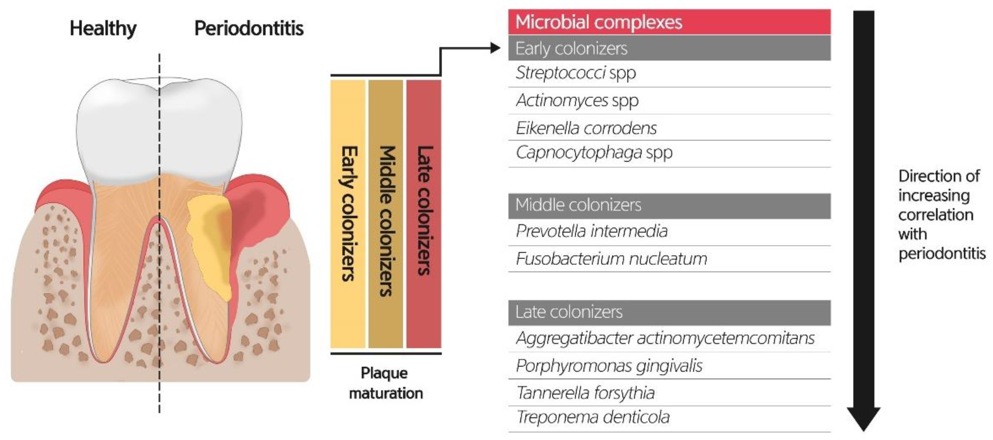
6.1. Role of Oral Microbiome on Periodontitis

Periodontal disease is one of the most prevalent oral diseases worldwide. Periodontitis is characterized by the extension of inflammation into the supporting tissues of the teeth, causing the loss of attachment and bone. Progression of the disease may involve local, systemic, or environmental factors, and subsequent immune-inflammatory responses can occur in both hypo- and hyper-responsive manners [62]. It is a chronic non-communicable disease (NCD) with common risk factors with other NCDs, including cardiovascular disease, chronic obstructive pulmonary disease, diabetes, and cancer, which contribute to an increase in the global burden [63,64,65]. Gingivitis is an inflammation within the gingival tissue induced by the accumulation of bacterial deposits at the gingival crevice, which can be resolved by removing the deposits [66]. Despite the complex multifactorial features of the host response, the essential role of bacteria in the etiopathogenesis of gingivitis and periodontitis has long been identified [67,68,69].
Among the microbial habitats in the oral cavity, tooth surfaces harbor dental plaque biofilms in both supragingival and subgingival areas, and the bacterial deposits might vary depending on the anatomic factors such as surface topography, location, and shape of the teeth that affect bacterial stagnation and oral hygiene [70]. Individual variations in the biomass and composition of plaque are found between different sites of the same tooth or same sites of different teeth. More posterior locations and interdental space accumulated higher levels of plaque, whereas lingual surfaces showed limited debris compared to the buccal surface [71]. Further research and information on the spatial patterning of the oral microbiome associated with periodontal disease will further the understanding of the site-specific characteristics of the disease.
The crevicular epithelium and gingival crevice are oral microbial habitats critical for the initiation and development of gingivitis and periodontitis. The microbial community on the root surface can be protected from shear forces; the microenvironment is nourished by the gingival crevicular fluid (GCF), a serum-like exudate from the adjacent tissue, and a low redox potential to maintain anaerobic conditions. It has been estimated that more than 500 species exist in the subgingival plaque [72], and the current methods of high-throughput molecular technologies provide extended knowledge and understanding of the highly diverse microbial community in the oral cavity. The healthy subgingival microbiome is characterized by the dominant Gram-positive cocci and rods, Actinomyces spp. and Streptococcus spp., as early colonizers that co-aggregate and form early dental plaque [72]. The Gram-negative rod Fusobacterium nucleatum is the second most abundant species that acts as a secondary colonizer to bridge multiple bacteria as the plaque matures. Other Gram-negative species, such as Veillonella spp. and Capnocytophaga spp., are also important components of biofilms. Recently, Gram-positive Rothia spp. and Corynebacteria have emerged for their roles in spatial arrangements [73]. With the development of gingivitis, the subgingival microbiome shifts to an increase in Gram-negative bacteria, including Prevotella spp., Selenomonas spp., and F. nucleatum ss. polymorphum with a decrease in Gram-positive species and is involved in the elevation of inflammatory cytokines in GCF [74,75,76]. The total biomass of the bacteria, as well as compositional changes, increased by 3-log.
The periodontitis-associated subgingival microbiome can be described as the enrichment of diverse groups of Gram-negative species. In the classic fundamental study of Socransky et al., red complex bacteria comprising three species, Porphyromonas gingivalis, Treponema denticola, and Tannerella forsythia, are strongly associated with diseased sites and possess virulence factors such as gingipain, dentilisin, and PrtH, respectively, which showed high protease activity [6]. Aggregatibacter actinomycetemcomitans appears to be associated with aggressive periodontitis rather than chronic periodontitis, and it secretes leukotoxin to damage host cells. In terms of dysbiosis, P. gingivalis can subvert or avoid host immune components such as Toll-like receptors and complements, which triggers an imbalance in host–bacterial interactions and relative abundance of other bacteria compared with that in the healthy state [68]. Several keystone pathogens, including the red complex triad, orchestrated inflammatory disease by altering the microbiota associated with the disease state and through the optimization of the acquisition of nutrition from the host, and further facilitate the growth of pathobionts that stimulate host immune responses, resulting in bone loss [77,78] (Figure 4).

6.2. Role of Oral Microbiome and Dental Caries

Dental caries is characterized by the demineralization of susceptible dental hard tissues by acidic bacterial by-products from sugar metabolism that leads to cavitation [79] and a chronic continuous process from sub-clinical decay to dentinal involvement [80]. Dental caries results from an ecological imbalance towards acidogenic and aciduric bacterial shifts and environmental acidification within the dental biofilm, and can therefore be explained as a dysbiosis-associated disease [81]. Microbiota adapt to the reduced pH microenvironment along with the prolonged maturation of the biofilms; the cavitated lesions allow for advanced ecological niches [82].
Epidemiologic findings revealed a more frequent occurrence of dental caries in the occlusal and proximal surfaces of the first molars [83]. Pit and fissure structures or interdental spaces are anatomical factors that are protected from host defense mechanisms and avoid adequate removal of the plaque biofilm. Site-to-site variabilities in salivary film velocity and salivary clearance might affect the environmental selection and biogeography of the microbiome community, which in turn implies that the tooth surface is susceptible to demineralization [84].
Bacterial diversity was considerably different according to the disease conditions, as the supragingival plaque on the sound surface included 500–600 species, which was reduced to 200 in dentin caries and approximately 125 in non-cavitated enamel lesions [71]. In addition, the bacterial composition was variable among the different carious sites and lesions within the individual or among individuals [85,86]. Despite the complex microbial variability, the bacterial diversity of the caries-associated microbiome decreases due to the competitiveness of the microorganisms; the characterization of the cariogenic consortia should be further identified [87]. Streptococcus mutans are the initial colonizers of the supragingival biofilm, which produce water-insoluble glucans that promote bacterial adhesion and only comprise <1% of the total community [88]. In addition, mutans streptococci, especially Streptococcus mutans and Streptococcus sobrinus, were first identified as prominent pathogens because of their extensive acidogenic and aciduric properties [89,90]. The mutan-centric paradigm in caries-etiopathogenesis has been challenged by the identification of other acidogenic species, including Bifidobacterium [91], Lactobacillus [92] and Scardovia wiggsiae [93], which showed a strong association with caries. It was also revealed that a complex community associated with caries included other prominent species such as Atopobium, Prevotella, Corynebacterium, non-mutans streptococci, Veillonella, and Capnocytophaga [71,88]. These species produce weak organic acids after carbohydrate fermentation and decrease the local pH to demineralize the tooth tissues. Nevertheless, its potential role in triggering dysbiosis should not be underestimated.

6.3. Role of Oral Microbiome on Oral Lichen Planus

Oral lichen planus (OLP) is a chronic inflammatory mucocutaneous disease that mainly involves the oral mucosa. The etiology and pathogenesis of OLP are not clearly understood. However, OLP has been linked to multiple disease processes and agents, such as autoimmune diseases, allergic reactions to dental restorative materials, viral and bacterial infections, vaccinations, and medications [94]. So far, there has been little research on the oral microbiome in OLP. Bornstein et al. [95] reported higher bacterial counts of C. sputigena, E. corrodens, L. crispatus, M. curtisii, N. mucosa, P. bivia, P. intermedia, S. agalactiae and S. haemolyticus at the OLP lesion sites. Ertugrul et al. also reported that OLP patients had higher levels of infection with A. actinomycetemcomitans, P. gingivalis, P. intermedia, T. forsythia, and T. denticola than non-OLP patients [96]. Choi et al. found a decrease in Streptococcus and an increase in gingivitis/periodontitis-associated bacteria in OLP lesions [97]. Furthermore, they demonstrated that intracellular bacteria in the tissue and bacterial LPS might induce the production of T cell chemokines C-X-C motif chemokine ligand 10 and C-C motif chemokine ligand [98]. To date, the association between OLP and HCV viral infection appears to be dependent on geographical heterogeneity. This was first suggested by Mokni et al. in 1991, and Carrozzo et al. demonstrated a strong association between hepatitis C viral infection and OLP [99,100]. However, changes in the oral microbiome in patients with OLP remain unclear.

6.4. Role of the Oral Microbiome on Pre-Malignancy and Oral Cancers

Oral cancer is a cancer of the lips, mouth, or oropharynx. In an effort to elucidate the pathogenesis of oral cancers, oral microbiota has come into the spotlight and has been suggested to be involved through three possible mechanisms [101]. The chronic inflammatory responses provoked by bacteria could be responsible for this, since chronic inflammatory mediators facilitate cell proliferation, mutagenesis, and oncogene activation.
Anaerobic oral bacteria are known to cause chronic inflammatory processes in periodontal tissue by increasing interleukin-1β (IL-1β), IL-6, tumor necrosis factor-α, and matrix metalloproteinases MMP-8 and MMP-9 [102]. The bacterial effector proteins using type 3 or type 4 secretion systems might influence cell proliferation, cytoskeletal rearrangements, activation of NF-κB, and inhibition of cellular apoptosis [18,101]. In particular, P. gingivalis has been suggested to be anti-apoptotic. Additionally, the purinergic receptor P2X7 receptors activated by ATP are involved in cell death and apoptosis, and are affected by a nucleoside-diphosphate-kinase homolog, which is an ATP-consuming enzyme in P. gingivalis [103,104].
Carcinogenic substances such as acetaldehyde converted from ethanol, reactive oxygen species, reactive nitrogen species, and volatile sulfur compounds by bacteria might facilitate carcinogenic processes [105]. S. gordonii, S. mitis, S. oralis, S. salivarius, and S. sanguinis [91], and Candida can metabolize alcohol to acetaldehyde using the enzyme alcohol dehydrogenase [106]. ROS and RNS produced by peroxigenic oral bacteria, including S. mitis, S. gordonii, S. sanguinis, S. oralis, L. fermentum, and Ljensenii, have been identified in various oral cancers. VSCs have genotoxic effects and can cause genomic mutations [107]. P. gingivalis, Pr. intermedia, A. actinomycetemcomitans, and F. nucleatum are mainly responsible for the production of hydrogen sulfide (H2S), methyl mercaptan (CH3SH), and dimethyl sulfide ((CH3)2S) [108,109].
Recent data have indicated an association between human cytomegalovirus and carcinogenesis [110]. Human cytomegalovirus belongs to the beta subfamily of Herpesviridae and is frequently detected in periodontal tissues [111,112,113]. Proangiogenic and anti-apoptotic mechanisms and immunosuppressive effects demonstrate the oncogenic potential of human cytomegalovirus, and a non-lytic infection of the virus can also promote the oncogenic transformation of epithelial cells [110]. These data suggest that the virus may contribute to the oncogenic process of oral malignancies, although direct evidence is yet to be provided.
More studies investigating the pathophysiology of oral cancers are required to confirm the association between oral cancer and oral microbiota; however, evidence advocating for the association is accumulating, and oral cancer surfaces harbor significantly higher numbers of oral aerobes and anaerobes compared to the healthy mucosa surface [114]. Therefore, the detection and management of the oral microenvironment are essential for the control of oral malignancy.

7. Oral Microbiome and Systemic Diseases

The oral microbiome can also be a pathogenic factor in systemic diseases (Figure 5). The association between the oral microbiome and systemic diseases has been reported because the mouth is the entrance connecting the external environment and the inside of the body. A bidirectional relationship may exist between the oral microbial community and systemic diseases [9].
The oral microbiome and inflammatory molecules can invade the systemic organs in two primary ways: the bloodstream or the digestive tract. First, bloodstream invasion is possible because the periodontal pockets are anatomically close to the bloodstream. Second, by means of alimentary dissemination, the oral microbiome reaches the digestive tract. However, the precise mechanisms of invasion must be determined [8].

7.1. Autoimmune Disease

Dysbiosis of the oral microbiome plays a prominent role in several autoimmune diseases such as rheumatoid arthritis (RA), systemic lupus erythematosus (SLE), and primary Sjogren syndrome (SS) [115]. In autoimmune diseases, the most studied connection between the oral microbiome and disease is RA. The RA disease activity scores, DAS 28, were higher in patients with RA with more serum antibodies of P. gingivalis, an oral anaerobe involved in the development of periodontitis [116,117,118]. P. gingivalis produces gingipains and peptidylarginine deiminase, which enable protein citrullination, an important trigger for RA anticitrullinated peptide antibody [119,120]. In addition, many epidemiological studies have shown a correlation between RA and periodontitis [121,122]. In a recent meta-analysis of 21 studies, periodontitis was more frequent in patients with RA than in healthy controls, with a risk ratio of 1.13 [123].
Dysbiosis of the oral microbiome in SS has been reported in several studies [124,125,126,127,128,129,130]. A decrease in salivary secretion is the most important factor in oral dysbiosis in SS [128,129]. However, the impact of the oral microbiome on SS pathogenesis remains unclear. van der Meulen et al. reported higher Firmicutes/Proteobacteria ratios compared to those of healthy controls and higher abundances of 19 genera in SS patients. SLE is also associated with changes in the oral microbiome. In 2017, Corrêa et al. reported the influence of SLE on the subgingival microbiota, that the Lachnospiraceae family was increased compared to controls at healthy gingival sites, and the proportions of Prevotella oulorum and Prevotella pleuritidis, Pseudomonas spp., Treponema maltophilum, and Actinomyces found in healthy individuals [131]. Bacteria were elevated at periodontal sites compared with the controls. Periodontal disease is also associated with SLE, which increases the risk or severity of SLE [132,133].

7.2. Systemic Malignancies

In a recent longitudinal study of hospitalized cancer patients, increased variability in the oral microbiome was associated with adverse clinical outcomes [91]. In colorectal cancer (CRC), the role of microbiota in carcinogenesis and its clinical significance has been reported in many studies. The identification of the Fusobacterium genus in about 30% of CRC cases by 16S rRNA sequencing has initiated research on the impact of the oral microbiota in CRC [134,135,136]. Fusobacterium nucleatum, an oral commensal species, is more frequently identified in CRC than in colorectal adenoma, suggesting that Fusobacterium may contribute to later progression rather than an earlier stage along the colorectal adenoma-carcinoma sequence [137,138]. However, Kato et al. reported no association between Fusobacterium and CRC; instead, they observed associations with the genera Lactobacillus and Rothia by means of an NGS-based study [139]. They explained that Lactobacillus and Rothia were related to oral hygiene, and poor oral hygiene was related to CRC. Other microbes of oral microbiota, such as Porphyromonas, Peptostreptococcus, Prevotella, Parvimonas, and Gemella, are often associated with CRC. However, the carcinogenic potential and virulence factors of these genera are unknown [138].
Oral microbiota variation was associated with pancreatic cancer, and the combination of decreased Neisseria elongata with Streptococcus mitis was suggested to distinguish factors for pancreatic cancer [140]. Michaud et al. reported that the risk of pancreatic cancer was significantly increased in the presence of elevated serum antibodies against P. gingivalis [141]. Both P. gingivalis and Aggregatibacter actinomycetemcomitans have the potential to initiate Toll-like receptor pathways, which have been shown to be drivers of pancreatic carcinogenesis [142]. In 2016, a prospective large cohort study supported that P. gingivalis and Aggregatibacter actinomycetemcomitans may contribute to a higher risk of pancreatic cancer [143].
Esophageal cancer has also been reported to be related to Tannerella forsythia and P. gingivalis [144]. The study showed that the genus Neisseria was associated with a lower risk of esophageal cancer, as was the carotenoid biosynthesis pathway, to which a number of Neisseria species can potentially contribute.
Some studies have shown that periodontal diseases are associated with lung cancer risk [145,146,147]; however, it was difficult to analyze the association between oral microbiota and lung cancer because smoking, one of the biggest risk factors for lung cancer, can also affect the oral microbiota. In a small cross-sectional study, the abundance of Capnocytophaga and Veillonella was elevated, together with a reduced number of Neisseria, as found by means of 16S rRNA gene sequencing [148]. Thus, the current data are insufficient to conclude that oral microbial variations contribute to lung cancer.

7.3. Pregnancy Outcomes

Infection-related preterm birth is the leading cause of infant mortality and morbidity. Evidence indicates that ~40% of preterm births are vaginal and intrauterine infection-related, and ~50% are associated with intra-amniotic infections [149]. Therefore, an understanding of the origin of the offending bacteria and routes of invasion of the placenta and amniotic cavity is required. A review by Mendez et al. concluded that the most common intra-amniotic bacterial taxa were species associated with the vagina, although other species were commonly associated with the oral cavity, gastrointestinal tract, and respiratory tract [150].
Significant evidence supports an association between periodontal pathogenic bacteria, preterm birth, and preeclampsia. For example, Fusobacterium nucleatum is associated with adverse pregnancy complications [151], and Porphyromonas gingivalis has been detected in the amniotic fluid of pregnant women at risk for premature delivery and in the placentas of patients with preeclampsia [52,152].

7.4. Other Systemic Diseases

There is still conflict regarding the relationship between chronic inflammatory conditions, such as diabetes, and changes in the oral microbiome. In 2013, Chapple et al. reported that there is no compelling evidence that diabetes has a significant impact on the oral microbiota [153]. However, several studies have reported the impact of diabetes on changes in the oral microbiome. da Cruz et al. reported an increase in P. gingivalis and Tannerella forsythia in diabetes [154], and Ganesan et al. reported increased levels of Capnocytophaga, Pseudomonas, Bergeyella, Sphingomonas, Corynebacterium, Propionibacterium, and Neisseria in hyperglycemic individuals [155]. One NGS study reported that diabetes reduces P. gingivalis, Tannerella forsythia, and Treponema denticola [156]. To obtain a clearer conclusion, large-scale longitudinal studies are needed in the future.
The direct effect of oral bacteria on cardiovascular disease (CVD) is even less well-known, and the involvement of bacteria in atherosclerotic plaques in atherogenesis is not clear. However, since periodontitis and oral dysbiosis are related, oral dysbiosis is thought to be related to systemic inflammation. In a number of studies, the oral microbiota affected the outcome of CVD. The periodontal pathogen burden has been linked with acute coronary syndrome (ACS) and subclinical atherosclerosis [157,158,159], and Fak et al. reported that Anaeroglobus was more abundant in patients with symptomatic atherosclerosis than in controls [160]. Oral Streptococcus sanguinis moves from the oral cavity into the human bloodstream, colonizes the cardiac endothelium, and is a risk factor for infective endocarditis [161,162]. Lipopolysaccharide (LPS), endotoxin, and virulence factors of bacteria are considered a molecular link between the microbiome and cardiometabolic disorders [163]. Serum LPS activity correlates with the levels of the P. gingivalis antibody [164]. In addition, proteins secreted by P. gingivalis, such as gingipains, are implicated in their pathogenicity [165] and subsequently activate cytokine production [166]. There appear to be fewer studies using NGS techniques of oral cavity samples to determine the associations between the oral microbiome composition and cardiovascular disease.
Neurodegenerative disorders, such as Alzheimer’s disease [167] and Parkinson’s disease [168], have also been reported to be associated with the oral microbiome. The most complete study in this regard is the association between P. gingivalis and Alzheimer’s disease [167]. P. gingivalis infection in mice resulted in brain colonization, and gingipain proteases produced by P. gingivalis have neurotoxic characteristics. It has also been shown that typical oral species of the phylum Spirochaetes, including multiple species of the genus Treponema, often comprise amyloid plaques [169]. The potential mechanisms of their action by oral taxa make this another attractive area for investigation.

8. Conclusions

There is a dynamic interaction between the oral and systemic environments and the composition of the resident oral microbiome (Figure 6). Accumulated evidence suggests that the oral microbiome is individualized and relatively stable over time, as long as oral and general health are maintained. Substantial changes in key environmental parameters that affect microbial growth can disrupt the natural balance of the oral microbiome and select potentially pathogenic organisms. In addition, the presence of systemic diseases and oral microbiota seems to have a bidirectional effect. Thus, the oral microbiome reflects the oral and general health status of individuals. However, future studies are needed to determine whether changes in the oral microbiome precede clinical signs of disease, which would enable the use of the oral microbiome in the prediction of future disease risk. In our extensive and comprehensive narrative review, there may be a bias in the selection of papers based on keywords and for papers written in English. Accordingly, follow-up studies, including meta-analyses, are needed to reach a clearer conclusion. In addition, prospective longitudinal studies are urgently needed to reveal the full potential of using the oral microbiome in the field of precision medicine.

Author Contributions

All authors, including Y.-H.L., S.W.C., Q.-S.A., S.-J.H., Y.-A.L., J.J., G.-J.L., H.J.P., S.-I.S. and J.-Y.H. participated in paper writing—original draft preparation. Conceptualization, Y.-H.L.; methodology, Y.-H.L. and J.-Y.H.; software, Y.-H.L.; validation, Q.-S.A., S.-J.H., Y.-A.L., J.J., G.-J.L. and H.J.P.; formal analysis, Y.-H.L. and J.-Y.H. Investigation, Y.-H.L., S.W.C., Q.-S.A., S.-J.H., Y.-A.L., J.J., G.-J.L., H.J.P., S.-I.S. and J.-Y.H.; resources, Y.-H.L., S.W.C., Q.-S.A., S.-J.H., Y.-A.L., J.J., G.-J.L., H.J.P., S.-I.S. and J.-Y.H.; data curation, Y.-H.L.; writing—review and editing, Y.-H.L.; visualization, Y.-H.L. and J.-Y.H.; supervision, Y.-H.L.; project administration, Y.-H.L.; funding acquisition, Y.-H.L. All authors have read and agreed to the published version of the manuscript.

Funding

This work was supported by the Korea Medical Device Development Fund grant funded by the Korean government (the Ministry of Science and ICT, the Ministry of Trade, Industry and Energy, the Ministry of Health and Welfare, Republic of Korea, the Ministry of Food and Drug Safety) (Project Number: KMDF_PR_20200901_0023, 9991006696).

Conflicts of Interest

The authors declare no conflict of interest.

References

  1. Sender, R.; Fuchs, S.; Milo, R. Are we really vastly outnumbered? Revisiting the ratio of bacterial to host cells in humans. Cell 2016, 164, 337–340. [Google Scholar] [CrossRef] [PubMed] [Green Version]
  2. Delgado, S.; Cabrera-Rubio, R.; Mira, A.; Suárez, A.; Mayo, B. Microbiological survey of the human gastric ecosystem using culturing and pyrosequencing methods. Microb. Ecol. 2013, 65, 763–772. [Google Scholar] [CrossRef] [PubMed]
  3. Malla, M.A.; Dubey, A.; Kumar, A.; Yadav, S.; Hashem, A.; Abd Allah, E.F. Exploring the human microbiome: The potential future role of next-generation sequencing in disease diagnosis and treatment. Front. Immunol. 2019, 9, 2868. [Google Scholar] [CrossRef] [PubMed]
  4. Kilian, M.; Chapple, I.L.; Hannig, M.; Marsh, P.D.; Meuric, V.; Pedersen, A.M.; Tonetti, M.S.; Wade, W.G.; Zaura, E. The oral microbiome—An update for oral healthcare professionals. Br. Dent. J. 2016, 221, 657–666. [Google Scholar] [CrossRef] [PubMed]
  5. Marsh, P.D. Are dental diseases examples of ecological catastrophes? Microbiology 2003, 149, 279–294. [Google Scholar] [CrossRef] [PubMed]
  6. Dewhirst, F.E.; Chen, T.; Izard, J.; Paster, B.J.; Tanner, A.C.; Yu, W.H.; Lakshmanan, A.; Wade, W.G. The human oral microbiome. J. Bacteriol. 2010, 192, 5002–5017. [Google Scholar] [CrossRef] [PubMed] [Green Version]
  7. Gao, L.; Xu, T.; Huang, G.; Jiang, S.; Gu, Y.; Chen, F. Oral microbiomes: More and more importance in oral cavity and whole body. Protein Cell 2018, 9, 488–500. [Google Scholar] [CrossRef] [Green Version]
  8. Bourgeois, D.; Inquimbert, C.; Ottolenghi, L.; Carrouel, F. Periodontal pathogens as risk factors of cardiovascular diseases, diabetes, rheumatoid arthritis, cancer, and chronic obstructive pulmonary disease—Is there cause for consideration? Microorganisms 2019, 7, 424. [Google Scholar] [CrossRef] [Green Version]
  9. Willis, J.R.; Gabaldón, T. The human oral microbiome in health and disease: From sequences to ecosystems. Microorganisms 2020, 8, 308. [Google Scholar] [CrossRef] [Green Version]
  10. Maukonen, J.; Mättö, J.; Suihko, M.L.; Saarela, M. Intra-individual diversity and similarity of salivary and faecal microbiota. J. Med. Microbiol. 2008, 57, 1560–1568. [Google Scholar] [CrossRef] [Green Version]
  11. Espinoza, J.L.; Harkins, D.M.; Torralba, M.; Gomez, A.; Highlander, S.K.; Jones, M.B.; Leong, P.; Saffery, R.; Bockmann, M.; Kuelbs, C.; et al. Supragingival plaque microbiome ecology and functional potential in the context of health and disease. mBio 2018, 9, e01631-18. [Google Scholar] [CrossRef] [PubMed] [Green Version]
  12. Camelo-Castillo, A.J.; Mira, A.; Pico, A.; Nibali, L.; Henderson, B.; Donos, N.; Tomás, I. Subgingival microbiota in health compared to periodontitis and the influence of smoking. Front. Microbiol. 2015, 6, 119. [Google Scholar] [CrossRef] [PubMed] [Green Version]
  13. Inquimbert, C.; Bourgeois, D.; Bravo, M.; Viennot, S.; Tramini, P.; Llodra, J.C.; Molinari, N.; Dussart, C.; Giraudeau, N.; Carrouel, F. The oral bacterial microbiome of interdental surfaces in adolescents according to carious risk. Microorganisms 2019, 7, 319. [Google Scholar] [CrossRef] [PubMed] [Green Version]
  14. Carrouel, F.; Viennot, S.; Santamaria, J.; Veber, P.; Bourgeois, D. Quantitative molecular detection of 19 major pathogens in the interdental biofilm of periodontally healthy young adults. Front. Microbiol. 2016, 7, 840. [Google Scholar] [CrossRef] [PubMed]
  15. Asakawa, M.; Takeshita, T.; Furuta, M.; Kageyama, S.; Takeuchi, K.; Hata, J.; Ninomiya, T.; Yamashita, Y. Tongue microbiota and oral health status in community-dwelling elderly adults. mSphere 2018, 3, e00332-18. [Google Scholar] [CrossRef] [PubMed] [Green Version]
  16. Faran Ali, S.M.; Tanwir, F. Oral microbial habitat a dynamic entity. J. Oral Biol. Craniofac. Res. 2012, 2, 181–187. [Google Scholar] [CrossRef] [Green Version]
  17. Deo, P.N.; Deshmukh, R. Oral microbiome: Unveiling the fundamentals. J. Oral Maxillofac. Pathol. 2019, 23, 122–128. [Google Scholar] [CrossRef]
  18. Lim, Y.; Totsika, M.; Morrison, M.; Punyadeera, C. Oral microbiome: A new biomarker reservoir for oral and oropharyngeal cancers. Theranostics 2017, 7, 4313–4321. [Google Scholar] [CrossRef]
  19. Baliga, S.; Muglikar, S.; Kale, R. Salivary pH: A diagnostic biomarker. J. Indian Soc. Periodontol. 2013, 17, 461–465. [Google Scholar] [CrossRef]
  20. Dickinson, B.C.; Moffatt, C.E.; Hagerty, D.; Whitmore, S.E.; Brown, T.A.; Graves, D.T.; Lamont, R.J. Interaction of oral bacteria with gingival epithelial cell multilayers. Mol. Oral Microbiol. 2011, 26, 210–220. [Google Scholar] [CrossRef]
  21. Peters, B.A.; Wu, J.; Hayes, R.B.; Ahn, J. The oral fungal mycobiome: Characteristics and relation to periodontitis in a pilot study. BMC Microbiol. 2017, 17, 157. [Google Scholar] [CrossRef] [PubMed]
  22. Bachrach, G.; Leizerovici-Zigmond, M.; Zlotkin, A.; Naor, R.; Steinberg, D. Bacteriophage isolation from human saliva. Lett. Appl. Microbiol. 2003, 36, 50–53. [Google Scholar] [CrossRef]
  23. Ly, M.; Abeles, S.R.; Boehm, T.K.; Robles-Sikisaka, R.; Naidu, M.; Santiago-Rodriguez, T.; Pride, D.T. Altered oral viral ecology in association with periodontal disease. mBio 2014, 5, e01133-14. [Google Scholar] [CrossRef] [Green Version]
  24. Pride, D.T.; Salzman, J.; Haynes, M.; Rohwer, F.; Davis-Long, C.; White, R.A., 3rd; Loomer, P.; Armitage, G.C.; Relman, D.A. Evidence of a robust resident bacteriophage population revealed through analysis of the human salivary virome. ISME J. 2012, 6, 915–926. [Google Scholar] [CrossRef]
  25. Naidu, M.; Robles-Sikisaka, R.; Abeles, S.R.; Boehm, T.K.; Pride, D.T. Characterization of bacteriophage communities and CRISPR profiles from dental plaque. BMC Microbiol. 2014, 14, 175. [Google Scholar] [CrossRef] [PubMed] [Green Version]
  26. Pérez-Brocal, V.; Moya, A. The analysis of the oral DNA virome reveals which viruses are widespread and rare among healthy young adults in Valencia (Spain). PLoS ONE 2018, 13, e0191867. [Google Scholar] [CrossRef] [PubMed] [Green Version]
  27. Edlund, A.; Santiago-Rodriguez, T.M.; Boehm, T.K.; Pride, D.T. Bacteriophage and their potential roles in the human oral cavity. J. Oral Microbiol. 2015, 7, 27423. [Google Scholar] [CrossRef] [PubMed]
  28. To, K.K.; Tsang, O.T.; Yip, C.C.; Chan, K.H.; Wu, T.C.; Chan, J.M.; Leung, W.S.; Chik, T.S.; Choi, C.Y.; Kandamby, D.H.; et al. Consistent detection of 2019 novel coronavirus in Saliva. Clin. Infect. Dis. 2020, 71, 841–843. [Google Scholar] [CrossRef] [PubMed] [Green Version]
  29. Yoon, J.G.; Yoon, J.; Song, J.Y.; Yoon, S.Y.; Lim, C.S.; Seong, H.; Noh, J.Y.; Cheong, H.J.; Kim, W.J. Clinical significance of a high SARS-CoV-2 viral load in the Saliva. J. Korean Med. Sci. 2020, 35, e195. [Google Scholar] [CrossRef] [PubMed]
  30. Sharma, N.; Bhatia, S.; Sodhi, A.S.; Batra, N. Oral microbiome and health. AIMS Microbiol. 2018, 4, 42–66. [Google Scholar] [CrossRef] [PubMed]
  31. Lai, C.C.; Shih, T.P.; Ko, W.C.; Tang, H.J.; Hsueh, P.R. Severe acute respiratory syndrome coronavirus 2 (SARS-CoV-2) and coronavirus disease-2019 (COVID-19): The epidemic and the challenges. Int. J. Antimicrob. Agents 2020, 55, 105924. [Google Scholar] [CrossRef] [PubMed]
  32. Wang, Y.; Kang, H.; Liu, X.; Tong, Z. Combination of RT-qPCR testing and clinical features for diagnosis of COVID-19 facilitates management of SARS-CoV-2 outbreak. J. Med. Virol. 2020, 92, 538–539. [Google Scholar] [CrossRef] [Green Version]
  33. Sabino-Silva, R.; Jardim, A.C.G.; Siqueira, W.L. Coronavirus COVID-19 impacts to dentistry and potential salivary diagnosis. Clin. Oral Investig. 2020, 24, 1619–1621. [Google Scholar] [CrossRef] [Green Version]
  34. Medeiros da Silva, R.C.; Nogueira Marinho, L.C.; de Araújo Silva, D.N.; Costa de Lima, K.; Pirih, F.Q.; Luz de Aquino Martins, A.R. Saliva as a possible tool for the SARS-CoV-2 detection: A review. Travel Med. Infect. Dis. 2020, 38, 101920. [Google Scholar] [CrossRef] [PubMed]
  35. Zaura, E. Next-generation sequencing approaches to understanding the oral microbiome. Adv. Dent. Res. 2012, 24, 81–85. [Google Scholar] [CrossRef]
  36. Caselli, E.; Fabbri, C.; D’Accolti, M.; Soffritti, I.; Bassi, C.; Mazzacane, S.; Franchi, M. Defining the oral microbiome by whole-genome sequencing and resistome analysis: The complexity of the healthy picture. BMC Microbiol. 2020, 20, 120. [Google Scholar] [CrossRef] [PubMed]
  37. Clarridge, J.E., 3rd. Impact of 16S rRNA gene sequence analysis for identification of bacteria on clinical microbiology and infectious diseases. Clin. Microbiol. Rev. 2004, 17, 840–862. [Google Scholar] [CrossRef] [PubMed] [Green Version]
  38. Lochman, J.; Zapletalova, M.; Poskerova, H.; Izakovicova Holla, L.; Borilova Linhartova, P. Rapid multiplex real-time PCR method for the detection and quantification of selected cariogenic and periodontal bacteria. Diagnostics 2019, 10, 8. [Google Scholar] [CrossRef] [PubMed] [Green Version]
  39. Melendez, J.H.; Frankel, Y.M.; An, A.T.; Williams, L.; Price, L.B.; Wang, N.Y.; Lazarus, G.S.; Zenilman, J.M. Real-time PCR assays compared to culture-based approaches for identification of aerobic bacteria in chronic wounds. Clin. Microbiol. Infect. 2010, 16, 1762–1769. [Google Scholar] [CrossRef] [PubMed]
  40. Sakamoto, M.; Umeda, M.; Benno, Y. Molecular analysis of human oral microbiota. J. Periodontal Res. 2005, 40, 277–285. [Google Scholar] [CrossRef]
  41. Huang, C.; Shi, G. Smoking and microbiome in oral, airway, gut and some systemic diseases. J. Transl. Med. 2019, 17, 225. [Google Scholar] [CrossRef] [Green Version]
  42. Martinon, P.; Fraticelli, L.; Giboreau, A.; Dussart, C.; Bourgeois, D.; Carrouel, F. Nutrition as a key modifiable factor for periodontitis and main chronic diseases. J. Clin. Med. 2021, 10, 197. [Google Scholar] [CrossRef] [PubMed]
  43. Lamont, R.J.; Koo, H.; Hajishengallis, G. The oral microbiota: Dynamic communities and host interactions. Nat. Rev. Microbiol. 2018, 16, 745–759. [Google Scholar] [CrossRef] [PubMed]
  44. Lönnroth, I.; Falsen, E.; Westin, J.; Lindholm, L. Two cases of “heavy chain disease”. Acta Pathol. Microbiol. Scand. Sect. B Microbiol. Immunol. 1971, 79, 443. [Google Scholar]
  45. Nociti, F.H., Jr.; Casati, M.Z.; Duarte, P.M. Current perspective of the impact of smoking on the progression and treatment of periodontitis. Periodontol. 2000 2015, 67, 187–210. [Google Scholar] [CrossRef]
  46. Wu, J.; Peters, B.A. Cigarette smoking and the oral microbiome in a large study of American adults. ISME J. 2016, 10, 2435–2446. [Google Scholar] [CrossRef] [PubMed]
  47. Kumar, P.S.; Matthews, C.R.; Joshi, V.; de Jager, M.; Aspiras, M. Tobacco smoking affects bacterial acquisition and colonization in oral biofilms. Infect. Immun. 2011, 79, 4730–4738. [Google Scholar] [CrossRef] [PubMed] [Green Version]
  48. Saadaoui, M.; Singh, P.; Al Khodor, S. Oral microbiome and pregnancy: A bidirectional relationship. J. Reprod. Immunol. 2021, 145, 103293. [Google Scholar] [CrossRef]
  49. Tettamanti, L.; Lauritano, D.; Nardone, M.; Gargari, M.; Silvestre-Rangil, J.; Gavoglio, P.; Tagliabue, A. Pregnancy and periodontal disease: Does exist a two-way relationship? ORAL Implantol. 2017, 10, 112–118. [Google Scholar] [CrossRef]
  50. Barak, S.; Oettinger-Barak, O.; Oettinger, M.; Machtei, E.E.; Peled, M.; Ohel, G. Common oral manifestations during pregnancy: A review. Obstet. Gynecol. Surv. 2003, 58, 624–628. [Google Scholar] [CrossRef]
  51. Madianos, P.N.; Bobetsis, Y.A.; Offenbacher, S. Adverse pregnancy outcomes (APOs) and periodontal disease: Pathogenic mechanisms. J. Periodontol. 2013, 84, S170–S180. [Google Scholar] [CrossRef] [PubMed]
  52. León, R.; Silva, N.; Ovalle, A.; Chaparro, A.; Ahumada, A.; Gajardo, M.; Martinez, M.; Gamonal, J. Detection of porphyromonas gingivalis in the amniotic fluid in pregnant women with a diagnosis of threatened premature labor. J. Periodontol. 2007, 78, 1249–1255. [Google Scholar] [CrossRef] [PubMed] [Green Version]
  53. Fujiwara, N.; Tsuruda, K.; Iwamoto, Y.; Kato, F.; Odaki, T.; Yamane, N.; Hori, Y.; Harashima, Y.; Sakoda, A.; Tagaya, A.; et al. Significant increase of oral bacteria in the early pregnancy period in Japanese women. J. Investig. Clin. Dent. 2017, 8, e12189. [Google Scholar] [CrossRef] [PubMed]
  54. Lin, W.; Jiang, W.; Hu, X.; Gao, L.; Ai, D.; Pan, H.; Niu, C.; Yuan, K.; Zhou, X.; Xu, C.; et al. Ecological shifts of supragingival microbiota in association with pregnancy. Front. Cell. Infect. Microbiol. 2018, 8, 24. [Google Scholar] [CrossRef] [PubMed]
  55. Lazarevic, V.; Manzano, S.; Gaïa, N.; Girard, M.; Whiteson, K.; Hibbs, J.; François, P.; Gervaix, A.; Schrenzel, J. Effects of amoxicillin treatment on the salivary microbiota in children with acute otitis media. Clin. Microbiol. Infect. 2013, 19, e335–e342. [Google Scholar] [CrossRef] [PubMed] [Green Version]
  56. Raju, S.C.; Viljakainen, H.; Figueiredo, R.A.O.; Neuvonen, P.J.; Eriksson, J.G.; Weiderpass, E.; Rounge, T.B. Antimicrobial drug use in the first decade of life influences saliva microbiota diversity and composition. Microbiome 2020, 8, 121. [Google Scholar] [CrossRef]
  57. Gupta, P.; Gupta, N.; Pawar, A.P.; Birajdar, S.S.; Natt, A.S.; Singh, H.P. Role of sugar and sugar substitutes in dental caries: A review. ISRN Dent. 2013, 2013, 519421. [Google Scholar] [CrossRef] [PubMed]
  58. Takahashi, N.; Nyvad, B. The role of bacteria in the caries process: Ecological perspectives. J. Dent. Res. 2011, 90, 294–303. [Google Scholar] [CrossRef] [PubMed]
  59. Gross, E.L.; Leys, E.J.; Gasparovich, S.R.; Firestone, N.D.; Schwartzbaum, J.A.; Janies, D.A.; Asnani, K.; Griffen, A.L. Bacterial 16S sequence analysis of severe caries in young permanent teeth. J. Clin. Microbiol. 2010, 48, 4121–4128. [Google Scholar] [CrossRef] [Green Version]
  60. Han, S.J.; Bae, K.H.; Lee, H.J.; Kim, S.J.; Cho, H.J. Association between regular walking and periodontitis according to socioeconomic status: A cross-sectional study. Sci. Rep. 2019, 9, 12969. [Google Scholar] [CrossRef]
  61. Schwendicke, F.; Dörfer, C.E.; Schlattmann, P.; Foster Page, L.; Thomson, W.M.; Paris, S. Socioeconomic inequality and caries: A systematic review and meta-analysis. J. Dent. Res. 2015, 94, 10–18. [Google Scholar] [CrossRef] [PubMed]
  62. Pihlstrom, B.L.; Michalowicz, B.S.; Johnson, N.W. Periodontal diseases. Lancet 2005, 366, 1809–1820. [Google Scholar] [CrossRef] [Green Version]
  63. FDI World Dental Federation. FDI policy statement on oral health and the social determinants of health. Adopted by the FDI General Assembly: 30 August 2013—Istanbul, Turkey. Int. Dent. J. 2013, 63, 287–288. [Google Scholar] [CrossRef]
  64. Kassebaum, N.J.; Smith, A.G.C.; Bernabe, E.; Fleming, T.D.; Reynolds, A.E.; Vos, T.; Murray, C.J.L.; Marcenes, W.; GBD 2015 Oral Health Collaborators. Global, regional, and national prevalence, incidence, and disability-adjusted life years for oral conditions for 195 countries, 1990–2015: A systematic analysis for the global burden of diseases, injuries, and risk factors. J. Dent. Res. 2017, 96, 380–387. [Google Scholar] [CrossRef] [PubMed]
  65. Tonetti, M.S.; Jepsen, S.; Jin, L.; Otomo-Corgel, J. Impact of the global burden of periodontal diseases on health, nutrition and wellbeing of mankind: A call for global action. J. Clin. Periodontol. 2017, 44, 456–462. [Google Scholar] [CrossRef] [PubMed] [Green Version]
  66. Loe, H.; Theilade, E.; Jensen, S.B. Experimental gingivitis in man. J. Periodontol. 1965, 36, 177–187. [Google Scholar] [CrossRef] [PubMed]
  67. Darveau, R.P. Periodontitis: A polymicrobial disruption of host homeostasis. Nat. Rev. Microbiol. 2010, 8, 481–490. [Google Scholar] [CrossRef] [PubMed]
  68. Hajishengallis, G.; Liang, S.; Payne, M.A.; Hashim, A.; Jotwani, R.; Eskan, M.A.; McIntosh, M.L.; Alsam, A.; Kirkwood, K.L.; Lambris, J.D.; et al. Low-abundance biofilm species orchestrates inflammatory periodontal disease through the commensal microbiota and complement. Cell Host Microbe 2011, 10, 497–506. [Google Scholar] [CrossRef] [Green Version]
  69. Socransky, S.S.; Haffajee, A.D.; Cugini, M.A.; Smith, C.; Kent, R.L., Jr. Microbial complexes in subgingival plaque. J. Clin. Periodontol. 1998, 25, 134–144. [Google Scholar] [CrossRef]
  70. Samaranayake, L.; Matsubara, V.H. Normal oral flora and the oral ecosystem. Dent. Clin. N. Am. 2017, 61, 199–215. [Google Scholar] [CrossRef]
  71. Simón-Soro, A.; Tomás, I.; Cabrera-Rubio, R.; Catalan, M.D.; Nyvad, B.; Mira, A. Microbial geography of the oral cavity. J. Dent. Res. 2013, 92, 616–621. [Google Scholar] [CrossRef] [PubMed]
  72. Moore, W.E.; Moore, L.V. The bacteria of periodontal diseases. Periodontology 1994, 5, 66–77. [Google Scholar] [CrossRef] [PubMed]
  73. Abusleme, L.; Dupuy, A.K.; Dutzan, N.; Silva, N.; Burleson, J.A.; Strausbaugh, L.D.; Gamonal, J.; Diaz, P.I. The subgingival microbiome in health and periodontitis and its relationship with community biomass and inflammation. ISME J. 2013, 7, 1016–1025. [Google Scholar] [CrossRef] [PubMed] [Green Version]
  74. Curtis, M.A.; Diaz, P.I.; Van Dyke, T.E. The role of the microbiota in periodontal disease. Periodontology 2020, 83, 14–25. [Google Scholar] [CrossRef]
  75. Kistler, J.O.; Booth, V.; Bradshaw, D.J.; Wade, W.G. Bacterial community development in experimental gingivitis. PLoS ONE 2013, 8, e71227. [Google Scholar] [CrossRef] [PubMed] [Green Version]
  76. Schincaglia, G.P.; Hong, B.Y.; Rosania, A.; Barasz, J.; Thompson, A.; Sobue, T.; Panagakos, F.; Burleson, J.A.; Dongari-Bagtzoglou, A.; Diaz, P.I. Clinical, immune, and microbiome traits of gingivitis and peri-implant mucositis. J. Dent. Res. 2017, 96, 47–55. [Google Scholar] [CrossRef]
  77. Hajishengallis, G.; Darveau, R.P.; Curtis, M.A. The keystone-pathogen hypothesis. Nat. Rev. Microbiol. 2012, 10, 717–725. [Google Scholar] [CrossRef] [PubMed]
  78. Jiao, Y.; Hasegawa, M.; Inohara, N. The role of oral pathobionts in dysbiosis during periodontitis development. J. Dent. Res. 2014, 93, 539–546. [Google Scholar] [CrossRef] [Green Version]
  79. Selwitz, R.H.; Ismail, A.I.; Pitts, N.B. Dental caries. Lancet 2007, 369, 51–59. [Google Scholar] [CrossRef]
  80. Featherstone, J.D. The continuum of dental caries—Evidence for a dynamic disease process. J. Dent. Res. 2004, 83 (Suppl. 1), C39–C42. [Google Scholar] [CrossRef]
  81. Takahashi, N.; Nyvad, B. Caries ecology revisited: Microbial dynamics and the caries process. Caries Res. 2008, 42, 409–418. [Google Scholar] [CrossRef]
  82. Kidd, E.A.; Fejerskov, O. What constitutes dental caries? Histopathology of carious enamel and dentin related to the action of cariogenic biofilms. J. Dent. Res. 2004, 83 (Suppl. 1), C35–C38. [Google Scholar] [CrossRef] [PubMed]
  83. Hopcraft, M.S.; Morgan, M.V. Pattern of dental caries experience on tooth surfaces in an adult population. Community Dent. Oral. Epidemiol. 2006, 34, 174–183. [Google Scholar] [CrossRef] [PubMed]
  84. Almståhl, A.; Carlén, A.; Eliasson, L.; Lingström, P. Lactobacillus species in supragingival plaque in subjects with hyposalivation. Arch. Oral. Biol. 2010, 55, 255–259. [Google Scholar] [CrossRef]
  85. Simon-Soro, A.; Guillen-Navarro, M.; Mira, A. Metatranscriptomics reveals overall active bacterial composition in caries lesions. J. Oral. Microbiol. 2014, 6, 25443. [Google Scholar] [CrossRef] [PubMed] [Green Version]
  86. Simon-Soro, A.; Mira, A. Solving the etiology of dental caries. Trends Microbiol. 2015, 23, 76–82. [Google Scholar] [CrossRef] [PubMed]
  87. Bowen, W.H.; Burne, R.A.; Wu, H.; Koo, H. Oral biofilms: Pathogens, matrix, and polymicrobial interactions in microenvironments. Trends Microbiol. 2018, 26, 229–242. [Google Scholar] [CrossRef] [PubMed]
  88. Gross, E.L.; Beall, C.J.; Kutsch, S.R.; Firestone, N.D.; Leys, E.J.; Griffen, A.L. Beyond streptococcus mutans: Dental caries onset linked to multiple species by 16S rRNA community analysis. PLoS ONE 2012, 7, e47722. [Google Scholar] [CrossRef] [PubMed]
  89. Loesche, W.J. Role of streptococcus mutans in human dental decay. Microbiol. Rev. 1986, 50, 353–380. [Google Scholar] [CrossRef] [PubMed]
  90. Tanzer, J.M.; Livingston, J.; Thompson, A.M. The microbiology of primary dental caries in humans. J. Dent. Educ. 2001, 65, 1028–1037. [Google Scholar] [CrossRef]
  91. Mantzourani, M.; Fenlon, M.; Beighton, D. Association between bifidobacteriaceae and the clinical severity of root caries lesions. Oral Microbiol. Immunol. 2009, 24, 32–37. [Google Scholar] [CrossRef]
  92. Badet, C.; Thebaud, N.B. Ecology of lactobacilli in the oral cavity: A review of literature. Open Microbiol. J. 2008, 2, 38–48. [Google Scholar] [CrossRef] [Green Version]
  93. Tanner, A.C.; Mathney, J.M.; Kent, R.L.; Chalmers, N.I.; Hughes, C.V.; Loo, C.Y.; Pradhan, N.; Kanasi, E.; Hwang, J.; Dahlan, M.A.; et al. Cultivable anaerobic microbiota of severe early childhood caries. J. Clin. Microbiol. 2011, 49, 1464–1474. [Google Scholar] [CrossRef] [Green Version]
  94. Boorghani, M.; Gholizadeh, N.; Taghavi Zenouz, A.; Vatankhah, M.; Mehdipour, M. Oral lichen planus: Clinical features, etiology, treatment and management; a review of literature. J. Dent. Res. Dent. Clin. Dent. Prospect. 2010, 4, 3–9. [Google Scholar] [CrossRef]
  95. Bornstein, M.M.; Hakimi, B.; Persson, G.R.J. Microbiological findings in subjects with asymptomatic oral lichen planus: A cross-sectional comparative study. J. Periodontol. 2008, 79, 2347–2355. [Google Scholar] [CrossRef] [PubMed]
  96. Ertugrul, A.S.; Arslan, U.; Dursun, R.; Hakki, S.S. Periodontopathogen profile of healthy and oral lichen planus patients with gingivitis or periodontitis. Int. J. Oral Sci. 2013, 5, 92–97. [Google Scholar] [CrossRef] [PubMed] [Green Version]
  97. Choi, Y.S.; Kim, Y.; Yoon, H.-J.; Baek, K.J.; Alam, J.; Park, H.K.; Choi, Y. The presence of bacteria within tissue provides insights into the pathogenesis of oral lichen planus. Sci. Rep. 2016, 6, 1–13. [Google Scholar] [CrossRef]
  98. Metzemaekers, M.; Vanheule, V.; Janssens, R.; Struyf, S.; Proost, P. Overview of the mechanisms that may contribute to the non-redundant activities of interferon-inducible CXC chemokine receptor 3 ligands. Front. Immunol. 2018, 8, 1970. [Google Scholar] [CrossRef] [PubMed]
  99. Carrozzo, M.; Gandolfo, S.; Carbone, M.; Colombatto, P.; Broccoletti, R.; Garzino-Demo, P.; Ghisetti, V. Hepatitis C virus infection in Italian patients with oral lichen planus: A prospective case-control study. J. Oral Pathol. Med. 1996, 25, 527–533. [Google Scholar] [CrossRef]
  100. Mokni, M.; Rybojad, M.; Puppin, D., Jr.; Catala, S.; Venezia, F.; Djian, R.; Morel, P. Lichen planus and hepatitis C virus. J. Am. Acad. Dermatol. 1991, 24, 792. [Google Scholar] [CrossRef]
  101. Zhang, Y.; Wang, X.; Li, H.; Ni, C.; Du, Z.; Yan, F. Human oral microbiota and its modulation for oral health. Biomed. Pharmacother. 2018, 99, 883–893. [Google Scholar] [CrossRef] [PubMed]
  102. Herrero, E.R.; Fernandes, S.; Verspecht, T.; Ugarte-Berzal, E.; Boon, N.; Proost, P.; Bernaerts, K.; Quirynen, M.; Teughels, W. Dysbiotic biofilms deregulate the periodontal inflammatory response. J. Dent. Res. 2018, 97, 547–555. [Google Scholar] [CrossRef] [PubMed]
  103. Lee, J.; Roberts, J.S.; Atanasova, K.R.; Chowdhury, N.; Yilmaz, O. A novel kinase function of a nucleoside-diphosphate-kinase homologue in Porphyromonas gingivalis is critical in subversion of host cell apoptosis by targeting heat-shock protein 27. Cell Microbiol. 2018, 20, e12825. [Google Scholar] [CrossRef]
  104. Yilmaz, O.; Yao, L.; Maeda, K.; Rose, T.M.; Lewis, E.L.; Duman, M.; Lamont, R.J.; Ojcius, D.M. ATP scavenging by the intracellular pathogen Porphyromonas gingivalis inhibits P2X7-mediated host-cell apoptosis. Cell Microbiol. 2008, 10, 863–875. [Google Scholar] [CrossRef] [PubMed] [Green Version]
  105. Meurman, J.H.; Uittamo, J. Oral micro-organisms in the etiology of cancer. Acta Odontol. Scand. 2008, 66, 321–326. [Google Scholar] [CrossRef] [PubMed]
  106. Marttila, E.; Bowyer, P.; Sanglard, D.; Uittamo, J.; Kaihovaara, P.; Salaspuro, M.; Richardson, M.; Rautemaa, R. Fermentative 2-carbon metabolism produces carcinogenic levels of acetaldehyde in Candida albicans. Mol. Oral Microbiol. 2013, 28, 281–291. [Google Scholar] [CrossRef] [PubMed]
  107. Attene-Ramos, M.S.; Wagner, E.D.; Plewa, M.J.; Gaskins, H.R. Evidence that hydrogen sulfide is a genotoxic agent. Mol. Cancer Res. 2006, 4, 9–14. [Google Scholar] [CrossRef] [PubMed] [Green Version]
  108. Suzuki, N.; Yoneda, M.; Takeshita, T.; Hirofuji, T.; Hanioka, T. Induction and inhibition of oral malodor. Mol. Oral Microbiol. 2019, 34, 85–96. [Google Scholar] [CrossRef]
  109. Nakamura, S.; Shioya, K.; Hiraoka, B.Y.; Suzuki, N.; Hoshino, T.; Fujiwara, T.; Yoshinari, N.; Ansai, T.; Yoshida, A. Porphyromonas gingivalis hydrogen sulfide enhances methyl mercaptan-induced pathogenicity in mouse abscess formation. Microbiology 2018, 164, 529–539. [Google Scholar] [CrossRef]
  110. Naucler, C.S.; Geisler, J.; Vetvik, K. The emerging role of human cytomegalovirus infection in human carcinogenesis: A review of current evidence and potential therapeutic implications. Oncotarget 2019, 10, 4333–4347. [Google Scholar] [CrossRef]
  111. Foglio-Bonda, P.L.; Gabriele, M.; Graziani, F.; De Andrea, M.; Mondini, M.; Gariglio, M. High prevalence of human cytomegalovirus in a population of periodontally healthy subjects. Med. Oral Patol. Oral Cir. Bucal. 2010, 15, e292–e296. [Google Scholar] [CrossRef] [PubMed] [Green Version]
  112. Saboia-Dantas, C.J.; Coutrin de Toledo, L.F.; Sampaio-Filho, H.R.; Siqueira, J.F., Jr. Herpesviruses in asymptomatic apical periodontitis lesions: An immunohistochemical approach. Oral Microbiol. Immunol. 2007, 22, 320–325. [Google Scholar] [CrossRef] [PubMed]
  113. Sunde, P.T.; Olsen, I.; Enersen, M.; Beiske, K.; Grinde, B. Human cytomegalovirus and Epstein-Barr virus in apical and marginal periodontitis: A role in pathology? J. Med. Virol. 2008, 80, 1007–1011. [Google Scholar] [CrossRef] [PubMed]
  114. Nagy, K.N.; Sonkodi, I.; Szoke, I.; Nagy, E.; Newman, H.N. The microflora associated with human oral carcinomas. Oral Oncol. 1998, 34, 304–308. [Google Scholar] [CrossRef]
  115. Rosenbaum, J.T.; Asquith, M.J. The microbiome: A revolution in treatment for rheumatic diseases? Curr. Rheumatol. Rep. 2016, 18, 62. [Google Scholar] [CrossRef] [PubMed]
  116. Bawadekar, M.; De Andrea, M.; Gariglio, M.; Landolfo, S. Mislocalization of the interferon inducible protein IFI16 by environmental insults: Implications in autoimmunity. Cytokine Growth Factor Rev. 2015, 26, 213–219. [Google Scholar] [CrossRef] [PubMed]
  117. Eriksson, K.; Fei, G.; Lundmark, A.; Benchimol, D.; Lee, L.; Hu, Y.O.; Kats, A.; Saevarsdottir, S.; Catrina, A.I.; Klinge, B. Periodontal health and oral microbiota in patients with rheumatoid arthritis. J. Clin. Med. 2019, 8, 630. [Google Scholar] [CrossRef] [Green Version]
  118. Arvikar, S.L.; Collier, D.S.; Fisher, M.C.; Unizony, S.; Cohen, G.L.; McHugh, G.; Kawai, T.; Strle, K.; Steere, A.C. Clinical correlations with Porphyromonas gingivalis antibody responses in patients with early rheumatoid arthritis. Arthritis Res. Ther. 2013, 15, R109. [Google Scholar] [CrossRef] [Green Version]
  119. Hitchon, C.A.; Chandad, F.; Ferucci, E.D.; Willemze, A.; Ioan-Facsinay, A.; van der Woude, D.; Markland, J.; Robinson, D.; Elias, B.; Newkirk, M. Antibodies to porphyromonas gingivalis are associated with anticitrullinated protein antibodies in patients with rheumatoid arthritis and their relatives. J. Rheumatol. 2010, 37, 1105–1112. [Google Scholar] [CrossRef] [PubMed]
  120. Mikuls, T.R.; Payne, J.B.; Reinhardt, R.A.; Thiele, G.M.; Maziarz, E.; Cannella, A.C.; Holers, V.M.; Kuhn, K.A.; O’Dell, J.R. Antibody responses to porphyromonas gingivalis (P. gingivalis) in subjects with rheumatoid arthritis and periodontitis. Int. Immunopharmacol. 2009, 9, 38–42. [Google Scholar] [CrossRef] [Green Version]
  121. Cheng, Z.; Meade, J.; Mankia, K.; Emery, P.; Devine, D.A. Periodontal disease and periodontal bacteria as triggers for rheumatoid arthritis. Best Pract. Res. Clin. Rheumatol. 2017, 31, 19–30. [Google Scholar] [CrossRef] [PubMed]
  122. Li, R.; Tian, C.; Postlethwaite, A.; Jiao, Y.; Garcia-Godoy, F.; Pattanaik, D.; Wei, D.; Gu, W.; Li, J. Rheumatoid arthritis and periodontal disease: What are the similarities and differences? Int. J. Rheum. Dis. 2017, 20, 1887–1901. [Google Scholar] [CrossRef] [PubMed]
  123. Fuggle, N.R.; Smith, T.O.; Kaul, A.; Sofat, N. Hand to mouth: A systematic review and meta-analysis of the association between rheumatoid arthritis and periodontitis. Front. Immunol. 2016, 7, 80. [Google Scholar] [CrossRef] [PubMed] [Green Version]
  124. Li, M.; Zou, Y.; Jiang, Q.; Jiang, L.; Yu, Q.; Ding, X.; Yu, Y. A preliminary study of the oral microbiota in Chinese patients with Sjögren’s syndrome. Arch. Oral Biol. 2016, 70, 143–148. [Google Scholar] [CrossRef] [PubMed] [Green Version]
  125. Rusthen, S.; Kristoffersen, A.K.; Young, A.; Galtung, H.; Petrovski, B.; Palm, Ø.; Enersen, M.; Jensen, J.L. Dysbiotic salivary microbiota in dry mouth and primary Sjögren’s syndrome patients. PLoS ONE 2019, 14, e0218319. [Google Scholar] [CrossRef]
  126. Sharma, D.; Sandhya, P.; Vellarikkal, S.K.; Surin, A.K.; Jayarajan, R.; Verma, A.; Kumar, A.; Ravi, R.; Danda, D.; Sivasubbu, S. Saliva microbiome in primary Sjögren’s syndrome reveals distinct set of disease-associated microbes. Oral Dis. 2020, 26, 295–301. [Google Scholar] [CrossRef] [PubMed]
  127. Siddiqui, H.; Chen, T.; Aliko, A.; Mydel, P.M.; Jonsson, R.; Olsen, I. Microbiological and bioinformatics analysis of primary Sjögren’s syndrome patients with normal salivation§. J. Oral Microbiol. 2016, 8, 31119. [Google Scholar] [CrossRef] [Green Version]
  128. Van der Meulen, T.A.; Harmsen, H.J.; Bootsma, H.; Liefers, S.C.; Vich Vila, A.; Zhernakova, A.; Fu, J.; Wijmenga, C.; Spijkervet, F.K.; Kroese, F.G. Dysbiosis of the buccal mucosa microbiome in primary Sjögren’s syndrome patients. Rheumatology 2018, 57, 2225–2234. [Google Scholar] [CrossRef]
  129. Van der Meulen, T.A.; Harmsen, H.J.; Bootsma, H.; Liefers, S.C.; Vila, A.V.; Zhernakova, A.; Weersma, R.K.; Spijkervet, F.K.; Kroese, F.G.; Vissink, A. Reduced salivary secretion contributes more to changes in the oral microbiome of patients with primary Sjögren’s syndrome than underlying disease. Ann. Rheum. Dis. 2018, 77, 1542–1544. [Google Scholar] [CrossRef]
  130. De Paiva, C.S.; Jones, D.B.; Stern, M.E.; Bian, F.; Moore, Q.L.; Corbiere, S.; Streckfus, C.F.; Hutchinson, D.S.; Ajami, N.J.; Petrosino, J.F. Altered mucosal microbiome diversity and disease severity in Sjögren syndrome. Sci. Rep. 2016, 6, 1–11. [Google Scholar] [CrossRef]
  131. Corrêa, J.D.; Calderaro, D.C.; Ferreira, G.A.; Mendonça, S.M.S.; Fernandes, G.R.; Xiao, E.; Teixeira, A.L.; Leys, E.J.; Graves, D.T.; Silva, T.A. Subgingival microbiota dysbiosis in systemic lupus erythematosus: Association with periodontal status. Microbiome 2017, 5, 34. [Google Scholar] [CrossRef] [Green Version]
  132. Fabbri, C.; Fuller, R.; Bonfá, E.; Guedes, L.K.; D’Alleva, P.S.R.; Borba, E.F. Periodontitis treatment improves systemic lupus erythematosus response to immunosuppressive therapy. Clin. Rheumatol. 2014, 33, 505–509. [Google Scholar] [CrossRef]
  133. Kobayashi, T.; Ito, S.; Yasuda, K.; Kuroda, T.; Yamamoto, K.; Sugita, N.; Tai, H.; Narita, I.; Gejyo, F.; Yoshie, H. The combined genotypes of stimulatory and inhibitory Fcγ receptors associated with systemic lupus erythematosus and periodontitis in Japanese adults. J. Periodontol. 2007, 78, 467–474. [Google Scholar] [CrossRef] [PubMed]
  134. Ahn, J.; Sinha, R.; Pei, Z.; Dominianni, C.; Wu, J.; Shi, J.; Goedert, J.J.; Hayes, R.B.; Yang, L. Human gut microbiome and risk for colorectal cancer. J. Natl. Cancer Inst. 2013, 105, 1907–1911. [Google Scholar] [CrossRef] [PubMed] [Green Version]
  135. Castellarin, M.; Warren, R.L.; Freeman, J.D.; Dreolini, L.; Krzywinski, M.; Strauss, J.; Barnes, R.; Watson, P.; Allen-Vercoe, E.; Moore, R.A. Fusobacterium nucleatum infection is prevalent in human colorectal carcinoma. Genome Res. 2012, 22, 299–306. [Google Scholar] [CrossRef] [PubMed] [Green Version]
  136. Kostic, A.D.; Gevers, D.; Pedamallu, C.S.; Michaud, M.; Duke, F.; Earl, A.M.; Ojesina, A.I.; Jung, J.; Bass, A.J.; Tabernero, J. Genomic analysis identifies association of Fusobacterium with colorectal carcinoma. Genome Res. 2012, 22, 292–298. [Google Scholar] [CrossRef] [Green Version]
  137. Feng, Q.; Liang, S.; Jia, H.; Stadlmayr, A.; Tang, L.; Lan, Z.; Zhang, D.; Xia, H.; Xu, X.; Jie, Z. Gut microbiome development along the colorectal adenoma–carcinoma sequence. Nat. Commun. 2015, 6, 6528. [Google Scholar] [CrossRef] [PubMed] [Green Version]
  138. Zackular, J.P.; Rogers, M.A.; Ruffin, M.T.; Schloss, P.D. The human gut microbiome as a screening tool for colorectal cancer. Cancer Prev. Res. 2014, 7, 1112–1121. [Google Scholar] [CrossRef] [PubMed] [Green Version]
  139. Kato, I.; Vasquez, A.A.; Moyerbrailean, G.; Land, S.; Sun, J.; Lin, H.-S.; Ram, J.L. Oral microbiome and history of smoking and colorectal cancer. J. Epidemiol. Res. 2016, 2, 92. [Google Scholar] [CrossRef]
  140. Farrell, J.J.; Zhang, L.; Zhou, H.; Chia, D.; Elashoff, D.; Akin, D.; Paster, B.J.; Joshipura, K.; Wong, D.T. Variations of oral microbiota are associated with pancreatic diseases including pancreatic cancer. Gut 2012, 61, 582–588. [Google Scholar] [CrossRef]
  141. Michaud, D.S.; Izard, J.; Wilhelm-Benartzi, C.S.; You, D.-H.; Grote, V.A.; Tjønneland, A.; Dahm, C.C.; Overvad, K.; Jenab, M.; Fedirko, V. Plasma antibodies to oral bacteria and risk of pancreatic cancer in a large European prospective cohort study. Gut 2013, 62, 1764–1770. [Google Scholar] [CrossRef]
  142. Zambirinis, C.P.; Levie, E.; Nguy, S.; Avanzi, A.; Barilla, R.; Xu, Y.; Seifert, L.; Daley, D.; Greco, S.H.; Deutsch, M. TLR9 ligation in pancreatic stellate cells promotes tumorigenesis. J. Exp. Med. 2015, 212, 2077–2094. [Google Scholar] [CrossRef] [PubMed]
  143. Fan, X.; Alekseyenko, A.V.; Wu, J.; Peters, B.A.; Jacobs, E.J.; Gapstur, S.M.; Purdue, M.P.; Abnet, C.C.; Stolzenberg-Solomon, R.; Miller, G. Human oral microbiome and prospective risk for pancreatic cancer: A population-based nested case-control study. Gut 2018, 67, 120–127. [Google Scholar] [CrossRef] [PubMed] [Green Version]
  144. Peters, B.A.; Wu, J.; Pei, Z.; Yang, L.; Purdue, M.P.; Freedman, N.D.; Jacobs, E.J.; Gapstur, S.M.; Hayes, R.B.; Ahn, J. Oral microbiome composition reflects prospective risk for esophageal cancers. Cancer Res. 2017, 77, 6777–6787. [Google Scholar] [CrossRef] [PubMed] [Green Version]
  145. Michaud, D.S.; Liu, Y.; Meyer, M.; Giovannucci, E.; Joshipura, K. Periodontal disease, tooth loss, and cancer risk in male health professionals: A prospective cohort study. Lancet Oncol. 2008, 9, 550–558. [Google Scholar] [CrossRef] [Green Version]
  146. Vogtmann, E.; Goedert, J.J. Epidemiologic studies of the human microbiome and cancer. Br. J. Cancer 2016, 114, 237–242. [Google Scholar] [CrossRef] [Green Version]
  147. Zeng, X.T.; Xia, L.Y.; Zhang, Y.G.; Li, S.; Leng, W.D.; Kwong, J.S. Periodontal disease and incident lung cancer risk: A meta-analysis of cohort studies. J. Periodontol. 2016, 87, 1158–1164. [Google Scholar] [CrossRef] [PubMed]
  148. Yan, X.; Yang, M.; Liu, J.; Gao, R.; Hu, J.; Li, J.; Zhang, L.; Shi, Y.; Guo, H.; Cheng, J. Discovery and validation of potential bacterial biomarkers for lung cancer. Am. J. Cancer Res. 2015, 5, 3111. [Google Scholar]
  149. Ganu, R.S.; Ma, J.; Aagaard, K.M. The role of microbial communities in parturition: Is there evidence of association with preterm birth and perinatal morbidity and mortality? Am. J. Perinatol. 2013, 30, 613–624. [Google Scholar] [CrossRef] [PubMed]
  150. Mendz, G.L.; Kaakoush, N.O.; Quinlivan, J.A. Bacterial aetiological agents of intra-amniotic infections and preterm birth in pregnant women. Front. Cell. Infect. Microbiol. 2013, 3, 58. [Google Scholar] [CrossRef] [PubMed] [Green Version]
  151. Han, Y.W.; Fardini, Y.; Chen, C.; Iacampo, K.G.; Peraino, V.A.; Shamonki, J.M.; Redline, R.W. Term stillbirth caused by oral Fusobacterium nucleatum. Obstet. Gynecol. 2010, 115, 442. [Google Scholar] [CrossRef] [Green Version]
  152. Katz, J.; Chegini, N.; Shiverick, K.; Lamont, R. Localization of P. gingivalis in preterm delivery placenta. J. Dent. Res. 2009, 88, 575–578. [Google Scholar] [CrossRef] [PubMed] [Green Version]
  153. Chapple, I.L.; Genco, R.; on behalf of working group 2 of the joint EFP/AAP workshop. Diabetes and periodontal diseases: Consensus report of the Joint EFP/AAP Workshop on Periodontitis and Systemic Diseases. J. Periodontol. 2013, 84, S106–S112. [Google Scholar] [CrossRef]
  154. Da Cruz, G.A.; de Toledo, S.; Sallum, E.A.; Sallum, A.W.; Ambrosano, G.M.; de Cassia Orlandi Sardi, J.; da Cruz, S.E.; Goncalves, R.B. Clinical and laboratory evaluations of non-surgical periodontal treatment in subjects with diabetes mellitus. J. Periodontol. 2008, 79, 1150–1157. [Google Scholar] [CrossRef]
  155. Ganesan, S.M.; Joshi, V.; Fellows, M.; Dabdoub, S.M.; Nagaraja, H.N.; O’Donnell, B.; Deshpande, N.R.; Kumar, P.S. A tale of two risks: Smoking, diabetes and the subgingival microbiome. ISME J. 2017, 11, 2075–2089. [Google Scholar] [CrossRef] [PubMed] [Green Version]
  156. Casarin, R.; Barbagallo, A.; Meulman, T.; Santos, V.; Sallum, E.; Nociti, F.; Duarte, P.; Casati, M.; Gonçalves, R. Subgingival biodiversity in subjects with uncontrolled type-2 diabetes and chronic periodontitis. J. Periodontal. Res. 2013, 48, 30–36. [Google Scholar] [CrossRef] [PubMed]
  157. Spahr, A.; Klein, E.; Khuseyinova, N.; Boeckh, C.; Muche, R.; Kunze, M.; Rothenbacher, D.; Pezeshki, G.; Hoffmeister, A.; Koenig, W. Periodontal infections and coronary heart disease: Role of periodontal bacteria and importance of total pathogen burden in the Coronary Event and Periodontal Disease (CORODONT) study. Arch. Intern. Med. 2006, 166, 554–559. [Google Scholar] [CrossRef] [PubMed] [Green Version]
  158. Renvert, S.; Pettersson, T.; Ohlsson, O.; Persson, G.R. Bacterial profile and burden of periodontal infection in subjects with a diagnosis of acute coronary syndrome. J. Periodontol. 2006, 77, 1110–1119. [Google Scholar] [CrossRef]
  159. Andriankaja, O.; Trevisan, M.; Falkner, K.; Dorn, J.; Hovey, K.; Sarikonda, S.; Mendoza, T.; Genco, R. Association between periodontal pathogens and risk of nonfatal myocardial infarction. Community Dent. Oral Epidemiol. 2011, 39, 177–185. [Google Scholar] [CrossRef] [PubMed]
  160. Fåk, F.; Tremaroli, V.; Bergström, G.; Bäckhed, F. Oral microbiota in patients with atherosclerosis. Atherosclerosis 2015, 243, 573–578. [Google Scholar] [CrossRef] [PubMed]
  161. Martini, A.M.; Moricz, B.S.; Ripperger, A.K.; Tran, P.M.; Sharp, M.E.; Forsythe, A.N.; Kulhankova, K.; Salgado-Pabón, W.; Jones, B.D. Association of novel streptococcus sanguinis virulence factors with pathogenesis in a native valve infective endocarditis model. Front. Microbiol. 2020, 11, 10. [Google Scholar] [CrossRef] [PubMed] [Green Version]
  162. Zhu, B.; Macleod, L.C.; Kitten, T.; Xu, P. Streptococcus sanguinis biofilm formation & interaction with oral pathogens. Future Microbiol. 2018, 13, 915–932. [Google Scholar] [CrossRef] [Green Version]
  163. Kallio, E. Lipopolysaccharide: A Link between Periodontitis and Cardiometabolic Disorders; University of Helsinki: Helsinki, Finland, 2014. [Google Scholar]
  164. Pussinen, P.J.; Tuomisto, K.; Jousilahti, P.; Havulinna, A.S.; Sundvall, J.; Salomaa, V. Endotoxemia, immune response to periodontal pathogens, and systemic inflammation associate with incident cardiovascular disease events. Arterioscler. Thromb. Vasc. Biol. 2007, 27, 1433–1439. [Google Scholar] [CrossRef] [Green Version]
  165. Haraguchi, A.; Miura, M.; Fujise, O.; Hamachi, T.; Nishimura, F. Porphyromonas gingivalis gingipain is involved in the detachment and aggregation of Aggregatibacter actinomycetemcomitans biofilm. Mol. Oral Microbiol. 2014, 29, 131–143. [Google Scholar] [CrossRef] [PubMed]
  166. Jayaprakash, K.; Khalaf, H.; Bengtsson, T. Gingipains from porphyromonas gingivalis play a significant role in induction and regulation of CXCL8 in THP-1 cells. BMC Microbiol. 2014, 14, 193. [Google Scholar] [CrossRef] [Green Version]
  167. Dominy, S.S.; Lynch, C.; Ermini, F.; Benedyk, M.; Marczyk, A.; Konradi, A.; Nguyen, M.; Haditsch, U.; Raha, D.; Griffin, C. Porphyromonas gingivalis in Alzheimer’s disease brains: Evidence for disease causation and treatment with small-molecule inhibitors. Sci. Adv. 2019, 5, eaau3333. [Google Scholar] [CrossRef] [PubMed] [Green Version]
  168. Mihaila, D.; Donegan, J.; Barns, S.; LaRocca, D.; Du, Q.; Zheng, D.; Vidal, M.; Neville, C.; Uhlig, R.; Middleton, F.A. The oral microbiome of early stage Parkinson’s disease and its relationship with functional measures of motor and non-motor function. PLoS ONE 2019, 14, e0218252. [Google Scholar] [CrossRef] [PubMed]
  169. Miklossy, J. Bacterial amyloid and DNA are important constituents of senile plaques: Further evidence of the spirochetal and biofilm nature of senile plaques. J. Alzheimer Dis. 2016, 53, 1459–1473. [Google Scholar] [CrossRef] [PubMed] [Green Version]
Figure 1. Oral cavity and the composition of oral microbiome.
Diagnostics 11 01283 g001
Figure 2. The oral microbiome is a crucial factor for systemic health.
Diagnostics 11 01283 g002
Figure 3. Various factors affecting the oral microbiome.
Diagnostics 11 01283 g003
Figure 4. Bacterial colonizers related to periodontitis (adapted from [69,72,73]).
Diagnostics 11 01283 g004
Figure 5. Exacerbation of oral microbial symbiosis to dysbiosis.
Diagnostics 11 01283 g005
Figure 6. Types of systemic diseases related to dysbiosis of the oral microbiome.
Diagnostics 11 01283 g006
Table 1. Various factors affecting the alteration of oral microbiome composition: analysis using 16S rRNA gene sequencing.
FactorsSamplesMajor Finding
SmokingSubgingival plaqueA high taxonomic diversity and richness in smokers
Higher abundances of anaerobes in smokers: Fusobacterium nucleatum, F. naviforme, Filifactor alocis, Dialister microaerophilus, Desulfobulbus sp. clone R004, Megasphaera sueciensis, M. geminatus, M. elsdenii, M. micronuciformis, Acinetobacter johnsonii, A. guillouiae, A. schindleri, A. baumannii, A. haemolyticus, Pseudomonas pseudoalcaligenes, and Pseudoramibacter alactolyticus
SalivaLower abundances of commensal microbes in smokers: Streptococcus sanguinis, S. parasanguinis, S. oralis, Granulicatella elegans, G. adiacens, Actinomyces viscosus, A. israelii, A. dentalis, Neisseria subflava and Hemophilus parainfluenzae
Buccal mucosaA lower taxonomic diversity in smokers
Marginal and subgingival plaque and gingival crevicular fluidHigh diversity in smokers
Relatively unstable initial colonization of marginal and subgingival biofilms in smokers
Periodontal pathogens belonging to the genera Fusobacterium, Cardiobacterium, Synergistes, and Selenomonas, as well as respiratory pathogens belonging to the genera Haemophilus and Pseudomonas, colonized the early biofilms of smokers and continued to persist over the observation period
Oral wash samplesLower abundance of the Proteobacteria phylum and genera belong to the Proteobacteria phylum in smokers
Taxa not belonging to Proteobacteria:
- increase: the Atopobium and Streptococcus genera in current smokers
- decrease: the Capnocytophaga, Peptostreptococcus and Leptotrichia genera in current smokers
SalivaIncrease: Streptococcus sobrinus and Eubacterium brachy in smokers
Antimicrobial agentSalivaReduced species richness and diversity
At the phylum level:
- increase: Proteobacteria at post-treatment
- decrease: Actinobacteria at the end of treatment
AntibioticsSalivaA linear inverse association between the use of azithromycin and Shannon index
Amoxicillin and amoxicillin-clavulanate use was associated with the largest decrease in abundance of Rikenellaceae
Phenoxymethylpenicillin were associated with a decrease in Paludibacter
PregnancySupragingival plaquesSignificantly higher Shannon diversity in pregnant women
More abundant: Neisseria, Porphyromonas, and Treponema in pregnant women
Less abundant: Streptococcus and Veillonella in pregnant women
Publisher’s Note: MDPI stays neutral with regard to jurisdictional claims in published maps and institutional affiliations.

