Review

Pancreatic Ductal Adenocarcinoma: Epidemiology and Risk Factors

1
Department of Medicine, Division of Gastroenterology, Jichi Medical University, 3311-1 Yakushiji, Shimotsuke, Tochigi 329-0498, Japan
2
Department of Surgery, Jichi Medical University, 3311-1 Yakushiji, Shimotsuke, Tochigi 329-0498, Japan
*
Author to whom correspondence should be addressed.
Diagnostics 2021, 11(3), 562; https://doi.org/10.3390/diagnostics11030562
Submission received: 28 February 2021 / Revised: 17 March 2021 / Accepted: 17 March 2021 / Published: 20 March 2021
(This article belongs to the Special Issue Diagnosis of Pancreatic Cancer)

Abstract

:
The number of new cases of pancreatic ductal adenocarcinoma is increasing with a cumulative total of 495,773 cases worldwide, making it the fourteenth most common malignancy. However, it accounts for 466,003 deaths per year and is the seventh leading cause of cancer deaths. Regional differences in the number of patients with pancreatic ductal adenocarcinoma appear to reflect differences in medical care, as well as racial differences. Compared to the prevalence of other organ cancers in Japan, pancreatic ductal adenocarcinoma ranks seventh based on the number of patients, eighth based on morbidity, and fourth based on the number of deaths, with a continuing increase in the mortality rate. Risk factors for developing pancreatic ductal adenocarcinoma include family history, genetic disorders, diabetes, chronic pancreatitis, and intraductal papillary mucinous neoplasms. An issue that hinders improvement in the prognosis of patients with pancreatic ductal adenocarcinoma is the development of a strategy to identify patients with these risk factors to facilitate detection of the disease at a stage when intervention will improve survival.

1. Introduction

The number of patients with pancreatic ductal adenocarcinoma (PDAC) continues to increase globally. Although the mortality rates of patients with gastric, colorectal, and hepatic cancers are decreasing because of advances in treatment, there has been no sign of a decrease in the mortality of patients with PDAC. There have been no concrete measures for improving the prognosis of patients with PDAC. Reducing the number of deaths due to PDAC is an urgent issue in global public health. This article describes the epidemiology of PDAC to provide an understanding of the current status of PDAC in the world and discusses the risk factors for the development of PDAC to understand and identify the groups at risk of developing it.

2. Epidemiology

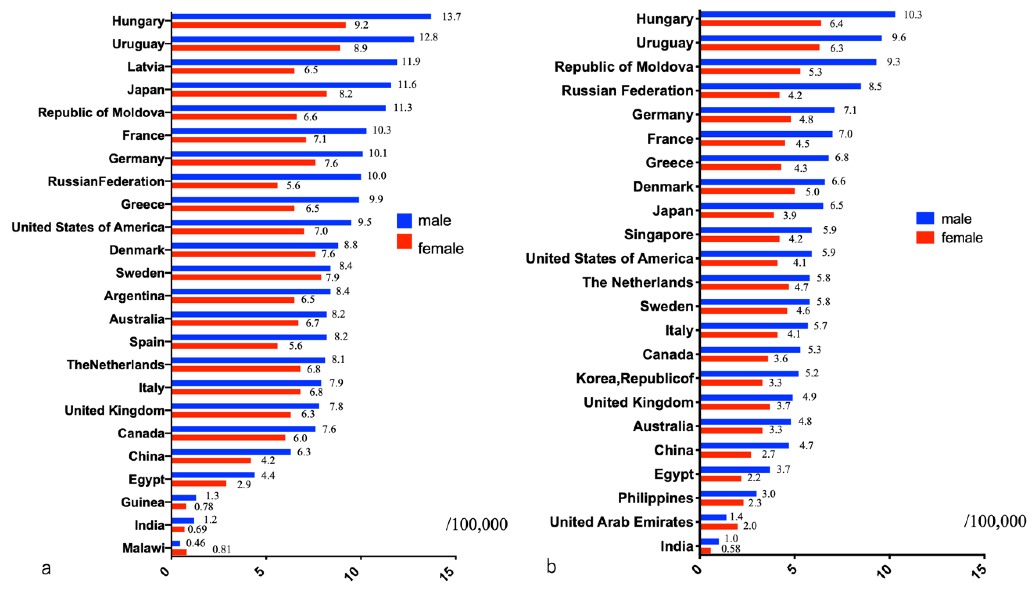
2.1. Status in the World

To compare the prevalence of PDAC and malignancies in other organs, it is important to consider the number of patients with PDAC, morbidity rate, number of deaths, and mortality rate. In particular, comparisons of global cancer surveys often reflect the state of healthcare in each country. According to a survey by GLOBOCAN in 2020, the number of new cases of PDAC was 495,773, placing it fourteenth on the list of most common cancers. However, the number of deaths secondary to PDAC was 466,003 per year, making it the seventh leading cause of cancer deaths [1]. The one-year overall survival (OS) rate of patients with PDAC is very low (24%), and the five-year OS rate is even lower (9%) [2]. The incidence of PDAC varies among countries, with the highest age-standardized incidence rates in Europe and North America, and the lowest rates in Africa and Central Asia [1]. The incidence of PDAC is slightly higher in males (5.7 per 100,000, with 262,865 cases) than in females (4.1 per 100,000, with 232,908 cases) [1]. Among males, the incidence is high in Eastern (9.3 per 100,000) and Western Europe (9.8 per 100,000)—particularly Hungary (13.7 per 100,000), Latvia (11.9 per 100,000), and Estonia (11.5 per 100,000)—followed by North America (9.9 per 100,000) and Southern Europe (8.4 per 100,000). The incidence is low in West (2.2 per 100,000), Middle (2.0 per 100,000), and East Africa (2.0 per 100,000), as well as in South Central Asia (1.5 per 100,000), particularly Guinea (1.3 per 100,000), India (1.2 per 100,000), and Malawi (0.46 per 100,000) [1] (Figure 1). Comparatively, among females, the incidence of PDAC is high in Western Europe (7.4 per 100,000), North America (6.7 per 100,000), Northern Europe (6.7 per 100,000), and Australia/New Zealand (6.7 per 100,000), particularly Hungary (9.2 per 100,000), the United States of America (7.0 per 100,000), and Sweden (7.9 per 100,000). It is low in West (1.8 per 100,000), East (1.7 per 100,000), and Middle Africa (1.2 per 100,000), as well as in South Central Asia (0.9 per 100,000), particularly Guinea (0.78 per 100,000), India (0.69 per 100,000), and Malawi (0.46 per 100,000) [1]. Wong et al. reported a higher incidence of PDAC in developed countries than in developing countries [3]. Saad et al. reported an annual increase of 1.03% in age-adjusted PDAC incidence according to a report using the Surveillance, Epidemiology, and End Results Program in the United States [4]. If PDAC continues to increase at this rate, it is expected to become the second leading cause of cancer death in the United States by 2030 [5].
The mortality rate associated with PDAC also varies among countries. Europe has the highest PDAC mortality rate (7.2 deaths per 100,000 people), followed by North America (6.5 per 100,000 people). East Africa has the lowest PDAC mortality rate (1.2 per 100,000 people). At least 90% of PDAC deaths are reported in patients aged ≥ 55 years, indicating increased onset of the disease with increasing age. When PDAC mortality was compared by country, mortality rates in males and females were found to be highest in Hungary (10.3 in males, 6.4 in females) and Uruguay (9.6 in males, 6.3 in females) (Figure 1). Comparatively, mortality rates in males were lowest in Botswana (0.07 per 100,000 people) and Malawi (0.47 per 100,000 people), whereas those in females were lowest in Botswana (0.29 per 100,000 people) and Pakistan (0.29 per 100,000 people) (data not shown). Racial differences in various risk factors and advances in diagnostic techniques and medical devices are assumed to be the reasons for regional differences in the morbidity and mortality of patients with PDAC. Mortality rates of patients with PDAC in low or medium human development index countries are about 4–5 times higher than those in high human development index countries [6]. PDAC treatment thus needs to be internationally standardized.

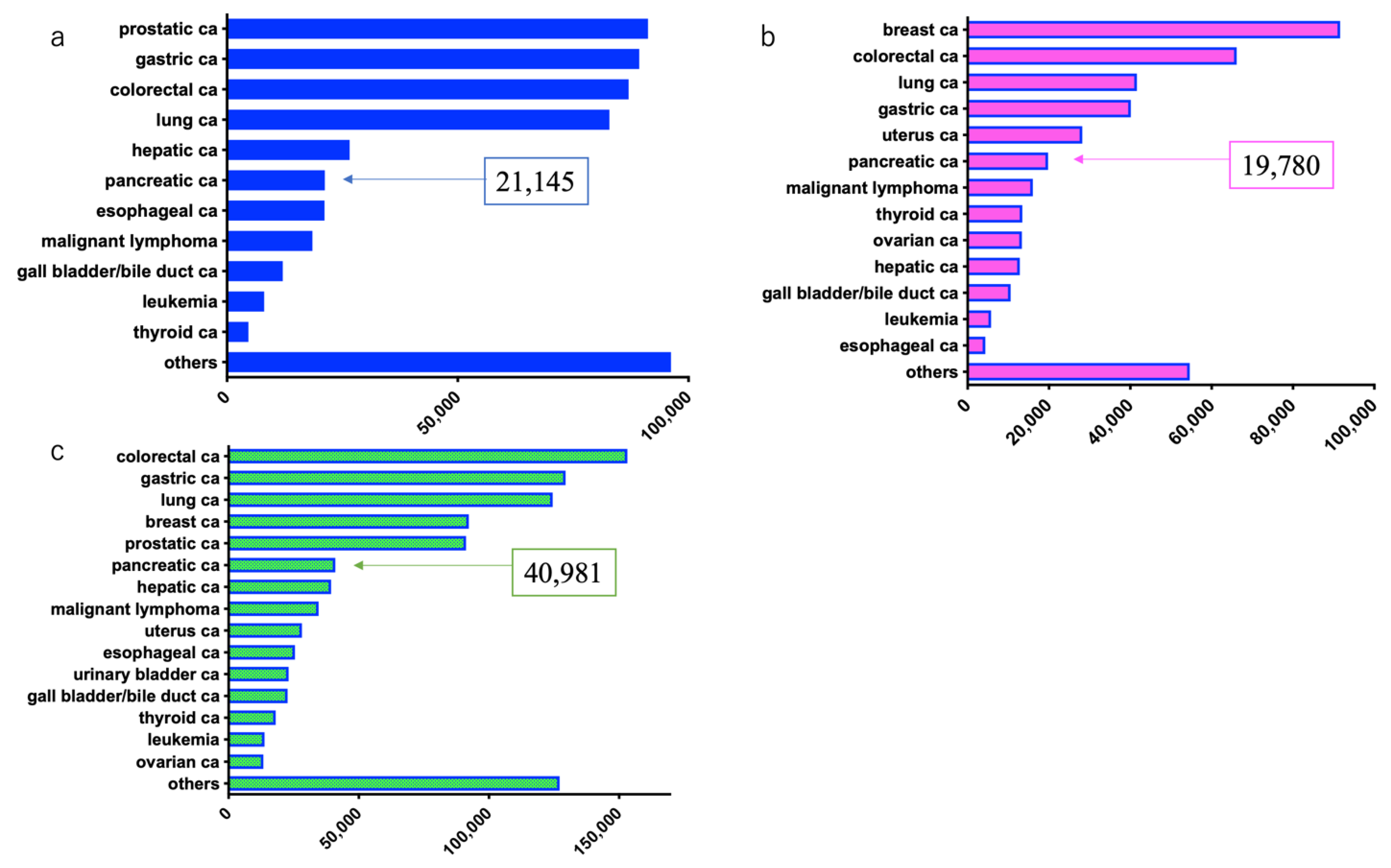
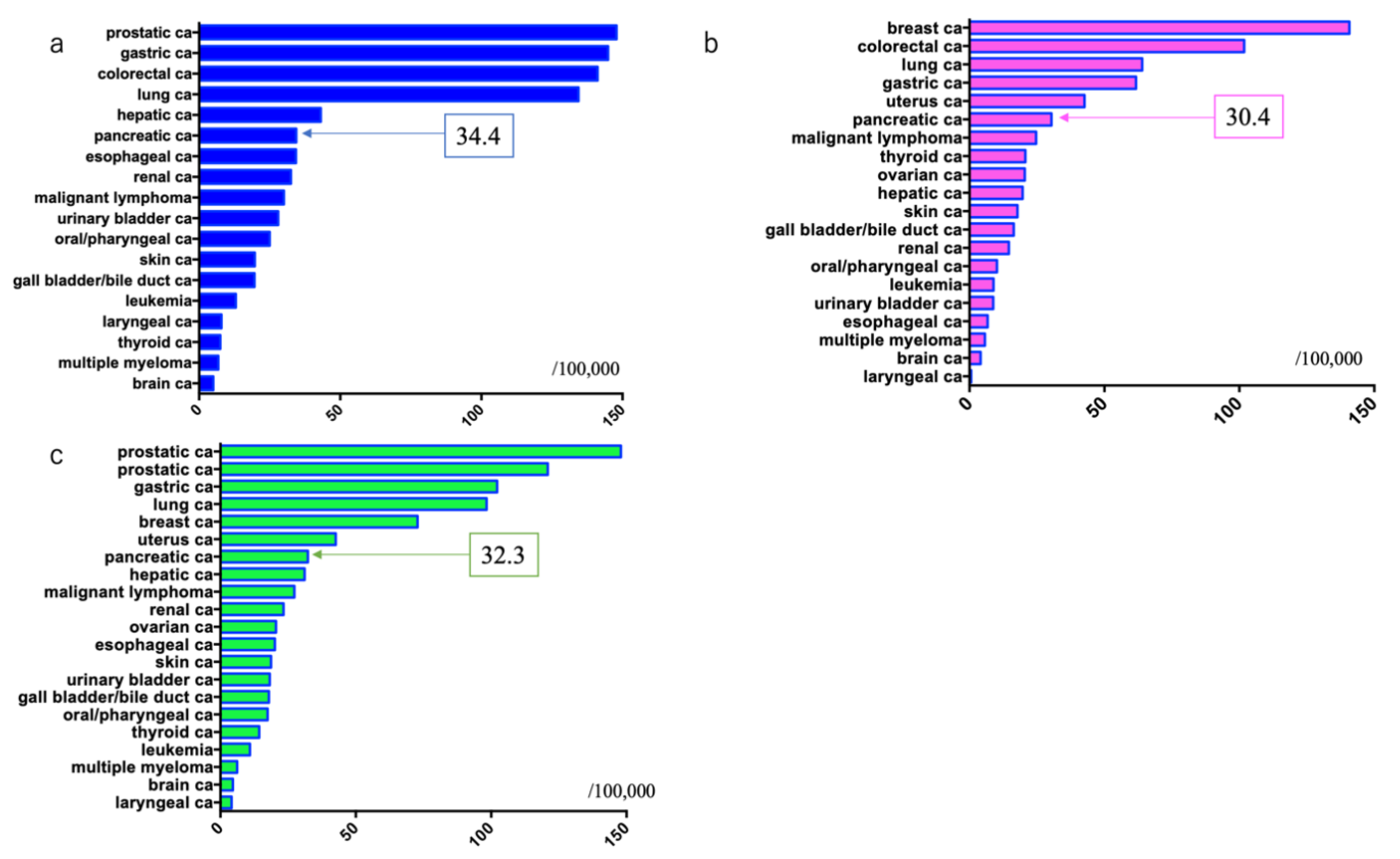
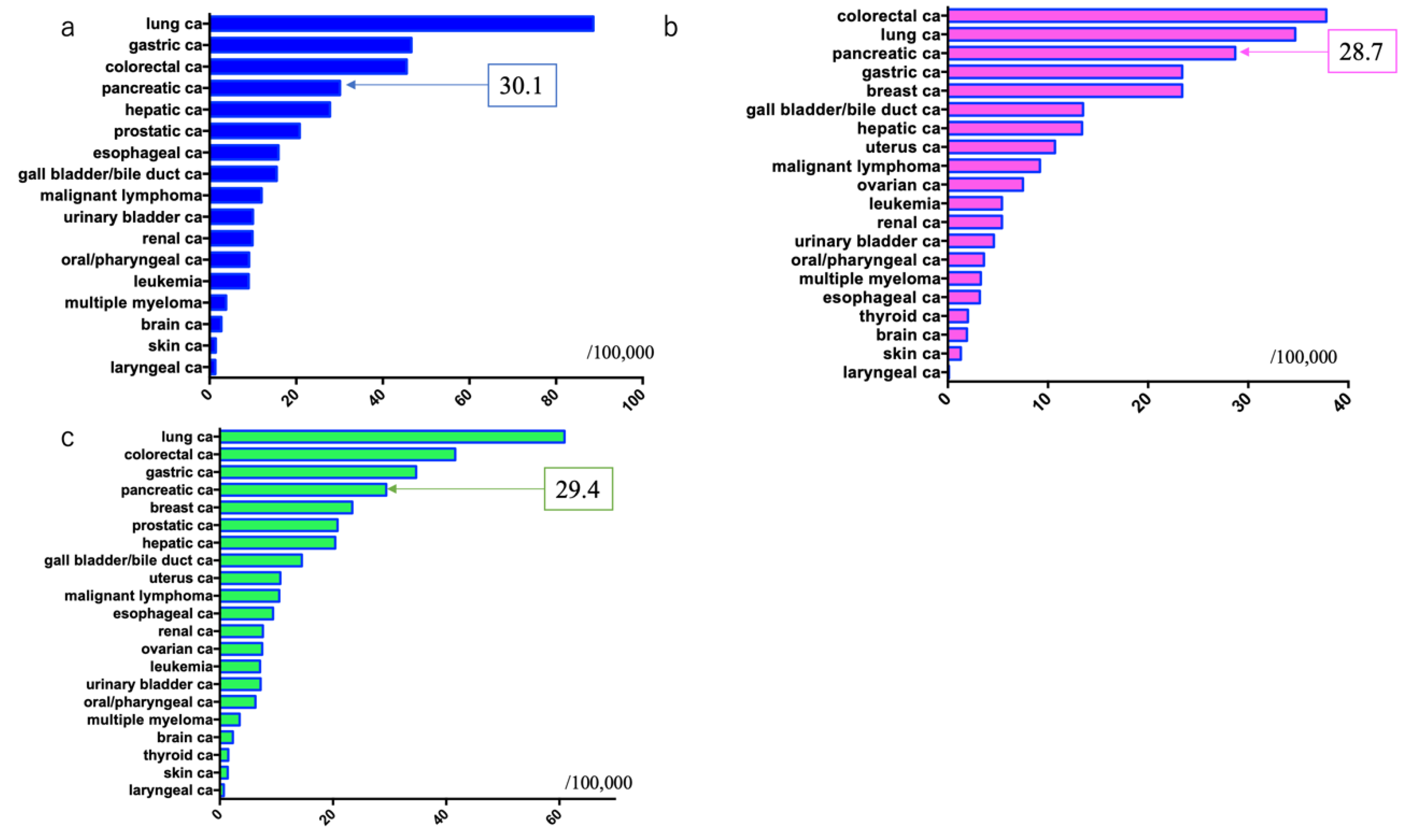
2.2. Current Status in Japan

2.2.1. Number and Incidence of Pancreatic Ductal Adenocarcinoma Cases

Japan has an original system for registering cancer cases. The incidence and mortality of PDAC in Japan have been increasing recently. Based on data in the Japanese cancer registry in 2017, the number of males with cancer was as follows: prostate cancer: 91,215, gastric cancer: 89,331, colorectal cancer: 87,019 (colon cancer: 54,358, rectal cancer: 32,661), lung cancer: 82,880, hepatic cancer: 26,570, PDAC: 21,200, and esophageal cancer: 21,145, with PDAC ranking sixth [7].
Comparatively, the number of females with cancer was as follows: breast cancer: 91,605, colorectal cancer: 66,170 (colon cancer: 47,593, rectal cancer: 18,577), lung cancer: 41,630, gastric cancer: 40,144, uterine cancer: 27,736 (uterine cervix cancer: 11,012, corpus uteri cancer: 16,724), and PDAC: 19,780, with PDAC ranking sixth [7].
The combined number of males and females with cancer included: colorectal cancer: 153,193 (colon cancer: 101,952, rectal cancer: 51,241), gastric cancer: 129,476, lung cancer: 124,510, breast cancer: 92,254, prostate cancer: 91,215, PDAC: 40,981, and hepatic cancer: 39,401, with PDAC ranking sixth [7] (Figure 2).
The incidence of cancer per 100,000 males was: prostate cancer: 147.9, gastric cancer: 144.9, colorectal cancer: 141.2 (colon cancer: 88.2, rectal cancer: 53.0), lung cancer: 134.4, hepatic cancer: 43.1, PDAC: 34.4, and esophageal cancer: 34.3, with PDAC ranking sixth [7].
The incidence of cancer per 100,000 females was: breast cancer: 140.8, colorectal cancer: 101.8 (colon cancer: 73.2, rectal cancer: 28.6), lung cancer: 64.0, gastric cancer: 61.7, uterine cancer: 42.6 (uterine cervix cancer: 16.9, corpus uteri cancer: 25.7), and PDAC: 30.4, with PDAC ranking sixth [7].
The overall incidence of cancer per 100,000 males and females was: prostate cancer: 147.9, colorectal cancer: 120.9 (colon cancer: 80.5, rectal cancer: 40.4), gastric cancer: 102.2, lung cancer: 98.3, breast cancer: 72.8, uterine cancer: 42.6 (uterine cervix cancer: 16.9, corpus uteri cancer: 25.7), PDAC: 32.3, and hepatic cancer: 31.1 [1], with PDAC ranking seventh [7] (Figure 3).

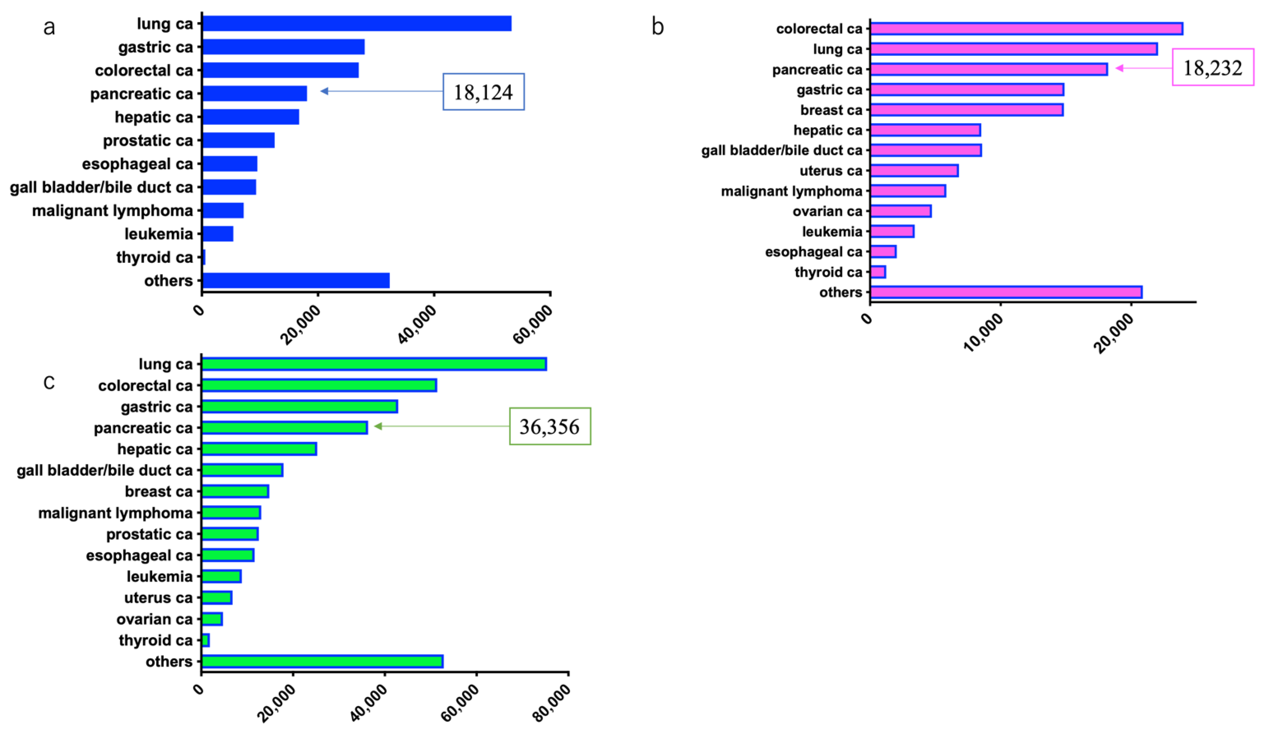
2.2.2. Number of Deaths and Mortality Rate of Patients with Pancreatic Ductal Adenocarcinoma

Based on data from the 2019 Japanese cancer registry, the number of cancer deaths in males was: lung cancer: 53,338, gastric cancer: 28,043, colorectal cancer: 27,026 (colon cancer: 17,517, rectal cancer: 9899), PDAC: 18,124, and hepatic cancer: 16,750, with PDAC ranking fourth [7].
The number of cancer deaths in females was: colorectal cancer: 24,004 (colon cancer: 18,082, rectal cancer: 5922), lung cancer: 22,056, PDAC: 18,232, gastric cancer: 14,888, and breast cancer: 14,839, with PDAC ranking third [7].
The combined number of deaths in males and females was: lung cancer: 75,394, colorectal cancer: 51,420 (colon cancer: 35,599, rectal cancer: 15,821), gastric cancer: 42,931, and PDAC: 36,356, with PDAC ranking fourth [7] (Figure 4).
Cancer mortality rates in males were: lung cancer: 88.6/100,000, gastric cancer: 46.6, colorectal cancer: 45.5 (colon cancer: 29.1, rectal cancer: 16.4), PDAC: 30.1, and hepatic cancer: 27.8, with PDAC ranking fourth [7] (Figure 5).
Cancer mortality rates in females were: colorectal cancer: 37.8 (colon cancer: 28.5, rectal cancer: 9.3), lung cancer: 34.7, PDAC: 28.7, and gastric cancer: 23.4, with PDAC ranking third [7] (Figure 5).
The overall combined cancer mortality rates in males and females were: lung cancer: 60.9, colorectal cancer: 41.6 (colon cancer: 28.8, rectal cancer: 12.8), gastric cancer: 34.7, and PDAC: 29.4, with PDAC ranking fourth [7] (Figure 5).
The five-year OS rate for patients with PDAC was 10.2%, whereas the 10-year OS rate was 6.2%. These prognoses are the poorest when compared with prognoses for malignancies in other organs [8].

3. Risk Factors

Early diagnosis is important to improve the prognosis of patients with PDAC. To achieve early diagnosis of patients with PDAC, it is extremely important to treat patients based on a solid understanding of associated risk factors. A variety of risk factors have been reported for the development of PDAC. The major factors can be broadly classified into family history, genetic disorders, complications, and preferences. Since PDAC does not present with specific symptoms, it is difficult to use clinical symptoms for early detection. Patients with risk factors for developing PDAC should be followed with blood tests, imaging techniques, and other screening methods.
It is rare for PDAC to be diagnosed at an early stage, with only about 2% of all PDAC being diagnosed at stages 0 and I [9]. However, according to a report compiled by the Pancreatic Cancer Registry of the Japan Pancreas Society, the five-year OS rates for patients with stage 0 disease was 85.8%, stage Ia—68.7%, and stage Ib—59.7%, which are greater than the OS rate at stage II or higher [10]. This demonstrates that to improve the prognosis of patients with PDAC, a system needs to be in place to efficiently identify patients who have risk factors associated with the disease and facilitate early diagnosis [9,10,11,12,13].

3.1. Family History

The proportion of patients with PDAC in their family history is reported to be 3–10% in Japan [14,15]. Familial pancreatic cancer is defined as the presence of two or more first-degree relatives with PDAC, such as a parent, sibling, or child. The risk ratio of developing PDAC is reportedly 6.79 times greater in families with a member who has PDAC. Additionally, if there are patients younger than 50 years of age with PDAC in the same family, the risk ratio increases to 9.31-fold [16]. Thus, it is critical to evaluate the family history of patients with PDAC in clinical practice. The disease has been associated with genetic mutations, including those in BRCA1/2, PALB2, CDKN2A, LKB1/STK11, PRSS1, and ATM [17,18,19,20,21,22,23,24,25]. In the United States and Europe, a registration system for familial pancreatic cancer has been established, and a prospective study and genetic analyses are being conducted. In Japan, the Japan Pancreas Society and the Pancreatic Cancer Action Network Japan (PanCAN, Sodegaura, Japan) have established a familial pancreatic cancer registry (http://jfpcr.com, accessed on 11 February 2021). In the future, this is expected to contribute to the identification and early diagnosis of patients with PDAC.

3.2. Genetic Disorders

Hereditary pancreatitis is a genetic condition characterized by recurrent episodes of acute pancreatitis starting in infancy. It is associated with mutations in the PRSS1 and SPINK1 genes. The risk of onset of PDAC in patients with hereditary pancreatitis reportedly increases by 60–87-fold with aging [26]. Hereditary breast and ovarian cancer syndrome is associated with germline mutations in BRCA1/2, leading to the development of breast and ovarian cancers that run in families [27,28,29]. Patients with hereditary breast and ovarian cancer syndrome also have a 3.5–10-fold increased risk of developing PDAC [17,30,31,32,33,34]. Additionally, the risk of developing PDAC is reportedly high in those with hereditary diseases, such as hereditary nonpolyposis colorectal cancer (Lynch syndrome) [35,36], familial adenomatous polyposis [22,36], Peutz–Jeghers syndrome [20,24,37], and familial atypical multiple mole melanoma syndrome [38]. Although the number of patients with these genetic disorders in Japan is low, they need to be considered in clinical practice (Table 1).

3.3. Diabetes Mellitus

The risk of developing PDAC is increased in patients with diabetes mellitus. The risk of onset in patients diagnosed with diabetes mellitus within one year is as high as 5.38-fold greater [39]. Thus, close examination of the pancreas is recommended for patients with new-onset diabetes mellitus or sudden poor glycemic control. In meta-analyses, the risk of PDAC in patients with type 2 diabetes mellitus was as high as 1.94-fold. The risk was 5.38-fold within one year, 1.95-fold within 1–4 years, 1.49-fold within 5–9 years, and 1.47-fold for ≥10 years from the onset of diabetes mellitus, showing PDAC presenting particularly after new-onset or rapid aggravation [39]. Smoking and chronic pancreatitis increase the risk of developing PDAC in patients with diabetes mellitus. No consensus has been reached regarding the effect of antidiabetic medications, and no significant effect was observed in the meta-analyses [40].

3.4. Intraductal Papillary Mucinous Neoplasms (IPMNs)

IPMN is characterized by ductal dilatation caused by the proliferation of a papillary epithelial neoplasm capable of producing mucus. An IPMN can evolve by itself into invasive cancer; however, primary PDAC can develop in the pancreas secondary to reasons other than IPMN [41]. The incidence of PDAC in branch-type IPMN is 2%–10%, while the mortality rate of PDAC in branch-type IPMN is 15.8–26-fold higher. In particular, this is reported to be 16.7-fold higher in patients aged ≥ 70 years and 22.5-fold higher in females [42,43].
In a retrospective study of 577 patients with branch-type IPMN who were followed up for a long period in the United States, malignant transformation was observed in 8% of patients within 10 years. The incidence of malignant transformation within five years was 4.3%, of which the incidence of invasive cancer was 2.4% [44]. The incidence of invasive cancer in patients with branch-type IPMN without high-risk stigmata or worrisome features is as high as 18.8-fold higher than that in the general population [44]. Some patients develop unresectable advanced PDAC even if they are followed up periodically at time points recommended by the International IPMN/mucinous cystic neoplasm clinical practice guidelines. Thus, there is a need to identify other factors that predict malignant transformation with high accuracy. A large prospective observational study conducted mainly by the Japan Pancreas Society is ongoing. More data regarding the incidence of PDAC in relation to IPMN are anticipated.

3.5. Chronic Pancreatitis

Chronic pancreatitis has been reported to be a risk factor for developing PDAC. In a meta-analysis by Malka et al., the risk of PDAC in patients with chronic pancreatitis was 13.3-fold greater [45]. According to a Danish cohort study of 11,972 patients with chronic pancreatitis in 2014, the hazard ratio for PDAC was 6.9-fold higher: 14.6-fold within 2–4 years and 4.8-fold between 5 and 16 years after being registered as having chronic pancreatitis [46]. The high hazard ratio for PDAC within two years of registration could be because patients with PDAC could have been included with those diagnosed with chronic pancreatitis. In a retrospective study of Japanese patients with chronic pancreatitis, excluding patients diagnosed within two years after diagnosis of chronic pancreatitis, the standardized incidence ratio of PDAC in patients with chronic pancreatitis (with a median follow-up of 5.6 years) was found to be 11.8-fold higher [47]. These results support abstinence from alcohol as an important factor in the prevention of PDAC in patients with chronic pancreatitis.
A new mechanistic definition of chronic pancreatitis was proposed for future studies based on a consensus of the definition, diagnostic criteria, classification, treatment, etc., of the disease [48]. In this definition, a conceptual model with a mechanistic definition proposed that acute pancreatitis and recurrent acute pancreatitis represent one stage on a path to chronic pancreatitis. There may be some cases with a sequential relationship between acute pancreatitis and chronic pancreatitis. A meta-analysis revealed a correlation between acute pancreatitis and the development of PDAC [49]. Patients with a past history of acute pancreatitis require especially careful evaluations from the perspective of pancreatic carcinogenesis.

3.6. Obesity

According to the results of a meta-analysis, an increase in body mass index (BMI) of 5 kg/m2 leads to a 1.10-fold higher increase in the risk of onset of PDAC, an increase in waist circumference of 10 cm leads to a 1.11-fold higher increase, and an increase in waist-to-hip ratio of 0.1 units leads to a 1.19-fold higher increased risk [50]. Additionally, the risk of PDAC increases 1.49-fold in males with BMI ≥ 35 kg/m2 and 2.76-fold in females with BMI ≥ 40 kg/m2 [51]. In cohort studies, obesity has also been identified as a risk factor for PDAC for those in their 20s. According to the results of a large-scale cohort study in Japan, in males with BMI ≥ 30 kg/m2 in their 20s, the risk of developing PDAC was 3.5-fold higher [52]. In other methods and studies, a BMI ≥ 30 kg/m2 increased the risk of onset of PDAC by 1.71-fold compared to BMIs 23–24.9 kg/m2 in males [53]. Taking into consideration conditions such as diabetes, there may be a need to improve modern eating habits.

3.7. Smoking

Cigarette smoking has been identified as a risk factor for the development of cancer in all organs. Smoking increases the risk of developing PDAC by 1.68-fold, [54] demonstrating an increased risk compared to that of nonsmokers. Moreover, cigarette smoking synergistically increases the risk of developing PDAC in patients with other risk factors [55,56]; thus, patients should be given an explanation on the risk of onset of arteriosclerosis and be advised to stop smoking.

3.8. Alcohol Consumption

The risk of developing PDAC reportedly increases 1.22-fold in heavy drinkers (ethanol equivalent of ≥37.5 g/day) [57], and significantly increases in moderate drinkers or those who drink less than moderate amounts. It is unknown whether the increase in risk of onset of PDAC is a direct effect of alcohol consumption or progression secondary to chronic pancreatitis.

3.9. Other

The number of patients with PDAC reportedly increases with occupational exposure to chemical substances (chlorinated hydrocarbon) [58], blood type (non-O) [59,60,61] history of Helicobacter pylori infection or gastric ulcer [62,63,64], and hepatitis B virus infection [65]. According to a recent study, the risk of PDAC increases 1.74-fold in patients with periodontal disease/periodontitis. Several dietary factors have been reported as risk factors for the development of PDAC. Consumption of red meat may increase the risk of developing PDAC due to the effect of carcinogens in red meat such as nitrates or N-nitroso compounds [66]. Foods with high glycemic indices, such as potatoes, have been reported as a risk factor for developing PDAC [67,68]. We summarized the risk factors in Table 2.

4. Conclusions

The epidemiology and risk factors for PDAC were summarized. The issues identified were that the incidence of PDAC is on the rise, and major regional differences exist globally. Differences in risk factors between races are assumed. Further, there are differences in the provision of medical care among regions. Determining risk factors for PDAC may enable early detection of PDAC by conducting periodic examinations in patients who are especially at risk. We anticipate that gaining a greater understanding of the overall picture of PDAC through epidemiology and risk factors will contribute to improving the prognosis of patients with this disease.

Author Contributions

Designed the study and wrote the manuscript: J.U. and A.K.; performed data analysis: J.U., A.K., E.I., K.A., H.N., T.M., Y.K., Y.T., K.Y., N.N., K.T., A.K.L. and H.Y. All authors have read and agreed to the published version of the manuscript.

Funding

This research received no external funding.

Institutional Review Board Statement

Ethical review and approval were waived for this study, due to the review.

Informed Consent Statement

Patient consent was waived due to the review.

Data Availability Statement

The data presented in this study are openly available in [GLOBOCAN and Cancer Registry and Statistics] at https://gco.iarc.fr/today (accessed on 28 February 2021) and https://ganjoho.jp/reg_stat/statistics/stat/summary.html (accessed on 28 February 2021), respectively, reference number [2,7,8].

Conflicts of Interest

The authors declare no conflict of interest.

References

  1. Sung, H.; Ferlay, J.; Siegel, R.L.; Laversanne, M.; Soerjomataram, I.; Jemal, A.; Bray, F. CA Global cancer statistics 2020, GLOBOCAN estimates of incidence and mortality worldwide for 36 cancers in 185 countries. Cancer J. Clin. 2021. [Google Scholar] [CrossRef]
  2. International Agency for Research on Cancer. Data Visualization Tools for Exploring the Global Cancer Burden in 2020. Available online: https://gco.iarc.fr/today (accessed on 17 February 2020).
  3. Wong, M.C.S.; Jiang, J.Y.; Liang, M.; Fang, Y.; Yeung, M.S.; Sung, J.J.Y. Global temporal patterns of pancreatic cancer and association with socioeconomic development. Sci Rep. 2017, 7, 3165. [Google Scholar] [CrossRef] [PubMed]
  4. Saad, A.M.; Turk, T.; Al-Husseini, M.J.; Abdel-Rahman, O. Trends in pancreatic adenocarcinoma incidence and mortality in the United States in the last four decades; A SEER-based study. BMC Cancer 2018, 18, 688. [Google Scholar] [CrossRef]
  5. Siegel, R.L.; Miller, K.D.; Jemal, A. Cancer statistics 2017. CA Cancer J. Clin. 2017, 67, 7–30. [Google Scholar] [CrossRef] [Green Version]
  6. Rahib, L.; Smith, B.D.; Aizenberg, R.; Rosenzweig, A.B.; Fleshman, J.M.; Matrisian, L.M. Projecting cancer incidence and deaths to 2030, the unexpected burden of thyroid, liver, and pancreas cancers in the United States. Cancer Res. 2014, 74, 2913–2921. [Google Scholar] [CrossRef] [Green Version]
  7. Cancer Registry and Statistics. Cancer Information Service, National Cancer Center, Japan (Ministry of Health, Labour and Welfare, National Cancer Registry). Available online: https://ganjoho.jp/reg_stat/statistics/stat/summary.html (accessed on 11 February 2021).
  8. Available online: http://www.zengankyo.ncc.go.jp/etc/seizonritsu/seizonritsu2012.html (accessed on 11 February 2021).
  9. Kanno, A.; Masamune, A.; Hanada, K.; Maguchi, H.; Shimizu, Y.; Ueki, T.; Hasebe, O.; Ohtsuka, T.; Nakamura, M.; Takenaka, M.; et al. Japan study group on the early detection of pancreatic cancer (JEDPAC). Multicenter study of early pancreatic cancer in Japan. Pancreatology 2018, 18, 61–67. [Google Scholar] [CrossRef]
  10. Egawa, S.; Toma, H.; Ohigashi, H.; Okusaka, T.; Nakao, A.; Hatori, T.; Maguchi, H.; Yanagisawa, A.; Tanaka, M. Japan pancreatic cancer registry; 30th year anniversary: Japan Pancreas Society. Pancreas 2012, 41, 985–992. [Google Scholar] [CrossRef]
  11. Kanno, A.; Masamune, A.; Hanada, K.; Kikuyama, M.; Kitano, M. Advances in early detection of pancreatic cancer. Diagnostics 2019, 9, 18. [Google Scholar] [CrossRef] [Green Version]
  12. Hanada, K.; Amano, H.; Abe, T. Early diagnosis of pancreatic cancer: Current trends and concerns. Ann. Gastroenterol. Surg. 2017, 1, 44–51. [Google Scholar] [CrossRef]
  13. Hanada, K.; Okazaki, A.; Hirano, N.; Izumi, Y.; Minami, T.; Ikemoto, J.; Kanemitsu, K.; Hino, F. Effective screening for early diagnosis of pancreatic cancer. Best Pract. Res. Clin. Gastroenterol. 2015, 29, 929–939. [Google Scholar] [CrossRef]
  14. Matsubayashi, H.; Maeda, A.; Kanemoto, H.; Uesaka, K.; Yamazaki, K.; Hironaka, S.; Miyagi, Y.; Ikehara, H.; Ono, H.; Klein, A.; et al. Risk factors of familial pancreatic cancer in Japan: Current smoking and recent onset of diabetes. Pancreas 2011, 40, 974–978. [Google Scholar] [CrossRef]
  15. Inoue, M.; Tajima, K.; Takezaki, T.; Hamajima, N.; Hirose, K.; Ito, H.; Tominaga, S. Epidemiology of pancreatic cancer in Japan: A nested case-control study from the Hospital-based Epidemiologic Research Program at Aichi Cancer Center (HERPACC). Int. J. Epidemiol. 2003, 32, 257–262. [Google Scholar] [CrossRef] [PubMed] [Green Version]
  16. Brune, K.A.; Lau, B.; Palmisano, E.; Canto, M.; Goggins, M.G.; Hruban, R.H.; Klein, A.P. Importance of age of onset in pancreatic cancer kindreds. J. Natl. Cancer Inst. 2010, 102, 119–126. [Google Scholar] [CrossRef] [PubMed] [Green Version]
  17. Couch, F.J.; Johnson, M.R.; Rabe, K.G.; Brune, K.; de Andrade, M.; Goggins, M.; Rothenmund, H.; Gallinger, S.; Klein, A.; Petersen, G.M.; et al. The prevalence of BRCA2 mutations in familial pancreatic cancer. Cancer Epidemiol. Biomark. Prev. 2007, 16, 342–346. [Google Scholar] [CrossRef] [PubMed] [Green Version]
  18. Jones, S.; Hruban, R.H.; Kamiyama, M.; Borges, M.; Zhang, X.; Parsons, D.W.; Lin, J.C.; Palmisano, E.; Brune, K.; Jaffee, E.M.; et al. Exomic sequencing identifies PALB2 as a pancreatic cancer susceptibility gene. Science 2009, 324, 217. [Google Scholar] [CrossRef] [PubMed] [Green Version]
  19. Slater, E.P.; Langer, P.; Niemczyk, E.; Strauch, K.; Butler, J.; Habbe, N.; Neoptolemos, J.P.; Greenhalf, W.; Bartsch, D.K. PALB2 mutations in European familial pancreatic cancer families. Clin. Genet. 2010, 78, 490–494. [Google Scholar] [CrossRef] [PubMed]
  20. Giardiello, F.M.; Brensinger, J.D.; Tersmette, A.C.; Goodman, S.N.; Petersen, G.M.; Booker, S.V.; Cruz-Corre, M.; Offerhaus, J.A. Very high risk of cancer in familial Peutz-Jeghers syndrome. Gastroenterology 2000, 119, 1447–1453. [Google Scholar] [CrossRef] [Green Version]
  21. Goldstein, A.M.; Fraser, M.C.; Struewing, J.P.; Hussussian, C.J.; Ranade, K.; Zametkin, D.P.; Fontaine, L.S.; Organic, S.M.; Dracopoli, N.C.; Clark Jr, W.H.; et al. Increased risk of pancreatic cancer in melanoma-prone kindreds with p16INK4mutations. N. Engl. J. Med. 1995, 333, 970–974. [Google Scholar] [CrossRef]
  22. Giardiello, F.M.; Offerhaus, G.J.; Lee, D.H.; Krush, A.J.; Tersmette, A.C.; Booker, S.V.; Kelley, N.C.; Hamilton, S.R. Increased risk of thyroid and pancreatic carcinoma in familial adenomatous polyposis. Gut 1993, 34, 1394–1396. [Google Scholar] [CrossRef] [Green Version]
  23. Kastrinos, F.; Mukherjee, B.; Tayob, N.; Wang, F.; Sparr, J.; Raymond, V.M.; Bandipalliam, P.; Stoffel, E.M.; Gruber, S.B.; Syngal, S. Risk of pancreatic cancer in families with Lynch syndrome. JAMA 2009, 302, 1790–1795. [Google Scholar] [CrossRef]
  24. Shi, C.; Hruban, R.H.; Klein, A.P. Familial pancreatic cancer. Arch. Pathol. Lab. Med. 2009, 133, 365–374. [Google Scholar] [CrossRef] [PubMed]
  25. Matsubayashi, H.; Takaori, K.; Morizane, C.; Maguchi, H.; Mizuma, M.; Takahashi, H.; Wada, K.; Hosoi, H.; Yachida, S.; Suzuki, M.; et al. Familial pancreatic cancer: Concept, management and issues. World J. Gastroenterol. 2017, 23, 935–948. [Google Scholar] [CrossRef] [PubMed]
  26. Rebours, V.; Boutron-Ruault, M.C.; Schnee, M.; Férec, C.; Maire, F.; Hammel, P.; Ruszniewski, P.; Lévy, P. Risk of pancreatic adenocarcinoma in patients with hereditary pancreatitis: A national exhaustive series. Am. J. Gastroenterol. 2008, 103, 111–119. [Google Scholar] [CrossRef]
  27. Friedenson, B. BRCA1 and BRCA2 pathways and the risk of cancers other than breast or ovarian. Med. Gen. Med. 2005, 7, 60. [Google Scholar]
  28. Yamauchi, H.; Takei, J. Management of hereditary breast and ovarian cancer. Int. J. Clin. Oncol. 2018, 23, 45–51. [Google Scholar] [CrossRef]
  29. Iqbal, J.; Ragone, A.; Lubinski, J.; Lynch, H.T.; Moller, P.; Ghadirian, P.; Foulkes, W.D.; Armel, S.; Eisen, A.; Neuhausen, S.L.; et al. The incidence of pancreatic cancer in BRCA1 and BRCA2 mutation carriers. Br. J. Cancer 2012, 107, 2005–2009. [Google Scholar] [CrossRef]
  30. Golan, T.; Kanji, Z.S.; Epelbaum, R.; Devaud, N.; Dagan, E.; Holter, S.; Aderka, D.; Paluch-Shimon, S.; Kaufman, B.; Hedley, D.; et al. Overall survival and clinical characteristics of pancreatic cancer in BRCA mutation carriers. Br. J. Cancer 2014, 111, 1132–1138. [Google Scholar] [CrossRef]
  31. Leung, K.; Saif, M.W. BRCA-associated pancreatic cancer: The evolving management. JOP J. Pancreas 2013, 14, 149–151. [Google Scholar]
  32. Murphy, K.M.; Brune, K.A.; Griffin, C.; Sollenberger, J.E.; Petersen, G.M.; Bansal, R.; Hruban, R.H. Evaluation of candidate genes MAP2K4, MADH4, ACVR1B, and BRCA2 in familial pancreatic cancer: Deleterious BRCA2 mutations in 17%. Cancer Res. 2002, 62, 3789–3793. [Google Scholar] [PubMed]
  33. Hahn, S.A.; Greenhalf, B.; Ellis, I.; Sina-Frey, M.; Rieder, H.; Korte, B.; Gerdes, B.; Kress, R.; Ziegler, A.; Raeburn, J.A.; et al. BRCA2 germline mutations in familial pancreatic carcinoma. J. Natl. Cancer Inst. 2003, 95, 214–221. [Google Scholar] [CrossRef]
  34. Leoz, M.L.; Sanchez, A.; Carballal, S.; Ruano, L.; Ocana, T.; Pellise, M.; Castells, A.; Balaguer, F.; Moreira, L. Hereditary gastric and pancreatic cancer predisposition syndromes. Gastroenterol. Hepatol. 2016, 39, 481–793. [Google Scholar] [CrossRef]
  35. Watson, P.; Vasen, H.F.; Mecklin, J.P.; Bernstein, I.; Aarnio, M.; Jarvinen, H.J.; Myrhoj, T.; Sunde, L.; Wijnen, J.T.; Lynch, H.T. The risk of extra-colonic, extra-endometrial cancer in the Lynch syndrome. Int. J. Cancer 2008, 123, 444–449. [Google Scholar] [CrossRef] [Green Version]
  36. Becker, A.E.; Hernandez, Y.G.; Frucht, H.; Lucas, A.L. Pancreatic ductal adenocarcinoma: Risk factors, screening, and early detection. World J. Gastroenterol. 2014, 20, 11182–11198. [Google Scholar] [CrossRef] [PubMed]
  37. Syngal, S.; Brand, R.E.; Church, J.M.; Giardiello, F.M.; Hampel, H.L.; Burt, R.W.; American College of Gastroenterology. ACG clinical guideline: Genetic testing and management of hereditary gastrointestinal cancer syndromes. Am. J. Gastroenterol. 2015, 110, 223–262. [Google Scholar] [CrossRef] [Green Version]
  38. Greer, J.B.; Lynch, H.T.; Brand, R.E. Hereditary pancreatic cancer: A clinical perspective. Best Pract. Res. Clin. Gastroenterol. 2009, 23, 159–170. [Google Scholar] [CrossRef]
  39. Ben, Q.; Xu, M.; Ning, X.; Liu, J.; Hong, S.; Huang, W.; Zhang, H.; Li, Z. Diabetes mellitus and risk of pancreatic cancer: A meta-analysis of cohort studies. Eur. J. Cancer 2011, 47, 1928–1937. [Google Scholar] [CrossRef]
  40. Singh, S.; Singh, P.P.; Singh, A.G.; Murad, M.H.; Sanchez, W. Anti-diabetic medications and the risk of hepatocellular cancer: A systematic review and meta-analysis. Am. J. Gastroenterol. 2013, 108, 881–891. [Google Scholar] [CrossRef] [PubMed]
  41. Yamaguchi, K.; Kanemitsu, S.; Hatori, T.; Maguchi, H.; Shimizu, Y.; Tada, M.; Nakagohri, T.; Hanada, K.; Osanai, M.; Noda, Y.; et al. Pancreatic ductal adenocarcinoma derived from IPMN and pancreatic ductal adenocarcinoma concomitant with IPMN. Pancreas 2011, 40, 571–580. [Google Scholar] [CrossRef] [PubMed]
  42. Uehara, H.; Nakaizumi, A.; Ishikawa, O.; Iishi, H.; Tatsumi, K.; Takakura, R.; Ishida, T.; Takano, Y.; Tanaka, S.; Takenaka, A. Development of ductal carcinoma of the pancreas during follow-up of branch duct intraductal papillary mucinous neoplasm of the pancreas. Gut 2008, 57, 1561–1565. [Google Scholar] [CrossRef]
  43. Tanno, S.; Nakano, Y.; Koizumi, K.; Sugiyama, Y.; Nakamura, K.; Sasajima, J.; Nishikawa, T.; Mizukami, Y.; Yanagawa, N.; Fujii, T.; et al. Pancreatic ductal adenocarcinomas in long-term follow-up patients with branch duct intraductal papillary mucinous neoplasms. Pancreas 2010, 39, 36–40. [Google Scholar] [CrossRef] [PubMed] [Green Version]
  44. Pergolini, I.; Sahora, K.; Ferrone, C.R.; Morales-Oyarvide, V.; Wolpin, B.M.; Mucci, L.A.; Brugge, W.R.; Mino-Kenudson, M.; Patino, M.; Sahani, D.V.; et al. Long-term risk of pancreatic malignancy in patients with branch duct intraductal papillary mucinous neoplasm in a referral center. Gastroenterology 2017, 153, 1284–1294. [Google Scholar] [CrossRef]
  45. Raimondi, S.; Lowenfels, A.B.; Morselli-Labate, A.M.; Maisonneuve, P.; Pezzilli, R. Pancreatic cancer in chronic pancreatitis; Aetiology, incidence, and early detection. Best Pract. Res. Clin. Gastroenterol. 2010, 24, 349–358. [Google Scholar] [CrossRef] [PubMed]
  46. Bang, U.C.; Benfield, T.; Hyldstrup, L.; Bendtsen, F.; Beck Jensen, J.E. Mortality, cancer, and comorbidities associated with chronic pancreatitis: A Danish nationwide matched-cohort study. Gastroenterology 2014, 146, 989–994. [Google Scholar] [CrossRef]
  47. Ueda, J.; Tanaka, M.; Ohtsuka, T.; Tokunaga, S.; Shimosegawa, T. Research Committee of Intractable Diseases of the Pancreas. Surgery for chronic pancreatitis decreases the risk for pancreatic cancer: A multicenter retrospective analysis. Surgery 2013, 153, 357–364. [Google Scholar] [CrossRef] [PubMed]
  48. Whitcomb, D.C.; Frulloni, L.; Garg, P.; Greer, J.B.; Schneider, A.; Yadav, D.; Shimosegawa, T. Chronic pancreatitis: An international draft consensus proposal for a new mechanistic definition. Pancreatology 2016, 16, 218–224. [Google Scholar] [CrossRef]
  49. Liu, J.; Wang, Y.; Yu, Y. Meta-analysis reveals an association between acute pancreatitis and the risk of pancreatic cancer. World J. Clin. Cases 2020, 8, 4416–4430. [Google Scholar] [CrossRef]
  50. Aune, D.; Greenwood, D.C.; Chan, D.S.; Vieira, R.; Vieira, A.R.; Navarro Rosenblatt, D.A.; Cade, J.E.; Burley, V.J.; Norat, T. Body mass index, abdominal fatness and pancreatic cancer risk: A systematic review and non-linear dose-response meta-analysis of prospective studies. Ann Oncol. 2012, 23, 843–852. [Google Scholar] [CrossRef]
  51. Stolzenberg-Solomon, R.Z.; Adams, K.; Leitzmann, M.; Schairer, C.; Michaud, D.S.; Hollenbeck, A.; Schatzkin, A.; Silverman, D.T. Adiposity, physical activity, and pancreatic cancer in the National Institutes of Health-AARP Diet and Health Cohort. Am. J. Epidemiol. 2008, 167, 586–597. [Google Scholar] [CrossRef] [PubMed]
  52. Lin, Y.; Kikuchi, S.; Tamakoshi, A.; Yagyu, K.; Obata, Y.; Inaba, Y.; Kurosawa, M.; Kawamura, T.; Motohashi, Y.; Ishibashi, T.; et al. Obesity, physical activity and the risk of pancreatic cancer in a large Japanese cohort. Int. J. Cancer. 2007, 120, 2665–2671. [Google Scholar] [CrossRef]
  53. Koyanagi, Y.N.; Matsuo, K.; Ito, H.; Tamakoshi, A.; Sugawara, Y.; Hidaka, A.; Wada, K.; Oze, I.; Kitamura, Y.; Liu, R.; et al. Body-mass index and pancreatic cancer incidence: A pooled analysis of nine population-based cohort studies with more than 340,000 Japanese subjects. J. Epidemiol. 2018, 28, 245–252. [Google Scholar] [CrossRef] [Green Version]
  54. Matsuo, K.; Ito, H.; Wakai, K.; Nagata, C.; Mizoue, T.; Tanaka, K.; Tsuji, I.; Tamakoshi, A.; Sasazuki, S.; Inoue, M.; et al. Cigarette smoking and pancreas cancer risk: An evaluation based on a systematic review of epidemiologic evidence in the Japanese population. Jpn. J. Clin. Oncol. 2011, 41, 1292–1302. [Google Scholar] [CrossRef] [Green Version]
  55. Luo, J.; Iwasaki, M.; Inoue, M.; Sasazuki, S.; Otani, T.; Ye, W.; Tsugane, S.; JPHC Study Group. Body mass index, physical activity and the risk of pancreatic cancer in relation to smoking status and history of diabetes: A large-scale population-based cohort study in Japan—The JPHC study. Cancer Causes Control. 2007, 18, 603–612. [Google Scholar] [CrossRef]
  56. Lynch, S.M.; Vrieling, A.; Lubin, J.H.; Kraft, P.; Mendelsohn, J.B.; Hartge, P.; Canzian, F.; Steplowski, E.; Arslan, A.A.; Gross, M.; et al. Cigarette smoking and pancreatic cancer: A pooled analysis from the pancreatic cancer cohort consortium. Am. J. Epidemiol. 2009, 170, 403–413. [Google Scholar] [CrossRef] [Green Version]
  57. Tramacere, I.; Scotti, L.; Jenab, M.; Bagnardi, V.; Bellocco, R.; Rota, M.; Corrao, G.; Bravi, F.; Boffetta, P.; La Vecchia, C. Alcohol drinking and pancreatic cancer risk: A meta-analysis of the dose-risk relation. Int. J. Cancer. 2010, 126, 1474–1486. [Google Scholar] [CrossRef]
  58. Ojajärvi, A.; Partanen, T.; Ahlbom, A.; Hakulinen, T.; Kauppinen, T.; Weiderpass, E.; Wesseling, C. Estimating the relative risk of pancreatic cancer associated with exposure agents in job title data in a hierarchical Bayesian meta-analysis. Scand. J. Work Environ. Health 2007, 33, 325–335. [Google Scholar] [CrossRef] [PubMed] [Green Version]
  59. Iodice, S.; Maisonneuve, P.; Botteri, E.; Sandri, M.T.; Lowenfels, A.B. ABO blood group and cancer. Eur. J. Cancer 2010, 46, 3345–3350. [Google Scholar] [CrossRef]
  60. Wolpin, B.M.; Chan, A.T.; Hartge, P.; Chanock, S.J.; Kraft, P.; Hunter, D.J.; Giovannucci, E.L.; Fuchs, C.S. ABO blood group and the risk of pancreatic cancer. J. Natl. Cancer Inst. 2009, 101, 424–431. [Google Scholar] [CrossRef] [PubMed] [Green Version]
  61. Wolpin, B.M.; Kraft, P.; Gross, M.; Helzlsouer, K.; Bueno-de-Mesquita, H.B.; Steplowski, E.; Stolzenberg-Solomon, R.Z.; Arslan, A.A.; Jacobs, E.J.; Lacroix, A.; et al. Pancreatic cancer risk and ABO blood group alleles: Results from the pancreatic cancer cohort consortium. Cancer Res. 2010, 70, 1015–1023. [Google Scholar] [CrossRef] [Green Version]
  62. Risch, H.A.; Yu, H.; Lu, L.; Kidd, M.S. ABO blood group, Helicobacter pylori seropositivity, and risk of pancreatic cancer: A case-control study. J. Natl. Cancer Inst. 2010, 102, 502–505. [Google Scholar] [CrossRef] [PubMed] [Green Version]
  63. Risch, H.A.; Lu, L.; Kidd, M.S.; Wang, J.; Zhang, W.; Ni, Q.; Gao, Y.T.; Yu, H. Helicobacter pylori seropositivities and risk of pancreatic carcinoma. Cancer Epidemiol. Biomark. Prev. 2014, 23, 172–178. [Google Scholar] [CrossRef] [PubMed] [Green Version]
  64. Bao, Y.; Spiegelman, D.; Li, R.; Giovannucci, E.; Fuchs, C.S.; Michaud, D.S. History of peptic ulcer disease and pancreatic cancer risk in men. Gastroenterology 2010, 138, 541–549. [Google Scholar] [CrossRef] [PubMed] [Green Version]
  65. Iloeje, U.H.; Yang, H.I.; Jen, C.L.; Su, J.; Wang, L.Y.; You, S.L.; Lu, S.N.; Chen, C.J. Risk of pancreatic cancer in chronic hepatitis B virus infection: Data from the REVEAL-HBV cohort study. Liver Int. 2010, 30, 423–429. [Google Scholar] [CrossRef] [PubMed]
  66. Beaney, A.J.; Banim, P.J.R.; Luben, R.; Lentjes, M.A.H.; Khaw, K.T.; Hart, A.R. Higher meat intake is positively associated with higher risk of developing pancreatic cancer in an age-dependent manner and are modified by plasma antioxidants: A prospective cohort study (EPIC-Norfolk) using data from food diaries. Pancreas 2017, 46, 672–678. [Google Scholar] [CrossRef]
  67. Rossi, M.; Lipworth, L.; Polesel, J.; Negri, E.; Bosetti, C.; Talamini, R.; McLaughlin, J.K.; La Vecchia, C. Dietary glycemic index and glycemic load and risk of pancreatic cancer: A case-control study. Ann. Epidemiol. 2010, 20, 460–465. [Google Scholar] [CrossRef]
  68. Polesel, J.; Talamini, R.; Negri, E.; Bosetti, C.; Boz, G.; Lucenteforte, E.; Franceschi, S.; Serraino, D.; La Vecchia, C. Dietary habits and risk of pancreatic cancer: An Italian case-control study. Cancer Causes Control. 2010, 21, 493–500. [Google Scholar] [CrossRef] [PubMed]
Figure 1. Estimated incidence and mortality from pancreatic ductal adenocarcinoma in 2020 in the world (source: GLOBOCAN 2020 ref. [2]). (a) incidence of pancreatic ductal adenocarcinoma; (b) mortality rate of patients with pancreatic ductal adenocarcinoma.
Diagnostics 11 00562 g001
Figure 2. Estimated number of new cases of pancreatic ductal adenocarcinoma in 2017 in Japan (source: Cancer Registry and Statistics ref. [7]). (a) Males; (b) Females; (c) Both males and females.
Diagnostics 11 00562 g002
Figure 3. Estimated incidence of pancreatic ductal adenocarcinoma in 2017 in Japan (source: Cancer Registry and Statistics ref. [7]). (a) Males; (b) Females; (c) Both males and females.
Diagnostics 11 00562 g003
Figure 4. Estimated number of deaths due to pancreatic ductal adenocarcinoma in 2019 in Japan (source: Cancer Registry and Statistics ref. [7]). (a) Males; (b) Females; (c) Both males and females.
Diagnostics 11 00562 g004
Figure 5. Estimated mortality rate of patients with pancreatic ductal adenocarcinoma in 2019 in Japan (source: Cancer Registry and Statistics ref. [7]). (a) Males; (b) Females; (c) Both males and females.
Diagnostics 11 00562 g005
Table 1. Hereditary pancreatic cancer syndrome.
DiseaseDisease GeneHereditary FormThe Risk of PDAC
Hereditary pancreatitisPRSS1Autosomal dominant60–87
Hereditary breast and ovarian cancer syndromeBRCA1/2Autosomal dominant4.1–5.8
Peutz–Jeghers syndromeSTK11/LKB1Autosomal dominant132
Familial atypical multiple mole melanoma syndromeCDKN2A/p16Autosomal dominant13–22
Hereditary colorectal adenomatous polyposisAPCAutosomal dominant4.4
Hereditary nonpolyposis colorectal cancerhMSH2, hMLH1Autosomal dominant8.6
Table 2. Risk factors for developing pancreatic ductal adenocarcinoma (PDAC).
Risk FactorsThe Risk of PDAC
Family historyPatients with PDAC in the family6.79-fold
Patients with family members with PDAC < 50 years old9.31-fold
Genetic disordersHereditary pancreatitis67–87-fold
Hereditary pancreatic cancer syndromeRefer to Table 1
ComplicationsDiabetes mellitus<1 year 5.38-fold,
1–4 years 1.95-fold,
5–9 years 1.49-fold,
≥10 years 1.47-fold
ObesityRisk of PDAC onset in males in their 20 s
with body mass index ≥ 30 kg/m2: 3.5-fold
Chronic pancreatitisWithin 4 years of diagnosis: 14.6-fold
≥5 years after diagnosis: 4.8-fold
Intraductal Papillary Mucinous Neoplasms (IPMNs)Branch-type IPMN: 15.8–26-fold
PreferencesSmoking1.68-fold
Alcohol1.22-fold
OccupationChlorinated hydrocarbon exposure2.21-fold
FoodRed meat1.25–1.76-fold
Publisher’s Note: MDPI stays neutral with regard to jurisdictional claims in published maps and institutional affiliations.

