
The Measurement of Stiffness for Major Muscles with Shear Wave Elastography and Myoton: A Quantitative Analysis Study


Abstract
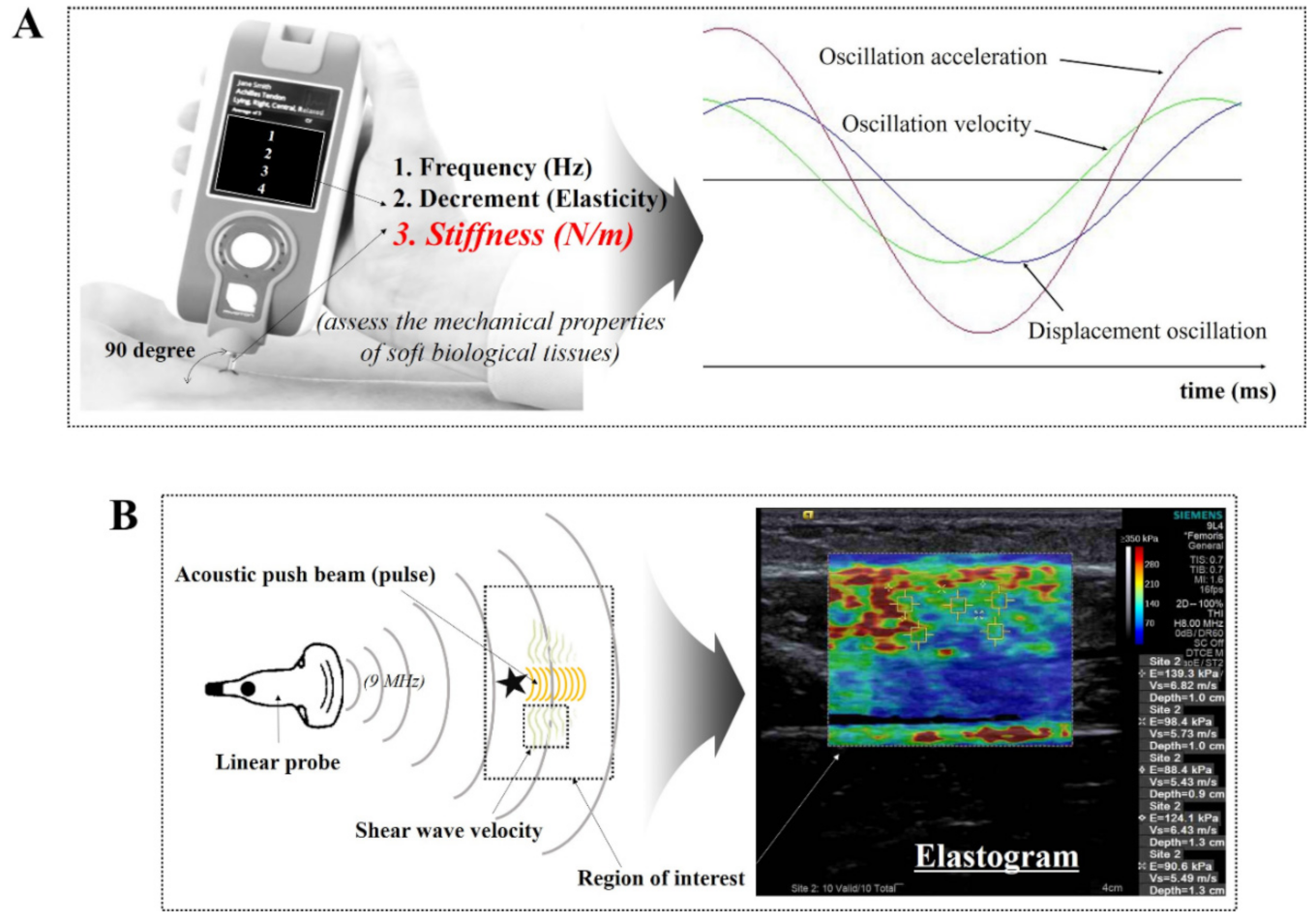
:1. Introduction
2. Materials and Methods
2.1. Ethical Approval
2.2. Participants
2.3. Experimental Protocol
2.4. Measurement Position
2.5. Quantitative Evaluation Method
2.6. Reliability Tests
2.7. Statistical Analysis
3. Results
3.1. Relationship between MyotonPRO and SWE for Measurement of Muscle Stiffness at Rest and during Contraction
3.2. The Difference in Muscle Stiffness between Men and Women
3.3. Intra-Rater Reliability of MyotonPro and SWE when Measuring Muscle Stiffness
4. Discussion
Author Contributions
Funding
Institutional Review Board Statement
Informed Consent Statement
Data Availability Statement
Acknowledgments
Conflicts of Interest
References
- Mannarino, P.; Matta, T.T.D.; Oliveira, L.F. An 8-week resistance training protocol is effective in adapting quadriceps but not patellar tendon shear modulus measured by Shear Wave Elastography. PLoS ONE 2019, 14, e0205782. [Google Scholar] [CrossRef] [Green Version]
- Lieber, R.L.; Roberts, T.J.; Blemker, S.S.; Lee, S.S.M.; Herzog, W. Skeletal muscle mechanics, energetics and plasticity. J. Neuroeng. Rehab. 2017, 14, 108. [Google Scholar] [CrossRef] [Green Version]
- Ando, R.; Suzuki, Y. Positive relationship between passive muscle stiffness and rapid force production. Hum. Mov. Sci. 2019, 66, 285–291. [Google Scholar] [CrossRef]
- Kelly, J.P.; Koppenhaver, S.L.; Michener, L.A.; Proulx, L.; Bisagni, F.; Cleland, J.A. Characterization of tissue stiffness of the infraspinatus, erector spinae, and gastrocnemius muscle using ultrasound shear wave elastography and superficial mechanical deformation. J. Electromyogr. Kinesiol. 2018, 38, 73–80. [Google Scholar] [CrossRef]
- Caliskan, E.; Akkoc, O.; Bayramoglu, Z.; Gozubuyuk, O.B.; Kural, D.; Azamat, S.; Adaletli, I. Effects of static stretching duration on muscle stiffness and blood flow in the rectus femoris in adolescents. Med. Ultrason. 2019, 21, 136–143. [Google Scholar] [CrossRef] [PubMed]
- Vena, P.; Royston, T.J. Dilatational and shear waves in poro-vioscoelastic media. J. Mech. Behav. Biomed. Mater. 2019, 97, 99–107. [Google Scholar] [CrossRef] [PubMed]
- Nordez, A.; Hug, F. Muscle shear elastic modulus measured using supersonic shear imaging is highly related to muscle activity level. J. Appl. Phys. (Bethesda) 2010, 108, 1389–1394. [Google Scholar] [CrossRef]
- Carlsen, J.F.; Pedersen, M.R.; Ewertsen, C.; Saftoiu, A.; Lonn, L.; Rafaelsen, S.R.; Nielsen, M.B. A comparative study of strain and shear-wave elastography in an elasticity phantom. AJR Am. J. Roentgenol. 2015, 204, W236–W242. [Google Scholar] [CrossRef] [PubMed]
- Xie, M.; Zhang, X.; Liu, J.; Ding, J.; Ren, Y.; Hua, K. Evaluation of levator ani with no defect on elastography in women with POP. Int. J. Clin. Exp. Med. 2015, 8, 10204–10212. [Google Scholar]
- Chen, T.L.; Agresta, C.E.; Lipps, D.B.; Provenzano, S.G.; Hafer, J.F.; Wong, D.W.; Zhang, M.; Zernicke, R.F. Ultrasound elastographic assessment of plantar fascia in runners using rearfoot strike and forefoot strike. J. Biomech. 2019, 89, 65–71. [Google Scholar] [CrossRef] [PubMed]
- Corrigan, P.; Zellers, J.A.; Balascio, P.; Silbernagel, K.G.; Cortes, D.H. Quantification of Mechanical Properties in Healthy Achilles Tendon Using Continuous Shear Wave Elastography: A Reliability and Validation Study. Ultrasound Med. Biol. 2019. [Google Scholar] [CrossRef] [PubMed]
- Feng, Y.N.; Li, Y.P.; Liu, C.L.; Zhang, Z.J. Assessing the elastic properties of skeletal muscle and tendon using shearwave ultrasound elastography and MyotonPRO. Sci. Rep. 2018, 8, 17064. [Google Scholar] [CrossRef] [PubMed]
- Lima, K.; Costa Junior, J.F.S.; Pereira, W.C.A.; Oliveira, L.F. Assessment of the mechanical properties of the muscle-tendon unit by supersonic shear wave imaging elastography: A review. Ultrasonography (Seoul) 2018, 37, 3–15. [Google Scholar] [CrossRef] [PubMed]
- Hsiao, M.Y.; Chen, Y.C.; Lin, C.Y.; Chen, W.S.; Wang, T.G. Reduced Patellar Tendon Elasticity with Aging: In Vivo Assessment by Shear Wave Elastography. Ultrasound Med. Biol. 2015, 41, 2899–2905. [Google Scholar] [CrossRef]
- Ahmadzadeh, S.M.H.; Chen, X.; Hagemann, H.; Tang, M.X.; Bull, A.M.J. Developing and using fast shear wave elastography to quantify physiologically-relevant tendon forces. Med. Eng. Phys. 2019. [Google Scholar] [CrossRef]
- Nowicki, A.; Dobruch-Sobczak, K. Introduction to ultrasound elastography. J. Ultrason. 2016, 16, 113–124. [Google Scholar] [CrossRef]
- Taljanovic, M.S.; Gimber, L.H.; Becker, G.W.; Latt, L.D.; Klauser, A.S.; Melville, D.M.; Gao, L.; Witte, R.S. Shear-Wave Elastography: Basic Physics and Musculoskeletal Applications. Radiographics 2017, 37, 855–870. [Google Scholar] [CrossRef] [Green Version]
- Liu, C.L.; Feng, Y.N.; Zhang, H.Q.; Li, Y.P.; Zhu, Y.; Zhang, Z.J. Assessing the viscoelastic properties of upper trapezius muscle: Intra- and inter-tester reliability and the effect of shoulder elevation. J. Electrom. Kinesiol. 2018, 43, 226–229. [Google Scholar] [CrossRef]
- Kong, P.W.; Chua, Y.H.; Kawabata, M.; Burns, S.F.; Cai, C. Effect of post-exercise massage on passive muscle stiffness measured using myotonometry—A double-blind study. J. Sports Sci. Med. 2018, 17, 599–606. [Google Scholar]
- Sigrist, R.M.; Liau, J.; El Kaffas, A.; Chammas, M.C.; Willmann, J.K. Ultrasound elastography: Review of techniques and clinical applications. Theranostics 2017, 7, 1303–1329. [Google Scholar] [CrossRef]
- Morgan, G.; Martin, R.; Welch, H.; Williams, L.; Morris, K. Objective assessment of stiffness in the gastrocnemius muscle in patients with symptomatic Achilles tendons. BMJ Open Sport Exerc. Med. 2019, 5, e000622. [Google Scholar] [CrossRef] [PubMed] [Green Version]
- Pruyn, E.C.; Watsford, M.L.; Murphy, A.J. Validity and reliability of three methods of stiffness assessment. J. Sport Health Sci. 2016, 5, 476–483. [Google Scholar] [CrossRef] [PubMed] [Green Version]
- Tas, S.; Salkin, Y. An investigation of the sex-related differences in the stiffness of the Achilles tendon and gastrocnemius muscle: Inter-observer reliability and inter-day repeatability and the effect of ankle joint motion. Foot (Edinb.) 2019, 41, 44–50. [Google Scholar] [CrossRef]
- Yang, F.; King, G.A.; Dillon, L.; Su, X. Controlled whole-body vibration training reduces risk of falls among community-dwelling older adults. J. Biomech. 2015, 48, 3206–3212. [Google Scholar] [CrossRef]
- MyotonAS. Available online: https://www.myoton.com/ (accessed on 8 March 2021).
- Yu, W.D.; Liu, S.H.; Hatch, J.D.; Panossian, V.; Finerman, G.A. Effect of estrogen on cellular metabolism of the human anterior cruciate ligament. Clin. Orthop. Relat. Res. 1999, 229–238. [Google Scholar] [CrossRef] [PubMed]
- Lee, H.; Petrofsky, J.S.; Daher, N.; Berk, L.; Laymon, M.; Khowailed, I.A. Anterior cruciate ligament elasticity and force for flexion during the menstrual cycle. Med. Sci. Monit. 2013, 19, 1080–1088. [Google Scholar] [CrossRef] [Green Version]
- Hansen, M.; Miller, B.F.; Holm, L.; Doessing, S.; Petersen, S.G.; Skovgaard, D.; Frystyk, J.; Flyvbjerg, A.; Koskinen, S.; Pingel, J.; et al. Effect of administration of oral contraceptives in vivo on collagen synthesis in tendon and muscle connective tissue in young women. J. Appl. Physiol. 2009, 106, 1435–1443. [Google Scholar] [CrossRef] [PubMed]
- Yim, J.; Petrofsky, J.; Lee, H. Correlation between Mechanical Properties of the Ankle Muscles and Postural Sway during the Menstrual Cycle. Tohoku J. Exp. Med. 2018, 244, 201–207. [Google Scholar] [CrossRef] [Green Version]
- Hu, X.; Lei, D.; Li, L.; Leng, Y.; Yu, Q.; Wei, X.; Lo, W.L.A. Quantifying paraspinal muscle tone and stiffness in young adults with chronic low back pain: A reliability study. Sci. Rep. 2018, 8, 14343. [Google Scholar] [CrossRef] [Green Version]


| Total (n = 40) | Men (n = 20) | Women (n = 20) | |
|---|---|---|---|
| Age (years) | 22.15 ± 2.29 | 22.75 ± 2.9 | 21.6 ± 1.28 |
| Height (Cm) | 168.10 ± 8.58 | 174.35 ± 5.91 | 161.85 ± 5.82 |
| Weight (kg) | 63.53 ± 12.53 | 70.45 ± 12.61 | 56.62 ± 7.90 |
| BMI (kg/m2) | 22.32 ± 2.88 | 23.06 ± 3.15 | 21.57 ± 2.44 |
| Muscle | MyotonPRO (N/m) (Mean ± SD) | SWE (kPa) (Mean ± SD) | r | |
|---|---|---|---|---|
| Rectus Femoris | R | 245.15 ± 36.84 | 13.99 ± 3.14 | 0.416 ** |
| C | 366.43 ± 88.94 | 79.76 ± 14.99 | 0.398 * | |
| Tibialis Anterior | R | 365.74 ± 35.67 | 21.10 ± 2.99 | 0.561 ** |
| C | 814.18 ± 170.58 | 138.97 ± 32.80 | 0.540 ** | |
| Biceps Femoris | R | 263.63 ± 57.00 | 13.13 ± 3.70 | 0.652 ** |
| C | 369.12 ± 144.80 | 80.45 ± 15.74 | 0.594 ** | |
| Medial Gastrocnemius | R | 278.25 ± 31.67 | 12.29 ± 2.84 | 0.669 ** |
| C | 381.55 ± 117.47 | 79.68 ± 18.45 | 0.551 ** |
| Muscle | MyotonPRO (N/m) (Mean ± SD) | SWE (kPa) (Mean ± SD) | |||
|---|---|---|---|---|---|
| Men | Women | Men | Women | ||
| Rectus Femoris | R | 269.20 ± 28.79 | 221.10 ± 27.20 ** | 15.47 ± 2.68 | 12.51 ± 2.91 ** |
| C | 441.80 ± 112.77 | 291.05 ± 66.05 ** | 84.66 ± 13.93 | 74.85 ± 14.71 * | |
| Tibialis Anterior | R | 374.50 ± 37.53 | 356.53 ± 32.02 | 22.27 ± 2.58 | 19.86 ± 2.95 ** |
| C | 920.85 ± 138.99 | 701.89 ± 122.83 ** | 150.19 ± 28.35 | 127.15 ± 26.48 * | |
| Biceps Femoris | R | 307.55 ± 38.88 | 219.7 ± 33.09 ** | 15.72 ± 2.86 | 10.54 ± 2.42 ** |
| C | 479.26 ± 124.89 | 258.98 ± 43.62 ** | 88.31 ± 12.86 | 72.17 ± 14.40 ** | |
| Medial Gastrocnemius | R | 269.90 ± 25.24 | 226.60 ± 20.86 ** | 13.85 ± 2.72 | 10.73 ± 2.00 ** |
| C | 456.69 ± 116.27 | 314.32 ± 68.66 ** | 83.72 ± 20.74 | 65.83 ± 15.54 * | |
| Muscle | MyotonPRO | SWE | |||
|---|---|---|---|---|---|
| ICC | 95% CI | ICC | 95% CI | ||
| Rectus Femoris | R | 0.938 | 0.153, 0.989 | 0.916 | 0.196, 0.994 |
| C | 0.872 | 0.036, 0.998 | 0.790 | −0.277, 0.998 | |
| Tibialis Anterior | R | 0.880 | 0.006, 0.992 | 0.852 | −0.171, 0.992 |
| C | 0.894 | 0.074, 0.993 | 0.814 | −0.073, 0.948 | |
| Biceps Femoris | R | 0.884 | −0.381, 0.981 | 0.842 | −0.140, 0.989 |
| C | 0.861 | −0.073, 0.990 | 0.715 | −0.439, 0.979 | |
| Medial Gastrocnemius | R | 0.904 | 0.122, 0.993 | 0.876 | −0.011, 0.991 |
| C | 0.856 | −0.091, 0.990 | 0.763 | −0.365, 0.990 | |
Publisher’s Note: MDPI stays neutral with regard to jurisdictional claims in published maps and institutional affiliations. |
© 2021 by the authors. Licensee MDPI, Basel, Switzerland. This article is an open access article distributed under the terms and conditions of the Creative Commons Attribution (CC BY) license (http://creativecommons.org/licenses/by/4.0/).
Share and Cite
Lee, Y.; Kim, M.; Lee, H. The Measurement of Stiffness for Major Muscles with Shear Wave Elastography and Myoton: A Quantitative Analysis Study. Diagnostics 2021, 11, 524. https://doi.org/10.3390/diagnostics11030524
Lee Y, Kim M, Lee H. The Measurement of Stiffness for Major Muscles with Shear Wave Elastography and Myoton: A Quantitative Analysis Study. Diagnostics. 2021; 11(3):524. https://doi.org/10.3390/diagnostics11030524
Chicago/Turabian StyleLee, Youngjin, Minkyoung Kim, and Haneul Lee. 2021. "The Measurement of Stiffness for Major Muscles with Shear Wave Elastography and Myoton: A Quantitative Analysis Study" Diagnostics 11, no. 3: 524. https://doi.org/10.3390/diagnostics11030524