Animal Models for Studying Stone Disease

 ,
,  and
and
Abstract
:1. Introduction
2. Animal Study for Stone Disease in Taiwan
3. Animal as Study Model
3.1. Ureter Peristalsis
3.2. Dog
3.3. Rat
3.4. Mouse
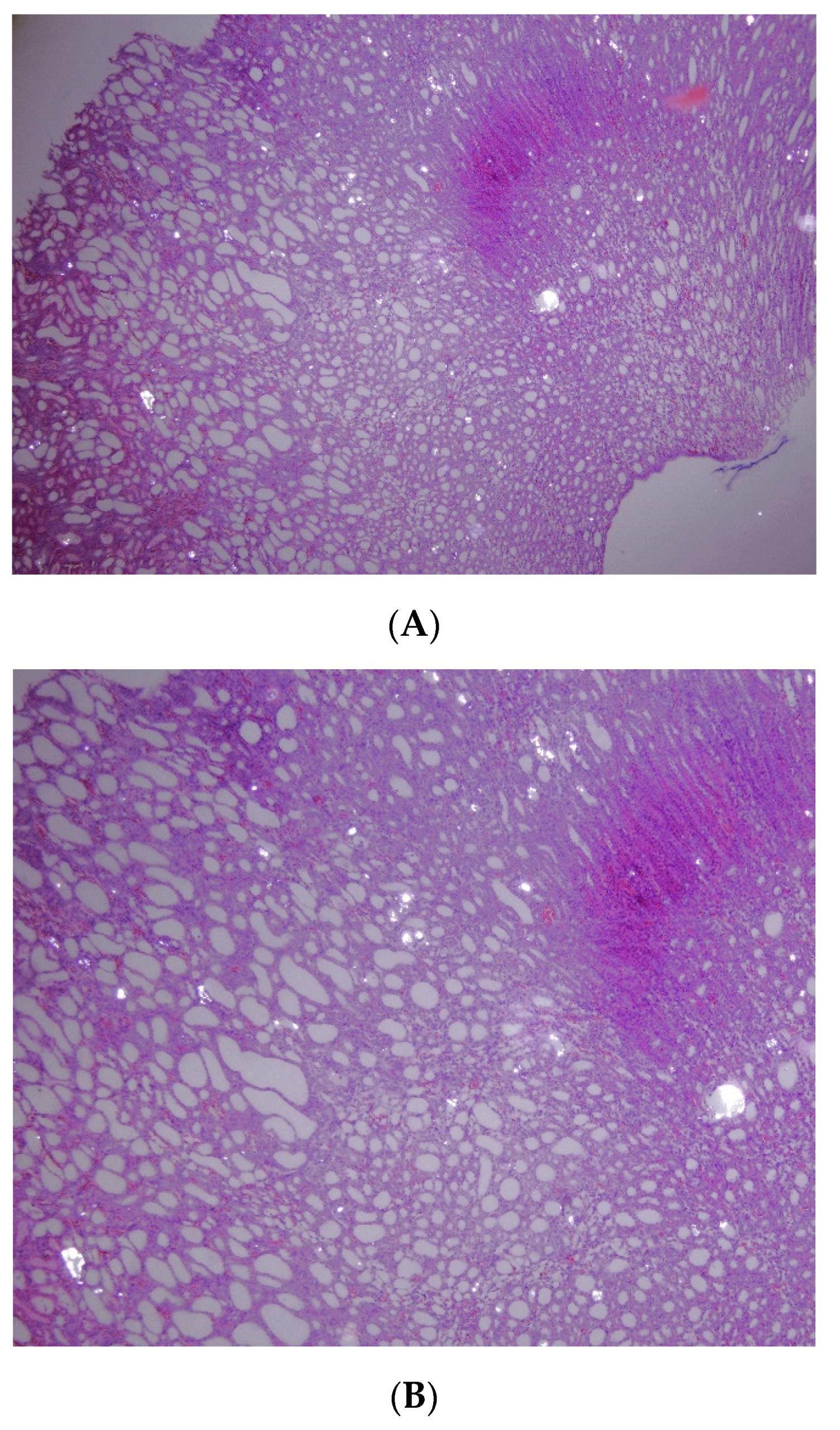
3.5. Porcine
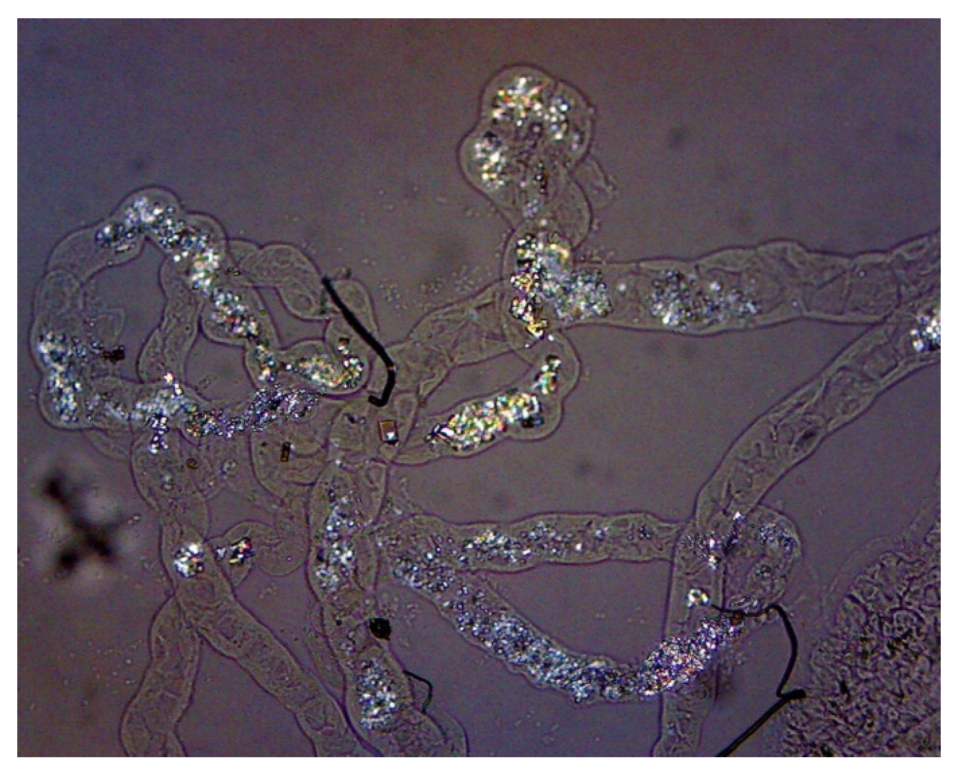
3.6. Drosophila (Fly)
4. Lithogenic Agents
4.1. Ethylene Glycol (EG)
4.2. Sodium Oxalate (NaOx)
4.3. l-Hydroxyproline (LHP)
5. Preventive Agent
6. Future Perspective
7. Conclusions
Author Contributions
Funding
Acknowledgments
Conflicts of Interest
References
- Robinson, M.R.; Norris, R.D.; Sur, R.L.; Preminger, G.M. Urolithiasis: Not just a 2-legged animal disease. J. Urol. 2008, 179, 46–52. [Google Scholar] [CrossRef]
- Pryor, W.H., Jr.; Chang, C.P.; Raulston, G.L. Urolithiasis in a Taiwan monkey (Macaca cyclopis). A literature review and case report. Lab. Anim. Care 1969, 19, 862–865. [Google Scholar]
- Keith, K.A.; Huang, J.H. Animal Models of Post-Traumatic Epilepsy. Diagnostics 2019, 10, 4. [Google Scholar] [CrossRef] [PubMed] [Green Version]
- Lee, Y.H.; Chang, L.S.; Chen, M.T.; Chiang, H.; Huang, J.K.; Huang, W.C. Characterization of ethylene glycol induced urolithiasis model in rats. J. Urol. ROC 1991, 2, 513–518. [Google Scholar]
- Lee, Y.H.; Huang, W.C.; Huang, J.K.; Chang, L.S. Testosterone enhances whereas estrogen inhibits calcium oxalate stone formation in ethylene glycol treated rats. J. Urol. 1996, 156 Pt 1, 502–505. [Google Scholar] [CrossRef]
- Huang, H.S.; Chen, C.F.; Chien, C.T.; Chen, J. Possible biphasic changes of free radicals in ethylene glycol-induced nephrolithiasis in rats. BJU Int. 2000, 85, 1143–1149. [Google Scholar] [CrossRef] [PubMed] [Green Version]
- Tsai, C.H.; Chen, Y.C.; Chen, L.D.; Pan, T.C.; Ho, C.Y.; Lai, M.T.; Tsai, F.J.; Chen, W.C. A traditional Chinese herbal antilithic formula, Wulingsan, effectively prevents the renal deposition of calcium oxalate crystal in ethylene glycol-fed rats. Urol. Res. 2008, 36, 17–24. [Google Scholar] [CrossRef]
- Chen, W.C.; Chen, H.Y.; Liao, P.C.; Wang, S.J.; Tsai, M.Y.; Chen, Y.H.; Lin, W.Y. Toward a new insight of calcium oxalate stones in Drosophila by micro-computerized tomography. Urolithiasis 2018, 46, 149–155. [Google Scholar] [CrossRef]
- Huang, H.S.; Ma, M.C.; Chen, J.; Chen, C.F. Changes in renal hemodynamics and urodynamics in rats with chronic hyperoxaluria and after acute oxalate infusion: Role of free radicals. Neurourol. Urodyn. 2003, 22, 176–182. [Google Scholar] [CrossRef]
- Tsai, C.H.; Pan, T.C.; Lai, M.T.; Lee, S.C.; Chen, M.L.; Jheng, J.R.; Chen, W.C. Prophylaxis of experimentally induced calcium oxalate nephrolithiasis in rats by Zhulingtang, a traditional Chinese herbal formula. Urol. Int. 2009, 82, 464–471. [Google Scholar] [CrossRef]
- Lin, W.C.; Lai, M.T.; Chen, H.Y.; Ho, C.Y.; Man, K.M.; Shen, J.L.; Lee, Y.J.; Tsai, F.J.; Chen, Y.H.; Chen, W.C. Protective effect of Flos carthami extract against ethylene glycol-induced urolithiasis in rats. Urol. Res. 2012, 40, 655–661. [Google Scholar] [CrossRef] [PubMed]
- Shen, J.-L.; Man, K.-M.; Chen, Y.-H.; Chang, C.-H.; Lee, Y.-J.; Chen, H.-Y.; Tsai, K.-S.; Tsai, F.-J.; Chen, W.-C.; Kuo, H.-F. Reduced albumin in renal cortex of ethylene glycol-treated rats. ScienceAsia 2014, 40, 35–41. [Google Scholar] [CrossRef] [Green Version]
- Chen, W.C.; Liu, H.P.; Wu, H.C.; Tsai, C.H.; Chen, H.Y.; Chen, H.Y.; Tasi, F.J.; Chang, C.H.; Liu, P.L.; Lin, F.Y.; et al. Preliminary Study of Ethylene Glycol-Induced Alanine-Glyoxylate Aminotransferase 2 Expression in Rat Kidney. Curr. Urol. 2009, 3, 129–135. [Google Scholar] [CrossRef]
- Wen, J.G.; Chang, Q.L.; Lou, A.F.; Li, Z.Z.; Lu, S.; Wang, Y.; Wang, Y.L.; Hu, J.H.; Mao, S.P.; Zhang, Y.; et al. Melamine-related urinary stones in 195 infants and young children: Clinical features within 2 years of follow-up. Urol. Int. 2011, 87, 429–433. [Google Scholar] [CrossRef] [PubMed]
- Liu, C.C.; Wu, C.F.; Shiea, J.; Cho, Y.T.; Hsieh, T.J.; Chou, Y.H.; Chen, B.H.; Huang, S.P.; Wu, W.J.; Shen, J.T.; et al. Detection of melamine in a human renal uric acid stone by matrix-assisted laser desorption/ionization time-of-flight mass spectrometry (MALDI-TOF MS). Clin. Chim. Acta 2012, 413, 1689–1695. [Google Scholar] [CrossRef] [PubMed]
- Chen, Y.T.; Jiann, B.P.; Wu, C.H.; Wu, J.H.; Chang, S.C.; Chien, M.S.; Hsuan, S.L.; Lin, Y.L.; Chen, T.H.; Tsai, F.J.; et al. Kidney stone distribution caused by melamine and cyanuric acid in rats. Clin. Chim. Acta 2014, 430, 96–103. [Google Scholar] [CrossRef] [PubMed]
- Chen, W.C.; Lin, W.Y.; Chen, H.Y.; Chang, C.H.; Tsai, F.J.; Man, K.M.; Shen, J.L.; Chen, Y.H. Melamine-induced urolithiasis in a Drosophila model. J. Agric. Food Chem. 2012, 60, 2753–2757. [Google Scholar] [CrossRef] [PubMed]
- Chen, Y.H.; Liu, H.P.; Chen, H.Y.; Tsai, F.J.; Chang, C.H.; Lee, Y.J.; Lin, W.Y.; Chen, W.C. Ethylene glycol induces calcium oxalate crystal deposition in Malpighian tubules: A Drosophila model for nephrolithiasis/urolithiasis. Kidney Int. 2011, 80, 369–377. [Google Scholar] [CrossRef] [Green Version]
- Ho, C.Y.; Chen, Y.H.; Wu, P.Y.; Chang, C.H.; Chen, H.Y.; Man, K.M.; Shen, J.L.; Tsai, F.J.; Lin, W.Y.; Lee, Y.J.; et al. Effects of commercial citrate-containing juices on urolithiasis in a Drosophila model. Kaohsiung J. Med. Sci. 2013, 29, 488–493. [Google Scholar] [CrossRef] [Green Version]
- Wu, S.-Y.; Shen, J.-L.; Man, K.-M.; Lee, Y.-J.; Chen, H.-Y.; Chen, Y.-H.; Tsai, K.-S.; Tsai, F.-J.; Lin, W.-Y.; Chen, W.-C. An emerging translational model to screen potential medicinal plants for nephrolithiasis, an independent risk factor for chronic kidney disease. Evid. Based Complement. Altern. Med. 2014, 2014, 972958. [Google Scholar] [CrossRef]
- Chen, W.C.; Wang, Y.C.; Shen, J.L.; Chen, H.Y.; Chang, C.H.; Tsai, F.J.; Lin, W.Y.; Chen, Y.H. Potential genitourinary toxicity and lithogenic effect of ractopamine. J. Food Nutr. Res. 2015, 3, 670–674. [Google Scholar]
- Tsai, K.-S.; Chen, Y.-H.; Shen, J.-L.; Man, K.-M.; Wu, S.-Y.; Chen, H.-Y.; Chang, C.-H.; Lee, Y.-J.; Hsu, T.-F.; Tsai, F.-J.; et al. Does chronic cola consumption increase urinary stone risk? Evidence from the Drosophila model of urolithiasis. J. Food Nutr. Res. 2015, 32, 109–113. [Google Scholar] [CrossRef] [Green Version]
- Chen, W.-C.; Chen, H.-Y.; Lin, W.-Y.; Yang, Y.-R.; Tsai, M.-Y.; Chen, Y.-H. Inhibitory Effect of Hydroxycitrate on Calcium Oxalate Crystal Formation in a Drosophila Model. J. Food Nutr. Res. 2018, 6, 706–709. [Google Scholar] [CrossRef] [Green Version]
- Hernandez, M.; Prieto, D.; Simonsen, U.; Rivera, L.; Barahona, M.V.; Garcia-Sacristan, A. Noradrenaline modulates smooth muscle activity of the isolated intravesical ureter of the pig through different types of adrenoceptors. Br. J. Pharmacol. 1992, 107, 924–931. [Google Scholar] [CrossRef] [Green Version]
- Wang, C.J.; Huang, S.W.; Chang, C.H. Efficacy of an alpha1 blocker in expulsive therapy of lower ureteral stones. J. Endourol. 2008, 22, 41–46. [Google Scholar] [CrossRef]
- Sigala, S.; Dellabella, M.; Milanese, G.; Fornari, S.; Faccoli, S.; Palazzolo, F.; Peroni, A.; Mirabella, G.; Cunico, S.C.; Spano, P.; et al. Evidence for the presence of alpha1 adrenoceptor subtypes in the human ureter. Neurourol. Urodyn. 2005, 24, 142–148. [Google Scholar] [CrossRef]
- Rajpathy, J.; Aswathaman, K.; Sinha, M.; Subramani, S.; Gopalakrishnan, G.; Kekre, N.S. An in vitro study on human ureteric smooth muscle with the alpha1-adrenoceptor subtype blocker, tamsulosin. BJU Int. 2008, 102, 1743–1745. [Google Scholar] [CrossRef]
- Wu, S.-Y.; Man, K.-M.; Shen, J.-L.; Chen, H.-Y.; Chang, C.-H.; Tsai, F.-J.; Hsieh, W.-T.; Winardi, D.; Lee, Y.-J.; Tsai, K.-S.; et al. Effect of Flos carthami extract and alpha1-adrenergic antagonists on the porcine proximal ureteral peristalsis. Evid. Based Complement. Altern. Med. 2014, 2014, 437803. [Google Scholar] [CrossRef]
- Chaussy, C.; Brendel, W.; Schmiedt, E. Extracorporeally induced destruction of kidney stones by shock waves. Lancet 1980, 2, 1265–1268. [Google Scholar] [CrossRef]
- Chaussy, C.G.; Tiselius, H.-G. How Can and Should We Optimize Extracorporeal Shockwave Lithotripsy? Urolithiasis 2018, 46, 3–17. [Google Scholar] [CrossRef] [Green Version]
- Khoo, S.Y.S. Justifiability and Animal Research in Health: Can democratisation help resolve difficulties? Animals 2018, 8, 28. [Google Scholar] [CrossRef] [PubMed] [Green Version]
- Morton, D.B. Ethics and animal research. Vet. Rec. 2020, 186, 356. [Google Scholar] [CrossRef] [PubMed]
- Khan, S.R.; Finlayson, B.; Hackett, R.L. Experimental calcium oxalate nephrolithiasis in the rat. Role of the renal papilla. Am. J. Pathol. 1982, 107, 59–69. [Google Scholar] [PubMed]
- Khan, S.R.; Finlayson, B.; Hackett, R.L. Experimental induction of crystalluria in rats using mini-osmotic pumps. Urol. Res. 1983, 11, 199–205. [Google Scholar] [CrossRef]
- Blood, F.R. Chronic toxicity of ethylene glycol in the rat. Food Cosmet. Toxicol. 1965, 3, 229–234. [Google Scholar] [CrossRef]
- Lyon, E.S.; Borden, T.A.; Vermeulen, C.W. Experimental oxalate lithiasis produced with ethylene glycol. Invest. Urol. 1966, 4, 143–151. [Google Scholar]
- Fan, J.; Glass, M.A.; Chandhoke, P.S. Impact of ammonium chloride administration on a rat ethylene glycol urolithiasis model. Scanning Microsc. 1999, 13, 299–306. [Google Scholar]
- Huang, H.S.; Ma, M.C.; Chen, C.F.; Chen, J. Changes in nitric oxide production in the rat kidney due to CaOx nephrolithiasis. Neurourol. Urodyn. 2006, 25, 252–258. [Google Scholar] [CrossRef]
- Huang, H.S.; Ma, M.C. High Sodium-Induced Oxidative Stress and Poor Anticrystallization Defense Aggravate Calcium Oxalate Crystal Formation in Rat Hyperoxaluric Kidneys. PLoS ONE 2015, 10, e0134764. [Google Scholar] [CrossRef] [Green Version]
- Cherng, J.H.; Hsu, Y.J.; Liu, C.C.; Tang, S.H.; Sartika, D.; Chang, S.J.; Fan, G.Y.; Wu, S.T.; Meng, E. Activities of Ca(2+)-related ion channels during the formation of kidney stones in an infection-induced urolithiasis rat model. Am. J. Physiol. Renal Physiol. 2019, 317, F1342–F1349. [Google Scholar] [CrossRef]
- Mo, L.; Huang, H.Y.; Zhu, X.H.; Shapiro, E.; Hasty, D.L.; Wu, X.R. Tamm-Horsfall protein is a critical renal defense factor protecting against calcium oxalate crystal formation. Kidney Int. 2004, 66, 1159–1166. [Google Scholar] [CrossRef] [PubMed] [Green Version]
- Salido, E.C.; Li, X.M.; Lu, Y.; Wang, X.; Santana, A.; Roy-Chowdhury, N.; Torres, A.; Shapiro, L.J.; Roy-Chowdhury, J. Alanine-glyoxylate aminotransferase-deficient mice, a model for primary hyperoxaluria that responds to adenoviral gene transfer. Proc. Natl. Acad. Sci. USA 2006, 103, 18249–18254. [Google Scholar] [CrossRef] [PubMed] [Green Version]
- Vernon, H.J.; Osborne, C.; Tzortzaki, E.G.; Yang, M.; Chen, J.; Rittling, S.R.; Denhardt, D.T.; Buyske, S.; Bledsoe, S.B.; Evan, A.P.; et al. Aprt/Opn double knockout mice: Osteopontin is a modifier of kidney stone disease severity. Kidney Int. 2005, 68, 938–947. [Google Scholar] [CrossRef] [PubMed] [Green Version]
- Tzortzaki, E.G.; Yang, M.; Glass, D.; Deng, L.; Evan, A.P.; Bledsoe, S.B.; Stambrook, P.J.; Sahota, A.; Tischfield, J.A. Impaired expression of an organic cation transporter, IMPT1, in a knockout mouse model for kidney stone disease. Urol. Res. 2003, 31, 257–261. [Google Scholar] [CrossRef] [PubMed]
- Khan, S.R.; Glenton, P.A. Experimentally induced hyperoxaluria in MCP-1 null mice. Urol. Res. 2011, 39, 253–258. [Google Scholar] [CrossRef] [PubMed] [Green Version]
- Dawson, P.A.; Russell, C.S.; Lee, S.; McLeay, S.C.; van Dongen, J.M.; Cowley, D.M.; Clarke, L.A.; Markovich, D. Urolithiasis and hepatotoxicity are linked to the anion transporter Sat1 in mice. J. Clin. Investig. 2010, 120, 706–712. [Google Scholar] [CrossRef] [Green Version]
- Kaplon, D.M.; Penniston, K.L.; Darriet, C.; Crenshaw, T.D.; Nakada, S.Y. Hydroxyproline-induced hyperoxaluria using acidified and traditional diets in the porcine model. J. Endourol. 2010, 24, 355–359. [Google Scholar] [CrossRef]
- Patel, S.R.; Penniston, K.L.; Iwicki, L.; Saeed, I.; Crenshaw, T.D.; Nakada, S.Y. Dietary induction of long-term hyperoxaluria in the porcine model. J. Endourol. 2012, 26, 433–438. [Google Scholar] [CrossRef]
- Trojan, B.P.; Trojan, S.J.; Navetta, A.; Staches, B.; Sutton, B.; Filleur, S.; Nelius, T. Novel porcine model for calcium oxalate stone formation. Int. Urol. Nephrol. 2017, 49, 1751–1761. [Google Scholar] [CrossRef]
- Bellen, H.J.; Yamamoto, S. Morgan’s legacy: Fruit flies and the functional annotation of conserved genes. Cell 2015, 163, 12–14. [Google Scholar] [CrossRef] [Green Version]
- Bellen, H.J.; Tong, C.; Tsuda, H. 100 years of Drosophila research and its impact on vertebrate neuroscience: A history lesson for the future. Nat. Rev. Neurosci. 2010, 11, 514–522. [Google Scholar] [CrossRef] [PubMed] [Green Version]
- Venken, K.J.; Simpson, J.H.; Bellen, H.J. Genetic manipulation of genes and cells in the nervous system of the fruit fly. Neuron 2011, 72, 202–230. [Google Scholar] [CrossRef] [PubMed] [Green Version]
- Pandey, U.B.; Nichols, C.D. Human disease models in Drosophila melanogaster and the role of the fly in therapeutic drug discovery. Pharmacol. Rev. 2011, 63, 411–436. [Google Scholar] [CrossRef] [PubMed] [Green Version]
- Landry, G.M.; Hirata, T.; Anderson, J.B.; Cabrero, P.; Gallo, C.J.; Dow, J.A.; Romero, M.F. Sulfate and thiosulfate inhibit oxalate transport via a dPrestin (Slc26a6)-dependent mechanism in an insect model of calcium oxalate nephrolithiasis. Am. J. Physiol. Renal Physiol. 2016, 310, F152–F159. [Google Scholar] [CrossRef] [PubMed] [Green Version]
- Fan, Q.X.; Gong, S.Q.; Hong, X.Z.; Feng, X.M.; Zhang, F.J. Clinical-grade Garcinia cambogia extract dissolves calcium oxalate crystals in Drosophila kidney stone models. Eur. Rev. Med. Pharmacol. Sci. 2020, 24, 6434–6445. [Google Scholar]
- Chen, W.C.; Wu, S.Y.; Liao, P.C.; Chou, T.Y.; Chen, H.Y.; Chiang, J.H.; Su, Y.C.; Man, K.M.; Tsai, M.Y.; Chen, Y.H. Treatment of Urolithiasis with Medicinal Plant Salvia miltiorrhiza: A Nationwide Cohort Study. Evid. Based Complement. Altern. Med. 2018, 2018, 8403648. [Google Scholar] [CrossRef]
- Chen, W.C.; Chou, T.Y.; Chen, H.Y.; Yang, Y.R.; Man, K.M.; Tsai, M.Y.; Chen, Y.H. Salvia miltiorrhiza Bunge (Danshen) for Treatment and Prevention of Urolithiasis: A Drosophila Animal Study. Evid. Based Complement. Altern. Med. 2019, 2019, 1408979. [Google Scholar] [CrossRef] [Green Version]
- Lin, L.-H.; Chiang, J.-H.; Chou, P.-C.; Liao, P.-C.; Wu, S.-Y.; Tsai, K.-S.; Chen, H.-Y.; Chen, Y.-H.; Chen, W.-C. Chinese Herbal Medicine Safflower (Flos carthami) Does Not Increase Bleeding Complications: A Population-based Cohort Study. J. Food Nutr. Res. 2016, 4, 108–114. [Google Scholar]
- Chung, J.; Granja, I.; Taylor, M.G.; Mpourmpakis, G.; Asplin, J.R.; Rimer, J.D. Molecular modifiers reveal a mechanism of pathological crystal growth inhibition. Nature 2016, 536, 446–450. [Google Scholar] [CrossRef]
- Fan, Q.; Feng, X.; Hong, X.; Gong, S.; Tian, J.; Hou, F.; Zhang, F. Garcinia cambogia extract removes calcium oxalate kidney stones in both genetic and non-genetic Drosophila models of nephrolithiasis. bioRxiv 2018, 477570. [Google Scholar]
- Knauf, F.; Preisig, P.A. Drosophila: A fruitful model for calcium oxalate nephrolithiasis? Kidney Int. 2011, 80, 327–329. [Google Scholar] [CrossRef] [PubMed] [Green Version]
- Miller, J.; Chi, T.; Kapahi, P.; Kahn, A.J.; Kim, M.S.; Hirata, T.; Romero, M.F.; Dow, J.A.; Stoller, M.L. Drosophila melanogaster as an emerging translational model of human nephrolithiasis. J. Urol. 2013, 190, 1648–1656. [Google Scholar] [CrossRef] [PubMed] [Green Version]
- Patel, K.V.; Guralnik, J.M.; Dansie, E.J.; Turk, D.C. Prevalence and impact of pain among older adults in the United States: Findings from the 2011 National Health and Aging Trends Study. Pain 2013, 154, 2649–2657. [Google Scholar] [CrossRef] [PubMed] [Green Version]
- Porter, W.H. Ethylene glycol poisoning: Quintessential clinical toxicology; analytical conundrum. Clin. Chim. Acta. 2012, 413, 365–377. [Google Scholar] [CrossRef] [PubMed]
- McQuade, D.J.; Dargan, P.I.; Wood, D.M. Challenges in the diagnosis of ethylene glycol poisoning. Ann. Clin. Biochem. 2014, 51, 167–178. [Google Scholar] [CrossRef] [PubMed]
- Jacobsen, D.; McMartin, K.E. Methanol and ethylene glycol poisonings. Mechanism of toxicity, clinical course, diagnosis and treatment. Med. Toxicol. 1986, 1, 309–334. [Google Scholar] [CrossRef] [PubMed]
- Khan, S.R.; Finlayson, B.; Hackett, R.L. Histologic study of the early events in oxalate induced intranephronic calculosis. Investig. Urol. 1979, 17, 199–202. [Google Scholar]
- Ogawa, Y.; Yamaguchi, K.; Morozumi, M. Effects of magnesium salts in preventing experimental oxalate urolithiasis in rats. J. Urol. 1990, 144 Pt 1, 385–389. [Google Scholar] [CrossRef]
- Takayama, T.; Fujita, K.; Suzuki, K.; Sakaguchi, M.; Fujie, M.; Nagai, E.; Watanabe, S.; Ichiyama, A.; Ogawa, Y. Control of oxalate formation from L-hydroxyproline in liver mitochondria. J. Am. Soc. Nephrol. 2003, 14, 939–946. [Google Scholar] [CrossRef] [Green Version]
- Wu, S.Y.; Chen, H.Y.; Tsai, K.S.; Chiang, J.H.; Muo, C.H.; Sung, F.C.; Chen, Y.H.; Chen, W.C. Long-Term Therapy With Wu-Ling-San, a Popular Antilithic Chinese Herbal Formula, Did Not Prevent Subsequent Stone Surgery: A Nationwide Population-Based Cohort Study. Inquiry 2016, 53. [Google Scholar] [CrossRef]
- Lin, E.; Ho, L.; Lin, M.S.; Huang, M.H.; Chen, W.C. Wu-Ling-San formula prophylaxis against recurrent calcium oxalate nephrolithiasis—A prospective randomized controlled trial. Afr. J. Tradit. Complement. Altern. Med. 2013, 10, 199–209. [Google Scholar] [CrossRef] [PubMed] [Green Version]
- Chung, V.Y.; Turney, B.W. A Drosophila genetic model of nephrolithiasis: Transcriptional changes in response to diet induced stone formation. BMC Urol. 2017, 17, 109. [Google Scholar] [CrossRef] [PubMed] [Green Version]
- Cohen, E.; Sawyer, J.K.; Peterson, N.G.; Dow, J.A.T.; Fox, D.T. Physiology, Development, and Disease Modeling in the Drosophila Excretory System. Genetics 2020, 214, 235–264. [Google Scholar] [CrossRef] [PubMed] [Green Version]


| Animal | Fly | Rat | Mouse | Pig | Dog |
|---|---|---|---|---|---|
| Cost | Low | Intermediate | High | Low | High |
| Research organ | Malphigian tubules | Kidney | Kidney | Ureter | Kidney |
| Preparation of crystal observation | Direct observe under Polarizing microscopy | H&E stain before microscopy | H&E stain before microscopy | H&E stain before microscopy | H&E stain before microscopy |
| Biochemical measurement | Not available | Available | Available | Available | Available |
| Lithogenic agent | EG, LHP, | EG, NaOx | EG, NaOx | EG+VD, LHP | Not available |
| Requirement of animal ethic | - | Yes | Yes | - | Highly recommended |
© 2020 by the authors. Licensee MDPI, Basel, Switzerland. This article is an open access article distributed under the terms and conditions of the Creative Commons Attribution (CC BY) license (http://creativecommons.org/licenses/by/4.0/).
Share and Cite
Chen, S.-J.; Chiu, K.-Y.; Chen, H.-Y.; Lin, W.-Y.; Chen, Y.-H.; Chen, W.-C. Animal Models for Studying Stone Disease. Diagnostics 2020, 10, 490. https://doi.org/10.3390/diagnostics10070490
Chen S-J, Chiu K-Y, Chen H-Y, Lin W-Y, Chen Y-H, Chen W-C. Animal Models for Studying Stone Disease. Diagnostics. 2020; 10(7):490. https://doi.org/10.3390/diagnostics10070490
Chicago/Turabian StyleChen, Szu-Ju, Kun-Yuan Chiu, Huey-Yi Chen, Wei-Yong Lin, Yung-Hsiang Chen, and Wen-Chi Chen. 2020. "Animal Models for Studying Stone Disease" Diagnostics 10, no. 7: 490. https://doi.org/10.3390/diagnostics10070490