Next Article in Journal
Post-Mortem Investigations for the Diagnosis of Sepsis: A Review of Literature
Next Article in Special Issue
Transvaginal Strain Elastosonography in the Differential Diagnosis of Rectal Endometriosis: Some Potentials and Limits
Previous Article in Journal
Persistent Left Superior Vena Cava with Absent Right Superior Vena Cava and Discrete Subaortic Stenosis Diagnosed in a Patient with Sick Sinus Syndrome: A Case Report and Brief Review of the Literature
Previous Article in Special Issue
Assessment of Coagulation Parameters in Women Affected by Endometriosis: Validation Study and Systematic Review of the Literature
 
 
Comment published on 9 January 2021, see Diagnostics 2021, 11(1), 100.
Review

Differential Diagnosis of Endometriosis by Ultrasound: A Rising Challenge

1
Department of Obstetrics and Gynecology, Policlinico Hospital, 35031 Abano Terme, PD, Italy
2
Department of Obstetrics and Gynecology, “Filippo Del Ponte” Hospital, University of Insubria, 21100 Varese, VA, Italy
3
Obstetrics and Gynecology, University of Cagliari, 09124 Cagliari, CA, Italy
4
Department of Obstetrics and Gynecology, Azienda Ospedaliero Universitaria, Policlinico Universitario Duilio Casula, 09045 Monserrato, CA, Italy
*
Author to whom correspondence should be addressed.
Diagnostics 2020, 10(10), 848; https://doi.org/10.3390/diagnostics10100848
Submission received: 6 October 2020 / Accepted: 15 October 2020 / Published: 20 October 2020
(This article belongs to the Special Issue Diagnosis of Endometriosis: Biomarkers and Clinical Methods)

Abstract

:
Ultrasound is an effective tool to detect and characterize endometriosis lesions. Variances in endometriosis lesions’ appearance and distorted anatomy secondary to adhesions and fibrosis present as major difficulties during the complete sonographic evaluation of pelvic endometriosis. Currently, differential diagnosis of endometriosis to distinguish it from other diseases represents the hardest challenge and affects subsequent treatment. Several gynecological and non-gynecological conditions can mimic deep-infiltrating endometriosis. For example, abdominopelvic endometriosis may present as atypical lesions by ultrasound. Here, we present an overview of benign and malignant diseases that may resemble endometriosis of the internal genitalia, bowels, bladder, ureter, peritoneum, retroperitoneum, as well as less common locations. An accurate diagnosis of endometriosis has significant clinical impact and is important for appropriate treatment.

1. Introduction

Endometriosis is a common, chronic, and debilitating gynecological condition that affects between 5–15% of women within their reproductive ages [1,2]. It is characterized by the growth of tissue that mimics endometrial tissue and exhibits the same responses to hormonal changes [1]. However, this abnormal tissue growth occurs outside the uterus, usually on other organs inside the pelvis and abdominal cavity, thus creating endometriosis implants. These endometriosis implants lead to local inflammatory reactions that promote fibrosis and adhesion formation, which create resistance between organs and may result in an altered pelvic anatomy [3]. This cascade of events often causes menstrual and/or chronic pelvic pain, infertility, or malfunction of the affected abdominopelvic organs [1].
In the past decades, ultrasound has become a valuable tool to accompany pelvic bimanual examinations. Currently, both methods are used for the first-line examination and diagnosis of endometriosis [4]. When a patient presents with persistent symptoms of suspected endometriosis, a comprehensive and detailed evaluation of the pelvis by ultrasound is usually prescribed. However, the accuracy of the ultrasound in detecting deep-infiltrating endometriosis depends on the experience of the sonographer. Variances in endometriosis lesion appearance and distorted anatomy secondary to adhesions and fibrosis present as major difficulties during the complete sonographic evaluation of pelvic endometriosis [5].
Therefore, a trained ultrasound operator is needed to detect most of the endometriosis lesions, and is required to make sound judgments and to plan the appropriate treatment [5,6]. It is also important for expert evaluations to include a differential diagnosis (DD) of endometriosis to exclude all other abdominopelvic diseases. Although misdiagnoses represent a small number of cases with unknown prevalence (Figure 1), the clinical impact of a DD is great because the appropriate treatment largely varies based on the diagnosis.

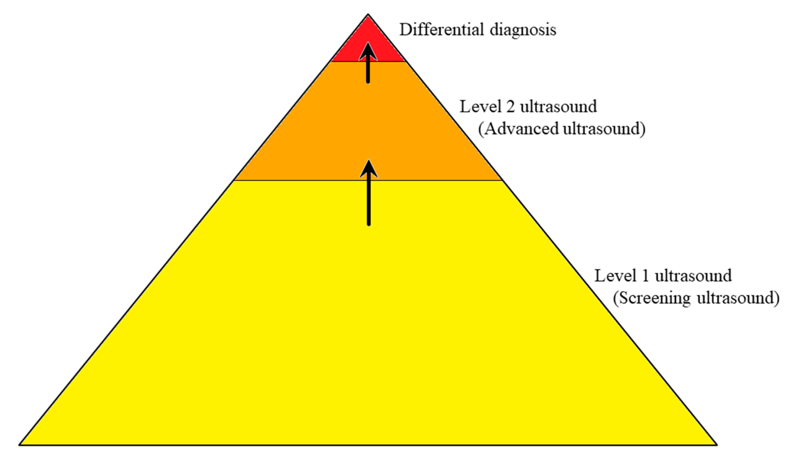
2. What a Comprehensive Ultrasound for Endometriosis Should Investigate

Traditionally, a routine pelvic ultrasound only evaluates uterine and ovarian lesions, which leads to the detection of elementary lesions, such as endometriomas [7]. However, the evaluation should include the anterior and posterior pelvic compartments to assess the mobility of pelvic organs [7,8,9,10] and to improve the assessment of endometriosis, as suggested by the International Deep Endometriosis Analysis (IDEA) consensus [11]. The use of a systematic ultrasound approach can enhance the detection of different endometriosis locations within the pelvis [12,13,14], with a detection rate that is similar to that of magnetic resonance [15,16].
Endometriosis can extend to organs other than internal genitalia, such as the bowels, bladder [1,16], and retroperitoneal structures (e.g., ureters, parametria, nerves) [17], which significantly complicates the ultrasound evaluation [18]. Although all these locations appear similar on an ultrasound (nodular, sometimes flat, hypoechoic lesions with the absence of colored Doppler spots), and they present specific features according to the organ or tissue involved. In fact, deep-infiltrating endometriosis (DIE) of hollow organs induces a retraction of margins with a subsequent irregular profile of both external and internal surfaces (i.e., bladder, bowels, and vagina), whereas DIE of dense organs (i.e., peritoneal and retroperitoneal organs) maintains a nodular hypoechoic structure [11]. Less frequently, some lesions may display vascularity with small cysts (i.e., bladder; Figure 2). Cystic endometriosis of retroperitoneal organs is a rare event that mainly occurs in patients with a history of pelvic surgery (Figure 3).

3. Sonographic Differential Diagnosis

During a comprehensive evaluation, the sonographer should be able to correctly identify all significant endometriosis lesions, the presence of other benign diseases (i.e., fibroids, uterine malformations, etc.), and any suspected malignancies. It is also important that the DD includes examination for adenomyosis [12,19,20,21] and endometrioma [22,23]. It is far more difficult for gynecologists to identify and characterize diseases of organs other than the internal genitalia. However, the ultrasound operator must be competent in identifying not only normal and altered pelvic anatomy due to endometriosis and other gynecological diseases, but also other diseases that are typically identified by other specialists (e.g., radiologist, gastroenterologist, urologist). Furthermore, gynecologists also have to consider that the presence of endometriosis does not exclude other clinical conditions that may require further evaluation by another specialist.
Several gynecological and non-gynecological conditions can mimic DIE, which are summarized in Table 1. In some cases, clinical signs can help differentiate diseases even when a pelvic bimanual examination is suggestive of endometriosis. Several studies have demonstrated the possibility of a DD of endometriosis lesions with benign (e.g., adenomyoma vs. myoma or endometrioma vs. other cysts) [19,22,24,25,26] and malignant genital diseases (e.g., ovarian cancer) [26,27].

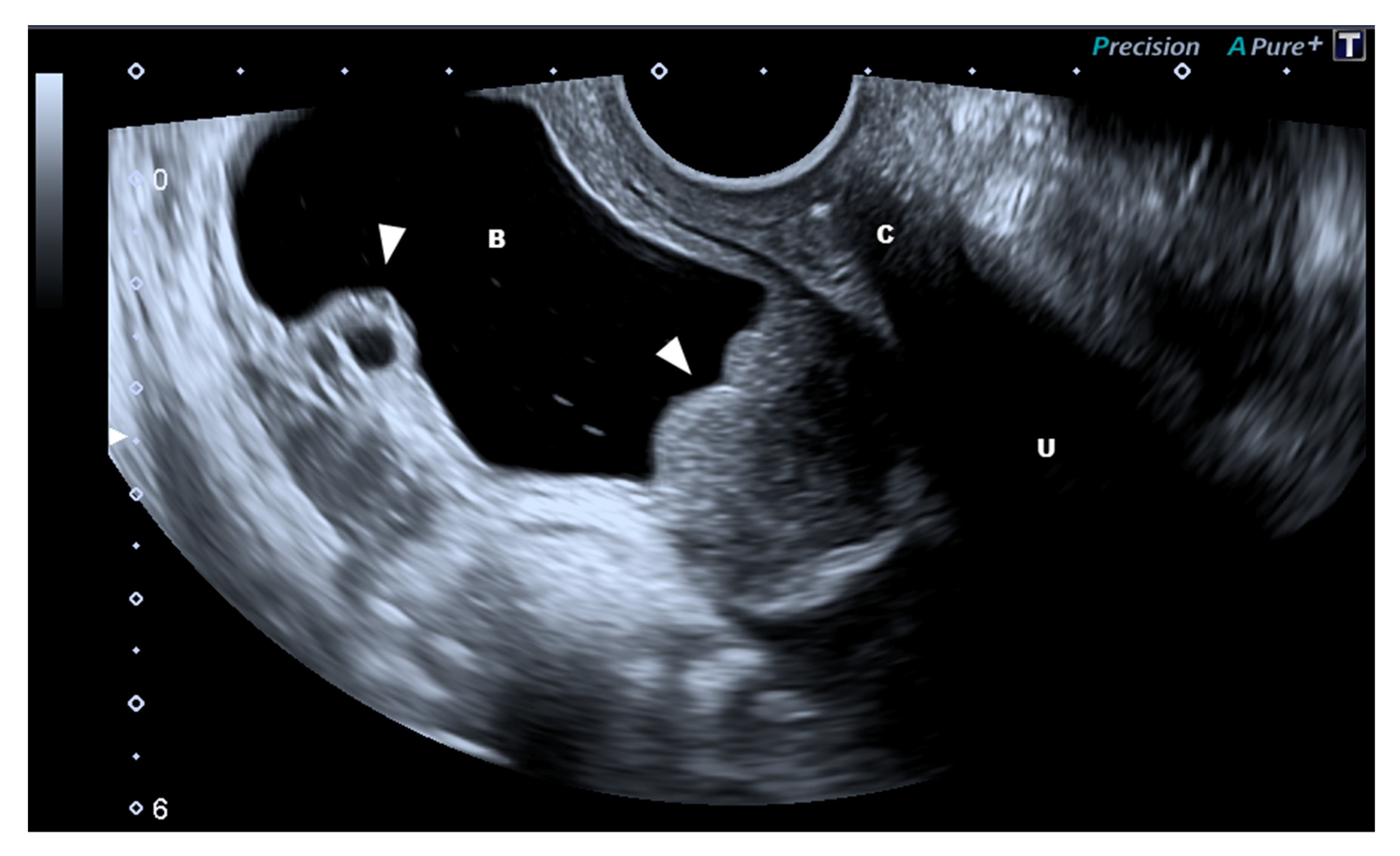
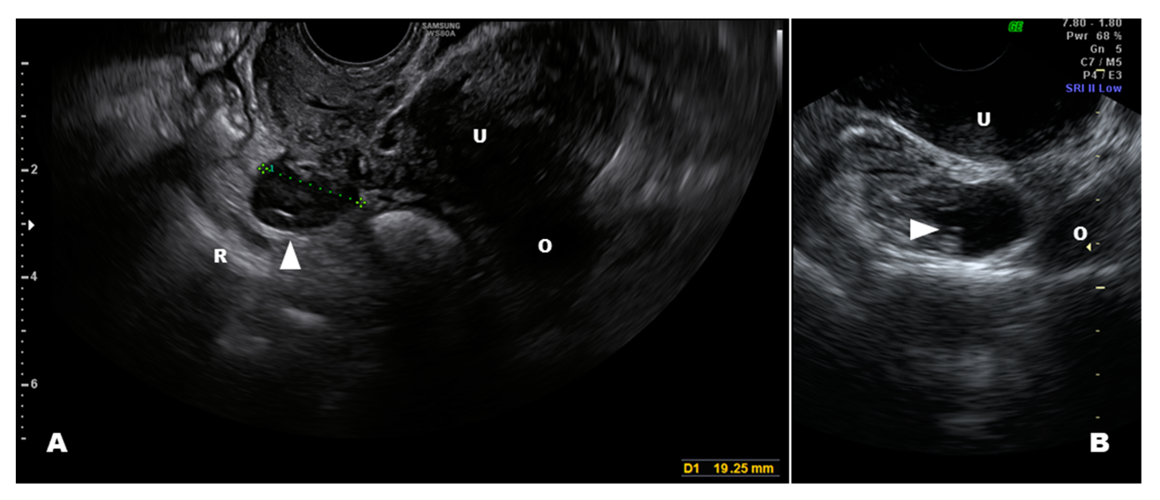
3.1. Internal Genitalia

The ovary is the most common site of endometriosis (Table 1). Additionally, the “typical” endometrioma in premenopausal women is the easiest to identify by ultrasound due to its specific characteristics (e.g., unilocular cysts with ground-glass echogenicity of the fluid content, polar clot/debris, and poor vascularization on color Doppler evaluation) [28,29]. This should be differentiated by hemorrhagic corpora lutea especially when fine strands of fibrin are seen in the fluid content of the cyst, through the evidence of rich peripheral vascularization and its transient nature. Sometimes a correct diagnosis is not easy, especially in the presence of multiple cysts, because it can be very difficult to distinguish an endometrioma from other adnexal masses, such as some dermoid cysts, hemorrhagic cysts, tubovarian abscesses, mucinous cystadenomas, or ovarian cystic adenomyomas [24,25,26]. The presence of intracystic vascularization certainly poses doubts about malignancy (e.g., borderline tumor, endometrioid cancer) [23] also because 1% of the masses presumed to be endometriomas are malignant [30]. Furthermore, the ultrasound characteristics of pre- and postmenopausal endometriomas differ. Postmenopausal endometriomas are less frequently unilocular and are less likely to have ground-glass content; however, those with typical ground-glass content have a high risk of malignancy [28].
Superficial endometriosis of the fallopian tubes is undetectable, but it can be suspected by the presence of a tubovarian complex with tube dilatation at the location of the endometriomas. Sonographic diagnosis of tubovarian abscesses is not easy in such cases, but the clinical presentations of tube wall edema and positive color/power Doppler intensities are suggestive [31], regardless of the presence of an endometrioma (abscess superimposed to the endometriosis tubovarian complex). Solid localizations of the fallopian tubes are certainly difficult to detect but they do not require a DD in the presence of other pelvic endometriosis lesions.
Adenomyosis is characterized by an altered junctional zone with an area that is isoechoic to the endometrial tissue (for the deepening of ectopic endometrial glands and stroma), and its ultrasound classification has been well described [19,32]. It can often present as an irregularity of the junctional zone (e.g., echogenic subendometrial lines and buds, an interrupted junctional zone) with or without myometrial cystic lesions, which appear either as focal or diffuse lesions or as adenomyomas. Solid lesions, which are referred to as buds, that originate from the endometrium must be differentiated from non-endophytic endometrial cancers, such as uterine clear-cell carcinoma and uterine papillary serous carcinoma. Buds do not develop from endometrial hyperplasia, but instead they arise from an atrophic endometrium [33]. The age of onset, which largely overlaps [34], clinical presentation [35], and endometrial thickness (women with symptomatic adenomyosis or perimenopausal menstrual alterations are often on hormonal therapy) may not help in the DD; however, the myometrial vascularization may provide some clues. In adenomyosis cases, the vessel distribution (i.e., arcuate, radial, and basal arteries) within the myometrium and the lesion is not altered [19,32], whereas the neoangiogenesis that occurs in tumoral lesions is detected by color/power Doppler and the identification of aberrant vessels, even in cases of endometrial cancer in which the endometrium does not appear particularly irregular [36].
Adenomyomas are defined as benign tumors that include components derived from endometrial glands, stroma, muscular cells, and fibrosis [20]. These lesions are mostly found within the myometrium, and their sonographic characteristics resemble those of myomas. Nevertheless, in some cases they can be differentiated from myomas [21]. The correct preoperative diagnosis between myomas and adenomyomas is of key importance for surgeons because the surgical removal of adenomyomas is very difficult, if not impossible. The ultrasound characteristics of adenomyomas have been reported [12,19], and their DD mainly consists of the absence of both the pseudocapsule and vascularization compared with that of myomas. In fact, non-encapsulated myomas that lack the typical refraction at the mass border maintain the typical circumferential vascularization of fibroids, whereas adenomyomas exhibit an anatomical myometrial pattern of vessels (i.e., arcuate, radial, and basal arteries). Lacunas are not necessarily a sign of adenomyomas because uterine fibroids may exhibit cystic degeneration secondary to a limited blood supply. In fact, the thick hyperechoic wall of the cystic glands of adenomyomas is poorly represented during hormonal therapy, thus a clear DD may not be possible. Clinical symptoms may support the findings of the sonographic examination because uterine myomas with cystic degeneration are poorly symptomatic, unless they are submucosal. However, adenomyomas may present with abnormal uterine bleeding and periodic pain during ovulation and menstruations. In cases of uterine masses with strong vascularity, usually hypervascular myomas, a DD that includes adenomyosis, occult degeneration (leiomyosarcoma or smooth uterine muscle of uncertain malignant potential [STUMP]), or a benign fibroid is not possible [21].
Endometriosis of the vagina occurs in approximately 12% of women with revised American Society for Reproductive Medicine (rASRM) stage IV endometriosis [17]. It presents as a nodular thickening of the vaginal wall that causes pain under a gentle pressure with the probe [37] and that does not modify with probe compression [22]. It infiltrates the recto-vaginal septum, usually in the posterior or lateral-posterior upper third of the vagina, and it presents as a hypoechoic lesion with a negative/minimal color Doppler signaling. Its DD must include advanced cervical cancer with vaginal infiltration. The normal appearance of the uterine cervix with different vascularization as well as symptoms after intercourse (endometriosis is associated to dyspareunia, whereas cervical cancer is associated with postcoital bleeding) can lead to a correct diagnosis. A DD that includes rare tumors, such as those of primary vaginal cancer, is mainly clinical because of its clinical appearance and the age of diagnosis (more frequent in postmenopausal women).
Endometriosis of the vagina rarely presents as cystic and is usually a part of a larger solid nodulus. Vaginal cysts have been reported to occur in approximately 1% of all women, and they are located in the anterolateral vaginal wall [38]. These cysts are asymptomatic remnants of the Wolffian duct and present as regular hypo- or anechoic cysts. Less frequently, they can be located on the posterolateral vaginal wall within vaginal scars (i.e., episiotomy and birth lacerations). The key aspects for a DD are the location (endometriosis is always on the posterior vaginal wall) and the absence of tenderness.

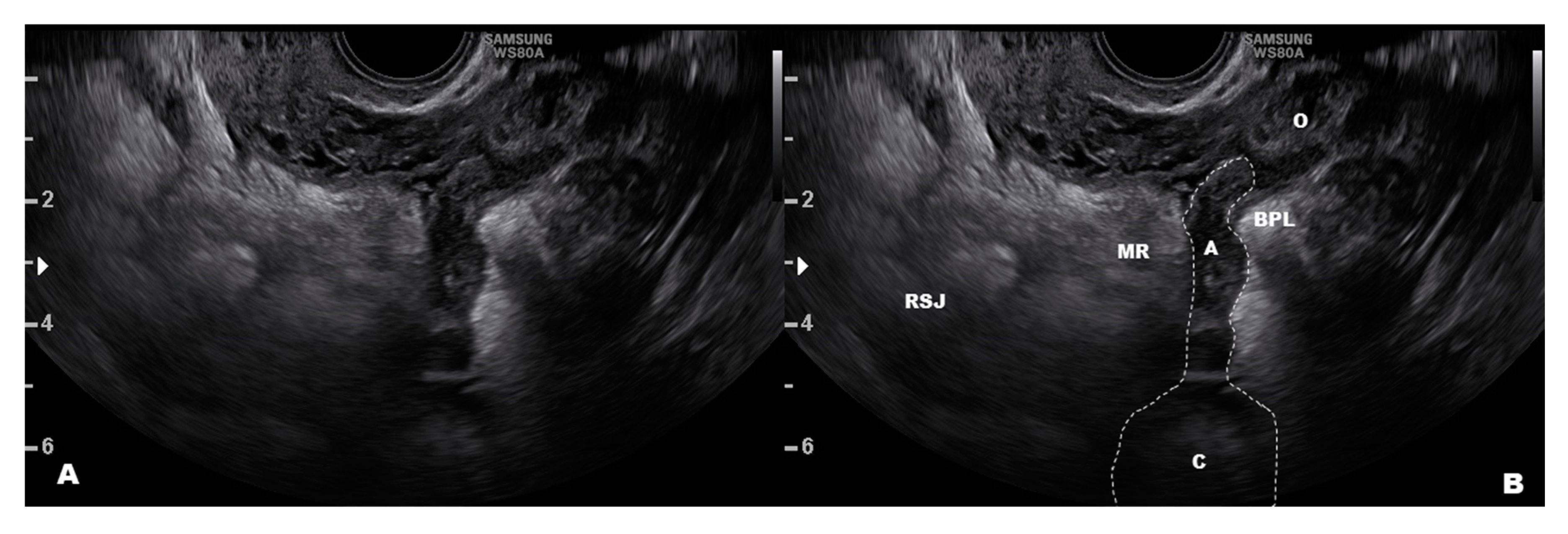
3.2. Peritoneal and Retroperitoneal Endometriosis Lesions

The pelvic peritoneum is almost always involved in advanced-stage endometriosis [3,17]. The typical appearance is a nodular thickening of the peritoneum, which is sometimes nodular or flat, like a plaque (this is very often found beneath the ovary on the ovarian fossa), and hypoechoic with negative color Doppler signaling. These lesions may grow inward into tissues from a superficial site of origin, deepening further into the mesometrium (endophytic growth) and reaching subperitoneal structures, such as ligaments (e.g., uterosacral and round), ureters, and superficial nerves (e.g., hypogastric nerve). The DD includes peritoneal carcinomatosis that presents as nodular or sheet-like, exophytic, hypoechoic, vascularized structures and is almost always associated with ascites [39].
The peritoneum over the uterus and bladder can be clearly evaluated by a transvaginal scan with an unemptied bladder, and any lesions with enough thickness (nodule) can be identified [40]. Although superficial, plain lesions cannot be detected, alterations in the peritoneal profile due to retractive fibrosis and adhesions have sometimes been reported [22]. The presence of adhesions in prevesical peritoneum due to a cesarean section with the absence of the uterine sliding sign are often misleading and impossible to differentiate from endometriosis. The two clinical conditions (adhesions and endometriosis) can coexist, and it is important to evaluate if a peritoneal lesion of endometriosis deepens into the vesicovaginal septum to the bladder.
Retroperitoneal endometriosis near the uterus (i.e., paracervical and parametrial) is common [17,41], and ultrasound is a valid tool to identify these lesions [11,42,43,44,45]. A DD that includes the spread of cervical cancer warrants attention [46]. Endometriosis of the broad posterior ligaments may infiltrate deeply into the mesometrium and paracervical tissues, reaching organs, such as the ureter [47], and appearing as a solid nodule in most cases. Retroperitoneal cystic endometriosis is not very common (Figure 3B), and it typically occurs in women who previously had pelvic surgery for endometriosis. However, it should be differentiated from a pelvic varicocele. A misplaced appendix may be easily confused with endometriosis of the mesorectum, which is usually superficial and presents as a peritoneal implant of endometriosis (Figure 4) [48]. Cystic endometriosis beneath the rectum is very rare [49] compared with benign cysts of sacral nerve roots.

3.3. The Bowels

Colonic endometriosis is not a rare condition. It affects 37% of women with severe endometriosis [13], and its correct diagnosis is essential for the proper planning of treatment [15,48,50,51]. Recently, the ultrasound detection rate of endometriosis foci of the bowels has increased, exhibiting both high sensitivity and specificity [46,52]. Additionally, its accuracy is as high as other imaging techniques, such as magnetic resonance, rectal endoscopy sonography, and double contrast barium enema [11,12,15,53]. Bowel lesions can present as different shapes; however, they exhibit an anechoic appearance without posterior enhancement, they can invade the bowel lumen, and they can have digitiform, irregular, or smooth limits. The sliding sign with the uterus and near organs is often negative because the nodule may involve the Douglas pouch or the uterosacral ligaments, or it can be attached to the posterior wall of the uterus.
The DD of bowel endometriosis using ultrasound is particularly challenging for gynecologists who are not used to examining lesions other than those of endometriosis. In some cases, bowel disease may resemble endometriosis, and the DD is more difficult [54]. It is important to note that rectosigmoid endometriosis always involves the anterior or the lateral rectosigmoid wall and never the posterior wall that is directed toward the mesorectum/mesosigmoid [54]. The most common bowel diseases that can be mistaken for endometriosis include appendicitis, appendiceal tumors [48], colonic polyps, diverticulosis, and colonic malignancy [54]. Specific factors of the internal surface of the gut should be considered during a DD, such as colonic polyps and malignancies. As we reported in a previous article [54], colon cancer growths typically extend outward and reach the serosa, whereas endometriosis lesions grow inward. A transrectal ultrasound with a radial probe that scans the entire 360 degrees of the rectal lumen is a useful tool to detect and confirm the presence of polyps or malignancies; however, a colonoscopy remains the gold standard for diagnosis. In advanced-stage rectal cancer, there may be infiltration of the entire bowel wall, but the presence of specific symptoms (e.g., rectal bleeding, unexplained weight loss, gas pains or cramps) may help with the correct diagnosis.
It may also be difficult to distinguish bowel endometriosis from colonic diverticula [54]. Diverticula are mainly characterized as outward growths on a thick bowel wall with hyperechoic content, whereas endometriosis is endophytic and hypoechoic. The imaging may even be more complex if inflammation occurs within a closed diverticular abscess. In this case, an undefined oval mass that is growing outward beyond the bowel surface may be identified. Additionally, a transverse section may exhibit intense Doppler positivity in acute diverticulitis. Clinical symptoms (e.g., constant abdominal tenderness, nausea and vomiting, and pyrexia) and inflammatory blood tests may support the sonographic findings and lead to the correct diagnosis and treatment.

3.4. The Bladder and Ureter

Urinary tract endometriosis occurs in 0.3–12% of women with endometriosis [55,56], with an incidence of 43% in women with stage IV endometriosis [17]. Endometriosis of the muscularis propria of the bladder and the ureters are the rarest conditions, with incidences of 4.3% and 9.5%, respectively [17]. The bladder wall can be examined by ultrasound because it is surrounded by a low-echogenic tissue (i.e., perivesical loose connective tissue) and the peritoneum, which appears as a hyperechoic line. In most cases, a hypoechoic lesion that alters the profile of the peritoneum of the bladder and infiltrates into the detrusor muscle indicates bladder endometriosis and originates from uterine adenomyosis [50]. Endometriosis of the bladder is often symptomatic (e.g., urgency, frequency, and pain on passing urine, which worsen days before and during menstruation, and recurrent urinary tract infections) and appears as a consistently solid mass that involves the prevesical space, the serosa (adventitia), and the detrusor muscle, sometimes up to the urothelium [22,42,57]. These lesions always involve the bladder dome on the sagittal plane, and sometimes the nodule may also extend laterally. Additionally, they exhibit color Doppler positivity. The vascularity of these lesions may suggest a malignant origin of the nodule; however, only advanced-stage cancer (either a bladder or cervical carcinoma) invades the prevesical peritoneum. Furthermore, bladder cancer is typically found in postmenopausal women and originates from the internal surface (urothelium) of the lateral wall of the bladder. In cases of advanced-stage cervical cancer, the lesion clearly involves the cervix and quite often the lateral parametrial and paracervical tissues [50,58]. Endometriosis of the bladder may present some uncommon features, such as cystic lesions and multifocal endometriosis. In such cases, it is very difficult to differentiate from bladder cancer that may be both cystic and multifocal [59,60]. Vesical cancer usually presents as a solid papillary mass that projects into the bladder, with a normal thickness of the bladder wall at the site of the lesion [60]. Nevertheless, large carcinomas may exhibit anechoic cystic degeneration within the mass and infiltration of the detrusor muscle [60]. Color Doppler imaging is not useful for this condition because cystic endometriosis of the bladder usually shows no vascularization. When making the DD, the facts that bladder carcinomas typically occur in elderly women and that urinary cytology is the simplest, less-invasive, and least expensive method to obtain a correct diagnosis warrants consideration [50].
We recently reviewed the DD of urinary tract endometriosis [50]. The DD may sometimes be difficult due to the presence of external masses that encroach the bladder dome (i.e., uterine fibroids or an attached small bowel from a previous surgery, such as a cesarean section), an intravesical ureterocele that may resemble deep lesions of endometriosis, or hypertrophic areas of the bladder wall due to a chronic, complete cystocele and recurrent cystitis [50]. In such cases, although bladder endometriosis very often appears as an endophytic lesion, it may sometimes appear flat and may be confused with hypertrophic areas due to chronic mechanical trauma (cystocele) or recurrent cystitis (Figure 5).
Endometriosis of the ureter affects the pelvic segment of the organ as a deepening of foci of the posterior broad ligament. Endometrial tissue may only invade the outer adventitia (extrinsic type) or the mucosal and/or muscular layers of the ureter (intrinsic type) [22,61]. It presents as a nodule along the course of the ureter that causes a dilatation of the ureteral tract proximal to the stenosis. The dilated ureter appears as a blood vessel in the parametrial tissue with negative color Doppler signaling and evidence of peristalsis [47], which is also often associated with hydronephrosis. The presence of ureteric calculi, which cause intense symptoms and appear as solid echogenic masses often with dilatation of the proximal part of the ureter, must be excluded [50].

3.5. Uncommon Endometriosis Locations

Uncommon locations of endometriosis are difficult to detect, and the accuracy of the ultrasound may be significantly reduced due to an inexperienced operator [62]. Endometriosis of the external genitalia (e.g., the perineum and mons pubis) may be easily confused with other conditions (i.e., abscesses, genital Crohn’s disease, skin trauma/swollen lesions, and hematomas) [63,64] because it may originate from abdominal wall lesions (i.e., muscular strains, heparin hematomas, muscular tumors, and hernias). In such cases, patient history in addition to the ultrasound findings is useful for identification of any conditions that resemble endometriosis [65,66,67]. The rarest sites of endometriosis include lesions of the liver, pancreas, kidney, and gallbladder. Hepatic endometriosis appears as superficial hypoechoic lesions and may mimic an abscess, a hematoma, an angioma, or malignancy [62]. Endometriosis of pancreas, kidney, and gallbladder show hypoechoic lesions that can be seen by ultrasound and they require a second evaluation by computerized tomography or magnetic resonance imaging [62]. These lesions do not present pathognomonic features that may differentiate endometriosis from malignancy or adenomas, so the definitive diagnosis relies on histology only [62].

4. Conclusions

The accuracy of ultrasound for the diagnosis of pelvic endometriosis has greatly improved over recent years, and it is widely regarded as the first-line diagnostic tool, despite a significant interobserver variability has been reported [68]. Certainly, the identification of extra-genitalia lesions requires a specific training [69]; however, ongoing practice is necessary to acquire and maintain competence [70].
DD by ultrasound for patients who have been referred for endometriosis may result in the detection of other gynecological diseases; therefore, it is important to consider other conditions when unusual lesions are detected. A skilled operator is key because his mental reconstruction of the distorted anatomy is necessary to correctly localize and evaluate the lesion of interest. Additional knowledge of the ultrasound appearances of lesions that do not occur within the reproductive system is needed to make a correct diagnosis. however, in such cases, further examinations should be required, or patients should be referred to other experts/specialists. A specific hands-on training for making DDs is not possible, but theoretical courses and off-line scanning sessions for sonographers with experience in gynecological and endometriosis ultrasound help to improve their background and may result in more accurate diagnoses.

Author Contributions

Conceptualization, M.S. and B.A.V.; methodology, S.G.; data curation, M.S., B.A.V., and T.B.; writing—original draft preparation, M.S.; writing—review and editing, A.S.L., N.F., and M.N.; visualization, M.S.; supervision, S.G. All authors have read and agreed to the published version of the manuscript.

Funding

This research received no external funding.

Conflicts of Interest

The authors declare no conflict of interest.

References

  1. Zondervan, K.T.; Becker, C.M.; Missmer, S.A. Endometriosis. N. Engl. J. Med. 2020, 382, 1244–1256. [Google Scholar] [CrossRef] [PubMed]
  2. Nnoaham, K.E.; Hummelshoj, L.; Webster, P.; d’Hooghe, T.; de Cicco Nardone, F.; de Cicco Nardone, C.; Jenkinson, C.; Kennedy, S.H.; Zondervan, K.T. World Endometriosis Research Foundation Global Study of Women’s Health consortium Impact of endometriosis on quality of life and work productivity: A multicenter study across ten countries. Fertil. Steril. 2011, 96, 366373.e8. [Google Scholar] [CrossRef] [Green Version]
  3. Burney, R.O.; Giudice, L.C. Pathogenesis and pathophysiology of endometriosis. Fertil. Steril. 2012, 98, 511–519. [Google Scholar] [CrossRef] [PubMed] [Green Version]
  4. Dunselman, G.A.J.; Vermeulen, N.; Becker, C.; Calhaz-Jorge, C.; D’Hooghe, T.; De Bie, B.; Heikinheimo, O.; Horne, A.W.; Kiesel, L.; Nap, A.; et al. ESHRE guideline: Management of women with endometriosis. Hum. Reprod. 2014, 29, 400–412. [Google Scholar] [CrossRef] [PubMed]
  5. Schliep, K.; Chen, Z.; Stanford, J.; Xie, Y.; Mumford, S.; Hammoud, A.; Boiman Johnstone, E.; Dorais, J.; Varner, M.; Buck Louis, G.; et al. Endometriosis diagnosis and staging by operating surgeon and expert review using multiple diagnostic tools: An inter-rater agreement study. BJOG Int. J. Obstet. Gynecol. 2017, 124, 220–229. [Google Scholar] [CrossRef] [PubMed]
  6. Chapron, C.; Marcellin, L.; Borghese, B.; Santulli, P. Rethinking mechanisms, diagnosis and management of endometriosis. Nat. Rev. Endocrinol. 2019, 15, 666–682. [Google Scholar] [CrossRef] [PubMed]
  7. Collins, B.G.; Ankola, A.; Gola, S.; McGillen, K.L. Transvaginal US of Endometriosis: Looking Beyond the Endometrioma with a Dedicated Protocol. Radiographics 2019, 39, 1549–1568. [Google Scholar] [CrossRef]
  8. Piessens, S.; Edwards, A. Sonographic Evaluation for Endometriosis in Routine Pelvic Ultrasound. J. Minim. Invasive Gynecol. 2020, 27, 265–266. [Google Scholar] [CrossRef] [PubMed] [Green Version]
  9. Fraser, M.A.; Agarwal, S.; Chen, I.; Singh, S.S. Routine vs. expert-guided transvaginal ultrasound in the diagnosis of endometriosis: A retrospective review. Abdom. Imaging 2015, 40, 587–594. [Google Scholar] [CrossRef] [Green Version]
  10. Fratelli, N.; Scioscia, M.; Bassi, E.; Musola, M.; Minelli, L.; Trivella, G. Transvaginal sonography for preoperative assessment of deep endometriosis. J. Clin. Ultrasound 2013, 41, 69–75. [Google Scholar] [CrossRef]
  11. Guerriero, S.; Condous, G.; van den Bosch, T.; Valentin, L.; Leone, F.P.G.; Van Schoubroeck, D.; Exacoustos, C.; Installé, A.J.F.; Martins, W.P.; Abrao, M.S.; et al. Systematic approach to sonographic evaluation of the pelvis in women with suspected endometriosis, including terms, definitions and measurements: A consensus opinion from the International Deep Endometriosis Analysis (IDEA) group. Ultrasound Obstet. Gynecol. 2016, 48, 318–332. [Google Scholar] [CrossRef] [PubMed]
  12. Van den Bosch, T.; Van Schoubroeck, D. Ultrasound diagnosis of endometriosis and adenomyosis: State of the art. Best Pract. Res. Clin. Obstet. Gynaecol. 2018, 51, 16–24. [Google Scholar] [CrossRef] [PubMed]
  13. Deslandes, A.; Parange, N.; Childs, J.T.; Osborne, B.; Bezak, E. Current Status of Transvaginal Ultrasound Accuracy in the Diagnosis of Deep Infiltrating Endometriosis Before Surgery: A Systematic Review of the Literature. J. Ultrasound Med. 2020. [Google Scholar] [CrossRef] [PubMed]
  14. Guerriero, S.; Ajossa, S. Transvaginal ultrasonography in superficial endometriosis. Aust. N. Z. J. Obstet. Gynaecol. 2020, 60, E5. [Google Scholar] [CrossRef] [PubMed]
  15. Noventa, M.; Scioscia, M.; Schincariol, M.; Cavallin, F.; Pontrelli, G.; Virgilio, B.; Vitale, S.G.; Laganà, A.S.; Dessole, F.; Cosmi, E.; et al. Imaging Modalities for Diagnosis of Deep Pelvic Endometriosis: Comparison between Trans-Vaginal Sonography, Rectal Endoscopy Sonography and Magnetic Resonance Imaging. A Head-to-Head Meta-Analysis. Diagnostics 2019, 9, 225. [Google Scholar] [CrossRef] [Green Version]
  16. Guerriero, S.; Saba, L.; Pascual, M.A.; Ajossa, S.; Rodriguez, I.; Mais, V.; Alcazar, J.L. Transvaginal ultrasound vs magnetic resonance imaging for diagnosing deep infiltrating endometriosis: Systematic review and meta-analysis. Ultrasound Obstet. Gynecol. 2018, 51, 586–595. [Google Scholar] [CrossRef] [Green Version]
  17. Scioscia, M.; Bruni, F.; Ceccaroni, M.; Steinkasserer, M.; Stepniewska, A.; Minelli, L. Distribution of endometriotic lesions in endometriosis stage IV supports the menstrual reflux theory and requires specific preoperative assessment and therapy. Acta Obstet. Gynecol. Scand. 2011, 90, 136–139. [Google Scholar] [CrossRef]
  18. Chamié, L.P.; Ribeiro, D.M.F.R.; Tiferes, D.A.; de Macedo Neto, A.C.; Serafini, P.C. Atypical Sites of Deeply Infiltrative Endometriosis: Clinical Characteristics and Imaging Findings. RadioGraphics 2018, 38, 309–328. [Google Scholar] [CrossRef]
  19. Van den Bosch, T.; Dueholm, M.; Leone, F.P.G.; Valentin, L.; Rasmussen, C.K.; Votino, A.; Van Schoubroeck, D.; Landolfo, C.; Installé, A.J.F.; Guerriero, S.; et al. Terms, definitions and measurements to describe sonographic features of myometrium and uterine masses: A consensus opinion from the Morphological Uterus Sonographic Assessment (MUSA) group. Ultrasound Obstet. Gynecol. 2015, 46, 284–298. [Google Scholar] [CrossRef]
  20. Paul, P.G.; Gulati, G.; Shintre, H.; Mannur, S.; Paul, G.; Mehta, S. Extrauterine adenomyoma: A review of the literature. Eur. J. Obstet. Gynecol. Reprod. Biol. 2018, 228, 130–136. [Google Scholar] [CrossRef]
  21. Testa, A.C.; Di Legge, A.; Bonatti, M.; Manfredi, R.; Scambia, G. Imaging techniques for evaluation of uterine myomas. Best Pract. Res. Clin. Obstet. Gynaecol. 2016, 34, 37–53. [Google Scholar] [CrossRef] [PubMed]
  22. Exacoustos, C.; Zupi, E.; Piccione, E. Ultrasound Imaging for Ovarian and Deep Infiltrating Endometriosis. Semin. Reprod. Med. 2017, 35, 005–024. [Google Scholar] [CrossRef]
  23. Na, K.; Park, S.Y.; Kim, H.-S. Clinicopathological Characteristics of Primary Ovarian Adenomyoma: A Single-institutional Experience. Anticancer Res. 2017, 37, 2565–2574. [Google Scholar] [CrossRef] [PubMed] [Green Version]
  24. Sokalska, A.; Timmerman, D.; Testa, A.C.; Van Holsbeke, C.; Lissoni, A.A.; Leone, F.P.G.; Jurkovic, D.; Valentin, L. Diagnostic accuracy of transvaginal ultrasound examination for assigning a specific diagnosis to adnexal masses. Ultrasound Obstet. Gynecol. 2009, 34, 462–470. [Google Scholar] [CrossRef]
  25. Valentin, L. Characterising acute gynaecological pathology with ultrasound: An overview and case examples. Best Pract. Res. Clin. Obstet. Gynaecol. 2009, 23, 577–593. [Google Scholar] [CrossRef]
  26. Sayasneh, A.; Ekechi, C.; Ferrara, L.; Kaijser, J.; Stalder, C.; Sur, S.; Timmerman, D.; Bourne, T. The characteristic ultrasound features of specific types of ovarian pathology (review). Int. J. Oncol. 2015, 46, 445–458. [Google Scholar] [CrossRef] [Green Version]
  27. Moro, F.; Zannoni, G.F.; Arciuolo, D.; Pasciuto, T.; Amoroso, S.; Mascilini, F.; Mainenti, S.; Scambia, G.; Testa, A.C. Imaging in gynecological disease (11): Clinical and ultrasound features of mucinous ovarian tumors. Ultrasound Obstet. Gynecol. 2017, 50, 261–270. [Google Scholar] [CrossRef] [Green Version]
  28. Van Holsbeke, C.; Van Calster, B.; Guerriero, S.; Savelli, L.; Paladini, D.; Lissoni, A.A.; Czekierdowski, A.; Fischerova, D.; Zhang, J.; Mestdagh, G.; et al. Endometriomas: Their ultrasound characteristics. Ultrasound Obstet. Gynecol. 2010, 35, 730–740. [Google Scholar] [CrossRef]
  29. Timmerman, D.; Valentin, L.; Bourne, T.H.; Collins, W.P.; Verrelst, H.; Vergote, I. International Ovarian Tumor Analysis (IOTA) Group Terms, definitions and measurements to describe the sonographic features of adnexal tumors: A consensus opinion from the International Ovarian Tumor Analysis (IOTA) Group. Ultrasound Obstet. Gynecol. 2000, 16, 500–505. [Google Scholar] [CrossRef]
  30. Van Holsbeke, C.; Van Calster, B.; Guerriero, S.; Savelli, L.; Leone, F.; Fischerova, D.; Czekierdowski, A.; Fruscio, R.; Veldman, J.; Van de Putte, G.; et al. Imaging in gynaecology: How good are we in identifying endometriomas? Facts Views Vis. ObGyn 2009, 1, 7–17. [Google Scholar]
  31. Timor-Tritsch, I.E.; Lerner, J.P.; Monteagudo, A.; Murphy, K.E.; Heller, D.S. Transvaginal sonographic markers of tubal inflammatory disease. Ultrasound Obstet. Gynecol. 1998, 12, 56–66. [Google Scholar] [CrossRef] [PubMed]
  32. Van den Bosch, T.; de Bruijn, A.M.; de Leeuw, R.A.; Dueholm, M.; Exacoustos, C.; Valentin, L.; Bourne, T.; Timmerman, D.; Huirne, J.A.F. Sonographic classification and reporting system for diagnosing adenomyosis. Ultrasound Obstet. Gynecol. 2019, 53, 576–582. [Google Scholar] [CrossRef] [PubMed]
  33. Gründker, C.; Günthert, A.R.; Emons, G. Hormonal heterogeneity of endometrial cancer. Adv. Exp. Med. Biol. 2008, 630, 166–188. [Google Scholar] [CrossRef] [PubMed]
  34. Yu, O.; Schulze-Rath, R.; Grafton, J.; Hansen, K.; Scholes, D.; Reed, S.D. Adenomyosis incidence, prevalence and treatment: United States population-based study 2006–2015. Am. J. Obstet. Gynecol. 2020, 223, 94.e1–94.e10. [Google Scholar] [CrossRef] [PubMed]
  35. Chapron, C.; Vannuccini, S.; Santulli, P.; Abrão, M.S.; Carmona, F.; Fraser, I.S.; Gordts, S.; Guo, S.-W.; Just, P.-A.; Noël, J.-C.; et al. Diagnosing adenomyosis: An integrated clinical and imaging approach. Hum. Reprod. Update 2020, 26, 392–411. [Google Scholar] [CrossRef]
  36. Scioscia, M.; Noventa, M.; Laganà, A.S. Abnormal uterine bleeding and the risk of endometrial cancer: Can subendometrial vascular ultrasound be of help to discriminate cancer from adenomyosis? Am. J. Obstet. Gynecol. 2020, 223, 605–606. [Google Scholar] [CrossRef]
  37. Guerriero, S.; Ajossa, S.; Gerada, M.; D’Aquila, M.; Piras, B.; Melis, G.B. “Tenderness-guided” transvaginal ultrasonography: A new method for the detection of deep endometriosis in patients with chronic pelvic pain. Fertil. Steril. 2007, 88, 1293–1297. [Google Scholar] [CrossRef]
  38. Shobeiri, S.A.; Rostaminia, G.; White, D.; Quiroz, L.H.; Nihira, M.A. Evaluation of Vaginal Cysts and Masses by 3-Dimensional Endovaginal and Endoanal Sonography. J. Ultrasound Med. 2013, 32, 1499–1507. [Google Scholar] [CrossRef]
  39. Testa, A.C.; Ludovisi, M.; Mascilini, F.; Di Legge, A.; Malaggese, M.; Fagotti, A.; Fanfani, F.; Salerno, M.G.; Ercoli, A.; Scambia, G.; et al. Ultrasound evaluation of intra-abdominal sites of disease to predict likelihood of suboptimal cytoreduction in advanced ovarian cancer: A prospective study. Ultrasound Obstet. Gynecol. 2012, 39, 99–105. [Google Scholar] [CrossRef]
  40. Savelli, L.; Manuzzi, L.; Pollastri, P.; Mabrouk, M.; Seracchioli, R.; Venturoli, S. Diagnostic accuracy and potential limitations of transvaginal sonography for bladder endometriosis. Ultrasound Obstet. Gynecol. 2009, 34, 595–600. [Google Scholar] [CrossRef]
  41. Chiantera, V.; Petrillo, M.; Abesadze, E.; Sozzi, G.; Dessole, M.; Catello Di Donna, M.; Scambia, G.; Sehouli, J.; Mechsner, S. Laparoscopic Neuronavigation for Deep Lateral Pelvic Endometriosis: Clinical and Surgical Implications. J. Minim. Invasive Gynecol. 2018, 25, 1217–1223. [Google Scholar] [CrossRef] [PubMed]
  42. Guerriero, S.; Ajossa, S.; Minguez, J.A.; Jurado, M.; Mais, V.; Melis, G.B.; Alcazar, J.L. Accuracy of transvaginal ultrasound for diagnosis of deep endometriosis in uterosacral ligaments, rectovaginal septum, vagina and bladder: Systematic review and meta-analysis. Ultrasound Obstet. Gynecol. 2015, 46, 534–545. [Google Scholar] [CrossRef] [PubMed]
  43. Mabrouk, M.; Raimondo, D.; Arena, A.; Iodice, R.; Altieri, M.; Sutherland, N.; Salucci, P.; Moro, E.; Seracchioli, R. Parametrial Endometriosis: The Occult Condition that Makes the Hard Harder. J. Minim. Invasive Gynecol. 2019, 26, 871–876. [Google Scholar] [CrossRef] [PubMed]
  44. Scioscia, M.; Virgilio, B.A.; Scardapane, A.; Pontrelli, G. Fusion Imaging: A Novel Diagnostic Tool for Nerve-sparing Surgery for Deep Infiltrating Endometriosis. J. Minim. Invasive Gynecol. 2020, 27, 246–247. [Google Scholar] [CrossRef]
  45. Leonardi, M.; Martins, W.P.; Espada, M.; Arianayagam, M.; Condous, G. Proposed technique to visualize and classify uterosacral ligament deep endometriosis with and without infiltration into parametrium or torus uterinus. Ultrasound Obstet. Gynecol. 2020, 55, 137–139. [Google Scholar] [CrossRef] [Green Version]
  46. Alcazar, J.L.; García, E.; Machuca, M.; Quintana, R.; Escrig, J.; Chacón, E.; Mínguez, J.A.; Chiva, L. Magnetic resonance imaging and ultrasound for assessing parametrial infiltration in cervical cancer. A systematic review and meta-analysis. Med. Ultrason. 2020, 22, 85–91. [Google Scholar] [CrossRef]
  47. Scioscia, M. Ureteral endometriosis: Correlation between ultrasonography and laparoscopy: Ureteral endometriosis. Ultrasound Obstet. Gynecol. 2019, 53, 706–708. [Google Scholar] [CrossRef]
  48. Virgilio, B.A.; Pontrelli, G.; Trevisan, P.; Sacchi, D.; Bernardini, T.; Scioscia, M. Incidental diagnosis on transvaginal ultrasound of appendiceal mucocele arising on low-grade appendiceal mucinous neoplasm. Ultrasound Obstet. Gynecol. 2019, 54, 412–414. [Google Scholar] [CrossRef]
  49. Sima, R.-M.; Radosa, J.C.; Zamfir, R.; Ionescu, C.-A.; Carp, D.; Iordache, I.-I.; Stănescu, A.-D.; Pleş, L. Novel diagnosis of mesenteric endometrioma: Case report. Medicine (Baltimore) 2019, 98, e16432. [Google Scholar] [CrossRef]
  50. Scioscia, M.; Zanetti, I.; Raspanti, X.; Spoto, E.; Portuese, A.; Noventa, M.; Pontrelli, G.; Greco, P.; Virgilio, B.A. Ultrasound Differential Diagnosis in Deep Infiltrating Endometriosis of the Urinary Tract. J. Ultrasound Med. 2020. [Google Scholar] [CrossRef]
  51. Scardapane, A.; Lorusso, F.; Francavilla, M.; Bettocchi, S.; Fascilla, F.D.; Angelelli, G.; Scioscia, M. Magnetic Resonance Colonography May Predict the Need for Bowel Resection in Colorectal Endometriosis. BioMed Res. Int. 2017, 2017, 1–7. [Google Scholar] [CrossRef] [PubMed] [Green Version]
  52. Kammann, S.; Menias, C.; Hara, A.; Moshiri, M.; Siegel, C.; Safar, B.; Brandes, S.; Shaaban, A.; Sandrasegaran, K. Genital and reproductive organ complications of Crohn disease: Technical considerations as it relates to perianal disease, imaging features, and implications on management. Abdom. Radiol. 2017, 42, 1752–1761. [Google Scholar] [CrossRef] [PubMed]
  53. Nezhat, C.; Li, A.; Falik, R.; Copeland, D.; Razavi, G.; Shakib, A.; Mihailide, C.; Bamford, H.; DiFrancesco, L.; Tazuke, S.; et al. Bowel endometriosis: Diagnosis and management. Am. J. Obstet. Gynecol. 2018, 218, 549–562. [Google Scholar] [CrossRef] [PubMed]
  54. Scioscia, M.; Orlandi, S.; Trivella, G.; Portuese, A.; Bettocchi, S.; Pontrelli, G.; Bocus, P.; Anna Virgilio, B. Sonographic Differential Diagnosis in Deep Infiltrating Endometriosis: The Bowel. BioMed Res. Int. 2019, 2019, 1–9. [Google Scholar] [CrossRef] [PubMed] [Green Version]
  55. Fujita, M.; Manabe, N.; Honda, K.; Murao, T.; Osawa, M.; Kawai, R.; Akiyama, T.; Shiotani, A.; Haruma, K.; Hata, J. Usefulness of Ultrasonography for Diagnosis of Small Bowel Tumors: A Comparison Between Ultrasonography and Endoscopic Modalities. Medicine 2015, 94, e1464. [Google Scholar] [CrossRef] [PubMed]
  56. Kołodziej, A.; Krajewski, W.; Dołowy, Ł.; Hirnle, L. Urinary Tract Endometriosis. Urol. J. 2015, 12, 2213–2217. [Google Scholar]
  57. Pateman, K.; Holland, T.K.; Knez, J.; Derdelis, G.; Cutner, A.; Saridogan, E.; Jurkovic, D. Should a detailed ultrasound examination of the complete urinary tract be routinely performed in women with suspected pelvic endometriosis? Hum. Reprod. 2015, 30, 2802–2807. [Google Scholar] [CrossRef] [Green Version]
  58. Testa, A.C.; Di Legge, A.; De Blasis, I.; Cristina Moruzzi, M.; Bonatti, M.; Collarino, A.; Rufini, V.; Manfredi, R. Imaging techniques for the evaluation of cervical cancer. Best Pract. Res. Clin. Obstet. Gynaecol. 2014, 28, 741–768. [Google Scholar] [CrossRef] [PubMed]
  59. Smereczyński, A.; Szopiński, T.; Gołąbek, T.; Ostasz, O.; Bojko, S. Sonography of tumors and tumor-like lesions that mimic carcinoma of the urinary bladder. J. Ultrason. 2014, 14, 36–48. [Google Scholar] [CrossRef]
  60. Pavlica, P.; Gaudiano, C.; Barozzi, L. Sonography of the bladder. World J. Urol. 2004, 22, 328–334. [Google Scholar] [CrossRef]
  61. Barra, F.; Scala, C.; Biscaldi, E.; Vellone, V.G.; Ceccaroni, M.; Terrone, C.; Ferrero, S. Ureteral endometriosis: A systematic review of epidemiology, pathogenesis, diagnosis, treatment, risk of malignant transformation and fertility. Hum. Reprod. Update 2018, 24, 710–730. [Google Scholar] [CrossRef] [PubMed] [Green Version]
  62. Guerriero, S.; Conway, F.; Pascual, M.A.; Graupera, B.; Ajossa, S.; Neri, M.; Musa, E.; Pedrassani, M.; Alcazar, J.L. Ultrasonography and Atypical Sites of Endometriosis. Diagnostics (Basel) 2020, 10, 345. [Google Scholar] [CrossRef] [PubMed]
  63. Mascilini, F.; Moruzzi, C.; Giansiracusa, C.; Guastafierro, F.; Savelli, L.; De Meis, L.; Epstein, E.; Timor-Tritsch, I.E.; Mailath-Pokorny, M.; Ercoli, A.; et al. Imaging in gynecological disease. 10: Clinical and ultrasound characteristics of decidualized endometriomas surgically removed during pregnancy. Ultrasound Obstet. Gynecol. 2014, 44, 354–360. [Google Scholar] [CrossRef] [PubMed]
  64. Noventa, M.; Saccardi, C.; Litta, P.; Vitagliano, A.; D’Antona, D.; Abdulrahim, B.; Duncan, A.; Alexander-Sefre, F.; Aldrich, C.J.; Quaranta, M.; et al. Ultrasound techniques in the diagnosis of deep pelvic endometriosis: Algorithm based on a systematic review and meta-analysis. Fertil. Steril. 2015, 104, 366–383.e2. [Google Scholar] [CrossRef] [PubMed] [Green Version]
  65. Grigore, M.; Socolov, D.; Pavaleanu, I.; Scripcariu, I.; Grigore, A.M.; Micu, R. Abdominal wall endometriosis: An update in clinical, imagistic features, and management options. Med. Ultrason. 2017, 19, 430. [Google Scholar] [CrossRef] [Green Version]
  66. Morales Martínez, C.; Tejuca Somoano, S. Abdominal wall endometriosis. Am. J. Obstet. Gynecol. 2017, 217, 701–702. [Google Scholar] [CrossRef]
  67. Ecker, A.M.; Donnellan, N.M.; Shepherd, J.P.; Lee, T.T.M. Abdominal wall endometriosis: 12 years of experience at a large academic institution. Am. J. Obstet. Gynecol. 2014, 211, 363.e1–363.e5. [Google Scholar] [CrossRef]
  68. Bean, E.; Chaggar, P.; Thanatsis, N.; Dooley, W.; Bottomley, C.; Jurkovic, D. Intra- and interobserver reproducibility of pelvic ultrasound for the detection and measurement of endometriotic lesions. Hum. Reprod. Open 2020, 2020, hoaa001. [Google Scholar] [CrossRef]
  69. Guerriero, S.; Pascual, M.A.; Ajossa, S.; Rodriguez, I.; Zajicek, M.; Rolla, M.; Rams Llop, N.; Yulzari, V.; Bardin, R.; Buonomo, F.; et al. Learning curve for ultrasonographic diagnosis of deep infiltrating endometriosis using structured offline training program. Ultrasound Obstet. Gynecol. 2019, 54, 262–269. [Google Scholar] [CrossRef]
  70. Eisenberg, V.H.; Alcazar, J.L.; Arbib, N.; Schiff, E.; Achiron, R.; Goldenberg, M.; Soriano, D. Applying a statistical method in transvaginal ultrasound training: Lessons from the learning curve cumulative summation test (LC-CUSUM) for endometriosis mapping. Gynecol. Surg. 2017, 14, 19. [Google Scholar] [CrossRef] [Green Version]
Figure 1. A pyramid chart that represents the distribution of ultrasound scans for endometriosis according to complexity. Although differential diagnosis must be considered in each exam, the number of cases where it makes a difference is relatively small, although it is fundamental.
Diagnostics 10 00848 g001
Figure 2. Bifocal endometriosis of the bladder (arrow). The two lesions present different characteristics as the one on the right-hand side presents a typical solid aspect while the left one is a cystic endometriosis location. Abbreviations: B, bladder; C, cervix; U, uterus.
Diagnostics 10 00848 g002
Figure 3. Cystic endometriosis (arrow) of the retroperitoneum in two patients on medical therapy with previous pelvic surgery for endometriosis. (A) shows an endometriosis cyst of the rectovaginal septum and (B) shows a cyst of the mesometrium. Abbreviations: U, uterus; R, rectum; O, Ovary.
Diagnostics 10 00848 g003
Figure 4. Appendix dislocated downward and attached to the ovary that may mimic mesorectal endometriosis. (A,B) show the clean and the labelled image. Abbreviations: A, appendix; O, Ovary; BPL, broad posterior ligament; C, caecum; MR, mesorectum; RSJ, rectosigmoid junction.
Diagnostics 10 00848 g004
Figure 5. A typical endometriosis nodule (arrow) of the bladder (A); figure (B) shows a flat nodule of endometriosis (arrow) with a subserosal myoma that distorts the bladder dome; a flat endometriosis nodule (arrow) of the bladder that can be seen at 3D ultrasound only when cut (C) while the internal surface of the bladder appears regular (D); figure (E) shows a thick bladder wall due to cystocele and figure (F) in case of recurrent cystitis. Abbreviations: U, uterus; B, bladder; M, myoma.
Diagnostics 10 00848 g005
Table 1. Endometriosis possible anatomic locations, anatomical features, and differential diagnosis.
Anatomic Locations Anatomical AspectPossible Differential Diagnosis
Internal genitalia
 OvaryCysticHemorrhagic corpus luteus
Hemorrhagic cyst
Dermoid cyst
Tubovarian abscess
Mucinous cystoadenoma
Ovarian cystic adenomyoma
Malignancy
 TubeCysticTubovarian abscess
 Uterus (adenomyosis)Focal, solid (buds)Endometrial malignancy
Solid adenomyomaNon-encapsulated myomas
Cystic adenomyomaMyomas with cystic degeneration/necrosis
Suspected sarcoma/STUMP
 VaginaSolid nodulus


Cystic
Cervical malignancy
Vaginal malignancy

Benign vaginal cyst
Peritoneum
 Broad anterior/posterior ligamentSolidPeritoneal carcinomatosis
 Douglas pouchSolidPeritoneal carcinomatosis
Advanced cervical malignancy
 Prevesical peritoneumSolidPost-cesarean adhesions
Peritoneal carcinomatosis
Advanced cervical malignancy
 Uterosacral ligamentsSolidPeritoneal carcinomatosis
Advanced cervical malignancy
 Round ligamentsSolidPeritoneal carcinomatosis
Advanced endometrial malignancy
Retroperitoneal tissues
 Paracervix/parametriumSolidAdvanced cervical malignancy
 MesometriumSolid


Cystic
Pedunculated infraligamentary fibroid
Ovarian malignancy

Varicocele
 MesorectumSolid


Cystic
Colonic malignancy
Downward dislocated appendix

Retrorectal dermoid cyst
 Presacral spaceCysticTarlov cyst
Ganglioneuroma
 Vesicovaginal septumSolidBladder malignancy
Bowel
 RectosigmoidSolidColonic polyp
Diverticulosis
Colonic malignancy
 Caecum and appendixSolidAppendicitis
Appendiceal tumors
 IleumSolidMalignancy
Urinary tract
 BladderSolid


Cystic
Bladder malignancy
Cervical malignancy
Uterine leiomyomas/adenomyomas
Hypertrophic traumatic bladder

Bladder malignancy
Intravesical ureterocele
Small bowel attached to the prevesical peritoneum
 UreterSolidUreteric stone
External genitalia
 PerineumSolidPostpatum scar
Chron disease
 Mons pubisSolidAbscess
Hematoma
Muscular strain
Abdominal wall
 UmbilicusSolidOmphalitis
Umbilical granuloma
 Abdominal muscleSolid


Cystic
Strain
Benign tumor (desmoid)

Hematoma
 Inguinal canalSolidComplete small hernia
Publisher’s Note: MDPI stays neutral with regard to jurisdictional claims in published maps and institutional affiliations.

