
Advanced Deep Learning Approaches for Accurate Brain Tumor Classification in Medical Imaging
 , , , , , ,
Abstract
:1. Introduction
2. Related Work
3. Materials and Methods
3.1. Data Collection
3.1.1. Data Preparation
3.1.2. Data Preprocessing
- CLAHE (Construct limited histogram equalization)
- Morphological analysis
3.1.3. Data Segmentation
- Resizing Images
- Data Augmentation
| Algorithm 1 BDA |
| Input: |
|
| Processing: |
|
| Output: |
| Save steps 1, 2, 3, 4, 5, 6 |
3.2. Methods
3.2.1. Aquila Optimizer (AQO): A Meta-Heuristic Optimization Algorithm
- Generation of Initial Population
- Updating Population
- Terminal Criteria
- Validation Stage
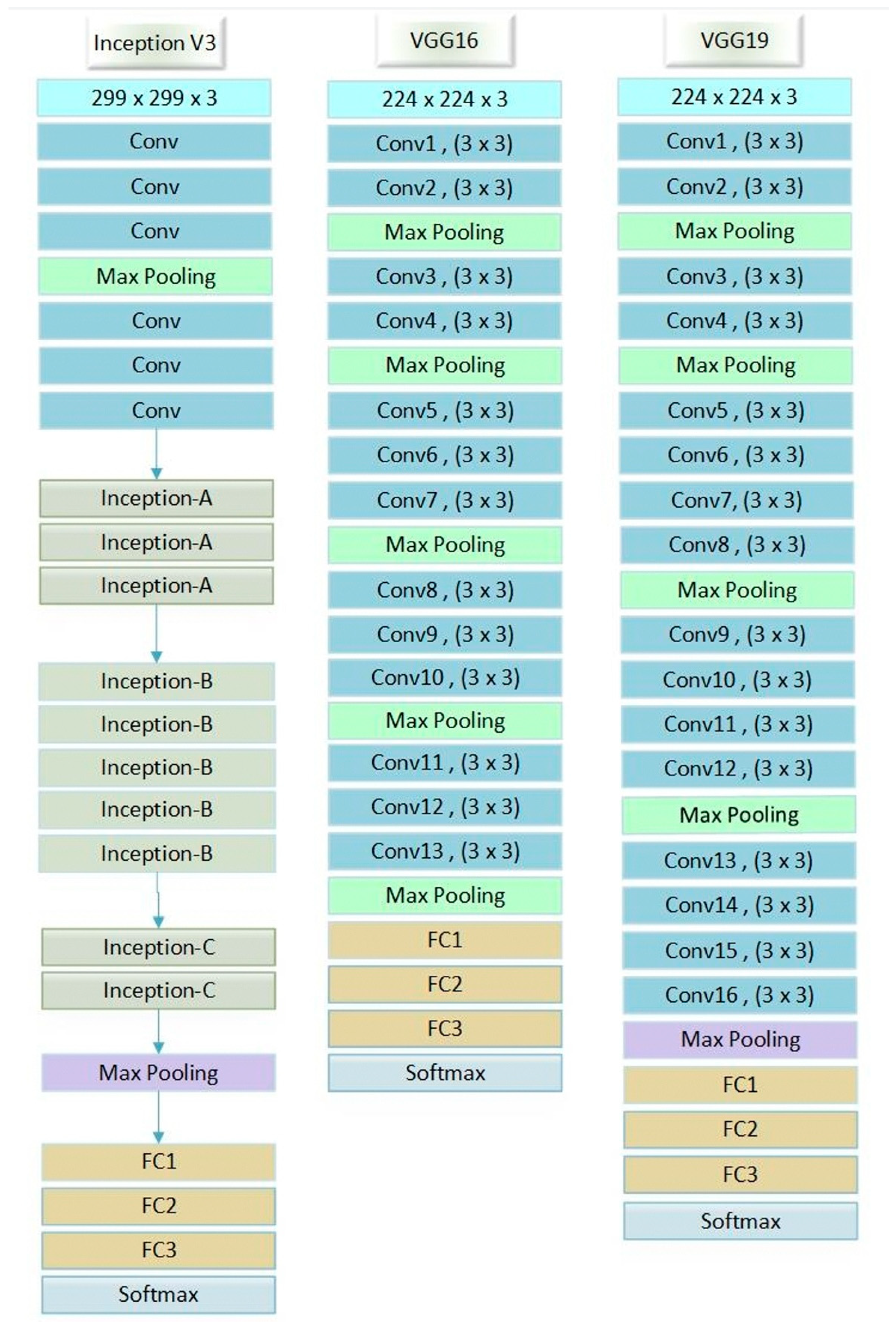
3.2.2. VGG-16 Model
- VGG-16 can recognize and categorize images for the purpose of medical imaging diagnostics, such as x-ray and MR images. Furthermore, it may be used in the ability to read street signs while in motion.
- Although its detection capabilities were not covered in the introduction, VGG-16 can achieve excellent results in image detection use cases: notably, it triumphed in 2014′s ImageNet detection contest (where it ended up as the first runner-up for the classification challenge).
- The model may be trained to generate image embedding vectors, which can then be utilized for tasks such as face verification inside a VGG-16-based Siamese network. This is made possible by removing the top output layer.
3.2.3. VGG-19 Model
3.2.4. Inception-V3 Model
4. Experimental Setup and Results
4.1. Experimental Design
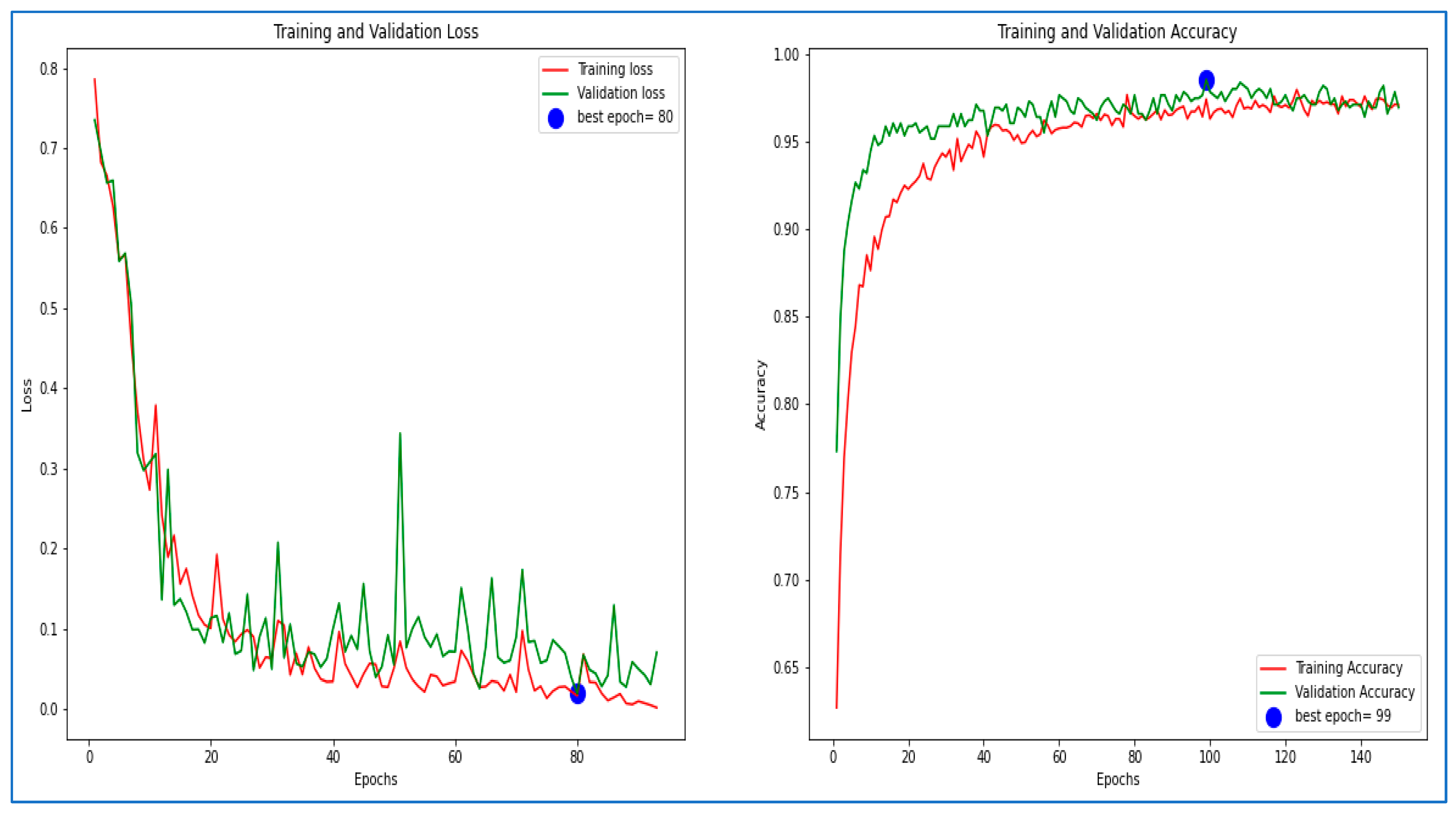
4.2. VGG-16 Model Validation
4.3. VGG-19 Model Validation
4.4. Inception-V3 Model Validation
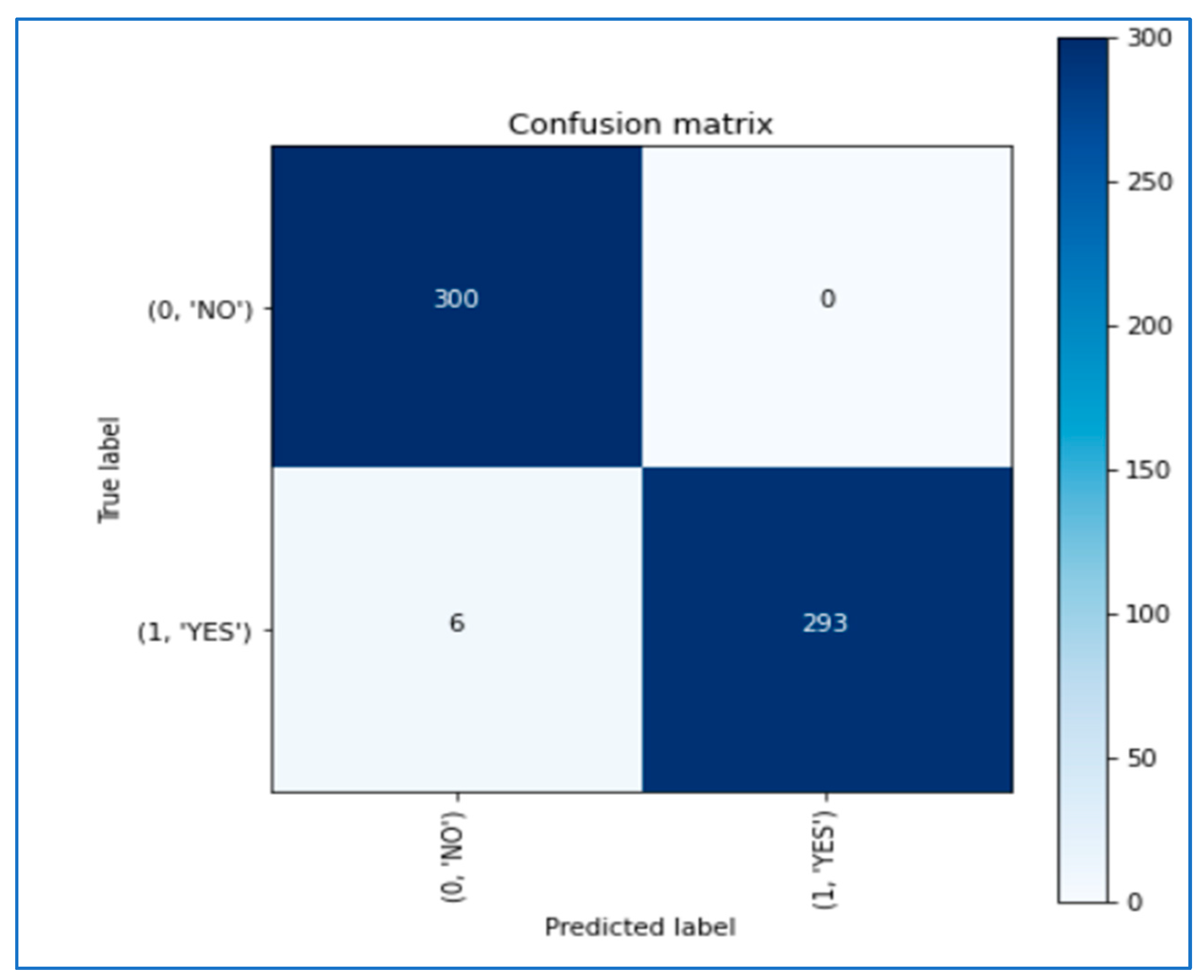
4.5. Results
5. Conclusions
Author Contributions
Funding
Institutional Review Board Statement
Informed Consent Statement
Data Availability Statement
Conflicts of Interest
References
- Zhang, J.; Lv, X.; Zhang, H.; Liu, B. AResU-Net: Attention Residual U-Net for Brain Tumor Segmentation. Symmetry 2020, 12, 721. [Google Scholar] [CrossRef]
- Nirmalapriya, G.; Agalya, V.; Regunathan, R.; Ananth, M.B.J. Fractional Aquila spider monkey optimization based deep learning network for classification of brain tumor. Biomed. Signal Process. Control 2023, 79, 104017. [Google Scholar] [CrossRef]
- Paul, J.S.; Plassard, A.J.; Landman, B.A.; Fabbri, D. Deep learning for brain tumor classification. In Proceedings of the SPIE 10137, Medical Imaging 2017: Biomedical Applications in Molecular, Structural, and Functional Imaging, Orlando, FL, USA, 12–14 February 2017. [Google Scholar]
- Masood, M.; Nazir, T.; Nawaz, M.; Mehmood, A.; Rashid, J.; Kwon, H.-Y.; Mahmood, T.; Hussain, A. A Novel Deep Learning Method for Recognition and Classification of Brain Tumors from MRI Images. Diagnostics 2021, 11, 744. [Google Scholar] [CrossRef] [PubMed]
- Ali, M.; Shah, J.H.; Khan, M.A.; Alhaisoni, M.; Tariq, U.; Akram, T.; Kim, Y.J.; Chang, B. Brain tumor detection and classification using pso and convolutional neural network. Comput. Mater. Contin. 2022, 73, 4501–4518. [Google Scholar] [CrossRef]
- Zhang, Y.; Liu, X.; Wa, S.; Liu, Y.; Kang, J.; Lv, C. GenU-Net++: An Automatic Intracranial Brain Tumors Segmentation Algorithm on 3D Image Series with High Performance. Symmetry 2021, 13, 2395. [Google Scholar] [CrossRef]
- Saber, A.; Keshk, A.; Abo-Seida, O.; Sakr, M. Tumor detection and classification in breast mammography based on fine-tuned convolutional neural networks. IJCI Int. J. Comput. Inf. 2022, 9, 74–84. [Google Scholar]
- Saber, A.; Sakr, M.; Abo-Seida, O.; Keshk, A.; Chen, H. A novel deep-learning model for automatic detection and classification of breast cancer using the transfer-learning technique. IEEE Access 2021, 9, 71194–71209. [Google Scholar] [CrossRef]
- Elmezain, M.; Mahmoud, A.; Mosa, D.; Said, W. Brain Tumor Segmentation Using Deep Capsule Network and Latent-Dynamic Conditional Random Fields. J. Imaging 2022, 8, 190. [Google Scholar] [CrossRef] [PubMed]
- Arora, S.; Sharma, M. Deep Learning for Brain Tumor Classification from MRI Images. In Proceedings of the Sixth International Conference on Image Information Processing (ICIIP), Shimla, India, 26–28 November 2021; pp. 409–412. [Google Scholar]
- Meenakshi, A.; Revathy, S. An Efficient Model for Predicting Brain Tumor using Deep Learning Techniques. In Proceedings of the 5th International Conference on Communication and Electronics Systems (ICCES), Coimbatore, India, 10–12 June 2020; pp. 1000–1007. [Google Scholar]
- Khan, M.A.; Ashraf, I.; Alhaisoni, M.; Damaševičius, R.; Scherer, R.; Rehman, A.; Bukhari, S.A.C. Multimodal Brain Tumor Classification Using Deep Learning and Robust Feature Selection: A Machine Learning Application for Radiologists. Diagnostics 2020, 10, 565. [Google Scholar] [CrossRef] [PubMed]
- Hossain, A.; Islam, M.T.; Abdul Rahim, S.K.; Rahman, M.A.; Rahman, T.; Arshad, H.; Khandakar, A.; Ayari, M.A.; Chowdhury, M.E.H. A Lightweight Deep Learning Based Microwave Brain Image Network Model for Brain Tumor Classification Using Reconstructed Microwave Brain (RMB) Images. Biosensors 2023, 13, 238. [Google Scholar] [CrossRef]
- Xenya, M.C.; Wang, Z. Brain Tumour Detection and Classification using Multi-level Ensemble Transfer Learning in MRI Dataset. In Proceedings of the International Conference on Artificial Intelligence, Big Data, Computing and Data Communication Systems (icABCD), Durban, South Africa, 5–6 August 2021; pp. 1–7. [Google Scholar]
- El-Feshawy, S.A.; Saad, W.; Shokair, M.; Dessouky, M. Brain Tumour Classification Based on Deep Convolutional Neural Networks. In Proceedings of the International Conference on Electronic Engineering (ICEEM), Menouf, Egypt, 3–4 July 2021; pp. 1–5. [Google Scholar]
- Asif, S.; Yi, W.; Ain, Q.U.; Hou, J.; Yi, T.; Si, J. Improving Effectiveness of Different Deep Transfer Learning-Based Models for Detecting Brain Tumors From MR Images. IEEE Access 2022, 10, 34716–34730. [Google Scholar] [CrossRef]
- Kibriya, H.; Masood, M.; Nawaz, M.; Rafique, R.; Rehman, S. Multiclass Brain Tumor Classification Using Convolutional Neural Network and Support Vector Machine. In Proceedings of the Mohammad Ali Jinnah University International Conference on Computing (MAJICC), Karachi, Pakistan, 15–17 July 2021; pp. 1–7. [Google Scholar]
- Yerukalareddy, D.R.; Pavlovskiy, E. Brain Tumor Classification based on MR Images using GAN as a Pre-Trained Model. In Proceedings of the IEEE Ural-Siberian Conference on Computational Technologies in Cognitive Science, Genomics and Biomedicine (CSGB), Novosibirsk, Russia, 26–28 May 2021; pp. 380–388. [Google Scholar]
- El kaitouni, S.E.I.; Tairi, H. Segmentation of medical images for the extraction of brain tumors: A comparative study between the Hidden Markov and Deep Learning approaches. In Proceedings of the International Conference on Intelligent Systems and Computer Vision (ISCV), Fez, Morocco, 9–11 June 2020; pp. 1–5. [Google Scholar]
- Arumaiththurai, T.; Mayurathan, B. The Effect of Deep Learning and Machine Learning Approaches for Brain Tumor Recognition. In Proceedings of the 10th International Conference on Information and Automation for Sustainability (ICIAfS), Negambo, Sri Lanka, 11–13 August 2021; pp. 185–193. [Google Scholar]
- Dasanayaka, S.; Silva, S.; Shantha, V.; Meedeniya, D.; Ambegoda, T. Interpretable Machine Learning for Brain Tumor Analysis Using MRI. In Proceedings of the 2nd International Conference on Advanced Research in Computing (ICARC), Belihuloya, Sri Lanka, 23–24 February 2022; pp. 212–219. [Google Scholar]
- Available online: https://www.kaggle.com/datasets/ahmedhamada0/brain-tumor-detection (accessed on 20 December 2022).
- Mishra, A. Contrast Limited Adaptive Histogram Equalization (CLAHE) Approach for Enhancement of the Microstructures of Friction Stir Welded Joints. 2021. Available online: https://arxiv.org/ftp/arxiv/papers/2109/2109.00886.pdf (accessed on 30 January 2023).
- Abualigaha, L.; Yousrib, D.; Elaziz, M.A.; Eweesd, A.A.; Al-qanesse, M.A.A.; Gandomi, A.H. Aquila Optimizer: A novel meta-heuristic optimization algorithm. Comput. Ind. Eng. 2021, 157, 107250. [Google Scholar] [CrossRef]
- Li, H.; Wang, X. Robustness Analysis for VGG-16 Model in Image Classification of Post-Hurricane Buildings. In Proceedings of the 2nd International Conference on Big Data & Artificial Intelligence & Software Engineering (ICBASE), Zhuhai, China, 24–26 September 2021; pp. 401–409. [Google Scholar]
- Shaha, M.; Pawar, M. Transfer Learning for Image Classification. In Proceedings of the Second International Conference on Electronics, Communication and Aerospace Technology (ICECA), Coimbatore, India, 29–31 March 2018; pp. 656–660. [Google Scholar]
- Zhenga, Y.; Yangb, C.; Merkulovb, A. Breast Cancer Screening Using Convolutional Neural Network and Follow-up Digital Mammography. Comput. Imaging III 2018, 10669, 1066905. [Google Scholar]
- Szegedy, C.; Vanhoucke, V.; Ioffe, S.; Shlens, J. Rethinking the inception architecture for computer vision. In Proceedings of the IEEE Conference on Computer Vision and Pattern Recognition, IEEE, Las Vegas, NV, USA, 27–30 June 2016; pp. 2818–2826. [Google Scholar]
- Jakhar, S.P.; Nandal, A.; Dixit, R. Classification and Measuring Accuracy of Lenses Using Inception Model V3. In Advances in Intelligent Systems and Computing; Book Series (AISC); Springer: Berlin/Heidelberg, Germany, 2020; p. 1189. [Google Scholar]
- Andrew, L.M.; Hannun, A.Y.; Ng, A.Y. Rectifier nonlinearities improve neural network acoustic models. In Proceedings of the 30th International Conference on International Conference on Machine Learning, Atlanta, GA, USA, 16–21 June 2013; Volume 30, p. 3. [Google Scholar]


| Folder | Description |
|---|---|
| Yes | Folder yes contains 1500 Brain MRI Images that are tumors |
| No | Folder no contains 1500 Brain MRI Images that are non-tumorous |
| Pred | This folder contains 60 Brain MRI Images that are both tumors and non-tumorous to be used to validate the model in the end |
| CNN Model | Accuracy | Sensitivity | Specificity |
|---|---|---|---|
| VGG-16 with AQO | 97.2 | 98.23 | 98.55 |
| VGG-16 with modified AQO | 98.66 | 99.05 | 99.4 |
| VGG-19 with AQO | 98.95 | 99.1 | 99.6 |
| Inception-V3 with AQO | 97.38 | 97.18 | 97.61 |
| CNN Model | Class | Classifier Performance | ||
|---|---|---|---|---|
| Accuracy (%) | Sensitivity | Specificity | ||
| VGG-16 with AQO | Normal | 96.89 | 97.46 | 98.9 |
| Abnormal | 97.52 | 99 | 98.2 | |
| VGG-16 with modified AQO | Normal | 99.12 | 99.2 | 99.2 |
| Abnormal | 98.5 | 98.9 | 99.6 | |
| VGG-19 with AQO | Normal | 99.52 | 98.8 | 100 |
| Abnormal | 98.39 | 99.41 | 99.2 | |
| Inception-V3 with AQO | Normal | 96.9 | 97.31 | 97.21 |
| Abnormal | 97.87 | 97.0 | 98.02 | |
Disclaimer/Publisher’s Note: The statements, opinions and data contained in all publications are solely those of the individual author(s) and contributor(s) and not of MDPI and/or the editor(s). MDPI and/or the editor(s) disclaim responsibility for any injury to people or property resulting from any ideas, methods, instructions or products referred to in the content. |
© 2023 by the authors. Licensee MDPI, Basel, Switzerland. This article is an open access article distributed under the terms and conditions of the Creative Commons Attribution (CC BY) license (https://creativecommons.org/licenses/by/4.0/).
Share and Cite
Mahmoud, A.; Awad, N.A.; Alsubaie, N.; Ansarullah, S.I.; Alqahtani, M.S.; Abbas, M.; Usman, M.; Soufiene, B.O.; Saber, A. Advanced Deep Learning Approaches for Accurate Brain Tumor Classification in Medical Imaging. Symmetry 2023, 15, 571. https://doi.org/10.3390/sym15030571
Mahmoud A, Awad NA, Alsubaie N, Ansarullah SI, Alqahtani MS, Abbas M, Usman M, Soufiene BO, Saber A. Advanced Deep Learning Approaches for Accurate Brain Tumor Classification in Medical Imaging. Symmetry. 2023; 15(3):571. https://doi.org/10.3390/sym15030571
Chicago/Turabian StyleMahmoud, Amena, Nancy Awadallah Awad, Najah Alsubaie, Syed Immamul Ansarullah, Mohammed S. Alqahtani, Mohamed Abbas, Mohammed Usman, Ben Othman Soufiene, and Abeer Saber. 2023. "Advanced Deep Learning Approaches for Accurate Brain Tumor Classification in Medical Imaging" Symmetry 15, no. 3: 571. https://doi.org/10.3390/sym15030571