
Retrodeformation of the Steinheim Cranium: Insights into the Evolution of Neanderthals

 , , , , and
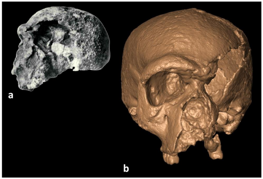
Abstract
:1. Introduction
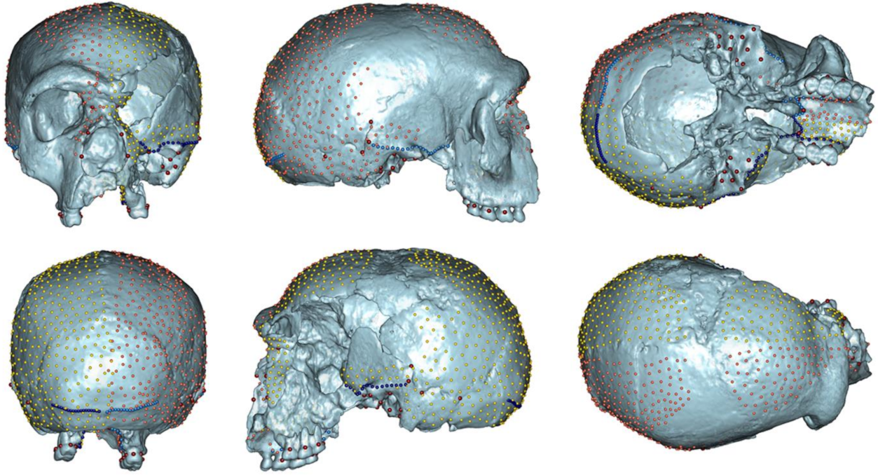
2. Materials and Methods
3. Results
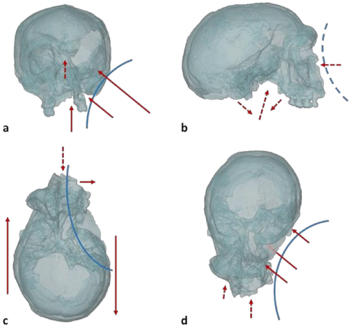
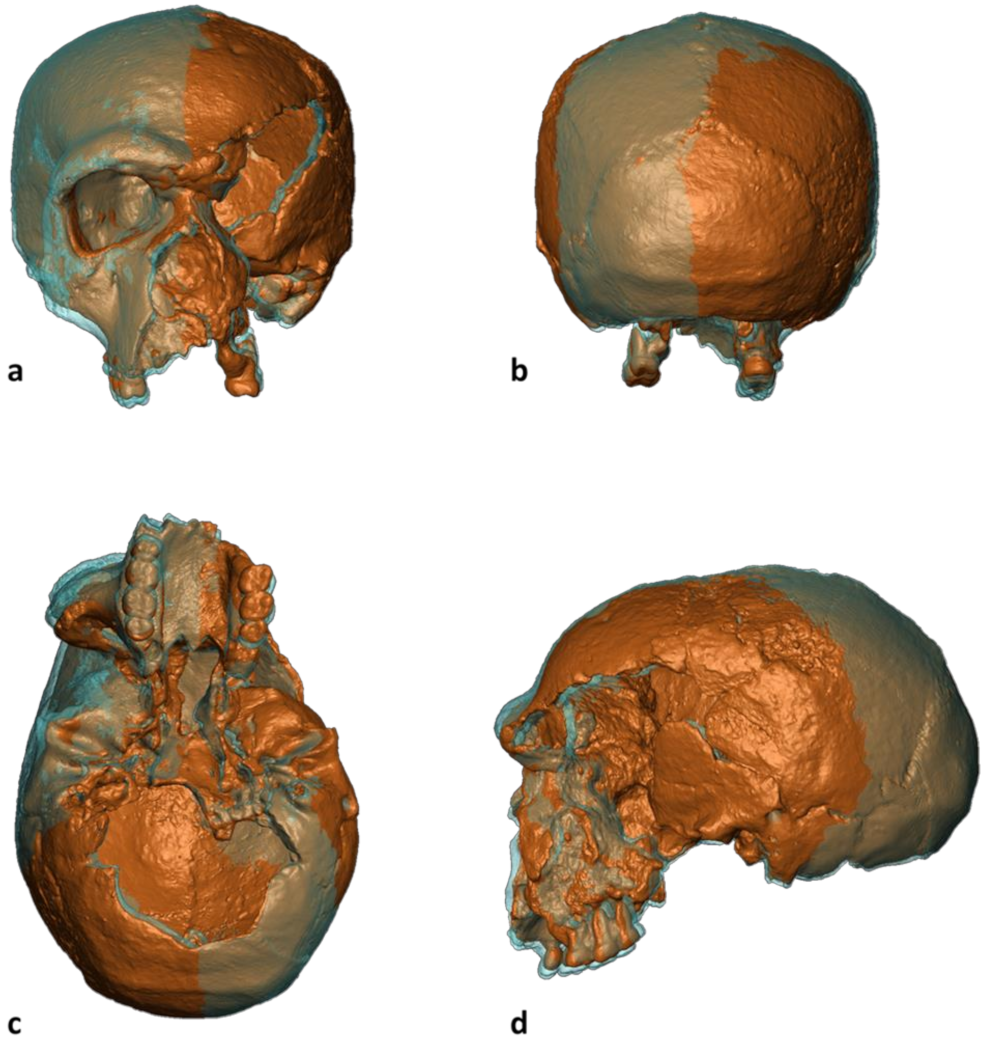
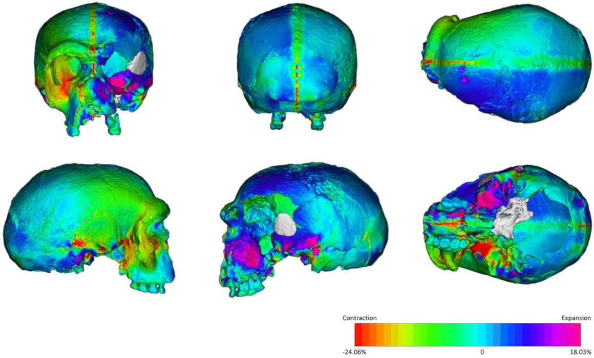
3.1. The Retrodeformation
3.2. Local Displacement
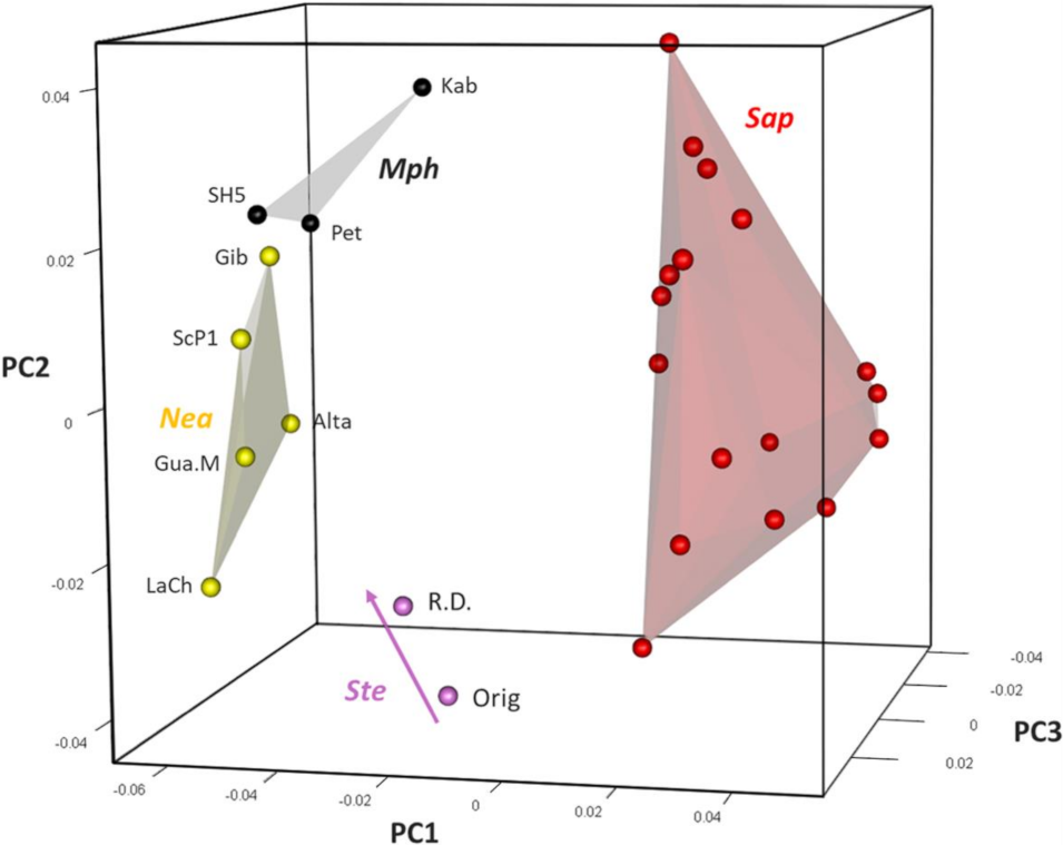
3.3. Principal Component Analysis
4. Discussion
5. Conclusions
Supplementary Materials
Author Contributions
Funding
Institutional Review Board Statement
Informed Consent Statement
Data Availability Statement
Acknowledgments
Conflicts of Interest
References
- Bookstein, F.L. The study of shape transformation after D’Arcy Thompson. Math. Biosci. 1977, 34, 177–219. [Google Scholar] [CrossRef] [Green Version]
- Klingenberg, C.P.; Barluenga, M.; Meyer, A. Shape analysis of symmetric structures: Quantifying variation among individuals and asymmetry. Evolution 2002, 56, 1909–1920. [Google Scholar] [CrossRef] [Green Version]
- Mardia, K.V.; Bookstein, F.L.; Moreton, I.J. Statistical assessment of bilateral symmetry of shapes. Biometrika 2000, 87, 285–300. [Google Scholar] [CrossRef]
- Schlager, S.; Profico, A.; Di Vincenzo, F.; Manzi, G. Retrodeformation of fossil specimens based on 3D bilateral semi-landmarks: Implementation in the R package “Morpho”. PLoS ONE 2018, 13, e0194073. [Google Scholar] [CrossRef]
- Fernández-Jalvo, Y.; Andrews, P. Atlas of Taphonomic Identifications; Springer: Dordrecht, The Netherlands, 2016; p. 4. [Google Scholar]
- Adams, D.C.; Rohlf, F.J.; Slice, D.E. A field comes of age: Geometric morphometrics in the 21st century. Hystrix 2013, 24, 7–14. [Google Scholar]
- Gunz, P.; Mitteroecker, P.; Neubauer, S.; Weber, G.W.; Bookstein, F.L. Principles for the virtual reconstruction of hominin crania. J. Hum. Evol. 2009, 57, 48–62. [Google Scholar] [CrossRef]
- Di Vincenzo, F.; Profico, A.; Bernardini, F.; Cerroni, V.; Dreossi, D.; Schlager, S.; Zaio, P.; Benazzi, S.; Biddittu, I.; Rubini, M.; et al. Digital reconstruction of the Ceprano calvarium (Italy), and implications for its interpretation. Sci. Rep. 2017, 7, 1–11. [Google Scholar] [CrossRef] [Green Version]
- White, T. Early hominids—Diversity or distortion? Science 2003, 299, 1994–1997. [Google Scholar] [CrossRef] [Green Version]
- Weber, G.W.; Schäfer, K.; Prossinger, H.; Gunz, P.; Mitteröcker, P.; Seidler, H. Virtual anthropology: The digital evolution in anthropological sciences. J. Physiol. Anthropol. Appl. Human Sci. 2001, 20, 69–80. [Google Scholar] [CrossRef] [Green Version]
- Weber, G.W. Another link between archaeology and anthropology: Virtual anthropology. DAACH 2014, 1, 3–11. [Google Scholar] [CrossRef] [Green Version]
- Weber, G.W. Virtual anthropology. Am. J. Phys. Anthropol. 2015, 156, 22–42. [Google Scholar] [CrossRef]
- Amano, H.; Kikuchi, T.; Morita, Y.; Kondo, O.; Suzuki, H.; Ponce de León, M.S.; Zollikofer, C.P.E.; Bastir, M.; Stringer, C.B.; Ogihara, N. Virtual reconstruction of the Neanderthal Amud 1 cranium. Am. J. Phys. Anthropol. 2015, 158, 185–197. [Google Scholar] [CrossRef] [PubMed] [Green Version]
- Bosman, A.M.; Buck, L.T.; Reyes-Centeno, H.; Mirazón Lahr, M.; Stringer, C.B.; Harvati, K. The Kabua 1 cranium: Virtual anatomical reconstructions. In Modern Human Origins and Dispersal; Sahle, Y., Reyes-Centeno, H., Bentz, C., Eds.; Kerns Verlag: Tübingen, Germany, 2019; pp. 137–170. [Google Scholar]
- Harvati, K.; Röding, C.; Bosman, A.M.; Karakostis, F.A.; Grün, R.; Stringer, C.B.; Karkanas, P.; Thompson, N.C.; Koutoulidis, V.; Moulopoulos, L.A.; et al. Apidima Cave fossils provide earliest evidence of Homo sapiens in Eurasia. Nature 2019, 571, 500–504. [Google Scholar] [CrossRef]
- Profico, A.; Buzi, C.; Davis, C.; Melchionna, M.; Veneziano, A.; Raia, P.; Manzi, G. A new tool for digital alignment in virtual anthropology. Anat. Rec. 2019, 302, 1104–1115. [Google Scholar] [CrossRef] [PubMed]
- Benazzi, S.; Bookstein, F.L.; Strait, D.S.; Weber, G.W. A new OH5 reconstruction with an assessment of its uncertainty. J. Hum. Evol. 2011, 61, 75–88. [Google Scholar] [CrossRef] [PubMed]
- Benazzi, S.; Gruppioni, G.; Strait, D.S.; Hublin, J.J. Virtual reconstruction of KNM-ER 1813 Homo habilis cranium. Am. J. Phys. Anthropol. 2014, 153, 154–160. [Google Scholar] [CrossRef] [PubMed]
- Berge, C.; Goularas, D. A new reconstruction of Sts 14 pelvis (Australopithecus africanus) from computed tomography and three-dimensional modeling techniques. J. Hum. Evol. 2010, 58, 262–272. [Google Scholar] [CrossRef]
- Gunz, P.; Neubauer, S.; Golovanova, L.; Doronichev, V.; Maureille, B.; Hublin, J.J. A uniquely modern human pattern of endocranial development. Insights from a new cranial reconstruction of the Neandertal newborn from Mezmaiskaya. J. Hum. Evol. 2012, 62, 300–313. [Google Scholar] [CrossRef]
- Hublin, J.J.; Ben-Ncer, A.; Bailey, S.E.; Freidline, S.E.; Neubauer, S.; Skinner, M.M.; Bergmann, I.; Le Cabec, A.; Benazzi, S.; Harvati, K.; et al. New fossils from Jebel Irhoud, Morocco and the pan-African origin of Homo sapiens. Nature 2017, 546, 289–292. [Google Scholar] [CrossRef] [Green Version]
- Vialet, A.; Guipert, G.; Alçiçek, M.C. Homo erectus found still further west: Reconstruction of the Kocabaş cranium (Denizli, Turkey). Comptes Rendus Palevol. 2012, 11, 89–95. [Google Scholar] [CrossRef]
- Arbour, V.M.; Currie, P.J. Analyzing taphonomic deformation of ankylosaur skulls using retrodeformation and finite element analysis. PLoS ONE 2012, 7, e39323. [Google Scholar] [CrossRef] [PubMed] [Green Version]
- Kazhdan, M.M.; Amenta, N.; Gu, S.; Wiley, D.F.; Hamann, B. Symmetry restoration by stretching. In Proceedings of the 21st Canadian Conference on Computational Geometry (CCCG2009), Vancouver, Canada, 17–19 August 2009; CCCG: Vancouver, BC, Canada, 2009; pp. 37–40. [Google Scholar]
- Lautenschlager, S. Reconstructing the past: Methods and techniques for the digital restoration of fossils. Roy. Soc. Open Sci. 2016, 3, 160342. [Google Scholar] [CrossRef] [Green Version]
- Ogihara, N.; Nakatsukasa, M.; Nakano, Y.; Ishida, H. Computerized restoration of nonhomogeneous deformation of a fossil cranium based on bilateral symmetry. Am. J. Phys. Anthropol. 2006, 130, 1–9. [Google Scholar] [CrossRef]
- Tallman, M.; Amenta, N.; Delson, E.; Frost, S.R.; Ghosh, D.; Klukkert, Z.S.; Morrow, A.; Sawyer, G.J. Evaluation of a new method of fossil retrodeformation by algorithmic symmetrization: Crania of papionins (Primates, Cercopithecidae) as a test case. PLoS ONE 2014, 9, e100833. [Google Scholar] [CrossRef] [Green Version]
- Tschopp, E.; Russo, J.; Dzemski, G. Retrodeformation as a test for the validity of phylogenetic characters: An example from diplodocid sauropod vertebrae. Palaeontol. Electron. 2016, 16, 2T. [Google Scholar] [CrossRef] [Green Version]
- Berckhemer, F. Der Urmenschenschädel aus den zwischeneiszeitlichen Fluss-Schottern von Steinheim an der Murr. Forsch u Fortschr 1936, 12, 349–350. [Google Scholar]
- Adam, K.D. The chronological and systematic position of the Steinheim skull. In Ancestors: The Hard Evidence; Delson, E., Ed.; Liss: New York, NY, USA, 1985; pp. 272–276. [Google Scholar]
- Granat, J.; Peyre, É. L’énigme odontologique du crâne de Steinheim 280 ka-Allemagne. Actes SFHAD 2008, 13, 14–18. [Google Scholar]
- Wahl, J.; König, H.; Ziegler, R. Die Defekt-und Verformungsspuren am Schädel des Urmenschen von Steinheim an der Murr. Fundber. Baden-Württ. 2009, 30, 7–28. [Google Scholar]
- Weinert, H. Der urmenschenschädel von Steinheim. Z. Morphol. Anthropol. 1936, 35, 463–518. [Google Scholar]
- Czarnetzki, A. Steinheim skull: A morphological comparison with Tautavel Man. In Proceedings of the L’Homo Erectus et la Place de l’homme de Tautavel Parmi les Hominidés Fossiles, Actes Congrès International de Paléontologie Humaine, Nice, France, 16–21 October 1982; de Lumley, H., de Lumley, M.A., Eds.; pp. 875–893. [Google Scholar]
- Street, M.; Terberger, T.; Orschiedt, J. A critical review of the German Paleolithic hominin record. J. Hum. Evol. 2006, 51, 551–579. [Google Scholar] [CrossRef] [PubMed]
- Schwartz, J.H.; Tattersall, I. The Human Fossil Record. Vol. 1: Terminology and Craniodental Morphology of Genus Homo (Europe); Wiley-Liss: New York, NY, USA, 2002; pp. 347–351. [Google Scholar]
- Prossinger, H.; Seidler, H.; Wicke, L.; Weaver, D.; Recheis, W.; Stringer, C.B.; Müller, G.B. Electronic removal of encrustations inside the Steinheim cranium reveals paranasal sinus features and deformations and provides a revised endocranial volume estimate. Anat. Rec. B 2013, 273, 132–142. [Google Scholar] [CrossRef]
- Wolpoff, M.H. Cranial remains of middle Pleistocene European hominids. J. Hum. Evol. 1980, 9, 339–358. [Google Scholar] [CrossRef]
- Stringer, C.B.; Hublin, J.J.; Vandermeersch, B. The origin of anatomically modern humans in Western Europe. In The Origins of Modern Humans: A World Survey of the Fossil Evidence; Smith, F.H., Spencer, F., Eds.; Liss: New York, NY, USA, 1984; pp. 51–135. [Google Scholar]
- Arsuaga, J.L.; Martinón-Torres, M.; Santos, E. Homo steinheimensis, a comparison between the Steinheim skull and the Atapuerca Sima de los Huesos fossils. In Proceedings of the European Society for the study of Human Evolution (ESHE) 8, Liège, Belgium, 19–21 September 2019; ESHE: Liège, Belgium, 2019; p. 7. [Google Scholar]
- Sergi, S. The palaeanthropi in Italy: The fossil men of Saccopastore and Circeo. Part II: Discussion and interpretation. Man 1948, 48, 76–79. [Google Scholar] [CrossRef]
- Vlček, E. A new discovery of Homo erectus in Central Europe. J. Hum. Evol. 1978, 7, 239–251. [Google Scholar] [CrossRef]
- Day, M.H. Guide to Fossil Man, 4th ed.; University of Chicago Press: Chicago, IL, USA, 1986. [Google Scholar]
- Dean, D.; Hublin, J.J.; Holloway, R.; Ziegler, R. On the phylogenetic position of the pre-Neandertal specimen from Reilingen, Germany. J. Hum. Evol. 1998, 34, 485–508. [Google Scholar] [CrossRef] [PubMed] [Green Version]
- Hublin, J.J. The origin of Neandertals. Proc. Natl. Acad. Sci. USA 1998, 106, 16022–16027. [Google Scholar] [CrossRef] [PubMed] [Green Version]
- Bermúdez de Castro, J.M.; Arsuaga, J.L.; Carbonell, E.; Rosas, A.; Martınez, I.; Mosquera, M. A hominid from the Lower Pleistocene of Atapuerca, Spain: Possible ancestor to Neandertals and modern humans. Science 1997, 276, 1392–1395. [Google Scholar] [CrossRef] [Green Version]
- Freidline, S.E.; Gunz, P.; Harvati, K.; Hublin, J.J. Evaluating developmental shape changes in Homo antecessor subadult facial morphology. J. Hum. Evol. 2013, 65, 404–423. [Google Scholar] [CrossRef]
- Trafí, F.R.; Bartual, M.G.; Wang, Q. The affinities of Homo antecessor—A review of craniofacial features and their taxonomic validity. Anthropol. Rev. 2018, 81, 225–251. [Google Scholar] [CrossRef] [Green Version]
- Stringer, C. The status of Homo heidelbergensis (Schoetensack 1908). Evol. Anthropol. 2012, 21, 101–107. [Google Scholar] [CrossRef]
- Manzi, G. Humans of the Middle Pleistocene: The controversial calvarium from Ceprano (Italy) and its significance for the origin and variability of Homo heidelbergensis. Quat. Int. 2016, 411, 254–261. [Google Scholar] [CrossRef]
- Manzi, G. Before the emergence of Homo sapiens: Overview on the Early-to-Middle Pleistocene fossil record (with a proposal about Homo heidelbergensis at the subspecific level). Int. J. Evol. Biol. 2011, 2011, 582678. [Google Scholar] [CrossRef] [Green Version]
- Stringer, C.B. Secrets of the Pit of the Bones. Nature 1993, 362, 501–502. [Google Scholar] [CrossRef]
- Tattersall, I. Before the Neanderthals: Hominid Evolution in Middle Pleistocene Europe. In Continuity and Discontinuity in the Peopling of Europe; Condemi, S., Weniger, G.C., Eds.; Springer: Dordrecht, The Netherlands, 2011; pp. 47–53. [Google Scholar]
- Mounier, A.; Caparrós, M. The phylogenetic status of Homo heidelbergensis—A cladistic study of Middle Pleistocene hominins. Bull. Mém. Soc. Anthropol. Paris 2015, 27, 110–134. [Google Scholar] [CrossRef]
- Braun, M.; Hublin, J.J.; Bouchet, P. New reconstruction of the Middle Pleistocene skull of Steinheim (Baden-Würtemberg, Germany). Am. J. Phys. Anthropol. 1998, S26, 113. [Google Scholar]
- Czarnetzki, A.; Schwaderer, E.; Pusch, C.M. Fossil record of meningioma. Lancet 2003, 362, 408. [Google Scholar] [CrossRef]
- Stalling, D.; Westerhoff, M.; Hege, H.C. Amira: A highly interactive system for visual data analysis. In The Visualization Handbook; Hansen, C., Johnson, C., Eds.; Elsevier: Amsterdam, The Netherlands, 2005; pp. 749–767. [Google Scholar]
- Schlager, S. Morpho and Rvcg—Shape Analysis in R: R-Packages for geometric morphometrics, shape analysis and surface manipulations. In Statistical Shape and Deformation Analysis; Zheng, G., Li, S., Székely, G., Eds.; Academic Press: London, UK, 2017; pp. 217–256. [Google Scholar]
- Profico, A.; Buzi, C.; Castiglione, S.; Melchionna, M.; Piras, P.; Veneziano, A.; Raia, P. Arothron: An R package for geometric morphometric methods and virtual anthropology applications. Am. J. Phys. Anthropol. 2021, 176, 144–151. [Google Scholar]
- Buzi, C.; Di Vincenzo, F.; Profico, A.; Manzi, G. The pre-modern human fossil record in Italy from the Middle to the Late Pleistocene: An updated reappraisal. Alp. Mediterr. Quat. 2021, 34, 1–16. [Google Scholar]
- Arsuaga, J.L.; Martínez, I.; Arnold, L.J.; Aranburu, A.; Gracia-Téllez, A.; Sharp, W.D.; Quam, R.M.; Falguères, C.; Pantoja-Pérez, A.; Bischoff, J.; et al. Neandertal roots: Cranial and chronological evidence from Sima de los Huesos. Science 2014, 344, 1358–1363. [Google Scholar] [CrossRef]
- Churchill, S.E. Thin on the Ground: Neandertal Biology, Archeology and Ecology; Wiley: Hoboken, NJ, USA, 2014; pp. 15–22. [Google Scholar]
- Harvati, K.; Hublin, J.J.; Gunz, P. Evolution of middle-late Pleistocene human cranio-facial form: A 3-D approach. J. Hum. Evol. 2010, 59, 445–464. [Google Scholar] [CrossRef]


Publisher’s Note: MDPI stays neutral with regard to jurisdictional claims in published maps and institutional affiliations. |
© 2021 by the authors. Licensee MDPI, Basel, Switzerland. This article is an open access article distributed under the terms and conditions of the Creative Commons Attribution (CC BY) license (https://creativecommons.org/licenses/by/4.0/).
Share and Cite
Buzi, C.; Profico, A.; Di Vincenzo, F.; Harvati, K.; Melchionna, M.; Raia, P.; Manzi, G. Retrodeformation of the Steinheim Cranium: Insights into the Evolution of Neanderthals. Symmetry 2021, 13, 1611. https://doi.org/10.3390/sym13091611
Buzi C, Profico A, Di Vincenzo F, Harvati K, Melchionna M, Raia P, Manzi G. Retrodeformation of the Steinheim Cranium: Insights into the Evolution of Neanderthals. Symmetry. 2021; 13(9):1611. https://doi.org/10.3390/sym13091611
Chicago/Turabian StyleBuzi, Costantino, Antonio Profico, Fabio Di Vincenzo, Katerina Harvati, Marina Melchionna, Pasquale Raia, and Giorgio Manzi. 2021. "Retrodeformation of the Steinheim Cranium: Insights into the Evolution of Neanderthals" Symmetry 13, no. 9: 1611. https://doi.org/10.3390/sym13091611