Communication

Isolation and Structure Elucidation of a Novel Symmetrical Macrocyclic Phthalate Hexaester

1
Department of Ocean Sciences, Tokyo University of Marine Science and Technology, Tokyo 108-8477, Japan
2
Fisheries Technology Institute, Japan Fisheries Research and Education Agency, Yokohama 236-8648, Japan
*
Author to whom correspondence should be addressed.
Symmetry 2021, 13(2), 361; https://doi.org/10.3390/sym13020361
Submission received: 9 February 2021 / Revised: 19 February 2021 / Accepted: 20 February 2021 / Published: 23 February 2021

Abstract

:
A novel symmetrical macrocyclic phthalate hexaester (1) and a known macrocyclic phthalate tetraester (2) were isolated during a natural product-exploring program on the cyanobacterium Moorea producens. Their structures were elucidated based on spectroscopic data, including nuclear magnetic resonance and high-resolution mass spectra. In the antibacterial activity test, compounds 1 and 2 showed no bioactivity at the concentrations tested.

1. Introduction

The phthalate esters found in the environment can be anthropogenic and petrogenic compounds or natural products. Petrogenic phthalate esters such as di(2-ethylhexyl) phthalate (DEHP), diisononyl phthalate (DINP), diisodecyl phthalate (DIDP), di(2-propylheptyl) phthalate (DPHP), and dibutyl phthalate (DBP) are used as plastic additives or solvents and leach from them into environmental water and sediments [1]. In the ocean, the amount of microplastics that potentially release these phthalate esters is increasing [2]. Thus, the majority of phthalate esters in the ocean are recognized as being anthropogenic. In contrast, phthalate esters have been isolated or detected as natural products from various organisms, including terrestrial bacteria belonging to the genus Streptomyces [3,4,5]; the pathogenic bacteria Helicobacter pylori [6]; the terrestrial fungi Fusarium merismoides [7]; Penicillium olsonii [8]; Penicillium lanosum; Trichoderma asperellum; Aspergillus niger [9]; the terrestrial plants Aloe vera [10,11] and Cardaria draba [12]; the freshwater cyanobacteria Anabena flos-aquae, Cylindrospermopsis raciborskii, Microcystis aeruginosa, Oscillatoria sp. and Phormidium sp.; the green algae Botryococcus braunii, Cladophora fracta, Chlorella sp., Hydrodictyon reticulatum and Spirogyra spp. [13]; the marine red alga Bangia atropurpurea [14]; the marine green alga Ulva sp.; the marine brown algae Undaria pinnatifida; and Laminaria japonica [15]. Most recently, a macrocyclic phthalate tetraester (2, Figure 1) was reported as a natural product from the scorpion Liocheles australasiae [16]. These phthalate esters are consumed by microorganisms including aerobic and anaerobic bacteria [17,18,19,20]. Thus, some of the phthalate esters are biologically produced and consumed in the biosphere.
The cyanobacterium Moorea producens (formerly Lyngbya majuscula) is a species known for producing many bioactive compounds [21]. Some of these compounds have been recognized as potential pharmaceutical compounds [22,23]. M. producens has been involved in causing contact dermatitis, also known as “swimmer’s itch”, in many Pacific areas [24]. The causative agents of this contact dermatitis have been reported to be aplysiatoxins and lyngbyatoxins produced by M. producens [25,26,27,28]. In addition, it has been reported that food tainted with aplysiatoxins that led to food poisoning was contaminated with M. producens [29]. Recently, we isolated and reported new toxic constituents from M. producens [30,31,32,33,34]. During the exploration of aplysiatoxin-related compounds from the cyanobacterium M. producens, we isolated compounds with interesting symmetrical structures: a novel symmetrical macrocyclic hexaester 1 (Figure 1) and a known macrocyclic tetraester 2 (Figure 1). The true origin of the phthalate esters is controversial, as we mentioned above; however, the record of finding of new phthalates is valuable. Thus, here, we report the isolation, structure elucidation and bioactivities of these cyclic phthalate compounds 1 and 2.

2. Materials and Methods

2.1. General Experimental Procedures

Reversed-phase high-performance liquid chromatography (RP-HPLC) was carried out using an HPLC system equipped with a UV-975 Intelligent Ultraviolet-visible (UV/VIS) Detector (JASCO Co., Tokyo, Japan). A HPLC Senshu Scientific SSC-1310 Recycle Unit (Senshu Scientific Co., Tokyo, Japan) was also used, which consisted of a LC-10AD VP pump (SHIMADZU Co., Kyoto, Japan) and a SPD-6AV detector. HR-ESI-MS spectra data were obtained using a Bruker MicrOTOF QII (Bruker Co., Billerica, MA, USA) mass spectrometer. NMR spectra were recorded in CD3OD at 800 MHz (or 600 MHz) on a Bruker AVANCE III 800 MHz (or 600 MHz, Bruker Co., Billerica, MA, USA) spectrometer. The chemical shifts were reported in δ units (ppm) using CD3OD solvent (δH at 3.31 ppm and δC at 49.0 ppm) as the internal standard signals. The UV spectra were measured on a HITACHI U-3000 (Hitachi High-Tech Fielding Co., Tokyo, Japan) spectrometer.

2.2. The Cyanobacterium

The cyanobacterium M. producens was collected from Kuba Beach, Nakagusuku, Okinawa, Japan, on 13 July 2010. The collected samples were immediately stored in a freezer (−30 °C) without lyophilization. The Okinawan collection was identified as M. producens. A voucher specimen (#20100713-a) was also retained.

2.3. Extraction and Isolation

A frozen sample of the cyanobacterium M. producens (10.1 kg wet wt.) was lyophilized and then sequentially extracted with ethanol, methanol (MeOH), and acetone at RT. The extracts were combined and evaporated to obtain the residue (492.7 g, dry wt.). Next, the residue was partitioned between 80% MeOH and hexane. After solvent evaporation of the 80% MeOH layer, the condensed fraction was partitioned using distilled water and ethyl acetate (EtOAc). The EtOAc layer was then evaporated to dryness. The EtOAc fraction (4.3 g, dry wt.) was purified using a 40 × 170 mm open glass column filled with ODS resin (Pegasil Prep ODS-7515-12A, Senshu Scientific Co., Tokyo, Japan) with a stepwise increase in aqueous MeOH (30%, 50%, 70%, 85%, and 100%). The 85% MeOH layer (333.3 mg, dry wt.) was purified using reversed-phase HPLC on a 10 × 250 mm column (COSMOSIL 5C18-AR-II, Nacalai Tesque Inc., Kyoto, Japan) under the following conditions: 80% MeOH isocratic for the first 95 min; 100% MeOH isocratic from 95 min to 150 min at 2 mL/min flow rate; UV-Vis detection at 254 nm, with the sample divided into 16 fractions using a fraction collector at 8-min intervals. Fraction 3 (tR 16–24 min, 19.1 mg, dry wt.) was then subjected to HPLC using an isocratic system (flow rate: 1 mL/min; detection: 254 nm) on a 10 × 250 mm column (COSMOSIL 5C18-AR-II) with 85% MeOH solvent. The resulting 33 fractions were collected at 2-min intervals using a fraction collector. Finally, a recycling HPLC (column: COSMOSIL C18-AR-II 10 × 250 mm, solvent: 88% MeOH for compound 2 and 82% MeOH for compound 1, flow rate: 1 mL/min, detection: 210 nm) was performed on fractions 3–19 (tR 36–38 min) and fractions 3–18 (tR 34–36 min) for the isolation of compound 1 (0.10 mg) and compound 2 (0.16 mg).

2.4. Bioactive Assays

The antibacterial assays of compounds 1 and 2 were carried out using Escherichia coli JCM No. 20135 and Pyricularia oryzae Ina 86–137. E. coli was cultured at 25 °C for 3 days in B-1 medium, consisting of 5.0 g/L Bactom TM Peptone, 3.0 g/L beef extract, 3.0 g/L NaCl, and 15.0 g/L agar powder (Kanto Chemical Co. Inc., Tokyo, Japan). P. oryzae was cultured at RT in Ottaviani and Agosti (OA) medium containing 50 g/L of oatmeal, 5 g/L sucrose, and 30 g/L agar powder. Both media were prepared using distilled water. Compounds 1 and 2 were dissolved in MeOH and then absorbed on paper discs (8 mm in diameter). After placing the discs on to the assay plates were incubated at 27 °C for 18 h (E. coli) and at RT for 13 days (P. oryzae).

3. Results and Discussion

Compound 1 (0.10 mg) and compound 2 (0.16 mg) were isolated during exploring natural products from the Okinawan cyanobacterium M. producens (10.1 kg wet wt.). Compound 1 possessed a molecular formula of C42H48O12, as shown by the mass spectrum with the [M + H]+ ion peak at m/z 745.3222 (calcd. for C42H49O12, 745.3219) (Figure S1) and the [M + Na]+ ion peak at m/z 767.3042 (calcd. for C42H48O12Na, 767.3038). The UV spectral data of compound 1 suggested the existence of a conjugated ring system (UV λ max (ethanol) nm (ε) 225 (27,788), 274 (5686)), the structure of which was predominantly determined by 1D and 2D nuclear magnetic resonance (NMR) spectral analyses. The 1H-NMR spectrum revealed the existence of an ethylene group at H-5 (δH 4.30, dd, J = 6.6 Hz, 6.6 Hz) connected to the oxygen of the carboxyl group. The proton signals with chemical shifts of δH 7.60 (H-1, dd, J = 3.3 Hz, 5.7 Hz) and δH 7.71 (H-2, dd, J = 3.3 Hz, 5.7 Hz) indicated a benzene ring. Furthermore, the existence of two ethylene groups was revealed by the proton signals of δH 1.77 (H-6, m) and δH 1.49 (H-7, m). From the 13C-NMR spectrum, the signals of C-1 (δC 132.3), C-2 (δC 129.9), and C-3 (δC 133.6) confirmed the existence of a benzene ring and identified three methylene groups at C-5 (δC 66.9), C-6 (δC 29.6), and C-7 (δC 26.9).
1H-1H COSY spectrum analysis (Figure S4) revealed the correlation between H-1 and H-2, which further confirmed the existence of a benzene ring. The correlations of H-5/H-6 and H-6/H-7 were also detected. Moreover, the correlations of H-5/C-4, C-6, and C-7; and H-6/C-5 and C-7, were detected from the 1H-13C HMBC spectrum (Figure S6), indicating the partial structure of compound 1 (Figure 2). Furthermore, the molecular weight of compound 1 was detected to be 744 Da, which corresponds to exactly three times of the molecular weight of the partial structure (MW 248 Da, C14H16O4) (Figure 2). Meanwhile, the correlation of H-7/C-7 was detected from the HMBC spectrum (Figure 3), indicating that C-7 was connected to a carbon with the same situation as C-7. Thus, compound 1 was revealed to be a novel macrocyclic hexaester (Figure 1). NMR data are summarized in Table 1.
Compound 2 possessed a molecular formula of C28H32O8, as shown in the high-resolution electrospray ionization mass spectra (HR-ESI-MS) according to the [M + H]+ ion peak at m/z 497.2120 (calcd. for C28H33O8, 497.2170) and the [M + Na]+ ion peak at m/z 519.1934 (calcd. for C28H32O8Na, 519.1987). Comparing the 1H-NMR data of compounds 1 and 2, the proton signals were found to be identical, revealing that compound 1 and 2 had the same partial structure. Furthermore, the molecular weight of compound 2 was measured to be 496 Da, which was exactly twice that of the partial structure (MW 248 Da) of compound 1 (Figure 2), indicating that compound 2 is a macrocyclic tetraester (See Figure 1). Compound 2 was recently reported as a natural compound [16]. Therefore, compounds 1 and 2 are possibly natural products.
Compounds 1 and 2 showed no antibacterial activity against E. coli JCM No 20135 and P. oryzae Ina 86–137 at the concentrations tested (1, 12 µg/disc; 2, 10 µg/disc; methanol was tested as the control).
A novel symmetrical macrocyclic hexaester (1) and a known macrocyclic tetraester (2) were isolated during a natural product-exploring program on the cyanobacterium Moorea producens. These molecules could be produced by the cyanobacterium. Further study is needed to determine the true origin of these symmetrical compounds.

Supplementary Materials

The following are available online at https://www.mdpi.com/2073-8994/13/2/361/s1, Figure S1. HR-ESI-MS spectrum of compound 1, Figure S2. 1H-NMR spectrum of compound 1 in CD3OD, Figure S3. 13C-NMR spectrum of compound 1 in CD3OD, Figure S4. 1H-1H COSY spectrum of compound 1 in CD3OD, Figure S5. 1H-13C HSQC spectrum of compound 1 in CD3OD, Figure S6. 1H-13C HMBC spectrum of compound 1 in CD3OD.

Author Contributions

H.N. conceived and designed the research. W.J. purified the compound. R.W., T.S. and H.U. performed the spectral measurement. W.J. carried out the structural identification. H.O. performed the antibacterial assay. M.F. taxonomically identified the cyanobacterium. M.K., W.J. and H.N. analyzed the data and wrote the paper. All authors have read and agreed to the published version of the manuscript.

Funding

The authors disclose the receipt of the following financial support for the research, authorship, and/or publication of this article: this work was partly supported by JSPS KAKENHI Grant Number 19K06220 (HN) from the Ministry of Education, Culture, Sport, Science, and Technology, Japan.

Acknowledgments

The authors gratefully acknowledge N. Oshiro, S. Iwanaga, and D. Kamiya of the Okinawa Prefectural Institute of Health and Environment for the cyanobacteria sample collection. The authors would like to thank Microbe Division (RIKEN BRC JCM) for providing E. coli, as well as T. Sone of Hokkaido University for providing P. oryzae Ina 86–137.

Conflicts of Interest

The authors declare no conflict of interest.

References

  1. Hermabessiere, L.; Dehaut, A.; Paul-Pont, I.; Lacroix, C.; Jezequel, R.; Soudant, P.; Duflos, G. Occurrence and effects of plastic additives on marine environments and organisms: A review. Chemosphere 2017, 182, 781–793. [Google Scholar] [CrossRef] [PubMed] [Green Version]
  2. Isobe, A.; Iwasaki, S.; Uchida, K.; Tokai, T. Abundance of non-conservative microplastics in the upper ocean from 1957 to 2066. Nat. Commun. 2019, 10, 41. [Google Scholar] [CrossRef] [PubMed] [Green Version]
  3. Lyutskanova, D.; Ivanova, V.; Stoilova-Disheva, M.; Kolarova, M.; Aleksieva, K.; Peltekova, V. Isolation and characterization of a psychrotolerant Streptomyces strain from permafrost soil in spitsbergen, producing phthalic acid ester. Biotechnol. Biotechnol. Equip. 2009, 23, 1220–1224. [Google Scholar] [CrossRef] [Green Version]
  4. Smaoui, S.; Mellouli, L.; Lebrihi, A.; Coppel, Y.; Fguira, L.F.B.; Mathieu, F. Purification and structure elucidation of three naturally bioactive molecules from the new terrestrial Streptomyces sp. TN17 strain. Nat. Prod. Res. 2011, 25, 806–814. [Google Scholar] [CrossRef] [Green Version]
  5. Belghit, S.; Bijani, C.; Zitouni, A.; Sabaou, N.; Mathieu, F.; Badji, B. A new Streptomyces strain isolated from Saharan soil produces di-(2-ethylhexyl) phthalate, a metabolite active against methicillin-resistant Staphylococcus aureus. Ann. Microbiol. 2015, 65, 1341–1350. [Google Scholar]
  6. Keire, D.A.; Anton, P.; Faull, K.F.; Ruth, E.; Walsh, J.H.; Chew, P.; Quisimoro, D.; Territo, M.; Reeve, J.R. Diethyl phthalate, a chemotactic factor secreted by Helicobacter pylori. J. Biol. Chem. 2001, 276, 48847–48853. [Google Scholar] [CrossRef] [PubMed] [Green Version]
  7. Miyoshi, T.; Sato, H.; Harada, T. New Metabolites, 2, 4, 6-triketosuberic acid and 2, 4, 6, 8-tetraketosebacic acid, formed from 2-butyne-1, 4-diol by Fusarium merismoides B11. Agric. Biol. Chem. 1974, 38, 1935–1939. [Google Scholar]
  8. Amade, P.; Mallea, M.; Bouaicha, N. Isolation, structural identification and biological activity of two metabolites produced by Penicillium olsonii Bainier and Sartory. J. Antibiot. 1994, 47, 201–208. [Google Scholar] [CrossRef] [Green Version]
  9. Tian, C.; Ni, J.; Chang, F.; Liu, S.; Xu, N.; Sun, W.; Xie, Y.; Guo, Y.; Ma, Y.; Yang, Z. Bio-source of di-n-butyl phthalate production by filamentous fungi. Sci. Rep. 2016, 6, 19791. [Google Scholar] [CrossRef] [PubMed] [Green Version]
  10. Yamaguchi, I.; Mega, N.; Sanada, H. Components of the gel of Aloe vera (L.) Bunn. f. Biosci. Biotechnol. Biochem. 1993, 57, 1350–1352. [Google Scholar] [CrossRef]
  11. Lee, K.H.; Kim, J.H.; Lim, D.S.; Kim, C.H. Anti-leukaemic and anti-mutagenic effects of di (2-ethylhexyl) phthalate Isolated from Aloe vera Linne. J. Pharm. Pharmacol. 2000, 52, 593–598. [Google Scholar] [CrossRef]
  12. Radonić, A.; Blažević, I.; Mastelić, J.; Zekić, M.; Skočibušić, M.; Maravić, A. Phytochemical analysis and antimicrobial activity of Cardaria draba (L.) Desv. volatiles. Chem. Biodivers. 2011, 8, 1170–1181. [Google Scholar] [CrossRef]
  13. Babu, B.; Wu, J.-T. Production of phthalate esters by nuisance freshwater algae and cyanobacteria. Sci. Total Environ. 2010, 408, 4969–4975. [Google Scholar] [CrossRef]
  14. Chen, C.Y. Biosynthesis of di-(2-ethylhexyl) phthalate (DEHP) and di-n-butyl phthalate (DBP) from red alga-Bangia atropurpurea. Water Res. 2004, 38, 1014–1018. [Google Scholar] [CrossRef] [PubMed]
  15. Namikoshi, M.; Fujiwara, T.; Nishikawa, T.; Ukai, K. Natural abundance 14C content of dibutyl phthalate (DBP) from three marine algae. Mar. Drugs 2006, 4, 290–297. [Google Scholar] [CrossRef] [Green Version]
  16. Yoshimoto, Y.; Tanaka, M.; Miyashita, M.; Abdel-Wahab, M.; Megaly, A.M.; Nakagawa, Y.; Miyagawa, H. A Fluorescent compound from the exuviae of the scorpion, Liocheles australasiae. J. Nat. Prod. 2020, 83, 542–546. [Google Scholar] [CrossRef]
  17. Sawers, R.G. o-Phthalate derived from plastics’ plasticizers and a bacterium’s solution to its anaerobic degradation. Mol. Microbiol. 2018, 108, 595–600. [Google Scholar] [CrossRef] [Green Version]
  18. Junghare, M.; Spiteller, D.; Schink, B. Anaerobic degradation of xenobiotic isophthalate by the fermenting bacterium Syntrophorhabdus aromaticivorans. ISME J. 2019, 13, 1252–1268. [Google Scholar] [CrossRef]
  19. Liang, D.-W.; Zhang, T.; Fang, H.H.; He, J. Phthalates biodegradation in the environment. Appl. Microbiol. Biotechnol. 2008, 80, 183. [Google Scholar] [CrossRef]
  20. Vega, D.; Bastide, J. Dimethylphthalate hydrolysis by specific microbial esterase. Chemosphere 2003, 51, 663–668. [Google Scholar] [CrossRef]
  21. Engene, N.; Rottacker, E.C.; Kaštovský, J.; Byrum, T.; Choi, H.; Ellisman, M.H.; Komárek, J.; Gerwick, W.H. Moorea producens gen. nov., sp. nov. and Moorea bouillonii comb. nov., tropical marine cyanobacteria rich in bioactive secondary metabolites. Int. J. Syst. Evol. Microbiol. 2012, 62, 1171. [Google Scholar] [CrossRef]
  22. Singh, R.K.; Tiwari, S.P.; Rai, A.K.; Mohapatra, T.M. Cyanobacteria: An emerging source for drug discovery. J. Antibiot. 2011, 64, 401. [Google Scholar] [CrossRef] [Green Version]
  23. Tan, L.T. Pharmaceutical agents from filamentous marine cyanobacteria. Drug Discov. Today 2013, 18, 863–871. [Google Scholar] [CrossRef]
  24. Osborne, N.J.; Webb, P.M.; Shaw, G.R. The toxins of Lyngbya majuscula and their human and ecological health effects. Environ. Int. 2001, 27, 381–392. [Google Scholar] [CrossRef]
  25. Mynderse, J.S.; Moore, R.E.; Kashiwagi, M.; Norton, T.R. Antileukemia activity in the Osillatoriaceae: Isolation of debromoaplysiatoxin from Lyngbya. Science 1977, 196, 538–540. [Google Scholar] [CrossRef]
  26. Cardellina, J.H.; Marner, F.-J.; Moore, R.E. Seaweed dermatitis: Structure of lyngbyatoxin A. Science 1979, 204, 193–195. [Google Scholar] [CrossRef]
  27. Moore, R.E.; Blackman, A.J.; Cheuk, C.E.; Mynderse, J.S.; Matsumoto, G.K.; Clardy, J.; Woodard, R.W.; Craig, J.C. Absolute stereochemistries of the aplysiatoxins and oscillatoxin A. J. Org. Chem. 1984, 49, 2484–2489. [Google Scholar] [CrossRef]
  28. Aimi, N.; Odaka, H.; Sakai, S.-I.; Fujiki, H.; Suganuma, M.; Moore, R.E.; Patterson, G.M.L. Lyngbyatoxins B and C, two new irritants from Lyngbya majuscula. J. Nat. Prod. 1990, 53, 1593–1596. [Google Scholar] [CrossRef]
  29. Nagai, H.; Yasumoto, T.; Hokama, Y. Aplysiatoxin and debromoaplysiatoxin as the causative agents of a red alga Gracilaria coronopifolia poisoning in Hawaii. Toxicon 1996, 34, 753–761. [Google Scholar] [CrossRef]
  30. Jiang, W.; Zhou, W.; Uchida, H.; Kikumori, M.; Irie, K.; Watanabe, R.; Suzuki, T.; Sakamoto, B.; Kamio, M.; Nagai, H. A new lyngbyatoxin from the Hawaiian cyanobacterium Moorea producens. Mar. Drugs 2014, 12, 2748–2759. [Google Scholar] [CrossRef] [Green Version]
  31. Jiang, W.; Tan, S.; Hanaki, Y.; Irie, K.; Uchida, H.; Watanabe, R.; Suzuki, T.; Sakamoto, B.; Kamio, M.; Nagai, H. Two new lyngbyatoxin derivatives from the canobacterium, Moorea producens. Mar. Drugs 2014, 12, 5788–5800. [Google Scholar] [CrossRef] [Green Version]
  32. Nagai, H.; Watanabe, M.; Sato, S.; Kawaguchi, M.; Xiao, Y.-Y.; Hayashi, K.; Watanabe, R.; Uchida, H.; Satake, M. New aplysiatoxin derivatives from the Okinawan cyanobacterium Moorea producens. Tetrahedron 2019, 75, 2486–2494. [Google Scholar] [CrossRef]
  33. Nagai, H.; Sato, S.; Iida, K.; Hayashi, K.; Kawaguchi, M.; Uchida, H.; Satake, M. Oscillatoxin I: A new aplysiatoxin derivative, from a marine cyanobacterium. Toxins 2019, 11, 366. [Google Scholar] [CrossRef] [Green Version]
  34. Kawaguchi, M.; Satake, M.; Zhang, B.-T.; Xiao, Y.-Y.; Fukuoka, M.; Uchida, H.; Nagai, H. Neo-aplysiatoxin A isolated from Okinawan cyanobacterium Moorea producens. Molecules 2020, 25, 457. [Google Scholar] [CrossRef] [PubMed] [Green Version]
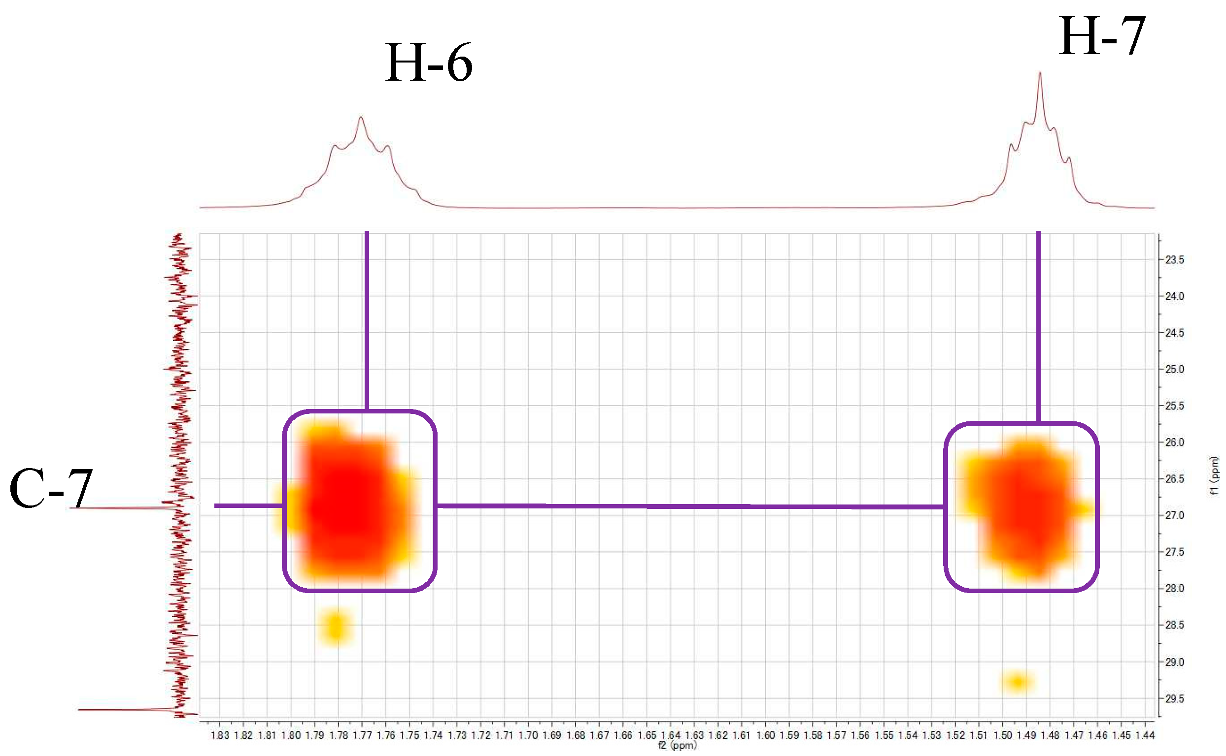
Figure 1. Chemical structures of compound 1 (left) and compound 2 (right).
Symmetry 13 00361 g001
Figure 2. Partial structure of compound 1.
Symmetry 13 00361 g002
Figure 3. Correlation of H-7/C-7 detected in the 1H-13C HMBC spectrum of compound 1 in CD3OD.
Symmetry 13 00361 g003
Table 1. NMR-assigned table for compound 1 in CD3OD.
Atom13C a1H, mult, J (Hz) bCOSYHMBC (H→C)
1132.3 7.60, dd (3.3, 5.7 Hz)H-2C-2
2129.9 7.71, dd (3.3, 5.7 Hz)H-1C-1, C-3
3133.6
4169.4
566.94.30, dd (6.6, 6.6 Hz)H-6C-4, C-6, C-7
629.6 1.77, m H-7C-5, C-7
726.9 1.49, mH-6C-7
a Recorded at 200 MHz; b recorded at 800 MHz. Coupling constants (Hz) are in parentheses. Abbreviations: dd, double-doublet; m, multiplet.
Publisher’s Note: MDPI stays neutral with regard to jurisdictional claims in published maps and institutional affiliations.

