Article

Direct Restorations for Anterior Esthetic Rehabilitation and Smile Symmetry Recovery: Two Case Reports

by
Inês Amaro
1,*,†,
José Saraiva
1,†,
Ana Catarina Gomes
1,
Ana Araújo
1,
Carlos Miguel Marto
2,3,4,5,
Ana Coelho
1,2,3,5,
Manuel Marques Ferreira
2,3,5,6,
Anabela Paula
1,2,3,5 and
Eunice Carrilho
1,2,3,5
1
Faculty of Medicine, Institute of Integrated Clinical Practice, University of Coimbra, 3000-075 Coimbra, Portugal
2
Coimbra Institute for Clinical and Biomedical Research, Area of Environment, Genetics and Oncobiology (CIMAGO), Faculty of Medicine, University of Coimbra, 3000-548 Coimbra, Portugal
3
Clinical Academic Center of Coimbra (CACC), 3004-561 Coimbra, Portugal
4
Faculty of Medicine, Institute of Experimental Pathology, University of Coimbra, 3004-548 Coimbra, Portugal
5
Centre for Innovative Biomedicine and Biotechnology (CIBB), University of Coimbra, 3000-075 Coimbra, Portugal
6
Faculty of Medicine, Institute of Endodontics, University of Coimbra, 3000-075 Coimbra, Portugal
*
Author to whom correspondence should be addressed.
These authors contributed equally to this work.
Symmetry 2021, 13(10), 1848; https://doi.org/10.3390/sym13101848
Submission received: 2 September 2021 / Revised: 17 September 2021 / Accepted: 28 September 2021 / Published: 2 October 2021
(This article belongs to the Special Issue Symmetry in Dentistry: From the Clinic to the Lab)

Abstract

:
Esthetics is an increasing concern for patients, particularly in anterior teeth. The current approaches to manage the esthetic demands include options such as prosthodontic rehabilitation, orthodontic treatment, and direct restorations. Direct restorative procedures for anterior teeth have become a reliable alternative, since current composite materials can mimic teeth’s esthetics and function perfectly. This article describes two clinical cases of anterior esthetic rehabilitations where the direct reshaping of the four maxillary incisors was performed using the buccolingual layering technique with the help of a silicone index. In Case 1, a diastema closure was also performed, whereas in Case 2, the replacement of old restorations was also carried out. It was possible to achieve excellent final esthetic results in both cases, recovering the smile symmetry, and both patients were satisfied. The literature supports the use of direct restorations in the anterior region with very good long-term prognosis. It is a minimally invasive approach that is reversible, repairable, cost-effective, and when combined with layering techniques and adequate restorative materials, it is possible to achieve good esthetic and functional outcomes. However, regular maintenance appointments are mandatory, and all cases should be carefully planned to ensure predictable and long-lasting results.

Graphical Abstract

1. Introduction

Esthetics is currently an increasing concern for patients, particularly in anterior teeth, and clinicians are being progressively requested to provide highly esthetic results without increasing costs and clinical working time [1]. Irregular shape and tooth position, and the presence of diastemas and discolorations in the anterior region, are the main aspects for facial and smile asymmetry and disharmony and are responsible for esthetic complaints amongst patients [2,3]. The current approach for managing such complaints includes a variety of treatment options, such as prosthodontic rehabilitation, orthodontic treatment, and direct restorations [1,3,4,5].
Direct dental restorations are one of the most common dental procedures executed in everyday clinical practice, namely the placement of new restorations and replacing old ones [6]. Nowadays, selecting these direct restorative approaches for anterior teeth rehabilitation has become a reliable option, since there are some advantages in selecting more conservative treatments [6]. They are minimally invasive restorative treatments, allowing for more tooth structure preservation, the possibility of easily reversing treatments and/or adding/removing material when needed, requiring fewer appointments, less treatment time, and lower treatment cost [1,7]. Recent esthetic composite materials have similar physical and mechanical properties to those of the natural tooth and offer a natural and pleasant appearance [4]. They can create an esthetic and symmetric outcome that perfectly mimics teeth’s esthetics, function, and biology, with promising clinical survival rates, which is near 85% after 5 years [8]. Combining these materials with the layering techniques and helpful but straightforward tools such as the diagnostic wax-up and the silicone index makes it possible to work with great predictability and achieve excellent clinical results [1].
In the context of restorative dentistry and smile esthetics, symmetry is the first detectable variable to the observer. It refers to the balance around the dental midline and plays an important role in direct restorations of anterior teeth. In a natural attractive smile, the ideal and pleasant symmetry is near the midline, while pleasant asymmetric irregularities are located far away from it, which makes the anterior region a more demanding area in terms of esthetic rehabilitation, where perfect symmetry is the ultimate goal [9,10].
This article aims to present two clinical cases that featured reshaping the four maxillary incisors as well as a diastema closure using resin-based composites with the help of a silicone index. All the clinical steps from color choice to restorative, finishing, and polishing procedures, as well as the final esthetic outcomes will be described as well.

2. Materials and Methods

2.1. Cases Selection and Description

This article describes two clinical cases of patients who reported to the Dentistry Department of the Faculty of Medicine, University of Coimbra presenting with esthetically compromised maxillary incisors in need of an esthetic rehabilitation. A complete assessment of all patients’ medical history was performed as well as an intraoral examination to evaluate the esthetic complaints and determine their etiology. All subjects gave their informed consent for inclusion in the study as well as for the case report and permission to use intraoral/extraoral photographs and relevant clinical information. The study was conducted in accordance with the Declaration of Helsinki, and the protocol was approved by the Ethics Committee of the Faculty of Medicine of the University of Coimbra (006/2019).

2.1.1. Case 1

In Case 1 (Figure 1), the fracture on tooth 21 resulted from a traumatic episode at age 9. It was unsuccessfully restored several times, as it fractured shortly after it was repaired. There was also a diastema between teeth 22 and 21. To achieve a more esthetic result and a better space distribution, it was suggested to restore all four maxillary incisors. Unfortunately, for financial reasons, ceramic veneers were rejected, so it was decided to carry on the treatment plan with direct composite restorations with the help of a silicone index and a new type of pre-contoured matrix.
First, preoperative extraoral and intraoral photographs were taken for future reference (Figure 1A,B). The color choice was performed to achieve a perfect color match between the natural teeth and the restorations. The color choosing process was performed through the “button try” technique (Figure 1C), where the enamel shade was selected based on incisal third matching, and the dentinal shade was selected based on cervical third matching. Afterwards, rubber dam isolation was performed, a retraction cord (Ultrapak Cleancut #00, Ultradent Products, Inc., South Jordan, UT, USA) was placed in all four maxillary incisors (Figure 1D), and the restorative process was initiated, guided by the previously prepared silicon index, which was made with putty polyvinyl siloxane (PVS) material (Lab-Putty®, Coltène, Altstätten, Switzerland) (Figure 1E), based on the diagnostic wax-up. First, all four teeth were etched using 35% phosphoric acid (Ultra-Etch, Ultradent Products, Inc., South Jordan, UT, USA) for 30 s. Then, the acid was removed by water rinse, and a universal adhesive system was applied, covering all surfaces (Scotchbond™ Universal, 3M™, St. Paul, MN, USA). Then, the palatal shells were made with the help of the silicone index, using enamel shade A2 (Filtek™ Supreme XTE, 3M™, St. Paul, MN, USA). Since all four palatal shells were performed simultaneously, small metal matrix pieces were fixed in the silicone index in the areas corresponding to the interproximal spaces to avoid the palatal shells sticking together during the photopolymerization phase (Figure 1F,G). Afterwards, for the dentinal body construction, a dentin shade A3 (Filtek™ Supreme XTE, 3M™, St. Paul, MN, USA) was used. A blue effect (MIRIS®2, Coltène, Altstätten, Switzerland) was also used near the incisal edge to create a slight opalescent effect to create a more natural-looking appearance. With the purpose of correctly and efficiently restoring interproximal areas, the Unica Anterior matrix system (Unica Anterior, Polydentia, Mezzovico-Vira, Switzerland) was used, allowing for a more accurate and regular shape of both lateral incisors (Figure 1H). Finally, an enamel shade A2 (Filtek™ Supreme XTE, 3M™, St. Paul, MN, USA) was used for the last layer (Figure 1I). Then, the rubber dam and retraction cord were removed. At the second appointment (3 weeks later), the finishing procedures were performed to restore shape and texture using abrasive discs (Sof-Lex™, 3M™, Germany), interproximal strips (Sof-Lex™, 3M™, St. Paul, MN, USA), and fine grain burs. As for the final polishing step, silicone spirals (DIATECH® ShapeGuard, Coltène, Altstätten, Switzerland) were used, which were followed by three polishing pastes (ENA Shiny, Micerium S.p.A., Genova, Italy) applied with a felt disc to get a final glossy, enamel-like surface (Figure 1J–L).

2.1.2. Case 2

In Case 2 (Figure 2), the patient presented with complaints about a fractured restoration on tooth 21. The patient also presented unsatisfactory restorations on teeth 12 and 22, with complaints regarding tooth shape and color. The patient also intended to perform dental bleaching. After careful clinical evaluation, a treatment plan was proposed comprising a dental bleaching treatment and ceramic veneers. However, the treatment plan was adapted to meet the patients’ esthetic and economic demands.
First, a home dental bleaching treatment was carried out for two weeks using custom-made bleaching trays and carbamide peroxide (Opalescence™ 10%, Ultradent Products, Inc., South Jordan, UT, USA) as a bleaching agent. Two weeks after the bleaching treatment, preoperative extraoral and intraoral photographs were taken for future reference (Figure 2A,B), and the color-choosing process was performed through the “button try” technique, where the enamel shade was selected based on incisal third matching, and the dentinal shade was selected based on cervical third matching. Afterwards, rubber dam isolation was performed, floss ligatures were placed (Figure 2C) to achieve better gingival retraction, and direct composite restorations were executed in both central and lateral incisors. In this particular case, the patient had time limitations, so the authors decided to go forward with freehand composite restorations on both central incisors and guided lateral incisors’ restorations with a silicon index (AFFINIS® putty super soft, Coltène, Altstätten, Switzerland) made from the old restorations (Figure 2D). Then, the previous composite resin restorations on teeth 12 and 22 were removed using spherical and conical diamond burs (Ref.: 801.314.009 and 856.314.014, Komet, Lemgo, Germany), and a small bevel was performed using a flame-shaped diamond bur (379.314.023, Komet, Lemgo, Germany) on the buccal face of all four maxillary incisors. Afterwards, all four teeth were etched using a 35% phosphoric acid (Ultra-Etch, Ultradent Products, Inc., South Jordan, UT, USA) for 30 s. Then, the acid was removed by water rinse, and a three-step total-etch adhesive system was applied, covering all surfaces (Optibond™ FL, Kerr™, Charlotte, NC, USA). Then, both central incisors were restored, freehand, using the buccolingual layering technique, with a combination of universal dentin (UD3) (ENA HRi®, Micerium S.p.A., Genova, Italy) and a high-value universal enamel (UE3) (ENA HRi®, Micerium S.p.A., Genova, Italy) (Figure 2E,F). Both lateral incisors were restored using the same composite system and using the silicone index to create both palatal shells (Figure 2G). All proximal areas were shaped using 6 mm/0.05 mm acetate matrices (Hawe Striproll, Kerr™, Charlotte, NC, USA). After all restorative procedures, the rubber dam and floss ligatures were removed (Figure 2H). At the second appointment (3 weeks later), the finishing procedures were carried out using abrasive discs (Sof-Lex™, 3M™, St. Paul, MN, USA), interproximal strips (Sof-Lex™, 3M™, St. Paul, MN, USA), and fine-grained burs. As for the polishing step, silicone spirals (DIATECH® ShapeGuard, Coltène, Altstätten, Switzerland) were used, which were followed by three polishing pastes (ENA Shiny, Micerium S.p.A., Genova, Italy) applied with a felt disc (Figure 2I,J).

3. Results

Regarding Case 1, the immediate postoperative photograph (Figure 1I) shows a significant improvement in shape, symmetry, and general esthetic appearance. The patient was already very satisfied with the preliminary final result, even without finishing and polishing procedures, which were only performed at a second appointment (Figure 1J–L). The same applies to Case 2, where the patient was already pleased after the dental bleaching treatment (Figure 2A,B), and the authors were able to improve further the overall esthetic appearance of the anterior region (Figure 2I). Both patients were satisfied with the final results, as all esthetic complaints were addressed, and all esthetic complications—fractures, diastema, and discoloration—were solved.

4. Discussion

Reshaping teeth and closing diastemas with direct resin composite has become a common practice in dentistry to achieve dental symmetry and improve esthetics [2,3]. This approach is particularly indicated when it involves restoring healthy teeth where there is a need to preserve as much dental structure as possible, namely enamel [3].
The diagnostic evaluation of each case when carrying out direct anterior restorative treatments using composite resins is based on the evaluation of clinical crown dimensions, gingival display and architecture, tooth position, wear factors, occlusion, and anterior guidance [4,5,11]. In addition, before initiating any restorative treatment, the periodontal tissues must be healthy and stable [5], which was the case in both clinical cases described. Therefore, all these aspects should be considered while planning these cases to ensure predictable and long-lasting esthetic results [11]. As for smile symmetry variables, even though the concept of esthetics is highly subjective, there are some aspects that the clinician should take into consideration, namely the smile arc, buccal corridors, and the existence or not of diastemas as well as of dental and gingival asymmetries [12]. In both clinical cases, all these features were carefully evaluated in order to achieve the most symmetric smile possible, which was what both patients intended.
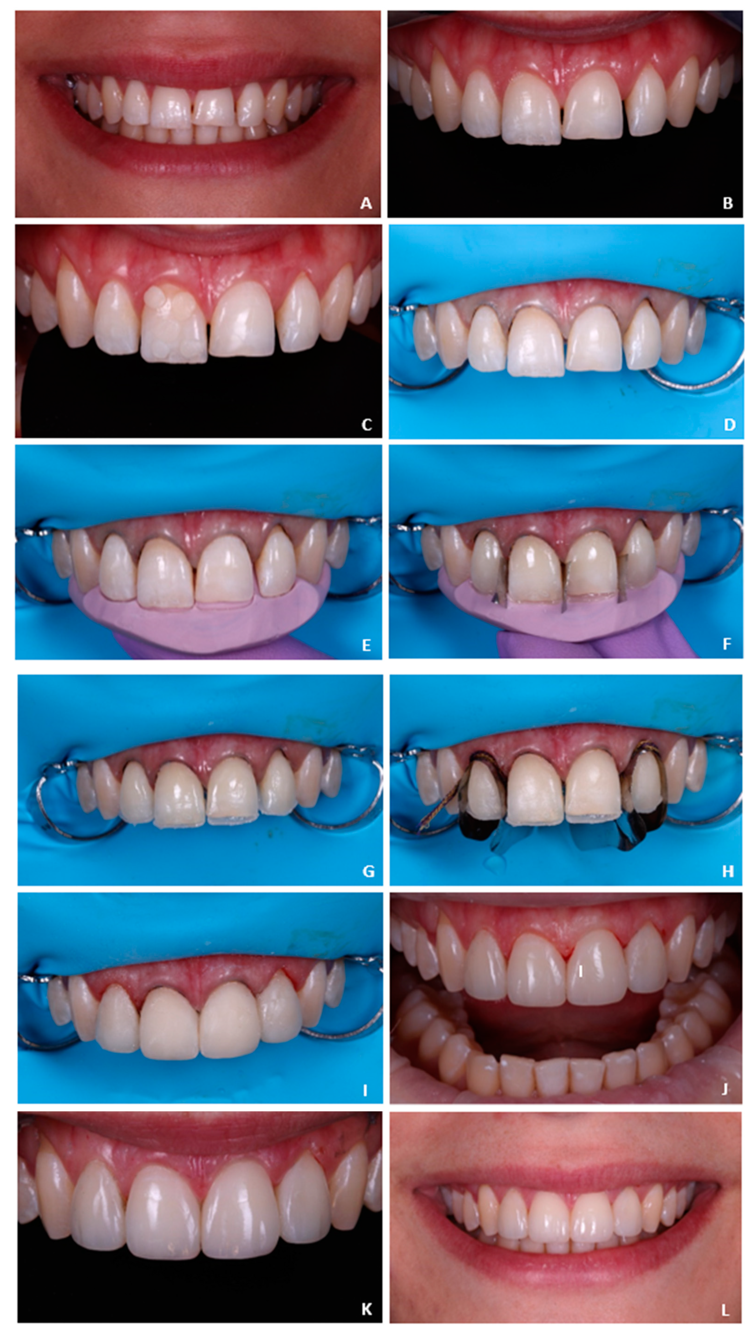
Obtaining a satisfactory and predictable esthetic and functional result is the main goal of restorative dental treatments. Specific tools, such as the diagnostic wax-up, may help improve the predictability of direct restorations. This technique consists of a dental diagnostic procedure in which planned restorations are developed in wax on a diagnostic cast to determine the necessary clinical procedures to achieve the desired clinical results [13]. It may assist in estimating the amount of restorative space available and how to manage it, and it can also help evaluate the occlusal scheme [13]. In addition, it can be used as a communication tool between the clinician and the patient and may help facilitate the acceptance and approval of the restorative treatment. From the diagnostic wax-up, it is possible to make a PVC matrix that will later serve as a guide throughout the restorative process, which was the case in the reported Case 1. The wax-up might also be a helpful tool when trying to restore smile symmetry, since the clinician may easily manage the available space in order to achieve perfect proportions and, therefore, smile symmetry [
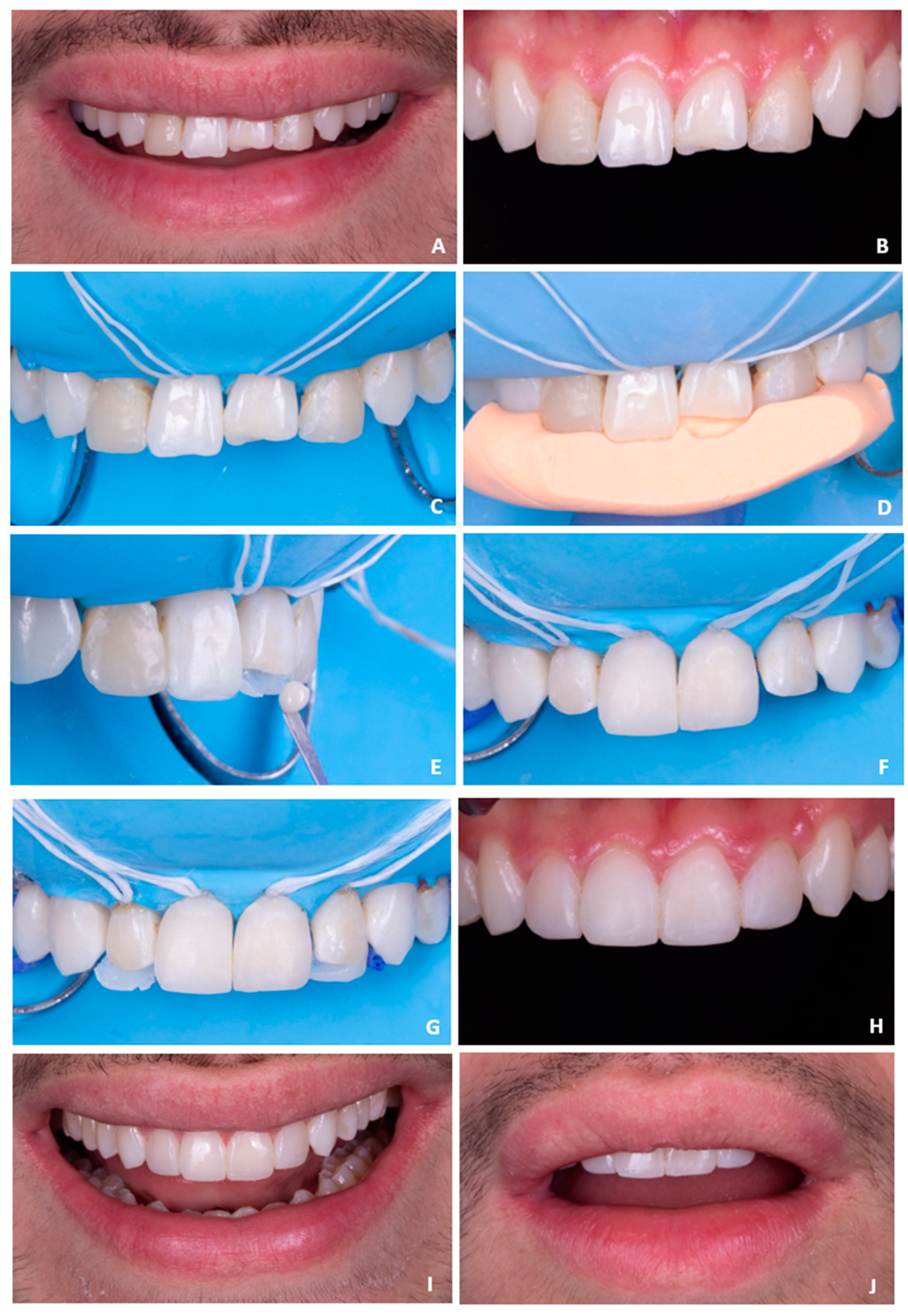
9]. In Case 1, the authors found it much easier to achieve an esthetic final result as compared to Case 2, where freehand restorations of both central incisors were performed.
The composite resin type, adhesive systems, and the entire restorative technique can influence both the longevity of the restorations and the esthetic results, which is the main concern when restoring anterior teeth [14]. Ceramic materials are an excellent option for anterior teeth rehabilitation [15]. Even though this is a more expensive treatment option, it offers great advantages as it allows for a better, more durable, and more predictable esthetic outcome [15]. However, composite resins have gained popularity as restorative materials, which is mainly due to the fact that they allow for maximum dental tissue conservation and possess excellent esthetic properties [6]. They are also a good alternative to ceramics when there are financial and time limitations, are easy to substitute or repair, and if well-executed, they allow for excellent esthetic results [7]. When working with dental composite materials, the main disadvantages relate to its color stability, possible resin chipping [16], and it requires a trained and skilled professional in restorative dentistry to optimize the results [1]. In fact, the failure behavior in anterior restorations is different from posterior restorations, presenting with less secondary caries and more restorations being replaced for other reasons such as chipping and color mismatch, which can many times be solved by repairing and polishing [6,16]. Therefore, regular maintenance appointments are mandatory, to correct any defects early or for a simple polishing procedure. Each material has its unique fabrication process, composition, and optical characteristics, which makes it predictable that the treatment outcome and longevity might differ according to the material used [15].
Dental composite materials have extensively evolved throughout the years, progressively improving their mechanical and optical properties [17]. Both nanohybrid and nanofilled composites are highly clinically successful and popular composites, since they combine good mechanical properties, such as clinical wear resistance, and allow excellent esthetic results since they are highly polishable and enable a smooth and lustrous surface [17,18]. The composite resin systems used in Case 1 and Case 2 are nanofilled and nanohybrid composites, respectively. They present similar characteristics, and the authors believe that the choice of these restorative materials positively influenced the final esthetic outcome. The main difference when using nanohybrid or nanofilled composites is regarding the polishing step, since there is evidence that nanofilled composites may show better polishability than hybrid composites [18]. However, in this case, the authors found this difference imperceptible in the final results. As for optical characteristics, it is known that composite optical features are different from those of the natural teeth, as teeth color results from the interaction between light with tooth tissues (enamel, dentin, and pulp) [19]. The optical properties (translucency, opalescence, and fluorescence) and thickness of these tissues determine the final tooth color [19]. In order to try to mimic teeth’s layered structure, two optically different restorative materials are usually the materials of choice. Generally, composite resins used for dentin restoration are less translucent and highly chromatic, whereas enamel composites are highly translucent and less chromatic [19]. By using restorative layering techniques using dentin and enamel composites, a more realistic final color can be achieved. However, since the optical properties of composite resins are not identical to the optical properties of the tooth tissues, it is not always advisable to place the enamel or dentin layers with the corresponding thickness to that of the natural structures [19]. For example, when placing the enamel layer with the same thickness as the natural enamel, the final restoration may result in a grayish appearance. One of the optical characteristics that may justify this is the refractive index (RI), which is usually lower in composites [19]. This means that using composite resins with a higher RI could result in better optical integration, with the possibility of using the same thickness as natural enamel [19]. The composite system used in Case 2, ENA HRi® (Micerium S.p.A., Genova, Italy), is an example of a high RI system (RI = 1.62) [19], which allowed the authors to use composite layers with a thickness closer to that of natural enamel without losing value. Since Case 2 was a more demanding case in terms of color matching as teeth had a higher value and little available space on tooth 11 for the restorative procedures, the authors decided to use this composite system to restore these teeth without taking the risk of losing value.
Along with improvements in their physicochemical properties, recent composites allow good esthetic results with relatively simple application and layering techniques, including the use of dentin- and enamel-like composites as well as effect materials [1]. There are two possible ways of performing the incremental application of composite resins: the centrifugal technique, which requires one or two oblique positioned layers of dentin shades, followed by an enamel layer as a final coat, or the buccolingual technique, first described by Didier Dietschi [8], which was the one used in both clinical cases reported in this study. This technique advocates using a silicon index made from a previously planned wax-up to facilitate the initial reshaping of the teeth [20]. The first layer of enamel is directly placed on the silicon index (to construct the palatal shell) followed by layers of dentin and/or different effect materials, which provides an optimal esthetic result, with a natural translucency and opalescence effect [20]. In Case 1 and 2, the combination of the incremental buccolingual technique with the use of recent composite materials allowed the creation of a natural and esthetic result in both cases, imperceptibly matching the remaining tooth structure.
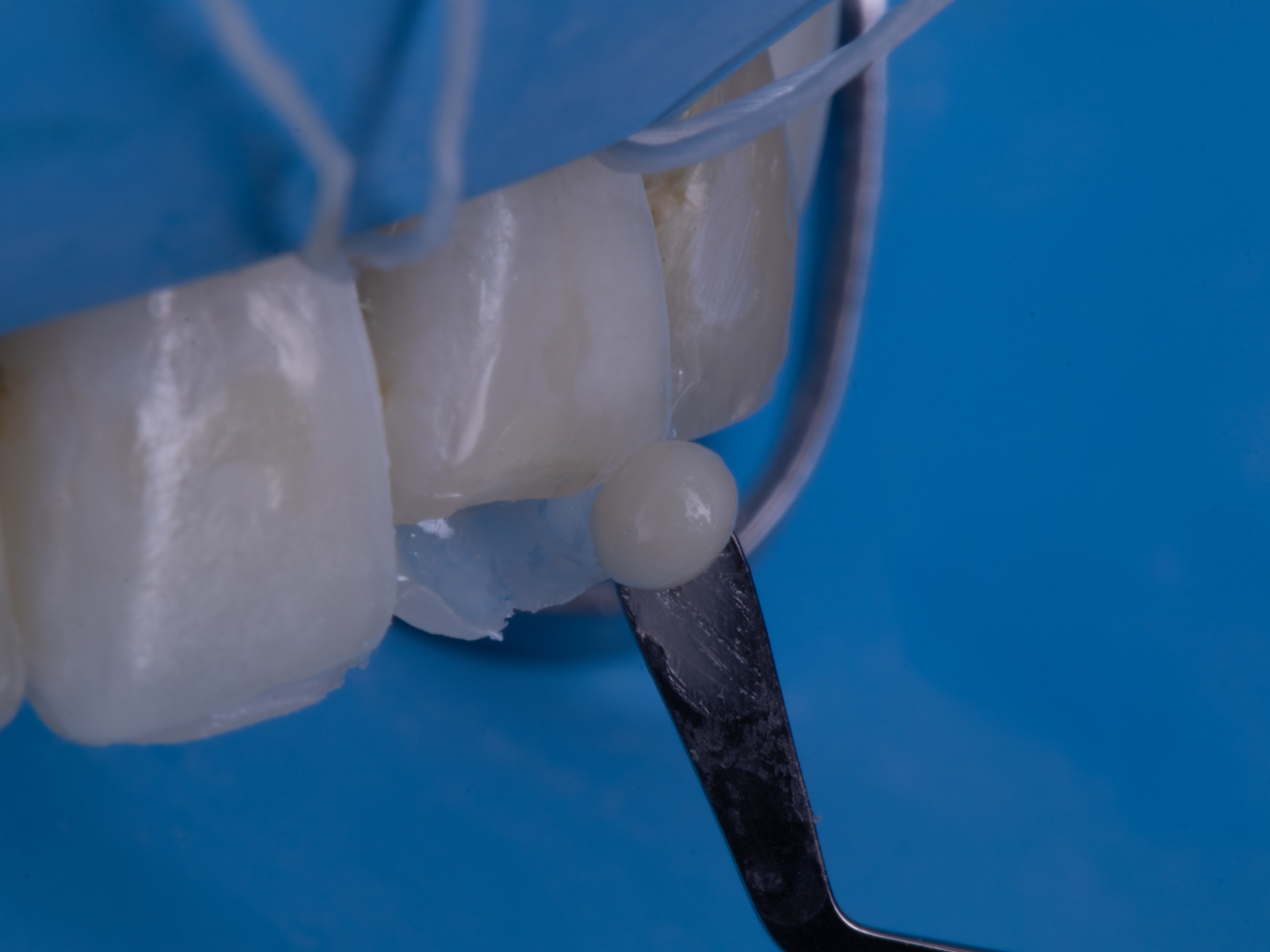
Accurate opposing and proximal contacts should also be correctly established when performing direct restorations. There are several ways of reshaping the proximal areas in anterior teeth. In Case 1, the authors used the Unica Anterior matrix (Unica Anterior, Polydentia, Mezzovico-Vira, Switzerland) to reshape the lateral incisors’ proximal areas. This particular matrix can be very helpful in more demanding cases, where perfect symmetry is the ultimate goal in anterior teeth. It is a pre-contoured metal matrix that can be easily adapted around the tooth, adjusting perfectly in the cervical area and allowing for the simultaneous shaping of both proximal areas. To the author’s knowledge, this is the first published clinical case using the Unica Anterior matrix system. Regarding diastema closure, it should be taken into consideration that the esthetic perception of the midline diastema varies according to culture, ethnic background, and socioeconomic and age groups [21]. For example, in Africans, the maxillary midline diastema is perceived as an attractive dental feature and as a sign of beauty, whereas in Caucasians, this is rarely the case, and so, these varying concepts may influence the clinical decision on how to manage the presence of midline diastemas [21]. In this case, the patient was unsatisfied with the presence of the diastema and aimed for its resolution. As for Case 2, the proximal areas were shaped using acetate matrices, which also allow a good proximal contour; however, it is more challenging to easily accomplish symmetry when comparing to the Unica Anterior matrix, for example. As for opposing contacts, these are as important as proximal contacts, and a careful evaluation should be previously carried out to avoid restorations fracture and interference with occlusal guides. In Case 1, the patient mentioned that tooth 21 was unsuccessfully restored several times before, as it fractured shortly after it was repaired. A carefully performed occlusal analysis suggested that anterior guidance was supported mainly on this tooth, justifying the recurrent fractures of the restoration. This highlights the need for a carefully planned case and the importance of analyzing all aspects involving extensive rehabilitations. In both clinical cases, the fractured central incisors (tooth 21 in both cases) were also a major complaint from both patients, which is understandable since the presence of maxillary incisor asymmetries strongly influences the perception of smile esthetics [12]. For example, central incisors rarely show asymmetries in one of the three dimensions (length, width, or thickness) bigger than 0.2 to 0.3 mm [9]. In addition, in a study conducted by Ribeiro et al. [12], the authors concluded that the closer to the midline, the greater the need for symmetry, while pleasant asymmetric irregularities, distant from the midline, are acceptable. The authors also concluded that other facial components such as eyes, nose, and hair do not significantly influence the assessment of smile esthetics. All of these clinical features were taken into consideration in both cases, and a careful evaluation was performed in order to achieve smile symmetry.
In esthetic dentistry, even if clinicians use the best restorative materials and layering techniques, the color-matching process is still one of the most challenging steps in direct anterior restorations [22]. The determination of dental color can be done visually, or employing instruments, such as the spectrophotometer, which allows a more objective evaluation [22]. However, even when using a more objective measuring tool, the color of identical shade designated esthetic restorative materials is usually not consistent among different brands of materials [23]. Therefore, the use of customized and individualized shade guides, layered in different colors and thicknesses, made from the same material as the future restoration is preferable [22]. Alternatively, the “button try” technique can also be an excellent option. It consists of placing small amounts of different shades of the resin composite that will be used in the restorative process on the vestibular surface of the tooth to be restored and then, after photopolymerization, the clinician may carefully observe the different shades and choose the best match between the composite and the natural tooth [22]. Furthermore, it involves analyzing each dental area (cervical, medium, and incisal third) to accurately choose the best color for every specific area [22]. This technique was the one used in Case 1 and 2 for the authors to achieve the best possible color match in both situations.
Regarding field isolation, the use of a rubber dam is still the gold standard in all dental restorative treatments, since it allows for the ideal conditions for an adhesive approach without any kind of contamination [1]. It also helps to achieve good gingival retraction, providing better access to the cervical area and permitting, when necessary, composite addition in areas previously occupied by gingival tissue and avoiding the undesirable black triangles [16]. In both clinical cases (1 and 2), a rubber dam isolation was performed even though, in Case 1, the split dam technique was preferred to allow the correct use of the Unica Anterior matrix since, as previously stated, this matrix needs to be adapted around the tooth, with a perfect fit in the cervical area. To achieve this perfect fit and avoid compromising the correct placement of the composite, the authors chose to use this modified rubber dam isolation technique. Importantly, even though the use of a rubber dam is crucial, its use makes the teeth drier, and it is not possible to accurately evaluate the final esthetic result, since teeth need time to rehydrate. The air that replaces the organic fluids when teeth become drier has a lower refractive index (RI air ≈ 1 < RI organic fluids = 1.33), and the color difference between the natural teeth (RI hydroxyapatite = 1.62) and the restorations becomes more obvious [24]. That is why the final result should be evaluated at a second appointment. Since patients should schedule this second session to fully evaluate the final integration of the restorations with the natural teeth, the clinician may also use this session to execute the finishing and polishing procedures with more accuracy. Since some direct rehabilitations, such as Cases 1 and 2, are extensive and esthetically challenging, they might benefit from a second appointment to look for aspects that can be improved and that can easily go unnoticed at the time of the restoration since, sometimes, it is a very long and tiring session, and small details might be overlooked. This was the case for both clinical cases, where the finishing and polishing procedures were only performed in a follow-up session. Finishing is related to the contouring, shaping, and smoothing of the restorations to restore anatomical contours and remove any excess material. After finishing, polishing is performed to achieve a surface with high luster and enamel-like texture [25]. These are important steps in the restorative protocol not only for esthetic reasons but also because the lack of surface smoothness can easily lead to staining, plaque accumulation, gingival irritation, recurrent caries, and tactile perception by the patient [25]. The main goal is to achieve the smoothest possible surface, since a surface roughness greater than 0.2 μm can not only be perceived by patients, but it is the value below which no further reduction in bacterial accumulation could be expected [18].
Even though both patients were very pleased with the final results, there were some details that could have been improved. In Case 1, the final smile symmetry might have been further improved, for example, by extending the restorative procedures to both canines and even by considering periodontal surgery on tooth 13 to cover up the gingival recession. However, these features did not oppose a problem to the patients’ notion of an ideal smile, as the main complaint was only focused on central and lateral incisors. Another possible improvement would be the substitution of the conventional diagnostic wax-up (performed by the authors) made for Case 1 for a digital wax-up, as it would be much easier to get a perfect symmetry when comparing to the use of a conventional wax-up. However, a digital wax-up requires additional costs when comparing to the alternative. Finally, Case 2 would probably have benefited from a diagnostic wax-up as was performed for Case 1. It would have made the treatment easier and more predictable. However, since the patient had time limitations, it was not possible to perform it.

5. Conclusions

The use of direct restorations in the anterior region has a very good long-term prognosis. It is a minimally invasive approach that allows for the correction of tooth shape, color, and position, and it is reversible, repairable, and cost-effective.
In both clinical cases, it was possible not only to correct tooth shape but also to improve color and overall smile symmetry. The fractured teeth were reshaped in both cases, and in Case 1, the existing diastema was also corrected, which the patient considered to be a very unaesthetic dental feature. As for Case 2, the old restorations were also successfully replaced with new ones. It was possible to achieve excellent final esthetic results in both cases, and both patients were satisfied.
Combining adequate restorative materials, proper layering techniques, and other helpful tools, such as the silicone index, it is possible to achieve optimal esthetic results and restore smile symmetry. However, regular maintenance appointments are mandatory, and all cases should be carefully planned to ensure predictable and long-lasting esthetic results.

Author Contributions

Conceptualization, I.A., J.S., C.M.M., A.C., A.P. and E.C.; methodology, J.S., A.P. and A.A.; writing—original draft preparation, A.C.G., I.A. and J.S.; writing—review and editing, I.A., J.S., C.M.M., A.C., A.P. and E.C.; supervision, M.M.F., A.P. and E.C. All authors have read and agreed to the published version of the manuscript.

Funding

This research received no external funding.

Institutional Review Board Statement

The study was conducted according to the guidelines of the Declaration of Helsinki, and approved by the Ethics Committee of the Faculty of Medicine of the University of Coimbra (006/2019; 28/01/2019).

Informed Consent Statement

Informed consent was obtained from all subjects involved in the study.

Conflicts of Interest

The authors declare no conflict of interest.

References

  1. Rosales, A.B.; Carvacho, D.N.; Cacciutolo, R.S.; Guineo, M.G.; Fuentes, C.G. Conservative Approach for the Esthetic Management of Multiple Interdental Spaces: A Systematic Approach. J. Esthet. Restor. Dent. 2015, 27, 344–354. [Google Scholar] [CrossRef]
  2. Ergin, E.; Kutuk, Z.B.; Cakir, F.Y.; Gurgan, S. Comparison of two different composite resins used for tooth reshaping and diastema closure in a 4-year follow-up. Niger. J. Clin. Pract. 2018, 21, 1098–1106. [Google Scholar] [CrossRef]
  3. Dietschi, D. Optimizing smile composition and esthetics with resin composites and other conservative esthetic procedures. Eur. J. Esthet. Dent. 2008, 3, 14–29. [Google Scholar]
  4. Goyal, A.; Nikhil, V.; Singh, R. Diastema Closure in Anterior Teeth Using a Posterior Matrix. Case Rep. Dent. 2016, 2016, 2538526. [Google Scholar] [CrossRef]
  5. Korkut, B.; Yanikoglu, F.; Tagtekin, D. Direct Midline Diastema Closure with Composite Layering Technique: A One-Year Follow-Up. Case Rep. Dent. 2016, 2016, 6810984. [Google Scholar] [CrossRef] [PubMed] [Green Version]
  6. Demarco, F.F.; Collares, K.; Coelho-de-Souza, F.H.; Correa, M.B.; Cenci, M.S.; Moraes, R.R.; Opdam, N.J. Anterior composite restorations: A systematic review on long-term survival and reasons for failure. Dent. Mater. 2015, 31, 1214–1224. [Google Scholar] [CrossRef] [PubMed]
  7. Demirci, M.; Tuncer, S.; Öztaş, E.; Tekçe, N.; Uysal, Ö. A 4-year clinical evaluation of direct composite build-ups for space closure after orthodontic treatment. Clin. Oral Investig. 2015, 19, 2187–2199. [Google Scholar] [CrossRef]
  8. Dietschi, D. Layering concepts in anterior compositerestorations. J. Adhes. Dent. 2001, 3, 71–80. [Google Scholar] [PubMed]
  9. Paolone, G. Direct composite restorations in anterior teeth. Managing symmetry in central incisors. Int. J. Esthet. Dent. 2014, 9, 12–25. [Google Scholar] [PubMed]
  10. Ormianer, Z.; Solodukhin, A.L.; Lauritano, D.; Segal, P.; Lavi, D.; Carinci, F.; Block, J. Bilateral symmetry of anterior maxillary incisors: Evaluation of a community-based population. J. Biol. Regul. Homeost. Agents 2017, 31, 37–43. [Google Scholar] [PubMed]
  11. Pena, C.E.; Viotti, R.G.; Dias, W.R.; Santucci, E.; Rodrigues, J.A.; Reis, A.F. Esthetic rehabilitation of anterior conoid teeth: Comprehensive approach for improved and predictable results. Eur. J. Esthet. Dent. 2009, 4, 210–224. [Google Scholar]
  12. Ribeiro, J.B.; Figueiredo, B.A.; Machado, A.W. Does the presence of unilateral maxillary incisor edge asymmetries influence the perception of smile esthetics? J. Esthet. Restor. Dent. 2017, 8, 291–297. [Google Scholar] [CrossRef] [PubMed]
  13. Simon, H.; Magne, P. Clinically based diagnostic wax-up for optimal esthetics: The diagnostic mock-up. J. Calif. Dent. Assoc. 2008, 36, 355–362. [Google Scholar] [PubMed]
  14. Heintze, S.D.; Rousson, V.; Hickel, R. Clinical effectiveness of direct anterior restorations—A meta-analysis. Dent. Mater. 2015, 31, 481–495. [Google Scholar] [CrossRef] [PubMed]
  15. Alothman, Y.; Bamasoud, M.S. The Success of Dental Veneers According to Preparation Design and Material Type. J. Med. Sci. 2018, 6, 2402–2408. [Google Scholar] [CrossRef] [PubMed] [Green Version]
  16. Kabbach, W.; Sampaio, C.S.; Hirata, R. Diastema closures: A novel technique to ensure dental proportion. J. Esthet. Restor. Dent. 2018, 30, 275–280. [Google Scholar] [CrossRef] [PubMed]
  17. Bayne, S.C.; Ferracane, J.L.; Marshall, G.W.; Marshall, S.J.; van Noort, R. The Evolution of Dental Materials over the Past Century: Silver and Gold to Tooth Color and Beyond. J. Dent. Res. 2019, 98, 257–265. [Google Scholar] [CrossRef]
  18. Yadav, R.D.; Raisingani, D.; Jindal, D.; Mathur, R. A Comparative Analysis of Different Finishing and Polishing Devices on Nanofilled, Microfilled, and Hybrid Composite: A Scanning Electron Microscopy and Profilometric Study. Int. J. Clin. Pediatr. Dent. 2016, 9, 201–208. [Google Scholar] [CrossRef]
  19. Ferraris, F.; Diamantopoulou, S.; Acunzo, R.; Alcidi, R. Influence of enamel composite thickness on value, chroma and translucency of a high and a nonhigh refractive index resin composite. Int. J. Esthet. Dent. 2014, 9, 382–401. [Google Scholar]
  20. Dietschi, D. Optimising aesthetics and facilitating clinicalapplication of free-hand bonding using the “naturallayering concept”. Br. Dent. J. 2008, 204, 181–185. [Google Scholar] [CrossRef]
  21. Akinboboye, B.; Umesi, D.; Ajayi, Y. Transcultural perception of maxillary midline diastema. Int. J. Esthet. Dent. 2015, 10, 610–617. [Google Scholar] [PubMed]
  22. Varvară, E.B.; Mesaroș, A.Ş.; Culic, b.; Varvară, A.M.; Dudea, D. Resin composite layering technique for direct anterior teeth Restorations. Palestrica Third Millenn. Civiliz. Sport 2018, 19, 234–238. [Google Scholar] [CrossRef]
  23. Lee, Y.-K.; Yu, B.; Lee, S.-H.; Cho, M.-S.; Lee, C.-Y.; Lim, H.-N. Shade compatibility of esthetic restorative materials–A review. Dent. Mater. 2010, 26, 1119–1126. [Google Scholar] [CrossRef] [PubMed]
  24. Denis, M.; Atlan, A.; Vennat, E.; Tirlet, G.; Attal, J.P. White defects on enamel: Diagnosis and anatomopathology: Two essential factors for proper treatment (part 1). Int. Orthod. 2013, 11, 139–165. [Google Scholar] [CrossRef] [Green Version]
  25. Silva, J.P.; Coelho, A.; Paula, A.; Amaro, I.; Saraiva, J.; Ferreira, M.M.; Marto, C.M.; Carrilho, E. The Influence of Irrigation during the Finishing and Polishing of Composite Resin Restorations—A Systematic Review of In Vitro Studies. Materials 2021, 14, 1675. [Google Scholar] [CrossRef]
Figure 1. Case 1—(A,B): Preoperative photographs showing fracture of tooth 21 and a diastema between tooth 22 and tooth 21. (C): Color-choosing process was performed through the “button try” technique. (D): Rubber dam isolation and placement of retraction cord on all four teeth. (E): Silicone index try-in. (F): Stabilization of small metal matrix pieces in the silicone index in the areas corresponding to the interproximal spaces and placement of the first enamel layer on all four maxillary incisors to obtain all four palatal shells. (G): Palatal shells after composite photopolymerization and removal of the silicone index. (H): Placement of the pre-contoured matrices (Unica Anterior, Polydentia, Switzerland) on teeth 12 and 22. (I): Postoperative photograph immediately after the restoration of all four maxillary incisors. (JL): Postoperative photographs immediately after finishing and polishing procedures.
Symmetry 13 01848 g001
Figure 2. Case 2—(A,B): Preoperative photographs showing fracture of the restoration on teeth 21 and unsatisfactory restorations on teeth 12 and 22. (C): Rubber dam isolation and placement of floss ligatures. (D): Silicone index try-in. (E): Freehand palatal shell definition on tooth 21. (F): Postoperative photograph immediately after the restoration of both central incisors. (G): Palatal shells’ definition on both lateral incisors. (H): Postoperative photograph immediately after the restoration of all four maxillary incisors. (I,J): Postoperative photographs immediately after finishing and polishing procedures.
Symmetry 13 01848 g002
Publisher’s Note: MDPI stays neutral with regard to jurisdictional claims in published maps and institutional affiliations.

