Article

Removal Performance of Faecal Indicators by Natural and Silver-Modified Zeolites of Various Particle Sizes under Dynamic Batch Experiments: Preliminary Results

by
Vasiliki I. Syngouna
1,2,* and
Apostolos Vantarakis
1
1
Environmental and Microbiology Unit, Department of Public Health, Medical School, University of Patras, 26504 Patras, Greece
2
Department of Environment, Ionian University, 29100 Zakynthos, Greece
*
Author to whom correspondence should be addressed.
Water 2021, 13(20), 2938; https://doi.org/10.3390/w13202938
Submission received: 20 September 2021 / Revised: 12 October 2021 / Accepted: 18 October 2021 / Published: 19 October 2021
(This article belongs to the Special Issue Water Quality and the Public Health)

Abstract

:
One of the oldest and most promising applications of natural zeolites (NZs) is in water and wastewater treatment processes. Modified zeolites (MZs), with improved ion exchange and adsorption capacities, have been extensively applied to the removal of pollutants from aqueous solutions. However, the application of MZs in pathogens or indicator organisms has not been extensively explored. This study examines the effect of both natural Greek zeolite (NZ), with a clinoptilolite content of up to 85% (OLYMPUS SA-INDUSTRIAL MINERALS), and modified Greek zeolite through incorporation with silver ions (Ag-MNZ), on the survival of two selected faecal indicator bacteria (Escherichia coli and Enterococcus faecalis). A series of dynamic batch experiments with a slow agitation of 12 rpm were conducted at a constant ambient temperature (22°C) in order to examine the inactivation of the above bacteria by NZ and Ag-MNZ. It was found that the Ag-MNZ resulted in a much higher reduction in the bacterial numbers when compared to the NZ and the control (absence of zeolites). Moreover, the reduction in bacterial numbers was affected by NZ particle size, with higher removal rates observed for coarse (1–3 mm) than for fine (0–1 mm) NZ. Finally, the E. faecalis was found to be more resistant than E. coli to Ag-MNZ.

1. Introduction

Water bodies, both surface and groundwater, may be contaminated by waterborne pathogens that pose significant health risks to humans and cause millions of deaths per year [1,2,3,4]. Therefore, the efficient removal of pathogens is urgently needed to guarantee the safety of water. Chlorination, the most used conventional water disinfection method in most countries, is ineffective against highly resistant bacteria and also forms carcinogenic disinfection by-products (DBPs). Thus, there is a necessity to develop a cost-effective, environmentally neutral disinfection solution with minimized chemical and energy footprint for pathogen removal.
Natural zeolites (NZs) are crystalline, hydrated alumina silicates of alkali and alkaline earth cations, with large deposits in many parts of the world. Their large reserves, the high ion exchange capacity and low market price make zeolites good potential materials for water and wastewater treatment and soil remediation [5,6,7,8]. Loosely bound cations (Na+, K+, Ca2+ and Mg2+) in zeolites can be easily exchanged with other cations such as silver ions.
Silver in the ionic form of Ag+ (soluble and ion doped or exchanged into organic and inorganic materials) or Ag0 in cluster formations (nanoparticles) exhibit good antibacterial properties [9,10,11,12,13,14,15]. Silver incorporated into other materials such as synthetic zeolites [16,17,18,19,20,21,22,23,24,25,26,27] and natural zeolites [27,28,29,30,31,32,33] seems to be the most common and low-cost way due to their easily handling, non-toxicity, and their ability to control the long-term release rate in bioactive applications [34].
The antibacterial behavior of Ag-zeolites on different microorganisms has been studied mainly for health and food field applications [35,36,37]. Despite previous research on the antibacterial effects of Ag-modified natural zeolites (Ag-MNZs), how bacterial inactivation in aqueous media occurs by Ag-MNZs of various particle sizes has not been explored.
The primary scope of this study is to compare the removal efficiency of NZs and Ag-MNZs against two water quality and wastewater process indicator bacteria. The most commonly tested indicators are total coliforms, faecal coliforms, and faecal indicator bacteria (FIB) such as Escherichia coli (E. coli), and enterococci (e.g., E. faecalis). The selected Gram-negative E. coli and Gram-positive E. faecalis are abundant in human and animal faeces and are also found in sewage, treated effluent, and all natural waters. Hence, a comprehensive understanding of the key processes of their removal is essential for public health protection through the development of more effective water purification and disinfection strategies.

2. Materials and Methods

2.1. Natural Zeolite

Natural zeolite is a low-cost mineral found worldwide in large amounts. The natural Greek zeolite (Olympus Industrial Minerals S.A.) is collected from a deposit in the northern region of Greece. The major component (>85%) is clinoptilolite in multicationic form [(Na,K,Ca)6(Si,Al)36O72∙20H2O] associated with impurities such as mica/illite, plagioclase feldspar, and quartz. Note that the Na+ cation in the structure of natural clinoptilolite, due to the similar hydrated ionic radii, is preferable in an exchange with the hydrated Ag+ ion to Ca2+, K+, and Mg2+ cations [38,39]. Moreover, the selectivity for the clinoptilolite was found to follow the sequence K+ > NH4+ > Ag+ ≥ Pb2+ > Na+ > Ca2+ > Li+ [40]. Natural Greek zeolite (NZ) was obtained in ground form and was then sieved to different fractions, of sizes 0–1 (fine) and 1–3 mm (coarse). Afterwards, the zeolite was thoroughly cleaned with deionized water (DIW) in order to eliminate any remaining dust and dried at 60 °C for desiccator storing.

2.2. Modification of Natural Zeolite with Silver

The Ag+ modification of natural zeolite was performed using silver nitrate (AgNO3) and the ion exchange method outlined by Boschetto et al. [41]. Briefly, 3 g of natural zeolite was added to 50 mL of AgNO3 aqueous solution (0.25% w/v) at pH 5 ± 0.2 (to prevent metal precipitation) and shaken at 300 rpm for 24 h in tubes wrapped with aluminium foil so that the formation of black silver oxide was avoided while achieving the maximum exchange of silver ions into the zeolites. Note that a pH value greater than 7.5–8 results in dark samples because Ag+ in the zeolite turns into Ag0 [42]. Then, the Ag-exchanged zeolite solution was centrifuged and, after repeated washings with DIW, the zeolite was dried at 60 °C for a duration of 24 h to obtain the silver-modified natural zeolite (0.25%Ag-MNZ). The selectivity of zeolites towards Ag+ tends to be higher with an increased Si/Al ratio [43].

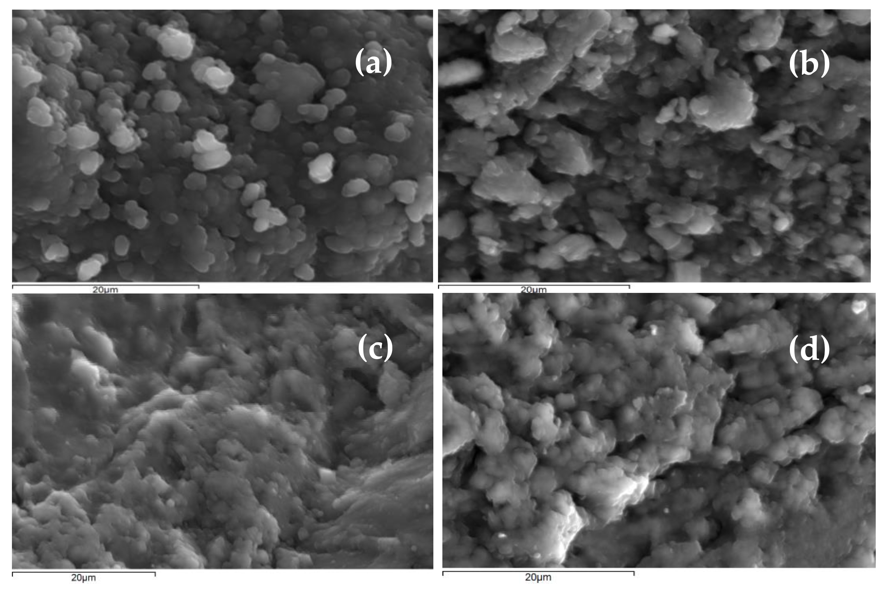
2.3. Natural and Silver-Modified Zeolite Characterization

The morphologies of selected NZs and Ag-MNZs were examined by a JSM 6300 JEOL Scanning Electron Microscope (SEM) at 20 kV. The energy dispersive X-ray (EDX) analysis of fine (0–1 mm) NZ, coarse (1–3 mm) NZ, fine (0–1 mm) Ag-MNZ, and coarse (1–3 mm) Ag-MNZ was also observed using this instrument. Clearly, the incorporation of Ag+ into the structure of Ag-MNZ did not change its morphology and thusly formations of NZ and Ag-MNZ were similar in particle size and appearance (see Figure 1). However, Boschetto et al. (2012) observed crystallinity and specific area loss after Ag incorporation in the structure of the zeolite Y [41]. The results of EDX analyses (data not shown) suggest that the molar ratio Si/Al of the NZ (≈6) does not change notably with Ag exchange. Note that high concentrations of Ag could cause significant changes in the zeolite structure [41]. SEM images also captured aggregates as finer crystal grains of the clinoptilolite mineral.

2.4. Bacterial Suspensions Preparation and Enumeration

The two model bacteria used in the batch experiments were the Gram-negative E. coli (NCTC 9001) and the Gram-positive E. faecalis (NCTC 775). Cultures were stored at −80 °C in growth media enhanced with 80% glycerol. Prior to each batch experiment, the bacteria were cultured on non-selective growth medium (Nutrient Agar) at 37 °C for 48 h. Subsequently, some of the sufficiently formed colonies of the microbial cultures were isolated and transferred to a test tube of sterilized DIW; bacterial concentration in the suspension was calculated based on the 0.5 McFarland turbidity scale, according to which, 0.1 optical absorbance at 600 nm equals to a concentration of 108 cfu/mL. Optical density measurements were conducted using a UV-visible spectrophotometer (U-2001, Hitachi, Tokyo, Japan). The dense bacterial suspensions were diluted to obtain the initial bacterial concentrations for the batch experiments. For the calculation of the bacteria concentrations, serial dilution of the samples was performed and aliquots of 100 μL were plated (in duplicates) on the surface of selective growth media: Harlequin Chromogenic Coliform Agar (NEOGEN NCM 1005A) and Slanetz and Bartley Agar (LAB166) for the growth of E. coli and E. faecalis, respectively. The plates were incubated at 37 °C for 48 h and the colonies were counted. Reliable dilutions for quantification were considered those that resulted in a reasonable number (30–300) of distinct colonies to count. Then, the concentration of bacteria in the sample was measured by taking into account the sample dilution and the volume plated out on the growth media, and given in colony-forming units per milliliter (cfu/mL) [44].

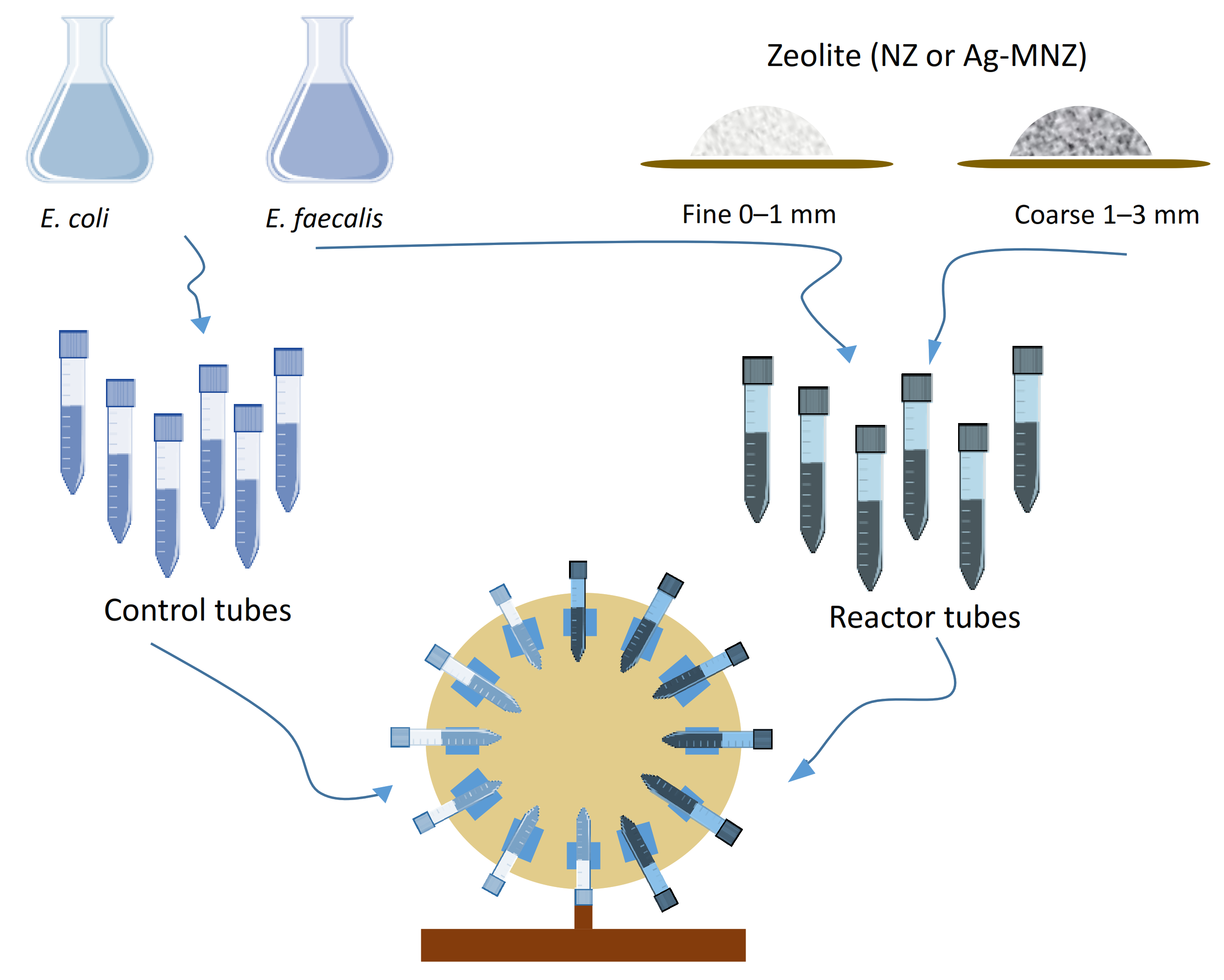
2.5. Batch Inactivation Experiments

For bacteria inactivation experiments with the ΝΖ and the Ag-MNZ, a standard procedure in the batch process was followed. In the batch process a solution of synthetic water inoculated with bacteria (E. coli, E. faecalis) containing a certain amount of NZ or Ag-MNZ (5 g/L) was used. The procedure was implemented under a constant ambient temperature and hydrodynamic conditions, while samples were obtained at specified intervals. Tubes with a volume of 50 mL were completely filled in a gentle manner with the above synthetic water inoculated with bacteria of two tested initial concentrations, 106 and 105 cfu/mL, so that no air remained upon their closure with caps. The tubes were attached onto a rotator. Then, a slow agitation of 12 rpm, associated with the dynamic conditions, was chosen in order to maximize the exposure of bacteria to NZ or Ag-MNZ.
For each experiment, 20 tubes divided into two sets were used. The first set of 10 tubes (controls) contained a 50 mL suspension of bacteria without NZ or Ag-MNZ in order to observe the time-dependent bacterium inactivation. The second set of 10 tubes (reactors) contained a 50 mL mixed suspension of bacteria with NZ or Ag-MNZ in order to observe any changes in bacterium inactivation provoked by the presence of zeolites. At various time intervals over a 1 h time period, a tube from each set of tubes was selected for sampling. The supernatant was sampled (a 2 mL collected sample) to determine the remaining bacteria concentration. Figure 2 illustrates the batch experimental procedure.

2.6. Theoretical Considerations

The log reduction in bacterial concentrations after the experimental time period was obtained by applying the following expression [45]:
Log ( Reduction ) = log 10 C total , 0 C total t = log 10 C total , 0 log 10 C total t
where Ctotal [cfu/mL] is the total bacterial concentration in the suspension at time t and Ctotal,0= Ctotal(t=0) is the initial total concentration of bacteria.
Additionally, the log reduction was converted to percent reduction (P) as follows:
P ( % ) = ( 1 10 log ( R e d u c t i o n ) ) × 100
Moreover, the below pseudo-first-order mathematical expression, accounting for time-dependent inactivation, was used to describe the experimental bacterial inactivation data [46,47,48]:
dC t o t a l t dt = λ t C t o t a l t
where λ [1/d] is the inactivation rate coefficient of bacteria which can be written as a function of time:
λ t = λ 0 e α t
where λ0 [1/d] is the initial inactivation rate coefficient, and α [1/d] is the resistivity coefficient.
The solution to Equation (3) is given as:
ln C t o t a l t C total , 0 = λ 0 α e α t 1
If λ is time independent λ(t) = λ, then the solution to Equation (3) is:
ln C t o t a l t C t o t a l , 0 = λ t
The unknown inactivation parameters λ, λ0 and α were estimated using the autonomous multipurpose fitting software: ColloidFit [49].

3. Results and Discussion

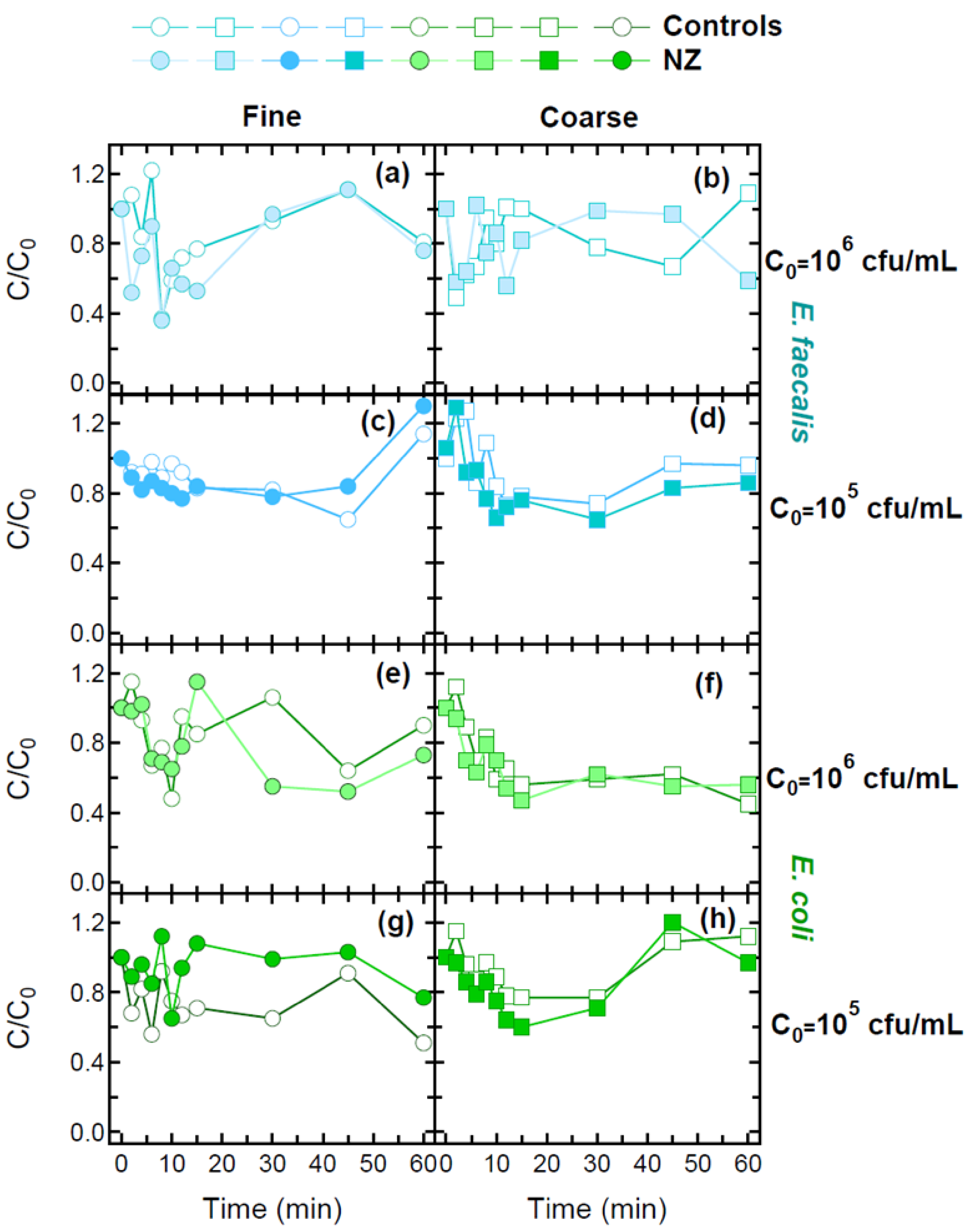
3.1. Bacterial Inactivation in the Presence of NZs

Figure 3 presents the inactivation results of both bacteria (E. coli, E. faecalis) in the absence (controls) and presence (reactors) of either fine (0–1 mm) or coarse (1–3 mm) NZs under dynamic conditions at 22 °C for two different initial bacteria concentrations (105 and 106 cfu/mL). Note that slight variations in the initial concentration of the bacteria suspensions present in each tube may result in normalized bacterial concentrations greater than unity. The experimental results show that in the absence of NZs, in most cases examined, the inactivation rates for both bacteria were higher for the higher initial bacterial concentration compared to the inactivation rates for bacteria with the lower initial concentration (Table 1, Figure 4). Although previous studies showed that bacteria inactivation rates decrease with an increase in initial bacteria concentration [50], in this study, such a clear trend was not observed.
Moreover, similar inactivation rates were observed in reactor and control tubes (absence of NZs) for both bacteria (E. coli, E. faecalis), suggesting that low concentration (5 g/L) of both fine (0–1 mm) and coarse (1–3 mm) NZs only slightly affects bacteria inactivation (no antibacterial effect) for both initial bacteria concentrations (105 and 106 cfu/mL). However, in most cases examined, higher inactivation rates were observed in the case of coarse rather than fine NZs.

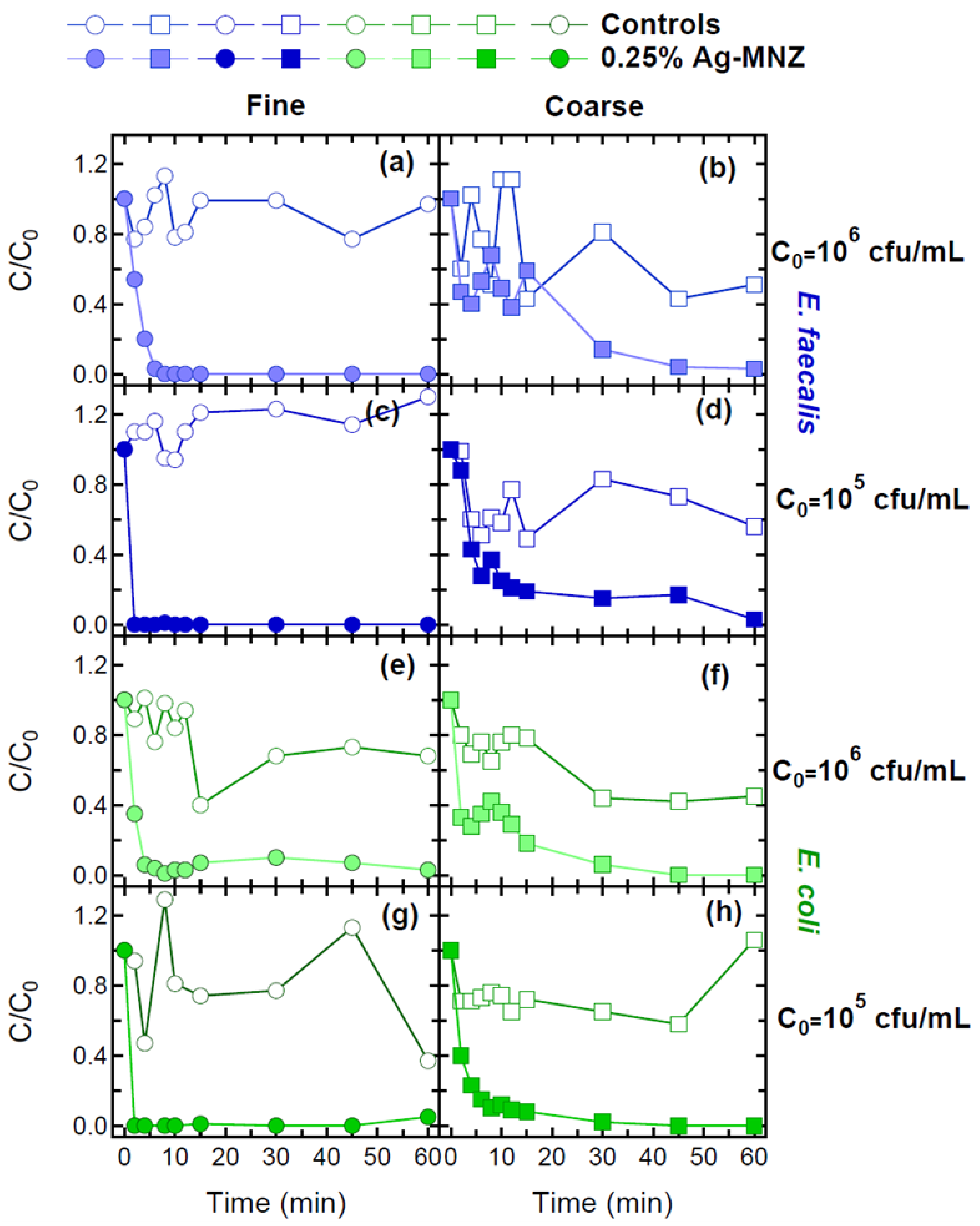
3.2. Bacterial Inactivation in the Presence of Ag-MNZs

Figure 5 shows the inactivation results of both bacteria (E. coli, E. faecalis) in the absence (controls) and presence (reactors) of either fine (0–1 mm) or coarse (1–3 mm) 0.25% Ag-MNZs under dynamic conditions at 22 °C for two different initial bacteria concentrations (105 and 106 cfu/mL). Clearly, higher inactivation rates are observed in the presence of 0.25% Ag-MNZs than NZs for both bacteria (E. coli, E. faecalis) and the two initial bacteria concentrations (105 and 106 cfu/mL) (Table 1, Figure 4). Similar inactivation rates were observed for the two initial concentrations under coarse (1–3 mm) 0.25% Ag-MNZs. In the presence of fine (0–1 mm) 0.25% Ag-MNZs, low compared to high initial bacteria concentrations yielded higher inactivation rates. Similar results for E. coli removal by natural zeolites and Mg2+-modified zeolites (MMZs) were found by Sang-Woo et al. (2016) [51]. The authors reported that increased initial E. coli concentration causes the zeolite adsorption capacity to decrease. An enormous E. coli adsorption capacity of MMZs compared to that of natural zeolite for different initial E. coli concentration was also observed. Note that zeolite morphology and particle size may alter bacteria-zeolite interactions and consequently the extent of bacterial inactivation. Zeolites with a smaller particle size are expected to result in faster bacterial inactivation due to faster ion exchange and release (shorter diffusion length) [27]. However, in this study no clear trend was observed. Note that significant variations in bacteria inactivation results among the replicates suggest the creation of bacterial aggregates. However, the bacterial inactivation data collected in this study cannot be compared directly with the results from previous studies, because of the differences in experimental conditions (e.g., silver modification of zeolites, zeolite and bacteria concentrations, reactor geometries, aqueous solutions, cultivation conditions etc.). Moreover, slight changes in water chemistry may result in different agglomeration and sedimentation behaviour, as well as in the toxic activity of Ag-MNZs.

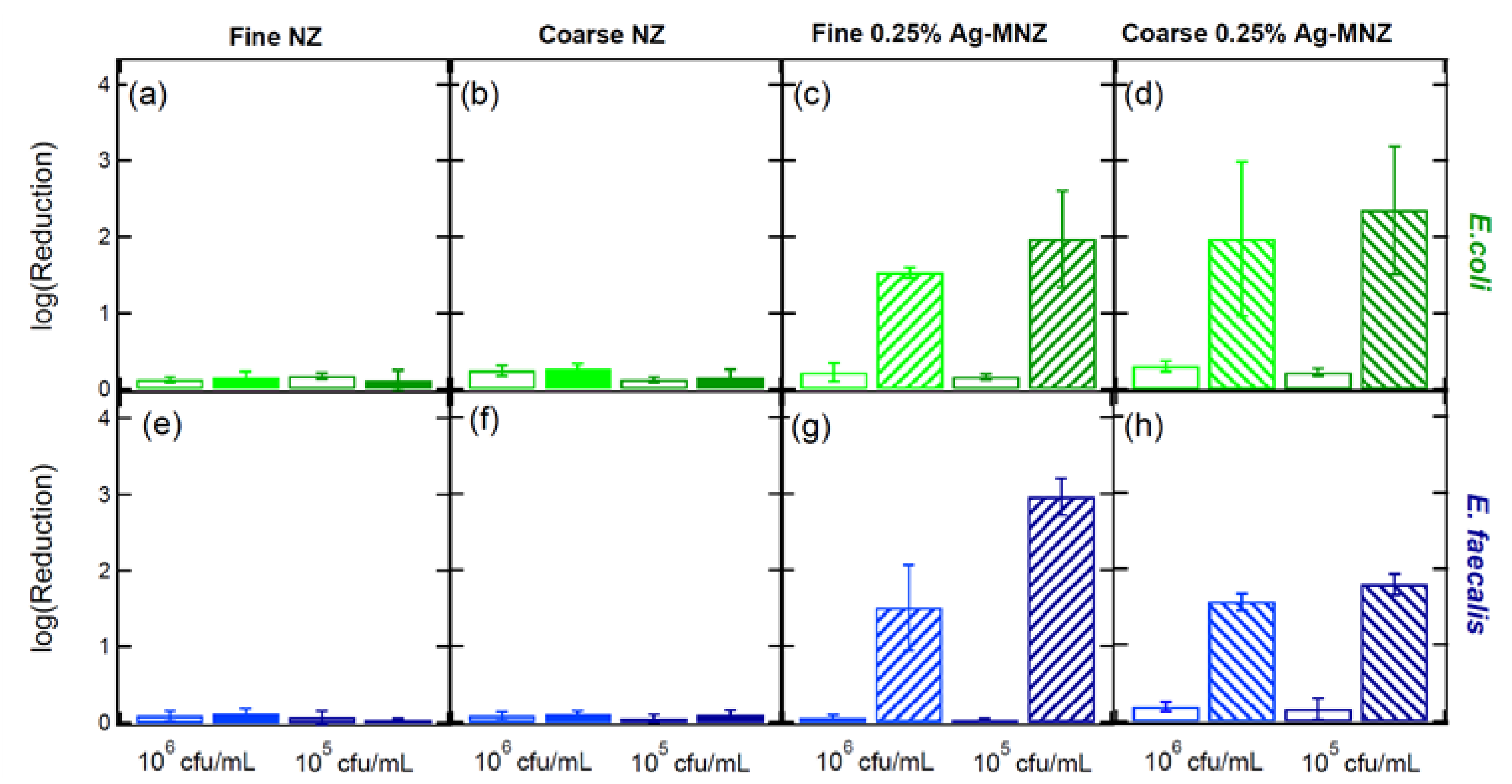
3.3. Bacteria Log Reduction

Figure 6 presents a graphical representation of the bacteria log reduction within a 1- h time period for all cases considered in this study. The calculated log and percent reduction values are presented in Table 2. In most cases examined under NZs, higher log reduction values were observed in the case of a higher initial concentration for both bacteria (E. coli, E. faecalis). However, in both control and reactor tubes within 1 h of the experiment, no substantial reduction in the bacteria’s initial concentrations were observed. In the presence of fine (0–1 mm) NZ, the reduction in E. faecalis by 0.12 ± 0.08 log cfu/mL (~23.58%) and 0.09 ± 0.02 log cfu/mL (~18.72%) was similar to that in E. coli (0.15 ± 0.08 log cfu/mL, ~29.21% and 0.11 ± 0.14 log cfu/mL, ~22.52%) for 106 and 105 cfu/mL initial concentrations, respectively. In the presence of coarse (1–3 mm) NZ, the E. coli population decreased by 0.26 ± 0.05 log cfu/mL (~44.87%) and 0.15 ± 0.11 log cfu/mL (~28.98%) within a 1 h time period for the 106 and 105 cfu/mL initial concentrations. E. faecalis was less sensitive to the presence of coarse (1–3 mm) NZ with a reduction of 0.10 ± 0.05 log cfu/mL (~21.04%) and 0.09 ± 0.07 log cfu/mL (~19.49%) for 106 and 105 cfu/mL initial concentrations, respectively.
The silver ions incorporated into NZs enhanced the bacterial population reduction in the presence of both fine (0–1 mm) and coarse (1–3 mm) 0.25% Ag-MNZs for both initial bacterial concentrations (106 and 105 cfu/mL) and both bacteria (Figure 6c,d,g,h). Clearly, the presence of fine (0–1 mm) 0.25% Ag-MNZs resulted in a greater population reduction for E. coli (1.53 ± 0.07, ~97.07% and 1.97 ± 0.63 log cfu/mL, ~98.93%) and for E. faecalis (∼1.50 ± 0.56 and 2.96 ± 0.24 log cfu/mL) for 106 and 105 cfu/mL initial concentrations, respectively. Coarse (1–3 mm) Ag-MNZs enhanced both E. coli (∼1.97 ± 1.01 and 2.35 ± 0.84 log cfu/mL) and E. faecalis population reduction (∼1.56 ± 0.11 and 1.79 ± 0.14 log cfu/mL) for the two above initial concentrations. Note that higher log reduction for both bacteria populations was observed in the case of lower initial bacterial concentration.
Top and Ülkü (2004) showed that the antibacterial activity of Ag+-clinoptilolite did not increase with increasing amounts of Ag+ in the zeolite, probably due to the formation of metallic Ag at high Ag+ concentrations [29]. Akhigbe et al. (2014) reported a significant reduction in E. coli growth by Ag+-modified clinoptilolite (4.34 wt%) with a 10 log10 reduction in 30 min [28]. The bactericidal action of silver-modified zeolite in the literature has been attributed to two mechanisms. The first one is based on the silver ion itself being released from the zeolite [52] and the second one on reactive oxygen species (ROS) generated from silver in the solid matrix [27]. Milenkovic et al. (2017) found that the bactericidal effect of Ag-NZs toward E. coli could be attributed not only to released Ag+ ions but also to Ag-Z itself [32]. Moreover, in most cases examined in this study, greater population reduction was observed for E. coli than E. faecalis, probably due to the higher resistance of Gram-positive bacteria (E. faecalis) than Gram-negative bacteria (E. coli) caused by the thicker peptidoglycan layer.

4. Conclusions

The results of this study show that under dynamic batch conditions, both E. coli and E. faecalis were removed in greater amounts by Ag-modified NZs than NZs for both particle sizes (0–1 mm and 1–3 mm) employed in this study at 22 °C. Furthermore, the log reduction in both bacteria was found to be dependent on the initial bacteria concentration. Moreover, a higher percent reduction was observed for fine than coarse NZ, while in the case of 0.25% Ag-MNZ, no clear trend was observed. Note that the 0.25% Ag-MNZ reduced both bacteria from 1.5 to 2.96 logs after 1 h. The estimated inactivation rates suggest that bacteria inactivation, in most cases considered in this study, is inversely correlated with zeolite particle size in the presence of 0.25% Ag-MNZs.
The preliminary results obtained in this research stage show the possibility of using NZs or Ag-MNZs of various particle sizes in the removal of pathogens or faecal indicators in water treatment processes. Modified zeolites, with exceptional ion-exchange and sorption properties, have the potential to be effective filtration media. The development of Ag-MNZ-based filters, with enhanced antimicrobial properties, lays the framework for new possibilities in water filtration. However, future work is needed to evaluate the removal of waterborne microbial pathogens by Ag-MNZs in fixed-bed columns under a broad range of conditions including: the Ag-MNZ incorporation method, solution pH, pathogen initial concentration, flow rate, and bed depth. Moreover, the potential of silver recovery for reuse should also evaluated. Based on the results of further research, it will be possible to economically assess the whole technological process and give a clear opinion on the use of NZ and Ag-MNZ in water treatment processes.

Author Contributions

V.I.S.: Conceptualization, Methodology, Formal analysis, Writing—review and editing, Funding acquisition, Writing—original draft. A.V.: Resources, Supervision, Writing—review and editing. All authors have read and agreed to the published version of the manuscript.

Funding

This research is co-financed by Greece and the European Union (European Social Fund-ESF) through the Operational Programme «Human Resources Development, Education and Lifelong Learning» in the context of the project “Reinforcement of Postdoctoral Researchers—2nd Cycle” (MIS-5033021), implemented by the State Scholarships Foundation (ΙΚΥ).

Institutional Review Board Statement

Not applicable.

Informed Consent Statement

Not applicable.

Data Availability Statement

Not applicable.

Conflicts of Interest

The authors declare no conflict of interest.

References

  1. Brombach, H.; Weiss, G.; Fuchs, S. A new database on urban runoff pollution: Comparison of separate and combined sewer systems. Water Sci. Technol. 2005, 51, 119–128. [Google Scholar] [CrossRef] [PubMed]
  2. Kim, T.J.; Silva, J.L.; Weng, W.L.; Chen, W.W.; Corbitt, M.; Jung, Y.S.; Chen, Y.S. Inactivation of Enterobacter sakazakii by water-soluble muscadine seed extracts. Int. J. Food Microbiol. 2008, 129, 295–299. [Google Scholar] [CrossRef] [PubMed]
  3. WHO; UNICEF. Progress on Drinking Water, Sanitation and Hygiene: Update and Sustainable Development Goal Baselines; License: CC BY-NC-SA 3.0 IGO, Launch Version July 12 Main Report; WHO: Geneva, Switzerland, 2017. [Google Scholar]
  4. Zazouli, M.A.; Kalankesh, L.R. Indicators and Causes of Environmental Health Inequalities. J. Maz. Univ. Med. Sci. 2018, 27, 218–229. [Google Scholar]
  5. Wang, S.; Peng, Y. Natural zeolites as effective adsorbents in water and wastewater treatment. Chem. Eng. J. 2010, 156, 11–24. [Google Scholar] [CrossRef]
  6. Zhao, Y. Review of the Natural, Modified, and Synthetic Zeolites for Heavy Metals Removal from Wastewater. Environ. Eng. Sci. 2016, 33, 443–454. [Google Scholar] [CrossRef]
  7. Galletti, C.; Dosa, M.; Russo, N.; Fino, D. Zn2+ and Cd2+ removal from wastewater using clinoptilolite as adsorbent. Environ. Sci. Pollut. Res. Int. 2021, 28, 24355–24361. [Google Scholar] [CrossRef]
  8. Morante-Carballo, F.; Montalván-Burbano, N.; Carrión-Mero, P.; Jácome-Francis, K. Worldwide Research Analysis on Natural Zeolites as Environmental Remediation Materials. Sustainability 2021, 13, 6378. [Google Scholar] [CrossRef]
  9. Feng, Q.L.; Wu, J.; Chen, G.Q.; Cui, F.Z.; Kim, T.N.; Kim, J.O. A mechanistic study of the antibacterial effect of silver ions on Escherichia coli and Staphylococcus aureus. J. Biomed. Mater. Res. 2000, 52, 662–668. [Google Scholar] [CrossRef]
  10. Sondi, I.; Salopek-Sondi, B. Silver nanoparticles as antimicrobial agent: A case study on E. coli as a model for Gram-negative bacteria. J. Colloid Interface Sci. 2004, 275, 177–182. [Google Scholar] [CrossRef]
  11. Baker, C.; Pradhan, A.; Pakstis, L.; Pochan, D.J.; Shah, S.I. Synthesis and antibacterial properties of silver nanoparticles. J. Nanosci. Nanotechnol. 2005, 5, 244–249. [Google Scholar] [CrossRef]
  12. Atiyeh, B.S.; Costagliola, M.; Hayek, S.N.; Dibo, S.A. Effect of silver on burn wound infection control and healing: Review of the literature. Burns 2007, 33, 139–148. [Google Scholar] [CrossRef]
  13. Birla, S.S.; Tiwari, V.V.; Gade, A.K.; Ingle, A.P.; Yadav, A.P.; Rai, M.K. Fabrication of silver nanoparticles by Phoma glomerata and its combined effect against Escherichia coli, Pseudomonas aeruginosa and Staphylococcus aureus. Lett. Appl. Microbiol. 2009, 48, 173–179. [Google Scholar] [CrossRef]
  14. Le Ouay, B.; Stellacci, F. Antibacterial activity of silver nanoparticles: A surface science insight. Nano Today 2015, 10, 339–354. [Google Scholar] [CrossRef] [Green Version]
  15. Deshmukh, S.P.; Patil, S.M.; Mullani, S.B.; Delekar, S.D. Silver nanoparticles as an effective disinfectant: A review. Mater. Sci. Eng. C Mater. Biol. Appl. 2019, 97, 954–965. [Google Scholar] [CrossRef]
  16. Galeano, B.; Korff, E.; Nicholson, W.L. Inactivation of vegetative cells, but not spores, of Bacillus anthracis, B. cereus, and B. subtilis on stainless steel surfaces coated with an antimicrobial silver- and zinc-containing zeolite formulation. Appl. Environ. Microbiol. 2003, 69, 4329–4331. [Google Scholar] [CrossRef] [Green Version]
  17. Kawahara, K.; Tsuruda, K.; Morishita, M.; Uchida, M. Antibacterial effect of silver-zeolite on oral bacteria under anaerobic conditions. Dent. Mater. 2000, 16, 452–455. [Google Scholar] [CrossRef]
  18. Matsumura, Y.; Yoshikata, K.; Kunisaki, S.; Tsuchido, T. Mode of bactericidal action of silver zeolite and its comparison with that of silver nitrate. Appl. Environ. Microbiol. 2003, 69, 4278–4281. [Google Scholar] [CrossRef] [Green Version]
  19. Zhang, Y.; Zhong, S.; Zhang, M.; Lin, Y. Antibacterial activity of silver-loaded zeolite A prepared by a fast microwave-loading method. J. Mater. Sci. 2009, 44, 457–462. [Google Scholar] [CrossRef]
  20. Hagiwara, Z.; Hoshino, S.; Ishino, H.; Nohara, S.; Tagawa, K.; Yamanaka, K. Zeolite Particles Retaining Silver Ions Having Antibacterial Properties. US Patent 4,911,898, 27 March 1990. [Google Scholar]
  21. Kwakye-Awuah, B.; Williams, C.; Kenward, M.A.; Radecka, I. Antimicrobial action and efficiency of silver-loaded zeolite X. J. Appl. Microbiol. 2008, 104, 1516–1524. [Google Scholar] [CrossRef]
  22. Lv, L.; Luo, Y.; Ng, W.J.; Zhao, X.S. Bactericidal activity of silver nanoparticles supported on microporous titanosilicate ETS-10. Microporous Mesoporous Mater. 2009, 120, 304–309. [Google Scholar] [CrossRef]
  23. Guerra, R.; Lima, E.; Viniegra, M.; Guzmán, A.; Lara, V. Growth of Escherichia coli and Salmonella typhi inhibited by fractal silver nanoparticles supported on zeolites. Microporous Mesoporous Mater. 2012, 147, 267–273. [Google Scholar] [CrossRef]
  24. Krishnani, K.K.; Zhang, Y.; Xiong, L.; Yan, Y.; Boopathy, R.; Mulchandani, A. Bactericidal and ammonia removal activity of silver ion-exchanged zeolite. Bioresour. Technol. 2012, 117, 86–91. [Google Scholar] [CrossRef]
  25. Demirci, S.; Ustaoğlu, Z.; Yılmazer, G.A.; Sahin, F.; Baç, N. Antimicrobial properties of zeolite-X and zeolite-A ion-exchanged with silver, copper, and zinc against a broad range of microorganisms. Appl. Biochem. Biotechnol. 2014, 172, 1652–1662. [Google Scholar] [CrossRef]
  26. Youssef, H.F.; Abdel-Aziz, M.S.; Fouda, F.K. Evaluation of antimicrobial activity of different silver-exchanged nano and micronized zeolites prepared by microwave technique. J. Porous Mater. 2017, 24, 947–957. [Google Scholar] [CrossRef]
  27. Dutta, P.; Wang, B. Zeolite-supported silver as antimicrobial agents. Coord. Chem. Rev. 2019, 383, 1–29. [Google Scholar] [CrossRef]
  28. Akhigbe, L.; Ouki, S.; Saroj, D.; Lim, X.M. Silver-modified clinoptilolite for the removal of Escherichia coli and heavy metals from aqueous solutions. Environ. Sci. Pollut. Res. Int. 2014, 21, 10940–10948. [Google Scholar] [CrossRef]
  29. Top, A.; Ülkü, S. Silver, zinc, and copper exchange in a Na-clinoptilolite and resulting effect on antibacterial activity. Appl. Clay Sci. 2004, 27, 13–19. [Google Scholar] [CrossRef] [Green Version]
  30. Rivera-Garza, M.; Olguín, M.T.; García-Sosa, I.; Alcántara, D.; Rodríguez-Fuentes, G. Silver supported on natural Mexican zeolite as an antibacterial material. Microporous Mesoporous Mater. 2000, 39, 431–444. [Google Scholar] [CrossRef]
  31. Rossainz-Castro, L.G.; De-La-Rosa-Gómez, I.; Olguín, M.T.; Alcántara-Díaz, D. Comparison between silver- and copper-modified zeolite-rich tuffs as microbicide agents for Escherichia coli and Candida albicans. J. Environ. Manag. 2016, 183, 763–770. [Google Scholar] [CrossRef] [PubMed]
  32. Milenkovic, J.; Hrenovic, J.; Matijasevic, D.; Niksic, M.; Rajic, N. Bactericidal activity of Cu-, Zn-, and Ag-containing zeolites toward Escherichia coli isolates. Environ. Sci. Pollut. Res. Int. 2017, 24, 20273–20281. [Google Scholar] [CrossRef] [PubMed]
  33. De La Rosa-Gómez, I.; Olguín, M.T.; Alcántara, D. Silver-modified Mexican clinoptilolite-rich tuffs with various particle sizes as antimicrobial agents against Escherichia coli. J. Mex. Chem. Soc. 2010, 54, 139–142. [Google Scholar] [CrossRef]
  34. Copcia, V.E.; Luchian, C.; Dunca, S.; Bilba, N.; Hristodor, C.M. Antibacterial activity of silver-modified natural clinoptilolite. J. Mater. Sci. 2011, 46, 7121–7128. [Google Scholar] [CrossRef]
  35. Chernousova, S.; Epple, M. Silver as antibacterial agent: Ion, nanoparticle, and metal. Angew. Chem. Int. Ed. Engl. 2013, 52, 1636–1653. [Google Scholar] [CrossRef]
  36. Cerrillo, J.L.; Palomares, A.E.; Rey, F.; Valencia, S.; Palou, L.; Pérez-Gago, M.B. Ag-zeolites as fungicidal material: Control of citrus green mold caused by Penicillium digitatum. Microporous Mesoporous Mater. 2017, 254, 69–76. [Google Scholar] [CrossRef]
  37. Ferreira, L.; Fonseca, A.M.; Botelho, G.; Aguiar, C.A.; Neves, I.C. Antimicrobial activity of faujasite zeolites doped with silver. Microporous Mesoporous Mater. 2012, 160, 126–132. [Google Scholar] [CrossRef]
  38. Koyama, K.; Takeuchi, Y. Clinoptilolite: The distribution of potassium atoms and its role in thermal stability. Z. Für Krist. -Cryst. Mater. 1977, 145, 216–239. [Google Scholar] [CrossRef]
  39. Arcoya, A.; González, J.A.; Llabre, G.; Seoane, X.L.; Travieso, N. Role of the countercations on the molecular sieve properties of a clinoptilolite. Microporous Mater. 1996, 7, 1–13. [Google Scholar] [CrossRef]
  40. Colella, C. Ion exchange equilibria in zeolite minerals. Miner. Depos. 1996, 31, 554–562. [Google Scholar] [CrossRef]
  41. Lúcia Boschetto, D.; Lerin, L.; Cansian, R.; Castellã Pergher, B.; Di Luccio, M. Preparation and antimicrobial activity of polyethylene composite films with silver exchanged zeolite-Y. Chem. Eng. J. 2012, 204–206, 210–216. [Google Scholar] [CrossRef]
  42. Akhigbe, L.; Ouki, S.; Saroj, D. Disinfection and removal performance for Escherichia coli and heavy metals by silver-modified zeolite in a fixed bed column. Chem. Eng. J. 2016, 295, 92–98. [Google Scholar] [CrossRef] [Green Version]
  43. Sherry, H.S. The Ion-Exchange Properties of Zeolites. I. Univalent Ion Exchange in Synthetic Faujasite. J. Phys. Chem. 1966, 70, 1158–1168. [Google Scholar] [CrossRef]
  44. Georgopoulou, M.P.; Syngouna, V.I.; Chrysikopoulos, C.V. Influence of graphene oxide nanoparticles on the transport and cotransport of biocolloids in saturated porous media. Colloids Surf. B Biointerfaces 2020, 189, 110841. [Google Scholar] [CrossRef]
  45. Bellou, M.I.; Syngouna, V.I.; Tselepi, M.A.; Kokkinos, P.A.; Paparrodopoulos, S.C.; Vantarakis, A.; Chrysikopoulos, C.V. Interaction of human adenoviruses and coliphages with kaolinite and bentonite. Sci. Total Environ. 2015, 517, 86–95. [Google Scholar] [CrossRef]
  46. Anders, R.; Chrysikopoulos, C. V Evaluation of the factors controlling the time-dependent inactivation rate coefficients of bacteriophage MS2 and PRD1. Environ. Sci. Technol. 2006, 40, 3237–3242. [Google Scholar] [CrossRef]
  47. Chrysikopoulos, C.V.; Aravantinou, A.F. Virus inactivation in the presence of quartz sand under static and dynamic batch conditions at different temperatures. J. Hazard. Mater. 2012, 233–234, 148–157. [Google Scholar] [CrossRef]
  48. Syngouna, V.I.; Chrysikopoulos, C.V. Inactivation of MS2 bacteriophage by titanium dioxide nanoparticles in the presence of quartz sand with and without ambient light. J. Colloid Interface Sci. 2017, 497, 117–125. [Google Scholar] [CrossRef]
  49. Katzourakis, V.E.; Chrysikopoulos, C.V. Fitting the Transport and Attachment of Dense Biocolloids in One-Dimensional Porous Media: ColloidFit. Groundwater 2017, 55, 156–159. [Google Scholar] [CrossRef]
  50. Furukawa, S.; Noma, S.; Shimoda, M.; Hayakawa, I. Effect of initial concentration of bacterial suspensions on their inactivation by high hydrostatic pressure. Int. J. Food Sci. Technol. 2002, 37, 573–577. [Google Scholar] [CrossRef]
  51. An, S.W.; Jeong, Y.C.; Cho, H.H.; Park, J.W. Adsorption of NH4+-N and E. coli onto Mg2+-modified zeolites. Environ. Earth Sci. 2016, 75, 437. [Google Scholar] [CrossRef]
  52. Im, K.C.; Takasaki, Y.; Endo, A.; Kuriyama, M. Antibacterial Activity of A-Type Zeolite Supporting Silver Ions in Deionized Distilled Water. J. Antibact. Antifung. Agents 1996, 24, 269–274. [Google Scholar]
Figure 1. SEM images taken at a magnification of 2500× of (a) 0–1 mm NZ, (b) 0–1 mm Ag-MNZ, (c) 1–3 mm NZ, and (d) 1–3 mm Ag-MNZ.
Water 13 02938 g001
Figure 2. Graphical representation of the dynamic batch experimental procedure. The control tubes were filled with bacteria suspension and the reactor tubes were filled with bacteria suspension as well as natural zeolite or Ag-modified natural zeolite. All tubes were attached to a tube rotator at 12 rpm and 22 °C.
Water 13 02938 g002
Figure 3. Experimental inactivation data of E. faecalis (ad) and E. coli (eh) removal by fine (0–1 mm) NZ (ag) and coarse (1–3 mm) NZ (bh) in the absence (empty symbols) and the presence (filled symbols) of natural zeolites. Bacterial initial concentrations: 106 and 105 cfu/mL.
Water 13 02938 g003
Figure 4. Estimated inactivation rates, λ, for E. coli (a,b) and E. faecalis (c,d) for the two initial bacteria concentrations: in the presence of NZ (a,c) and Ag-MNZ (b,d), in controls (empty columns) and reactors (filled and cross-shaded columns, respectively).
Water 13 02938 g004
Figure 5. Experimental inactivation data of E. faecalis (ad) and E. coli (eh) removal by fine (0–1 mm) Ag-MNZ (ag) and coarse (1–3 mm) Ag-MNZ (bh) in the absence (empty symbols) and the presence (filled symbols) of zeolites. Bacterial initial concentrations: 106 and 105 cfu/mL.
Water 13 02938 g005
Figure 6. Calculated log-reduced values of E. coli (ad) and E. faecalis (eh) in controls (empty columns) and interacting with fine (0–1 mm) (a,e) and coarse (1–3 mm) (b,f) NZ (filled columns), and fine (0–1 mm) (c,g) and coarse (1–3 mm) (d,h) Ag-MNZ (cross-shaded columns) for bacterial initial concentrations: 106 and 105 cfu/mL within 1 h of experiment.
Water 13 02938 g006
Table 1. Fitted inactivation parameter values (λ, λ0, and α) of E. coli and E. faecalis for the two initial concentrations (106 and 105 cfu/mL) under experimental conditions.
BacteriaExperimental Case
Absence/Presence of NZ or Ag-MNZBacteria Initial Concentrationλ (min−1)λ0 (min−1)α (min−1)
E. coliFine (0–1 mm) NZ
Controls106 cfu/mL0.38 × 10−2 ± 0.0037.28 × 10−2 ± 0.0340.111 ± 0.002
105 cfu/mL5.19 × 10−2 ± 0.0119.47 × 10−2 ± 0.0310.129 ± 0.016
Reactors106 cfu/mL4.92 × 10−2 ± 0.0111.12 × 10−2 ± 0.0070.026 ± 0.141
105 cfu/mL1.19 × 10−2 ± 0.0082.09 × 10−2± 0.0660.081 ± 0.436
Coarse (1–3 mm) NZ
Controls106 cfu/mL7.22 × 10−2± 0.0068.81 × 10−2± 0.0200.564 ± 1.230
105 cfu/mL1.20 × 10−2 ± 0.0087.71 × 10−2 ± 0.0110.148 ± 1.150
Reactors106 cfu/mL7.47 × 10−2 ± 0.0086.95 × 10−2± 0.07237.763 ± 83
105 cfu/mL2.6 × 10−2 ± 0.0122.49 × 10−2 ± 0.01070.047 ± 0.084
E. faecalisFine (0–1 mm) NZ
Controls106 cfu/mL3.07 × 10−2± 0.01718.3 × 10−2± 0.1940.015± 0.171
105 cfu/mL1.8 × 10−2 ± 0.0074.18 × 10−2 ± 0.0070.518± 0.153
Reactors106 cfu/mL4.57 × 10−2 ± 0.02230.9 × 10−2 ± 0.0490.916 ± 0.311
105 cfu/mL1.70 × 10−2 ± 0.00920.56 × 10−2 ± 0.1020.078 ± 0.117
Coarse (1–3 mm) NZ
Controls106 cfu/mL2.28 × 10−2 ± 0.01635.59 × 10−2± 0.0110.988 ± 0.066
105 cfu/mL1.81 × 10−2 ± 0.00910.91 × 10−2 ± 0.0211.142 ± 0.511
Reactors106 cfu/mL2.82 × 10−2 ± 0.01427.01 × 10−2 ± 0.0241.021 ± 0.202
105 cfu/mL3.62 × 10−2 ± 0.00813.84 × 10−2 ± 0.2881.475 ± 8.4
E. coliFine (0–1 mm) 0.25% Ag-MNZ
Controls106 cfu/mL4.75 × 10−2 ± 0.0113.55 × 10−2 ± 0.0170.130 ± 0.138
105 cfu/mL5.77 × 10−2 ± 0.02781.96 × 10−2 ± 0.0580.338± 0.419
Reactors106 cfu/mL43.97 × 10−2 ± 0.07670.43 × 10−2 ± 0.0900.191 ± 0.045
105 cfu/mL73.38 × 10−2 ± 0.157220.49 × 10−2 ± 0.0790.608 ± 0.041
Coarse (1–3 mm) 0.25% Ag-MNZ
Controls106 cfu/mL7.91 × 10−2 ± 0.00911.16 × 10−2 ± 0.0080.292 ± 0.038
105 cfu/mL4.09 × 10−2 ± 0.01316.07 × 10−2 ± 0.0120.471 ± 0.059
Reactors106 cfu/mL53.39 × 10−2± 0.09149.25 × 10−2 ± 0.0680.344± 0.081
105 cfu/mL16.34 × 10−2 ± 0.01343.25 × 10−2 ± 0.0230.142± 0.016
E. faecalisFine (0–1 mm) 0.25% Ag-MNZ
Controls106 cfu/mL1.42 × 10−2 ± 0.00812.91 × 10−2 ± 0.0141.326 ± 0.357
105 cfu/mL1.95 × 10−2± 0.0044.70 × 10−2 ± 0.0090.629± 0.219
Reactors106 cfu/mL15.82 × 10−2 ± 0.07175.14 × 10−2 ± 0.2760.035 ± 0.076
105 cfu/mL100.92 × 10−2 ± 0.142327.82 × 10−2 ± 0.2560.527 ± 0.074
Coarse (1–3 mm) 0.25% Ag-MNZ
Controls106 cfu/mL6.45 × 10−2 ± 0.01725.8 × 10−2± 0.04015.269 ± 6.665
105 cfu/mL6.40 × 10−2 ± 0.0148.01 × 10−2 ± 0.0290.136 ± 0.106
Reactors106 cfu/mL26.03 × 10−2 ± 0.03636.0 × 10−2 ± 0.0400.447 ± 0.087
105 cfu/mL28.35 × 10−2 ± 0.02616.44 × 10−2 ± 0.0360.083 ± 0.053
Table 2. Calculated log and percent reduction values of E. coli and E. faecalis in the presence of NZ and 0.25% Ag-MNZ within the period of 1 h under the experimental conditions.
Experimental ConditionsE. coliE. faecalis
Duration: 1 hControlsReactorsControlsReactors
Initial Bacterial Concentrations:log(Reduction)P (%)log(Reduction)P (%)log(Reduction)P (%)log(Reduction)P (%)
Fine (0–1 mm) NZ
106 cfu/mL0.12 ± 0.0424.14 ± 10.490.15 ± 0.0829.21 ± 6.860.09 ± 0.0719.24 ± 8.400.12 ± 0.0823.58 ± 17.46
105 cfu/mL0.17 ± 0.0432.39 ± 11.730.11 ± 0.1422.52 ± 13.380.07 ± 0.0714.89 ± 7.460.09 ± 0.0218.72 ± 3.42
Coarse (1–3 mm) NZ
106 cfu/mL0.24 ± 0.0742.46 ± 6.180.26 ± 0.0544.87 ± 5.830.08 ± 0.0616.82 ± 7.890.10 ± 0.0521.04 ± 10.21
105 cfu/mL0.12 ± 0.0423.30 ± 7.930.15 ± 0.1128.98 ± 18.920.05 ± 0.0510.87 ± 2.900.09 ± 0.0719.49 ± 10.6
Fine (0–1 mm) 0.25% Ag-MNZ
106 cfu/mL0.22 ± 0.1239.74 ± 15.011.53 ± 0.0797.07 ± 0.490.06 ± 0.0412.37 ± 8.801.50 ± 0.5696.81 ± 1.55
105 cfu/mL0.16 ± 0.0430.82 ± 6.231.97 ± 0.6398.93 ± 2.350.03 ± 0.026.67 ± 6.862.96 ± 0.2499.89 ± 0.09
Coarse (1–3 mm) 0.25% Ag-MNZ
106 cfu/mL0.3 ± 0.0749.88 ± 17.051.97 ± 1.0198.93 ± 3.910.19 ± 0.0635.43 ± 6.031.56 ± 0.1197.27 ± 0.84
105 cfu/mL0.22 ± 0.0539.74 ± 7.202.35 ± 0.8499.55 ± 1.170.16 ± 0.0930.82 ± 11.981.79 ± 0.1498.28 ± 0.44
Publisher’s Note: MDPI stays neutral with regard to jurisdictional claims in published maps and institutional affiliations.

