Article

The Role of Calcium and Strontium as the Most Dominant Elements during Combinations of Different Alkaline Earth Metals in the Synthesis of Crystalline Silica-Carbonate Biomorphs

by
Mayra Cuéllar-Cruz
1,2,* and
Abel Moreno
2,*
1
Departamento de Biología, División de Ciencias Naturales y Exactas, Campus Guanajuato, Universidad de Guanajuato, Noria Alta S/N, Col. Noria Alta, Guanajuato C.P. 36050, Mexico
2
Instituto de Química, Universidad Nacional Autónoma de México, Av. Universidad 3000, Ciudad Universitaria, Ciudad de México 04510, Mexico
*
Authors to whom correspondence should be addressed.
Crystals 2019, 9(8), 381; https://doi.org/10.3390/cryst9080381
Submission received: 22 June 2019 / Revised: 16 July 2019 / Accepted: 22 July 2019 / Published: 24 July 2019

Abstract

:
The origin of life from the chemical point of view is an intriguing and fascinating topic, and is of continuous interest. Currently, the chemical elements that are part of the different cellular types from microorganisms to higher organisms have been described. However, although science has advanced in this context, it has not been elucidated yet which were the first chemical elements that gave origin to the first primitive cells, nor how evolution eliminated or incorporated other chemical elements to give origin to other types of cells through evolution. Calcium, barium, and strontium silica-carbonates have been obtained in vitro and named biomorphs, because they mimic living organism structures. Therefore, it is considered that these forms can resemble the first structures that were part of primitive organisms. Hence, the objective of this work was to synthesize biomorphs starting with different mixtures of alkaline earth metals—beryllium (Be2+), magnesium (Mg2+), calcium (Ca2+), barium (Ba2+), and strontium (Sr2+)—in the presence of nucleic acids, RNA and genomic DNA (gDNA). Our results allow us to infer that the stability of calcium followed by strontium had played an important role in the evolution of life since the Precambrian era until our current age. In this way, the presence of these two chemical elements as well as silica (in the primitive life) and some organic molecules give origin to a great variety of life forms, in which calcium is the most common dominating element in many living organisms as we know nowadays.

Graphical Abstract

1. Introduction

Since ancient times, humans have been trying to define life, without yet reaching a universally accepted definition. However, there are diverse hypotheses and theories from several branches of science that emphasize the unique abilities, characteristics, and properties that distinguish living organisms from inanimate matter. Understanding how life originated and how it has been conserved for millions of years is an intriguing and interesting topic and of permanent discussion. The first hypothesis on life’s origin was conceived from a chemical point of view [1]. Oparin proposed that in the presence of a reducing atmosphere in the primitive Earth, the action of high temperatures and ultraviolet radiation, from the gradual chemical evolution of molecules based on carbon and contained in a primordial soup, favored the formation of more complex organic substances that gave origin to the first molecules, which eventually gave rise to life [1]. The order of the synthesis of biomolecules that allowed for the appearance of primitive cells from which life emerged resulted in a difficult to explain topic. Hence, in 1981, some researchers determined that the sequences of the ribosomal RNA of some protozoa, like Tetrahymena, have the ability to behave as enzymes, as they catalyze specific ruptures of sequences and allow linking reactions in substrates of RNA molecules [2,3]. Based on these results, it was determined that RNA was the first biomolecule that emerged in the primitive era. This knowledge has enabled us to understand cellular mechanisms for cell maintenance. Additionally, contemporary biology, specifically biochemistry, has allowed us to know the chemical elements that constitute living organisms. The elements that currently constitute mostly life are hydrogen (H), carbon (O), nitrogen (N), oxygen (O), and sulfur (S). In addition, organisms need other essential elements at a lower proportion, like sodium (Na), potassium (K), magnesium (Mg), calcium (Ca), iron (Fe), and/or manganese (Mn), cobalt (Co), zinc (Zn), molybdenum (Mo), selenium (Se), and chlorine (Cl) [4,5,6]. Interestingly, there are other life forms in which the required essential chemical elements are different, thus, they have incorporated some additional elements, like strontium (Sr), barium (Ba), boron (B), silicon (Si), arsenic (As), bromine (Br), iodine (I), vanadium (V), chromium (Cr), nickel (Ni), copper (Cu), cadmium (Cd), and wolfram (W) [5,6,7]. These data indicate that the different chemical elements’ requirements for each species must be the result of evolution in response to its adaptation to different habitats.
Currently it is known that the survival of a microorganism, a cell line, and an organism depends on its metabolome, which is defined as the set of biomolecules (RNA, DNA, proteins, carbohydrates, and lipids) with their elementary content [5]. Notwithstanding, although science has advanced in this route, the first elements that gave origin to the first primitive cell have not been described nor how evolution eliminated or incorporated other chemical elements to give origin to other types of cells along evolution. Identifying the first chemical elements that were part of the primitive cells structure is not an easy task, however, remnants of the first forms of life have been found in sedentary plates, which date back to the Precambrian era [8]. To be able to elucidate the elemental chemical composition of the first cells that existed in the Precambrian era is fundamental because it would help to understand evolution along the different eras. In this sense aiming at understanding which were the first structures formed in the Precambrian era with certain chemical elements in the presence of the first biomolecules reported to have existed in that era (RNA and DNA) [9], we decided to start with some chemical elements in the presence of RNA and DNA to assess the influence of nucleic acids in the formation of specific compounds. These compounds are the so-called silica-carbonates, because, in the last decades, silica-carbonates of calcium, barium, and strontium have been obtained in vitro; these have been called biomorphs because they mimic structures of living organisms. Therefore, it is considered that their form can resemble the first structures that were part of primitive organisms [10,11,12], because the chemical properties of a compound are determined by the arrangement, number, and types of atoms that constitute the compound. The synthesis mechanism of biomorphs is relatively simple as they are formed from self-assembled silica-carbonate nanocrystals to give origin to highly ordered complex structures in micrometers or millimeter scales, which mimic architectures of organisms obtained through biomineralization processes [13]. The differences in the type of links of molecules are of special relevance in living organisms. Even though, biomorphs have been obtained at different temperatures, reagent concentrations [14,15,16] and, recently, in the presence of nucleic acids (RNA, genomic DNA, and plasmid DNA) in a mixture of chemical elements, it has not been analyzed which is the chemical element that predominates over the others in the synthesis of biomorphs [14]. This determination would help to understand, partially, the reason why only some chemical elements have been conserved in living organisms along millions of years. With the aim of evaluating which is the predominating element in a metallic-type mixture containing several elements and a biomolecule, we synthesized biomorphs from different mixtures of alkaline earth metals—beryllium (Be2+), magnesium (Mg2+), calcium (Ca2+), barium (Ba2+), and strontium (Sr2+)—in the presence of nucleic acids, that is, RNA and genomic DNA (gDNA). The structure of the obtained biomorphs was identified through scanning electron microscopy (SEM). Chemical composition and the crystalline structure were determined through Raman microspectroscopy. Results showed that in both the obtained biomorphs and the solid structures formed, the most abundant element was calcium followed by strontium. These results allow us to infer that probably the stability of calcium followed by strontium has enabled the conservation and evolution of life since the Precambrian era until our current age. In this way, the presence of these two chemical elements as well as silica (in the primitive life) and some organic molecules give origin to a great variety of life forms, in which calcium is the most common dominating element in many living organisms as we know nowadays.

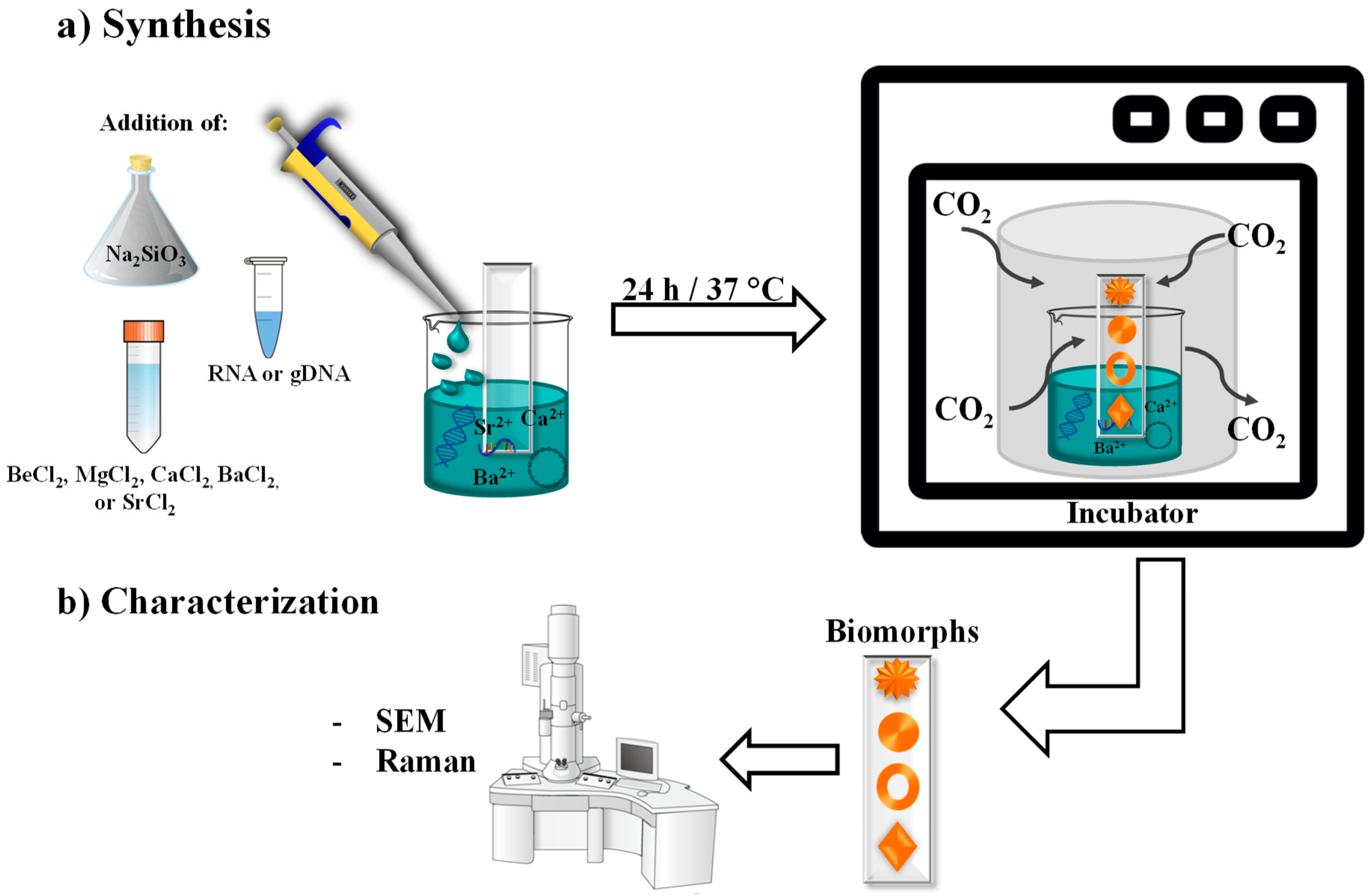
2. Materials and Methods

2.1. Strains Used for Nucleic Acids Extraction

Extraction of nucleic acids (genomic DNA and RNA) was performed from a liquid culture of Candida albicans (Department of Microbiology, ENCB-IPN, Mexico City, Mexico). The culture medium used was YPD (10 g/L yeast extract, 20 g/L peptone, and 20 g/L glucose).
The bacterial strain Escherichia coli JM109 was used to obtain plasmid DNA. The culture medium used was Luria-Bertani (LB: 5 g/L yeast extract, 10 g/L tryptone, and 5 g/L NaCl).

2.2. Extraction of Genomic DNA

The protocol used to extract the genomic DNA was in general as follows; C. albicans cells were suspended in 600 μL of a lysis buffer, containing 7 M urea, 50 mM Tris-HCl, pH 8.0, 1% N-lauroylsarcosine, 20 mM EDTA, and 0.35 M NaCl. The mixture was left to rest for 30 min at room temperature. Then, 600 μL of phenol-chloroform-isoamyl alcohol was added at a proportion of 25:24:1, mixed and centrifuged at 12,000× g for 10 min. Next, the aqueous phase was placed into a new assay tube, this last step was repeated until eliminating completely the organic phase. To precipitate the DNA, the clean aqueous phase was supplemented with an equal volume of isopropanol; the mixture was homogenized by inversion until observing the appearance of white strands. The homogeneous mixture was centrifuged at 14,000× g for 10 min, the supernatant was discarded, and the pellet was resuspended in 500 μL of 70% ethanol, homogenized in a vortex, and centrifuged at 12,000× g for 10 min. The supernatant was discarded, and the pellet was resuspended in sterile nuclease-free water, which contained 1 μL RNAsa (20 mg/mL) and incubated at 37 °C for 20 min to eliminate the RNA; the obtained gDNA was used in the synthesis of the biomorphs.

2.3. RNA Extraction

RNA was obtained with the Trizol technique. Briefly, the C. albicans cell pellet was washed five times with 1% diethyl pyrocarbonate (DEPC, Sigma-Aldrich, St. Louis, MO, USA). Then, cell rupture was achieved mechanically with the aid of a vortex and glass pearls. After cell lysis, Trizol and 200 μL of chloroform were added to the sample to allow for the completed dissociation of the nucleic-protein complex, the sample was incubated for 2–3 min at room temperature. The mixture was centrifuged at 12,000× g for 15 min at 4 °C, and the aqueous phase (which contains the RNA) was recovered and transferred to a new assay tube. The recovered aqueous phase was supplemented with 500 μL of cold isopropyl alcohol, shaken gently, and incubated for 10 min at room temperature. Then, the sample was centrifuged at 12,000× g for 10 min at 4 °C, the isopropanol was removed, adding immediately 1 mL of cold 75% ethanol, molecular biology grade, and centrifuging at 7500× g for 5 min at 4 °C. The ethanol was discarded, and the pellet was resuspended in DEPC water. To eliminate the DNA, the RNA samples were treated with DNAse (Invitrogen). The concentration of total RNA was determined through Nano Drop 2000/2000c (Thermo Scientific) at 260 nm. The obtained RNA was used to form the biomorphs.

2.4. Biomorphs Formation

Biomorphs were synthesized in different mixture of chemical alkaline earth elements: beryllium (Be), magnesium (Mg), calcium (Ca), barium (Ba), and strontium (Sr). In all the different combinations of mixtures, a 1:1 relation was maintained of each of the elements. Procurement of biomorphs was achieved through the gas diffusion method [17]. Synthesis was performed on glass squares of 1 mm thickness; the size of the glass square was of 5 mm × 5 mm height and width. The glass square was placed in a crystallization chamber, supplemented with 1000 ppm of sodium metasilicate, beryllium, magnesium, calcium, barium, or strontium chloride at a final concentration of 20 mM, 0.1 ng genomic DNA, plasmid DNA, or RNA, at pH 11.0, adjusted with sodium hydroxide (Figure 1). Table 1 summarizes the different assessed combinations. All reagents were from Sigma-Aldrich. Experiments were performed at 37 °C. Biomorphs formation was performed during 24 h. Experiments were repeated in triplicate.

2.5. Characterization of Biomorphs

Characterization of biomorphs was accomplished first by observing their morphology through SEM, using a model VEGA3 microscope (TESCAN, Brno, Czech Republic). Then, biomorphs were characterized through Raman spectroscopy (Figure 1). Raman measurements were performed with a confocal Raman microscope, WITec alpha300 R system, at room temperature. The Raman laser excitation wavelength was 532 nm and the spectral resolution of the spectrometer is 1.0 cm−1. Raman spectra were recorded with a WITec Alpha 300 Series Raman-AFM (WITec GmbH, Ulm, Germany) using a 672 lines/mm grating with 100x Zeiss objective (0.9 NA). A Nb:YVO4 green laser with wavelength of 532 nm was used as an excitations source with 14.4 mW of laser power. Punctual Raman spectra had 0.5 s of integration time and 10 accumulations and map Raman with 0.03 s of integration time.

3. Results and Discussion

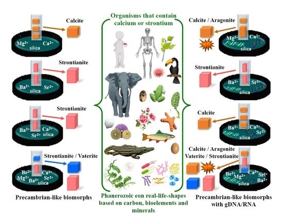
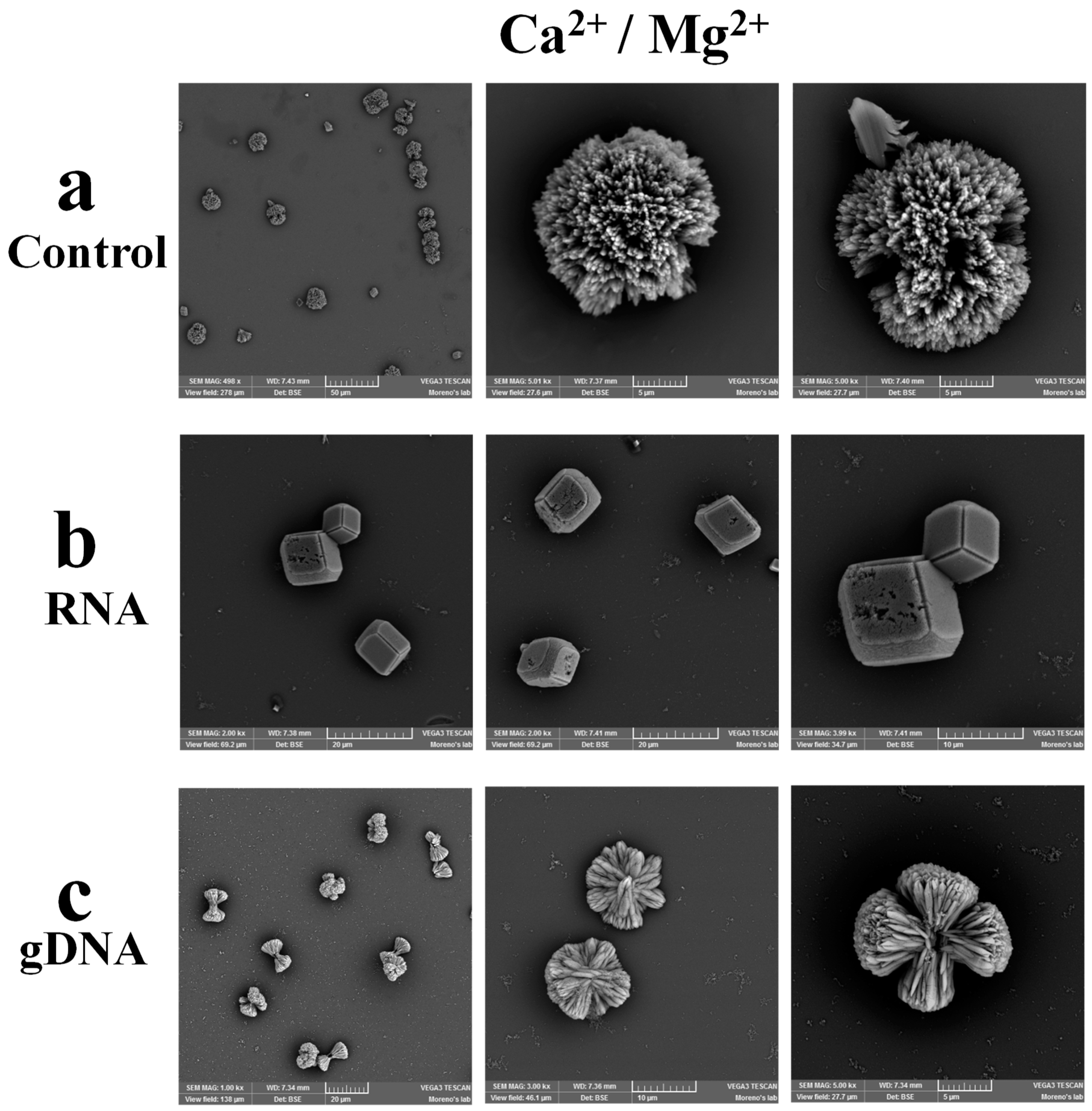
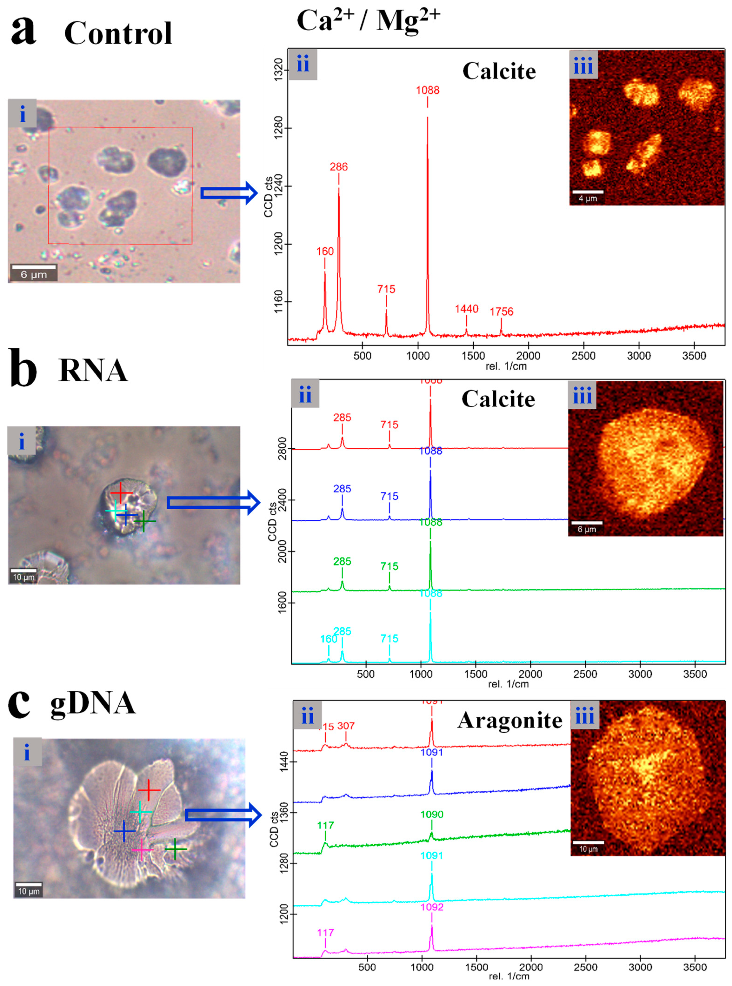
3.1. Composition of Biomorphs Depends on the Nature of the Chemical Elements, Whereas the Shape Depends on the Biomolecules

To contribute to understanding why some organisms have conserved certain chemical elements over others along the different geological eras, e.g., calcium is part of some bony structures, and in an attempt to elucidate how the first biomolecules (RNA and DNA), reported to have appeared in the Precambrian era in the presence of certain chemical elements, influenced the formation of certain structures as are the biomorphs, we evaluated the formation of these silica-carbonates biomorphs in different combinations. The first assessed cocktail combination was formed by Ca2+ and Mg2+ at a 1:1 proportion, in the presence or absence of RNA or gDNA. We chose these chemical elements because, aside from being alkaline earth metals, they are important constituents of current organisms. Calcium is the most abundant mineral in the human body, while magnesium is necessary for more than 300 biochemical reactions in the human body. As seen in Figure 2a, the biomorphs synthesized with calcium and magnesium presented a spherical morphology, but which also resembles a flower.
Biomorphs obtained in the presence of Ca2+, Mg2+, and RNA (Figure 2b) showed a rhombohedral morphology, in contrast to the morphology obtained without the nucleic acids (Figure 2a). This result is relevant because it indicates that biomolecules, such as RNA and gDNA, arrange the chemical elements to adopt a specific crystalline structure that is common when calcium is the sole chemical element forming the biomorph. This datum indicates that in the presence of the elements Ca2+ and Mg2+, the interaction of the nucleic acids with these elements favors calcium over magnesium, since the crystalline form found is common to Ca2+ (Figure 2b) with RNA, but different with gDNA (Figure 2c). These findings agree with previous results from our research team [14] and other groups that have shown that certain biomolecules, as are some peptides, modify the crystal habit of calcite [18]. Notwithstanding, although apparently both the gDNA and RNA direct the synthesis of the silica-carbonate biomorphs, it is necessary to know the chemical composition of the synthesized biomorphs, and to elucidate if a biomorph constituted by calcium and magnesium had been formed or rather one of these elements dominated the synthesis of the biomorph. To determine both the chemical composition and the crystalline phase of the obtained silica-carbonate biomorphs, Raman spectroscopy was used because it is a technique that enables to determine the polymorphs of crystalline compounds [19].
In the Raman spectrum for the biomorphs synthesized with Ca2+ and Mg2+, bands at 160, 288, 715, and 1088 cm−1 were identified (Figure 3a). These peaks correspond to calcium carbonate (CaCO3), specifically to the calcite polymorph with a trigonal structure, as reported by other authors [20,21]. In the analysis of biomorphs synthesized with the mixture of different ions/biomolecule made of Ca2+, Mg2+, and RNA (Figure 3b), spectra at different points of the biomorph were obtained to identify whether the chemical composition was the same or different in the whole structure of the biomorph. Punctual spectra presented vibrations at 160, 285, 715, and 1088 cm−1 (Figure 3b), which correspond to the calcite one polymorph of CaCO3. Whereas in the combination of Ca2+, Mg2+, and gDNA, the bands of the synthesized biomorphs were identified at 115, 117, 307, 1090, 1091, and 1092 cm−1 (Figure 3c). These vibrations correspond to the polymorph aragonite. These data are relevant because they show that calcium is the dominating element in the formation of the biomorphs over magnesium. Possibly, this event dominated in the organisms, and therefore their hard structures are formed by calcium and not magnesium. In this way, calcium in the form of CaCO3 is found as part of marine sponges, otoliths of all vertebrates, egg shells, seashells, bones, teeth, and nails [22,23,24,25,26]. Structures of organisms can present one or several of the three CaCO3 polymorphs (aragonite, calcite, and vaterite) [27,28]. For example, calcite and aragonite are the main phases found in marine organisms [29,30], whereas vaterite is found in few organisms [31,32]. The mechanisms by which some organisms synthesize preferentially one or another CaCO3 polymorph has not been elucidated yet; however, some authors refer that CaCO3 is initially precipitated as amorphous calcium carbonate to be later modified into stable polymorphs, as occurs in pearls that are formed by vaterite and aragonite [33,34]. It has also been reported that the formation of one or another polymorph depends on the environment; for example, it has been shown that vaterite can be generated in shells that are generally aragonitic but when found in fresh water change from aragonite to vaterite [35]. Our results agree with the above-cited information, because, even in the presence of magnesium, the prevalent polymorphs are calcite and aragonite (Figure 3). The prevalence of these polymorphs in the different structures of organisms is possibly because they have similar crystalline structures, in which the calcium ions are located in the same positions of the lattice layers (001), alternating with layers of carbonate ions [36]. In addition, the presence of Mg2+ favors the polymorph aragonite [36], as found in our mixture of different ions in the presence of gDNA (Figure 3c). As a whole, these data indicate that the composition of the organisms’ structures are controlled by the nature of the chemical elements, whereas the shape of the structures are controlled by biomolecules, as are RNA and gDNA, as shown in previous works [14].

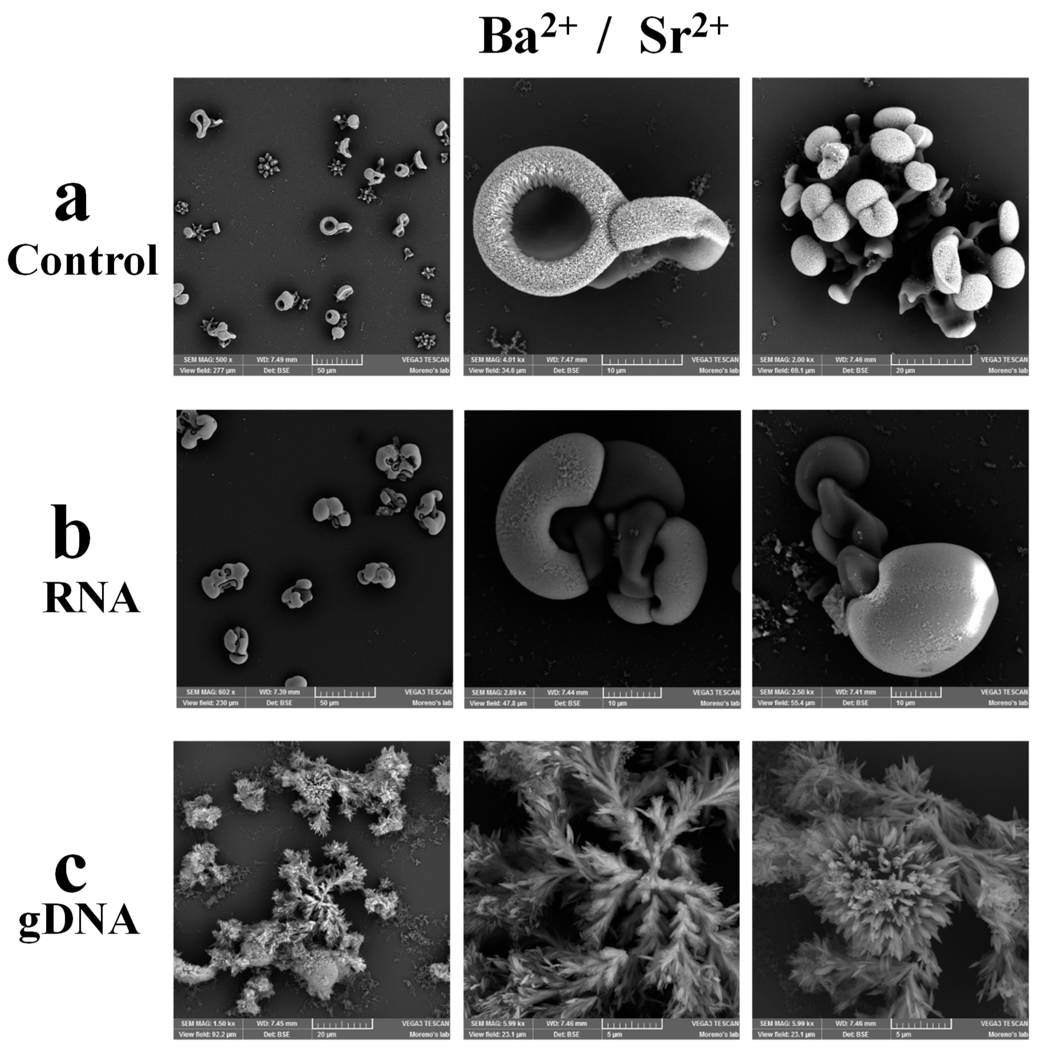
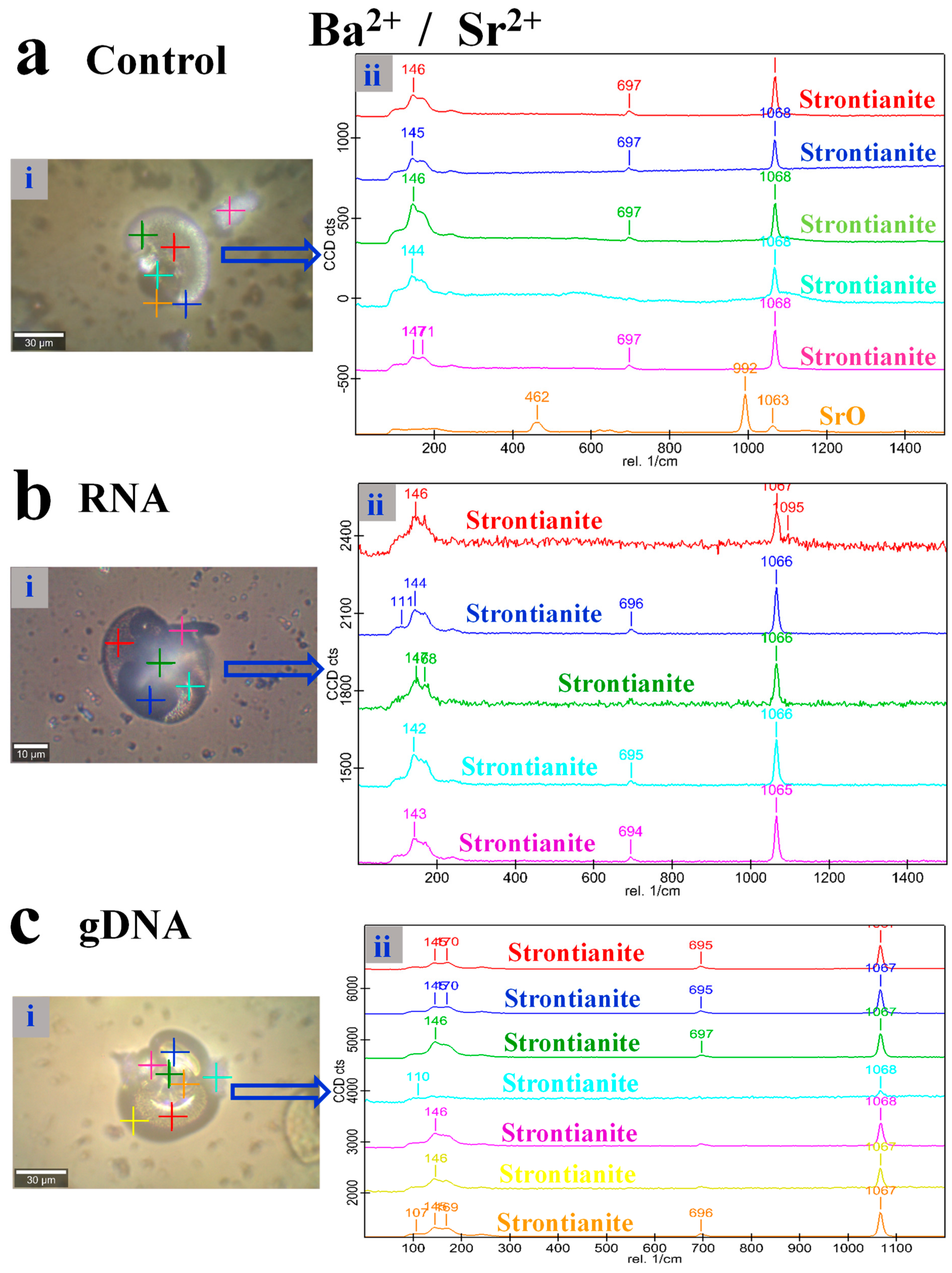
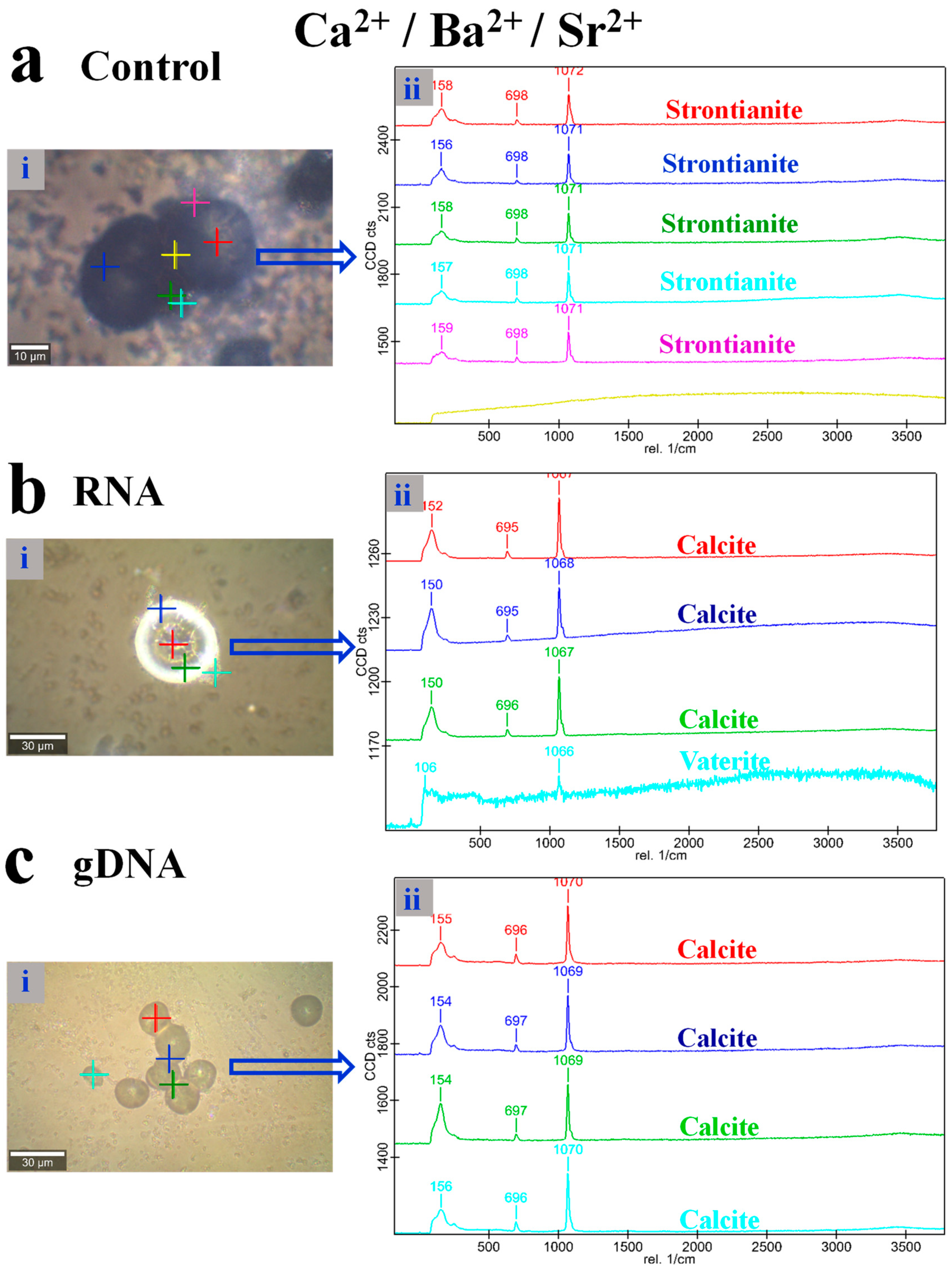
3.2. Strontium is the Dominant Element in the Biomorphs Synthesized in a Combination of Ba2+ and Sr2+

Other relevant elements to be analyzed are Ba2+ and Sr2+, because they have been conserved since ancient times until our current days. In this sense, strontium has been found in approximately 744 marine carbonates from the Phanerozoic era [37]. Currently, there are still organisms that contain strontium like algae, invertebrate organisms like Polyzoa, Crustacea, and Echinodermata; Ba2+ has also been found in algae, invertebrate species, and in fish of the Irish sea [38]. Based on the above information, we considered important to analyze, in a Ba2+ and Sr2+ mixture, which of the two is the predominant one in the formed biomorphs. For this, silica-carbonate biomorphs were obtained in the presence of a mixture of different ions made of Ba2+ and Sr2+ at a 1:1 proportion with or without RNA or gDNA. Biomorphs synthesized in the presence of only Ba2+ and Sr2+ were visualized through SEM. As seen in Figure 4a, biomorphs presented different morphologies that mimic flowers and bunches of flowers. The silica-carbonates obtained with the metallic combination of Ba2+, Sr2+, and RNA showed a morphology of circular and round structures linked through helix-type strands (Figure 4b). Interestingly, in these biomorphs a brilliant and an opaque part were observed, suggesting a possible different composition, as they were formed by a combination of two chemical elements and RNA. Some of the morphologies found in the obtained biomorphs have been previously described by other authors, such as cardioid structures (cardioid leaves), helicoidal forms (helicoidal filaments), funnel-like aggregates (circular and conic), worm form, braided structure, curly forms, framboidal forms, and fern-like forms [39]. The composition of the obtained structures was confirmed through Raman spectroscopy. In the biomorphs generated by the combination of Ba2+, Sr2+, and gDNA, morphologies similar to stems, leaves, and flowers were observed (Figure 4c).
Determination of the chemical composition as well as the crystalline phase of the silica-carbonate biomorphs generated with these chemical elements was performed through Raman spectroscopy. For this, multiple punctual spectra were obtained.
In five of the punctual spectra, corresponding to the sample with Ba2+ and Sr2+, vibrations at 144–147, 697, and 1068 cm−1 were identified (Figure 5a), which are characteristic of SrCO3 and correspond to the strontianite [40]. Also, in these biomorphs, one of the punctual spectra presented bands at 462, 902, and 1063 cm−1. These vibrations correspond to SrO [41]. Because the bands identified as of strontianite in the punctual spectra could be confused with BaCO3, we decided to perform a mapping of the complete biomorph and obtain a global spectrum, in which all bands could be distinguished and, thereby, confirm whether punctual spectra assignment had been correct. The spectrum corresponding to the mapping of the complete biomorph showed vibrations at 108, 146, 172, 242, 699, and 1067 cm−1 (Figure S1a in Supplementary Materials). These bands indeed correspond to strontianite because BaCO3 presents bands corresponding to the lattice vibration at 136, 155, 180, and 225, as well as two bands of internal vibrations at 691 and 1059 cm−1 [42]. In the biomorphs synthesized in the combination of Ba2+, Sr2+, and RNA (Figure 5b), as well as with Ba2+, Sr2+, and gDNA (Figure 5c), bands at 110, 111, 142–147, 694–697, 1065–1068, and 1095 cm−1 were identified, these vibrations correspond to the strontianite (SrCO3). In addition, as in the biomorphs obtained in the absence of RNA or DNA, a spectrum of complete biomorph mapping was also obtained from these biomorphs, to ensure their correct assignment. As shown in Figure S1b,c, spectra revealed the peaks corresponding to strontianite. This result is relevant and agrees with other previous works reporting that in several organisms like some algal species, invertebrates, and fishes, calcium and strontium have been identified, whereas barium has only been found in some cases [38]. These data indicate that although barium and strontium are similar chemical elements [43], strontium is the dominant element with respect to barium. Possibly, over time, this preference of strontium over barium gave these organisms an advantage over those organisms in which barium is part of their metabolism. The finding that the morphology is different for the three analyzed combinations (Figure 4), but their chemical composition is the same (Figure 5 and Figure S1), indicates that the arrangement of the crystalline networks of the obtained biomorphs depends on the nature of the elements, but the morphology depends on the nucleic acids, RNA and gDNA; the same was found for the Ca2+ and Mg2+ mixture (Figure 2 and Figure 3).

3.3. In the Presence of Nucleic Acids, Calcium is the Element that Controls the Synthesis of Biomorphs

Because barium and strontium present chemical similarity with calcium [44], we were interested in knowing which of these elements is the one to control the biomorphs synthesis in a mixture of different ions in which the three cations were present in the presence or absence of RNA and gDNA. In the silica-carbonate biomorphs obtained in the presence of only Ca2+, Ba2+, and Sr2+, SEM mainly revealed structures resembling stems and flowers and a minimal amount of rhombus-type morphologies (Figure 6a), whereas in the combinations of the three elements with RNA (Figure 6b) or gDNA (Figure 6c), the morphology was spherical. For these biomorphs, as done for the previous combinations, their chemical composition was also determined, as well as their crystalline structure through punctual spectra obtained with Raman spectroscopy; this type of spectra allow identifying the chemical composition in different points of the biomorph.
The punctual spectra of the sample obtained with the three elements without nucleic acid showed vibrations at 156–158, 698, and 1071–1072 cm−1 (Figure 7a). However, determining the compound to which these vibrations corresponded was no easy task, as there was the possibility of dealing with a calcium, barium, or strontium compound, or a mixture of two or three elements.
Faced with this complexity, we analyzed each vibration, and were able to determine that the formed compound was the strontianite (SrCO3) (Figure 7a), since none of the vibrations corresponded to a calcium or barium compound. Additionally, to confirm that the assignment had been correct, a global mapping was performed of the obtained biomorphs. The spectrum of the global mapping reveled peaks at 165, 255, 699, and 1070 cm−1 (Figure S2a), which correspond to strontianite, confirming that, indeed, the biomorphs produced in the presence of Ca2+, Ba2+, and Sr2+ are composed of the strontianite. This result is important, because it indicates that strontium controls the synthesis of biomorphs in the presence of Ca2+, Ba2+, and Sr2+ (Figure 7a and Figure S2a). This is probably related with the fact that several of the primitive organisms that have been conserved since the Precambrian era until now, conserved strontium as an essential element participating in their metabolism, as this possibly granted them an advantage allowing them to survive over time [5,6,7]. These primitive organisms, probably, incorporated strontium in their metabolism as it was available in the Precambrian era and, along the years, conserved it because they keep obtaining it from their habitat. For example, it has been reported that strontium can be found in most soils [45], wetlands, and aquatic environments [44]. The biomorphs synthesized in the presence of Ca2+, Ba2+, Sr2+, and RNA (Figure 6b) or gDNA (Figure 6c) presented spherical morphology. When determining their chemical composition, the punctual spectra showed bands at 150–156, 695–696, and 1066–1070 cm−1 (Figure 7b,c). These vibrations correspond to the polymorph calcite of CaCO3 for three punctual spectra of the biomorphs with RNA (Figure 7b), and for all punctual spectra of the sample with gDNA (Figure 6c). Additionally, in the case of biomorphs formed with RNA, one punctual spectrum corresponded to the polymorph vaterite of CaCO3 (Figure 7b). To confirm that the biomorphs synthesized in these mixtures corresponded mainly to calcite, global mappings of the biomorph were performed (Figure S2b,c). In the global mappings, peaks at 153–155, 697–696, and 1067–1070 cm−1 were identified, these vibrations indeed correspond to the polymorph calcite (Figure S2b,c). These results are very interesting, because in the presence of any of the nucleic acids (RNA or gDNA), calcium is the element that controls the synthesis of biomorphs over strontium and barium (Figure 7b,c and Figure S2b,c), which does not occur in their absence (Figure 7a and Figure S2a). This information contributes to the knowledge that there were organisms in which calcium already participated in their metabolism in the presence of RNA (which has been postulated as the first biomolecule that appeared in the Precambrian era [9,46]); this is probably because nucleic acids select the element that provides the best thermodynamic stability and viability for the organisms, ensuring survival of species along millions of years [46]. In this sense, it has been reported that the polymorph calcite is thermodynamically more stable than aragonite and vaterite [19,36]. Notwithstanding, the identification of vaterite (Figure 7b) is an interesting result, because it has been reported that this polymorph can adopt multiple structures [28,47,48,49]. Therefore, being calcite the most stable polymorph, RNA and gDNA induce mainly the formation of this polymorph in the different structures of organisms, ensuring in this way the biological perpetuation of a species. As indicated previously, the calcite polymorph is part of echinoderms, sea urchins, ophiuroids, sea cucumbers, crinoids; arthropods like trilobites; mollusks like bivalves, gastropods, polyplacophorans, ammonoids, or nautiloids; calcareous seashells; and spicules of sponges, algae, eggshells, teeth, bones, and otoliths [23,24,25,50,51]. Calcium, besides forming part of the support structures of organisms, is also very important for cellular metabolism. For example, it plays a role as second messenger in eukaryote cells, as it produces the largest number of intracellular signals with physiological and pathological implications. In addition, calcium is involved in neurotransmission, muscle contraction, control of ion channels, gene expression, and cell death [52,53,54]. In plants, calcium is required in structures like the cell wall and membranes, it also activates and regulates cell division and cell enlargement and influences the specialization of cell organs [55]. Hence, calcium is essential for root, fruits, and leaf differentiation, and, in consequence, in the absence of calcium there will be no new leaves or flowers in a plant. In bacteria, calcium is needed at a relatively high concentration as it participates in maintaining the cellular structure, motility, cell differentiation processes; besides, it has been suggested that there is also calcium-dependent signals transduction like in eukaryote cells [56]. All together, these data demonstrate that in the presence of nucleic acids, in a combination where more than two chemical elements coexist, the chemical element with the best stability and viability for the cell will always be chosen; this element is clearly calcium, which can be considered an evolutionary achievement that developed from the first cells in the Precambrian era and has been conserved until our current times. Our data provide support to this fact, because life as we know it would not be possible without calcium.

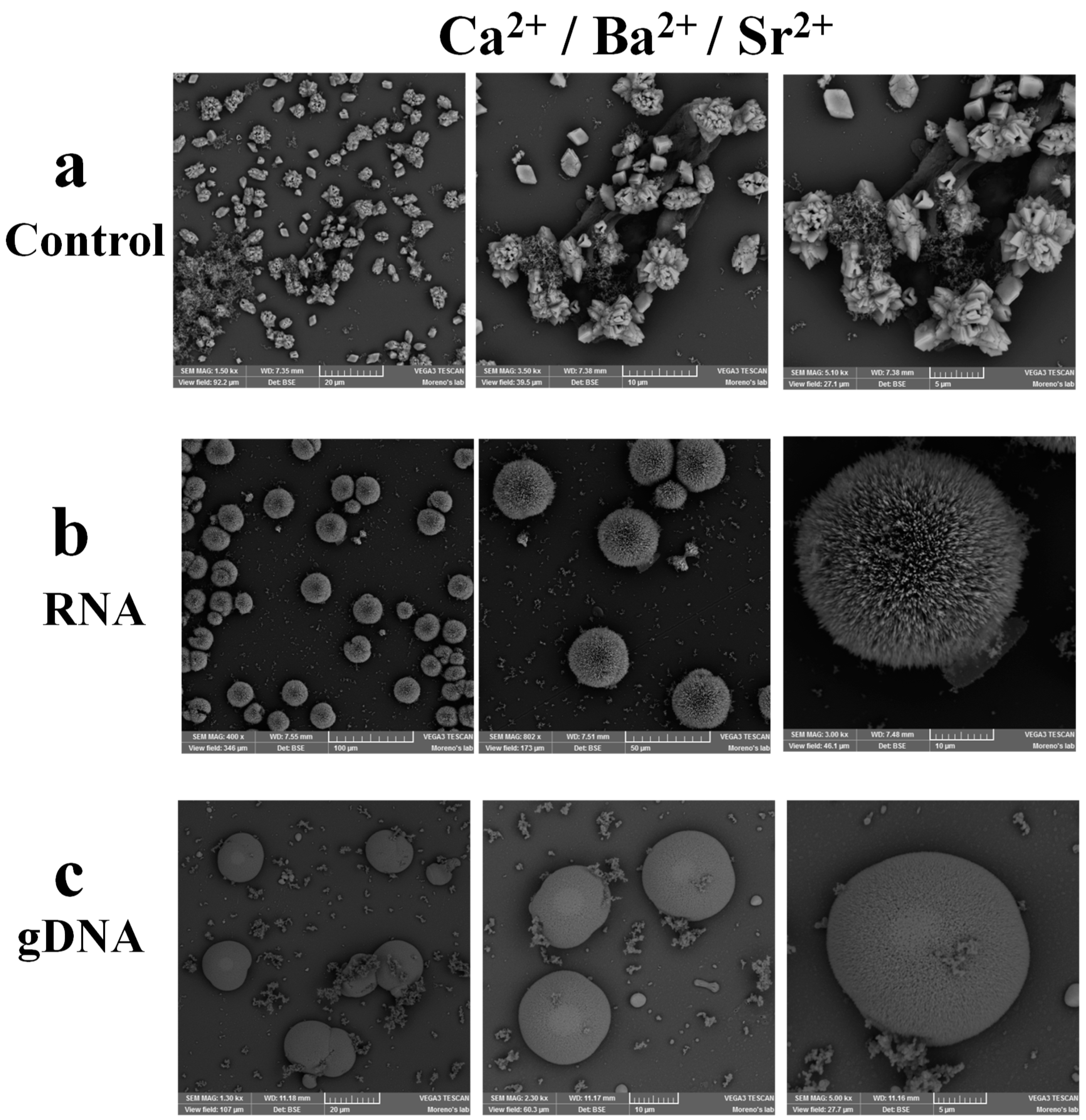
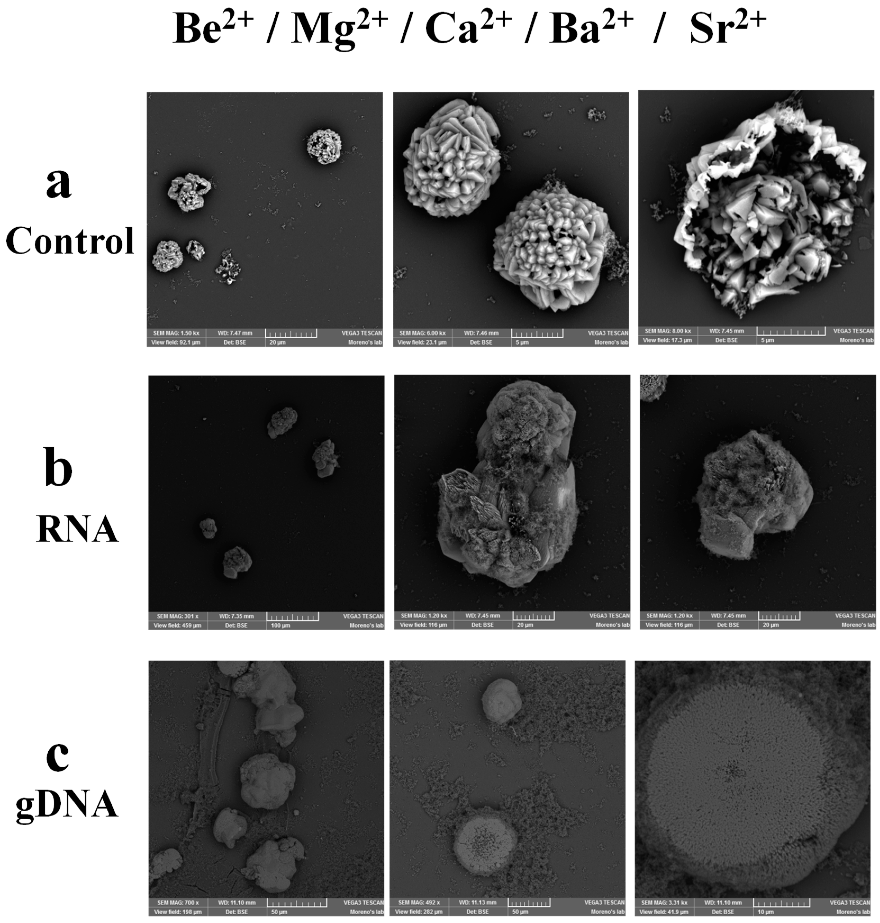
3.4. Calcium and Strontium in the Presence of Nucleic Acids are Part of Solid Structures Obtained from a Mixture with Multiple Chemical Elements

Although it had been evaluated before that calcium is the element regulating the synthesis of biomorphs in the presence of Ca2+, Ba2+, Sr2+, and nucleic acids, we also wanted to know what would happen in the synthesis of biomorphs in a mixture of different ions containing five alkaline earth elements: Be2+, Mg2+, Ca2+, Ba2+, and Sr2+, in the absence or presence of RNA and gDNA. The first assessed sample was constituted by only Be2+, Mg2+, Ca2+, Ba2+, and Sr2+, at an equimolecular relation. SEM observation of the formed biomorphs revealed a druse morphology (Figure 8a).
To determine the chemical composition of the obtained biomorphs, multiple punctual spectra were performed through Raman spectroscopy. Four of the punctual spectra showed peaks at 153–154, 699, and 1070–1074 cm−1 (Figure 9a and Figure S3a), these vibrations correspond to the strontianite (SrCO3).
Whereas the other four punctual spectra presented bands at 103–106, 108, 155, and 1072 cm−1 (Figure 9a) correspond to the polymorph vaterite of CaCO3. These results indicate that, in the presence of five elements, calcium and strontium are the ones that regulate the synthesis of biomorphs. Apparently, the presence of Be2+ and Mg2+ induces an effect so that both Ca2+ and Sr2+ become part of the formed biomorph. When Ca2+, Ba2+, and Sr2+ are present, strontium is the element that forms part of the biomorph (Figure 7a and Figure S2a). This result could be related to the fact that in nature there are species that contain calcium and strontium [38]. Whereas SEM observations of the metallic combinations that included Be2+, Mg2+, Ca2+, Ba2+, Sr2+, and RNA (Figure 8b) or gDNA (Figure 8c) revealed that no biomorphs were formed, but rather solid structures that mimic bones (Figure 8b), and a flower (Figure 8c). These structures are not hollow; therefore, these structures would not be fulfilling this important characteristic of biomorphs, hence they cannot be considered biomorphs. However, the formed structures became of interest because they were formed in a mixture of different ions containing five chemical elements and nucleic acids. Besides, these solid structures could be considered to be emulating the first structures formed in the Precambrian era, by the existence of the first biomolecule, i.e., RNA. These structures could have been the predecessors of the first cell formed in the Precambrian. Therefore, it was also important to determine the chemical composition of these solid structures through Raman spectroscopy. The multiple punctual spectra of the structure formed with Be2+, Mg2+, Ca2+, Ba2+, Sr2+, and RNA (Figure 8a) showed that it was composed of calcite, aragonite, and vaterite polymorphs of CaCO3, as well as the strontianite (SrCO3) (Figure 9b and Figure S3b). Whereas for the structure formed with Be2+, Mg2+, Ca2+, Ba2+, Sr2+, and gDNA (Figure 8b), the multiple punctual spectra showed vibrations corresponding to vaterite and aragonite polymorphs of CaCO3 and strontianite, and one punctual spectrum also corresponding to SrO (Figure 9c and Figure S3c). These results obtained with the different combinations, as shown in Table 2, indicate the availability of several elements to form a new structure in the presence of nucleic acids. Both the RNA and gDNA can interact with the different chemical elements, but possibly those elements with which they form the most stable interactions will be the elements that will specifically form part of organisms; hence, calcium is the most stable element to form the structure followed by strontium, as shown in this work.
This information can be correlated with other works that have described that both calcium and strontium are elements identified mainly in 15 algal species, 45 invertebrate species, and six fish species in the Iris sea [38].

4. Conclusions

Based on our data, we can infer that the stability presented by calcium, strontium, and certain biomolecules such as nucleic acids could have allowed conserving the first cells and primitive organisms generated in the Precambrian era to our present days and, on the other side, more complex life forms have evolved from relatively simple cells, produced since millions of years ago, such as the higher organisms but conserving calcium and, in some cases, strontium in their structures.

Supplementary Materials

The following are available online at https://www.mdpi.com/2073-4352/9/8/381/s1, Figure S1: Raman spectrum corresponding to the global mapping of biomorphs obtained with the combinations: (a) Ba2+/Sr2+, (b) Ba2+/Sr2+/RNA, and (c) Ba2+/Sr2+/gDNA. Figure S2: Raman spectrum corresponding to the global mapping of biomorphs obtained with (a) Ca2+/Ba2+/Sr2+, (b) Ca2+/Ba2+/Sr2+/RNA, and (c) Ca2+/Ba2+/Sr2+/gDNA. Figure S3: Raman spectrum corresponding to the global mapping of biomorphs obtained with (a) Be2+/Mg2+/Ca2+/Ba2+/Sr2+, (b) Be2+/Mg2+/Ca2+/Ba2+/Sr2+/RNA, and (c) Be2+/Mg2+/Ca2+/Ba2+/Sr2+/gDNA.

Author Contributions

Conceptualization, methodology, software, validation, formal analysis, investigation, writing—original draft preparation, and writing—review and editing: M.C.-C. and A.M.

Funding

This research was financed by DGAPA-UNAM project PAPIIT IG200218 (A.M.), and Secretaría de Educación Pública for PRODEP Sabbatical leave support (Oficio No. 511-6/18-5929).

Acknowledgments

Mayra Cuéllar-Cruz acknowledges the sabbatical leave support from SEP-PRODEP (Oficio No. 511-6/18-5929). We would like to thank Dra. Estela Ruiz-Baca for supplying gDNA, pDNA, and RNA. A. Moreno acknowledges to DGAPA-UNAM project PAPIIT IG200218 for the partial support of this project. Authors acknowledge to Dra. Selene Islas for Raman assistance. The authors thank the Laboratorio Universitario de Caracterización Espectroscópica, LUCE-ICAT-UNAM, for the Raman spectroscopic characterization of the samples. The authors acknowledge to Mtra. Ingrid Mascher for the English edition of this contribution.

Conflicts of Interest

The authors declare no conflict of interest.

References

  1. Oparin, A.I. Modern Views on the Origin of Life. Scientia. 1960, 95, 322–326. [Google Scholar]
  2. Cech, T.R.; Grabowski, P.J.; Zaug, A.J. Splicing of the Ribosomal-RNA Precursor and Cyclization of the Excised Intervening Sequence Rna in Nuclei of Tetrahymena. Fed. Proc. 1981, 40, 1651. [Google Scholar]
  3. Cech, T.R. Rna as an Enzyme. Biochem. Int. 1989, 18, 7–14. [Google Scholar] [CrossRef]
  4. Larsen, T.; Sandstrom, B. Effect of Calcium, Copper, and Zinc Levels in a Rapeseed Meal Diet on Mineral and Trace-Element Utilization in the Rat. Biol. Trace. Elem. Res. 1992, 35, 167–184. [Google Scholar] [CrossRef]
  5. Williams, R.J.P.; Fraústo da Silva, J.J.R. Bringing Chemistry to Life: From Matter to Man; Oxford University Press: New York, NY, USA, 1999; pp. 293–298. [Google Scholar]
  6. Fraústo da Silva, J.J.R.; Williams, R.J.P. The Biological Chemistry of the Elements: The Inorganic Chemistry of Life, 2nd ed.; Oxford University Press: New York, NY, USA, 1991; pp. 7–10. [Google Scholar]
  7. Turner, D.R.; Whitfield, M.; Dickson, A.G. The Equilibrium Speciation of Dissolved Components in Fresh-Water and Seawater at 25-Degrees-C and 1 Atm Pressure. Geochim. Cosmochim. Ac 1981, 45, 855–881. [Google Scholar] [CrossRef]
  8. Schopf, J.W. Microfossils of the Early Archean Apex Chert-New Evidence of the Antiquity of Life. Science 1993, 260, 640–646. [Google Scholar] [CrossRef]
  9. Pearce, B.K.D.; Pudritz, R.E.; Semenov, D.A.; Henning, T.K. Origin of the RNA world: The fate of nucleobases in warm little ponds. Proc. Natl. Acad. Sci. USA 2017, 114, 11327–11332. [Google Scholar] [CrossRef] [Green Version]
  10. Zhang, G.; Morales, J.; Garcia-Ruiz, J.M. Growth behaviour of silica/carbonate nanocrystalline composites of calcite and aragonite. J. Mater. Chem. B 2017, 5, 1658–1663. [Google Scholar] [CrossRef] [Green Version]
  11. Garcia-Ruiz, J.M.; Hyde, S.T.; Carnerup, A.M.; Christy, A.G.; Van Kranendonk, M.J.; Welham, N.J. Self-assembled silica-carbonate structures and detection of ancient microfossils. Science 2003, 302, 1194–1197. [Google Scholar] [CrossRef]
  12. Opel, J.; Wimmer, F.P.; Kellermeier, M.; Colfen, H. Functionalisation of silica-carbonate biomorphs. Nanoscale Horiz. 2016, 1, 144–149. [Google Scholar] [CrossRef]
  13. Eiblmeier, J.; Dankesreiter, S.; Ptitzner, A.; Schmalz, G.; Kunz, W.; Kellermeier, M. Crystallization of mixed alkaline-earth carbonates in silica solutions at high pH. Cryst. Growth Des. 2014, 14, 6177–6188. [Google Scholar] [CrossRef]
  14. Cuéllar-Cruz, M.; Islas, S.; González, G.; Moreno, A. The influence of nucleic acids on the synthesis of crystalline Ca(II), Ba(II) and Sr(II) silica-carbonate biomorphs: Implications to the chemical origin of life on primitive Earth. Cryst. Growth Des. 2019. Available online: https://pubs.acs.org/doi/10.1021/acs.cgd.9b00573. (accessed on 23 July 2019).
  15. Nakouzi, E.; Steinbock, O. Self-organization in precipitation reactions far from the equilibrium. Sci. Adv. 2016, 2, e160144. [Google Scholar] [CrossRef]
  16. Garcia-Ruiz, J.M.; Melero-Garcia, E.; Hyde, S.T. Morphogenesis of Self-Assembled Nanocrystalline Materials of Barium Carbonate and Silica. Science 2009, 323, 362–365. [Google Scholar] [CrossRef] [Green Version]
  17. Noorduin, W.L.; Grinthal, A.; Mahadevan, L.; Aizenberg, J. Rationally Designed Complex, Hierarchical Microarchitectures. Science 2013, 340, 832–837. [Google Scholar] [CrossRef] [Green Version]
  18. DeOliveira, D.B.; Laursen, R.A. Control of calcite crystal morphology by a peptide designed to bind to a specific surface. J. Am. Chem. Soc. 1997, 119, 10627–10631. [Google Scholar] [CrossRef]
  19. Carteret, C.; Dandeu, A.; Moussaoui, S.; Muhr, H.; Humbert, B.; Plasari, E. Polymorphism Studied by Lattice Phonon Raman Spectroscopy and Statistical Mixture Analysis Method. Application to Calcium Carbonate Polymorphs during Batch Crystallization. Cryst. Growth Des. 2009, 9, 807–812. [Google Scholar] [CrossRef]
  20. Vagenas, N.V.; Gatsouli, A.; Kontoyannis, C.G. Quantitative analysis of synthetic calcium carbonate polymorphs using FT-IR spectroscopy. Talanta 2003, 59, 831–836. [Google Scholar] [CrossRef]
  21. Agarwal, P.; Berglund, K.A. In situ monitoring of calcium carbonate polymorphs during batch crystallization in the presence of polymeric additives using Raman spectroscopy. Cryst. Growth Des. 2003, 3, 941–946. [Google Scholar] [CrossRef]
  22. Cuellar-Cruz, M. Synthesis of inorganic and organic crystals mediated by proteins in different biological organisms. A mechanism of biomineralization conserved throughout evolution in all living species. Prog. Cryst. Growth Charact. Mater. 2017, 63, 94–103. [Google Scholar] [CrossRef]
  23. Ruiz-Arellano, R.R.; Moreno, A. Obtainment of Spherical-Shaped Calcite Crystals Induced by Intramineral Proteins Isolated from Eggshells of Ostrich and Emu. Cryst. Growth Des. 2014, 14, 5137–5143. [Google Scholar] [CrossRef]
  24. Ruiz-Arellano, R.R.; Medrano, F.J.; Moreno, A.; Romero, A. Structure of struthiocalcin-1, an intramineral protein from Struthio camelus eggshell, in two crystal forms. Acta Crystallogr. D: Biol. Crystallogr. 2015, 71, 809–818. [Google Scholar] [CrossRef] [PubMed]
  25. Colfen, H. Biomineralization a crystal-clear view. Nat. Mater. 2010, 9, 960–961. [Google Scholar] [CrossRef] [PubMed]
  26. Xiang, L.; Su, J.T.; Zheng, G.L.; Liang, J.; Zhang, G.Y.; Wang, H.Z.; Xie, L.P.; Zhang, R.Q. Patterns of Expression in the Matrix Proteins Responsible for Nucleation and Growth of Aragonite Crystals in Flat Pearls of Pinctada fucata. PloS One 2013, 8, e66564. [Google Scholar] [CrossRef]
  27. Rudel, H.; Muller, J.; Jurling, H.; Bartel-Steinbach, M.; Koschorreck, J. Survey of patterns, levels, and trends of perfluorinated compounds in aquatic organisms and bird eggs from representative German ecosystems. Environ. Sci. Pollut. R 2011, 18, 1457–1470. [Google Scholar] [CrossRef] [PubMed]
  28. Demichelis, R.; Raiteri, P.; Gale, J.D.; Dovesi, R. The Multiple Structures of Vaterite. Cryst. Growth Des. 2013, 13, 2247–2251. [Google Scholar] [CrossRef]
  29. Lowenstam, H.A. Spicular Morphology and Mineralogy in Some Pyuridae (Ascidiacea). B Mar. Sci. 1989, 45, 243–252. [Google Scholar]
  30. Watabe, N. Crystal-Growth of Calcium-Carbonate in Biological-Systems. J. Cryst. Growth 1974, 24, 116–122. [Google Scholar] [CrossRef]
  31. Beniash, E.; Aizenberg, J.; Addadi, L.; Weiner, S. Amorphous calcium carbonate transforms into calcite during sea urchin larval spicule growth. Proc. Roy. Soc. B Biol. Sci. 1997, 264, 461–465. [Google Scholar] [CrossRef]
  32. Lowenstam, H.A.; Abbott, D.P. Vaterite - Mineralization Product of Hard Tissues of a Marine Organism (Ascidiacea). Science 1975, 188, 363–365. [Google Scholar] [CrossRef]
  33. Nehrke, G.; Nouet, J. Confocal Raman microscope mapping as a tool to describe different mineral and organic phases at high spatial resolution within marine biogenic carbonates: Case study on Nerita undata (Gastropoda, Neritopsina). Biogeosci. 2011, 8, 3761–3769. [Google Scholar] [CrossRef]
  34. Soldati, A.L.; Jacob, D.E.; Wehrmeister, U.; Hofmeister, W. Structural characterization and chemical composition of aragonite and vaterite in freshwater cultured pearls. Miner. Mag. 2008, 72, 579–592. [Google Scholar] [CrossRef]
  35. Spann, N.; Harper, E.M.; Aldridge, D.C. The unusual mineral vaterite in shells of the freshwater bivalve Corbicula fluminea from the UK. Naturwissenschaften 2010, 97, 743–751. [Google Scholar] [CrossRef] [PubMed]
  36. Falini, G.; Albeck, S.; Weiner, S.; Addadi, L. Control of aragonite or calcite polymorphism by mollusk shell macromolecules. Science 1996, 271, 67–69. [Google Scholar] [CrossRef]
  37. Veizer, J.; Ala, D.; Azmy, K.; Bruckschen, P.; Buhl, D.; Bruhn, F.; Carden, G.A.F.; Diener, A.; Ebneth, S.; Godderis, Y.; et al. Sr-87/Sr-86, delta C-13 and delta O-18 evolution of Phanerozoic seawater. Chem. Geol. 1999, 161, 59–88. [Google Scholar] [CrossRef]
  38. Templeton, W.L.; Brown, V.M. The Relationship between the Concentrations of Calcium, Strontium and Strontium-90 in Wild Brown Trout, Salmo-Trutta L and the Concentrations of the Stable Elements in Some Waters of the United-Kingdom, and the Implications in Radiological Health Studies. Air Water Pollut. 1964, 8, 49–75. [Google Scholar]
  39. Rouillard, J.; García-Ruiz, J.M.; Gong, J.; van Zuilen, M.A. A morphogram for silica-witherite biomorphs and its application to microfossil identification in the early earth rock record. Geobiology 2018, 16, 279–296. [Google Scholar] [CrossRef] [Green Version]
  40. Bonales, L.J.; Munoz-Iglesias, V.; Santamaria-Perez, D.; Caceres, M.; Fernandez-Remolar, D.; Prieto-Ballesteros, O. Quantitative Raman spectroscopy as a tool to study the kinetics and formation mechanism of carbonates. Spectrochim. Acta A 2013, 116, 26–30. [Google Scholar] [CrossRef]
  41. Rieder, K.H.; Migoni, R.; Renker, B. Lattice-Dynamics of Strontium Oxide. Phys. Rev. B 1975, 12, 3374–3379. [Google Scholar] [CrossRef]
  42. Lin, C.C.; Liu, L.G. High-pressure Raman spectroscopic study of post-aragonite phase transition in witherite (BaCO3). Eur. J. Miner. 1997, 9, 785–792. [Google Scholar] [CrossRef]
  43. Bowen, H.J.M. Strontium and Barium in Sea Water and Marine Organisms. J. Mar. Biol. Assoc. U.K. 1956, 35, 451–460. [Google Scholar] [CrossRef]
  44. Niedermeier, M.; Gierlinger, N.; Lutz-Meindl, U. Biomineralization of strontium and barium contributes to detoxification in the freshwater alga Micrasterias. J. Plant. Physiol. 2018, 230, 80–91. [Google Scholar] [CrossRef] [PubMed]
  45. Isermann, K.; Glatzle, A. Discrimination of Calcium against Strontium in Plants with a Cryptating Agent. Naturwissenschaften 1975, 62, 238–239. [Google Scholar] [CrossRef]
  46. Gilbert, W. Origin of Life: The RNA World. Nature 1986, 319, 618. [Google Scholar] [CrossRef]
  47. Christy, A.G. A review of the structures of vaterite: The Impossible, the possible and the likely. Cryst. Growth Des. 2017, 17, 3567–3578. [Google Scholar] [CrossRef]
  48. Currey, J.; Taylor, J. The mechanical behaviour of some molluscan hard tissues. J. Zool. 1974, 173, 395–406. [Google Scholar] [CrossRef]
  49. Burgess, K.M.N.; Bryce, D.L. On the crystal structure of the vaterite polymorph of CaCO3: A calcium-43 solid-state NMR and computational assessment. Solid State Nucl. Mag. Res. 2015, 65, 75–83. [Google Scholar] [CrossRef] [PubMed]
  50. Mann, K. The calcified eggshell matrix proteome of a songbird, the zebra finch (Taeniopygia guttata). Proteome. Sci. 2015, 13, 29. [Google Scholar] [CrossRef] [PubMed]
  51. Reyes-Grajeda, J.P.; Moreno, A.; Romero, A. Crystal structure of ovocleidin-17, a major protein of the calcified Gallus gallus eggshell - Implications in the calcite mineral growth pattern. J. Biol. Chem. 2004, 279, 40876–40881. [Google Scholar] [CrossRef] [PubMed]
  52. Vanhoutte, P.M.; Collis, M.G.; Janssens, W.J.; Verbeuren, T.J. Calcium Dependence of Prejunctional Inhibitory Effects of Adenosine and Acetylcholine on Adrenergic Neurotransmission in Canine Saphenous Veins. Eur. J. Pharmacol. 1981, 72, 189–198. [Google Scholar] [CrossRef]
  53. Douglas, W.W.; Rubin, R.P. The Role of Calcium in Secretory Response of the Adrenal Medulla to Acetylcholine. J. Physiol. 1961, 159, 40–57. [Google Scholar] [CrossRef] [PubMed]
  54. Katz, B. Neural transmitter release: From quantal secretion to exocytosis and beyond. J. Neurocytol. 1996, 25, 677–686. [Google Scholar] [CrossRef] [PubMed]
  55. White, P.J.; Broadley, M.R. Calcium in plants. Ann. Bot. 2003, 92, 487–511. [Google Scholar] [CrossRef] [PubMed]
  56. Dominguez, D.C. Calcium signalling in bacteria. Mol. Microbiol. 2004, 54, 291–297. [Google Scholar] [CrossRef] [PubMed]
Figure 1. Synthesis and characterization of biomorphs.
Crystals 09 00381 g001
Figure 2. SEM microphotographs of the biomorphs obtained with the mixture of different ion compositions. (a) Ca2+/Mg2+, (b) Ca2+/Mg2+/RNA, and (c) Ca2+/Mg2+/gDNA.
Crystals 09 00381 g002
Figure 3. Raman punctual spectra of the biomorphs synthesized with the combinations: (a) Ca2+/Mg2+, (b) Ca2+/Mg2+/RNA, and (c) Ca2+/Mg2+/gDNA. (i) Optical image, (ii) multiple spectra, and (iii) mapping of biomorphs.
Crystals 09 00381 g003
Figure 4. SEM microphotographs of the biomorphs obtained with the following mixtures of different ions/biomolecules: (a) Ba2+/Sr2+, (b) Ba2+/Sr2+/RNA, and (c) Ba2+/Sr2+/gDNA.
Crystals 09 00381 g004
Figure 5. Raman punctual spectra of the biomorphs synthesized with the mixture of different ions/biomolecules: (a) Ba2+/Sr2+, (b) Ba2+/Sr2+/RNA, and (c) Ba2+/Sr2+/gDNA. (i) Optical image and (ii) multiple spectra.
Crystals 09 00381 g005
Figure 6. SEM microphotographs of biomorphs obtained with: (a) Ca2+/Ba2+/Sr2+, (b) Ca2+/Ba2+/Sr2+/RNA, and (c) Ca2+/Ba2+/Sr2+/gDNA.
Crystals 09 00381 g006
Figure 7. Raman multiple punctual spectra of biomorphs synthesized with (a) Ca2+/Ba2+/Sr2+, (b) Ca2+/Ba2+/Sr2+/RNA, and (c) Ca2+/Ba2+/Sr2+/gDNA. (i) Optical image and (ii) multiple spectra.
Crystals 09 00381 g007
Figure 8. SEM microphotographs of biomorphs obtained with (a) Be2+/Mg2+/Ca2+/Ba2+/Sr2+, (b) Be2+/Mg2+/Ca2+/Ba2+/Sr2+/RNA, and (c) Be2+/Mg2+/Ca2+/Ba2+/Sr2+/gDNA.
Crystals 09 00381 g008
Figure 9. Raman multiple punctual spectra of biomorphs synthesized with (a) Be2+/Mg2+/Ca2+/Ba2+/Sr2+, (b) Be2+/Mg2+/Ca2+/Ba2+/Sr2+/RNA, and (c) Be2+/Mg2+/Ca2+/Ba2+/Sr2+/gDNA. (i) Optical image and (ii) multiple spectra.
Crystals 09 00381 g009
Table 1. Combinations of different chemical elements in the presence of RNA and DNA.
Number of CombinationsCombinations
1Ca2+ + Mg2+
2Ca2+ + Mg2+ + RNA
3Ca2+ + Mg2+ + gDNA
4Ba2+ + Sr2+
5Ba2+ + Sr2+ + RNA
6Ba2+ + Sr2+ + gDNA
7Ca2+ + Ba2+ + Sr2+
8Ca2+ + Ba2+ + Sr2+ + RNA
9Ca2+ + Ba2+ + Sr2+ + gDNA
10Be2+ + Mg2+ + Ca2+ + Ba2+ + Sr2+
11Be2+ + Mg2+ + Ca2+ + Ba2+ + Sr2+ + RNA
12Be2+ + Mg2+ + Ca2+ + Ba2+ + Sr2+ + gDNA
Table 2. Prevalent polymorphs obtained in the different assessed combinations.
Number CombinationsPrevalent Polymorphs
1Ca2+ + Mg2+Calcite
2Ca2+ + Mg2+ + RNACalcite
3Ca2+ + Mg2+ + gDNAAragonite
4Ba2+ + Sr2+Strontianite
5Ba2+ + Sr2+ + RNAStrontianite
6Ba2+ + Sr2+ + gDNAStrontianite
7Ca2+ + Ba2+ + Sr2+Strontianite
8Ca2+ + Ba2+ + Sr2+ + RNACalcite, Vaterite
9Ca2+ + Ba2+ + Sr2+ + gDNACalcite
10Be2+ + Mg2+ + Ca2+ + Ba2+ + Sr2+Strontianite, Vaterite
11Be2+ + Mg2+ + Ca2+ + Ba2+ + Sr2+ + RNACalcite, Strontianite, Aragonite, Vaterite
12Be2+ + Mg2+ + Ca2+ + Ba2+ + Sr2+ + gDNAStrontianite, Aragonite, Vaterite

