Article

Synthesis, Molecular and Supramolecular Structures of New Cd(II) Pincer-Type Complexes with s-Triazine Core Ligand

by
Saied M. Soliman
1,2,*,
Zainab Almarhoon
3 and
Ayman El-Faham
1,3,*
1
Department of Chemistry, Faculty of Science, Alexandria University, P.O. Box 426, Ibrahimia, Alexandria 21321, Egypt
2
Department of Chemistry, Rabigh College of Science and Art, King Abdulaziz University, P.O. Box 344, Rabigh 21911, Saudi Arabia
3
Department of Chemistry, College of Science, King Saud University, P. O. Box 2455, Riyadh 11451, Saudi Arabia
*
Authors to whom correspondence should be addressed.
Crystals 2019, 9(5), 226; https://doi.org/10.3390/cryst9050226
Submission received: 5 April 2019 / Revised: 23 April 2019 / Accepted: 24 April 2019 / Published: 27 April 2019
(This article belongs to the Section Crystalline Materials)

Abstract

:
The manuscript described the synthesis and characterization of the new [Cd(BDMPT)2](ClO4)2; 1 and [Cd2(MBPT)2(H2O)2Cl](ClO4)3.4H2O; 2 s-triazine pincer-type complexes, where BDMPT and MBPT are 2,4-bis(3,5-dimethyl-1H-pyrazol-1-yl)-6-methoxy-1,3,5-triazine and 2-methoxy-4,6-bis(2-(pyridin-2-ylmsethylene)hydrazinyl)-1,3,5-triazine respectively. The synthesized complexes were characterized using Fourier-transform infrared spectroscopy (FTIR), 1H and 13C NMR spectroscopy, and the single-crystal X-ray diffraction technique. The homoleptic mononuclear complex (1) contains a hexa-coordinated Cd(II) center with two tridentate N-pincer ligand (BDMPT) with a highly distorted octahedral coordination environment located as an intermediate case between the octahedron and trigonal prism. The heteroleptic dinuclear complex (2) contains two hepta-coordinated Cd(II) coordination spheres where each Cd(II) is coordinated with one pentadentate pincer N-chelate (MBPT), one water, and one bridged chloride ligand connecting the two metal ions. The different intermolecular interactions in the studied complexes were quantified using Hirshfeld analysis. Their thermal stabilities and FTIR spectra were compared with the corresponding free ligands. The strength and nature of Cd–N, Cd–O, and Cd–Cl coordination interactions were discussed in light of atoms in molecules calculations (AIM). The M(II)–BDMPT and M(II)–MBPT interaction energies revealed that such sterically hindered ligands have higher affinity toward large-size metal ions (M = Cd) compared to smaller ones (M = Ni or Mn).

1. Introduction

The self-assembly technique is the most simple and direct way used in inorganic chemistry to construct supramolecular extended metal organic frameworks [1,2,3], in which the molecular units are held together by strong metal–ligand interactions or weaker non-covalent interactions [4,5,6,7,8,9,10]. In this context, 1,3,5-triazine (s-triazine) derivatives are attractive building blocks to construct metal complexes due to their versatile coordination modes [11,12,13,14,15,16] and excellent ability to form coordination compounds with interesting extended supramolecular architectures [17,18,19].
Coordination compounds of Cd(II) attracted much interest due to their fluorescence properties where Cd(II) has the ability to change the emission characteristics of ligand to which it is coordinated [20]. Hence, Cd(II) complexes have interesting photochemical and photophysical properties [21,22,23]. In addition, Cd(II) complexes of organic ligands rich in nitrogen are important energetic materials [24]. In general, energetic metal–organic compounds [25,26,27] attracted a lot of interest because of their exciting structures, and good properties of explosion [28,29,30,31,32,33,34,35]. From a structural point of view, Cd(II) has a great ability to react with N-donor ligands leading to coordination compounds displaying versatile coordination numbers (4–8) and geometries. Selecting appropriate organic ligands is a critical step and the most valid strategy to build such interesting coordination compounds [36,37]. In this work, two new Cd(II) complexes of s-triazine-based ligands having different denticity (Figure 1) were synthesized and characterized by spectroscopic techniques. Their molecular and supramolecular structures were explored using single-crystal X-ray structure combined with Hirshfeld analysis of molecular packing. In addition, their thermal stabilities were discussed compared to the corresponding free ligands. Atoms in molecules calculations were used to assign the nature and strength of the Cd–N, Cd–O, and Cd–Cl coordination interactions.

2. Experimental

2.1. Materials and Methods

All chemicals were obtained from Sigma-Aldrich (Sigma-Aldrich Chemie GmbH, 82024 Taufkirchen, Germany). Fourier-transform infrared (FTIR) spectra (4000–400 cm−1) were measured on an Alpha Bruker spectrophotometer (Rheinstetten, Germany) in KBr pellets. Nuclear magnetic resonance (1H NMR and 13C NMR) spectra were measured on a 400-MHz JEOL (JEOL, Ltd., Tokyo, Japan) spectrometer. Thermogravimetric (TGA) analysis was performed on a TGA Q500 instrument (ELTRA GMbH, Retsch-Allee 1-5, 42781 Haan, Germany). Samples (3–5 mg) were taken in an Al-crucible under dry nitrogen flow (60 mL/min) at a heating rate of 7 °C/min.

2.2. Syntheses

2.2.1. Synthesis of BDMPT and MBPT Ligands

The ligands BDMPT [17,18] and MBPT [19] were prepared following the method reported by our research group.

2.2.2. Synthesis of [Cd(BDMPT)2](ClO4)2; (1) and [Cd2(MBPT)2(H2O)2Cl](ClO4)3.4H2O; (2)

Complexes 1 and 2 were synthesized by mixing a 5 mL methanolic solution of the organic ligand (0.5 mmol) with a 5 mL aqueous solution of CdCl2 (0.092 g, 0.5 mmol), followed by addition of 1 mL of 1:1 (v/v, volume/volume) 60% perchloric acid. The amounts of ligands used for the syntheses of complexes 1 and 2 were 0.150 g and 0.175 g, respectively. In both cases, the mixture was left to evaporate slowly at room temperature. Complexes 1 and 2 were formed as colorless crystals after 10 and 15 days, respectively.
Yield C28H34CdCl2N14O10 (1) 83%. IR (KBr, cm−1): 3162, 3122, 2935, 1619, 1543, 1136, 1106, 1056, 994; (Figure S1 in the Supplementary Materials). 1H NMR (dimethyl sulfoxide (DMSO)-d6) δ: 2.19 (s, 6Hb,b’, 2CH3), 2.65 (s, 6Ha,a’, 2CH3), 4.03 (s, 3H, OCH3), 6.24 (s, 2H2,2’, CH) ppm 13C NMR (DMSO-d6): δ 13.35 (Cb,b’), 15.38 (Ca,a’), 55.58 (OCH3), 111.79 (C2,2’), 144.20 (C3,3’), 152.12 (C1,1’), 164.08 (C4), 171.91 (C5,5’) ppm (Figure S2, Supplementary Materials).
Yield C32H42Cd2Cl4N18O20 (2) 69%. IR (KBr, cm−1): 3425, 3057, 3010, 2910, 1619, 1586, 1537, 1160, 1118, 1077 (Figure S1, Supplementary Materials).1H NMR (DMSO-d6) δ: 3.97 (s, 3H, OCH3), 7.66 (brs, 2Hb,b’), 7.88 (d, 2H, 2Hd,d’, J = 7.2 Hz), 8.0 (t, 2Hc,c’, J = 6.8 Hz), 8.28 (d, 2H, 2Hf,f’), 8.85 (s, 2H, 2Ha,a’), 13.06 (s, 2H, NH) ppm (Figure S3, Supplementary Materials); 13C NMR (DMSO-d6): δ 55.51 (OCH3), 127.33 (Cd,d’), 127.94 (Cb,b’), 139.74 (Cc,c’), 140.65 (Cf,f’), 147.51 (Ca,a’),162.98 (C-OMe), 171.81, 175.50 (C3,5) ppm (Figure S3, Supplementary Materials).
Caution: Although no explosion hazard was noted during the experimental work, caution should be considered when handling complexes containing perchlorate.

2.3. Crystal Structure Determination

The crystallographic measurements were made for complexes 1 and 2 using a Bruker D8 Quest diffractometer with monochromated graphite Mo-Kα radiation. Absorption corrections were performed by SADABS [38]. Using olex2 [39], the structure of complex 1 was solved and refined using the ShelXS [40] program package. The structure of complex 2 was solved using the Bruker APEX III program system and the SHELXTL program package [41,42]. The crystal data and structure refinement details are listed in Table 1. Quantitative analyses of molecular packing were performed using Hirshfeld analysis [43,44,45,46,47] with the aid of Crystal Explorer 17.5 program [48].

2.4. Computational Details

Single-point calculations on the X-ray structure of complexes 1 and 2 were performed using Gaussian 09 software [49]. The WB97XD [50] method combined with 6-311G(d,p) and LANL2DZ basis sets for nonmetals and Cd, respectively, were used for this task. Natural bond orbital (NBO) calculations were performed using the Gaussian 09 built-in NBO 3.1 [51] program. The Multiwfn [52] program was used to compute the atoms in molecules (AIM) topological parameters.

3. Results and Discussion

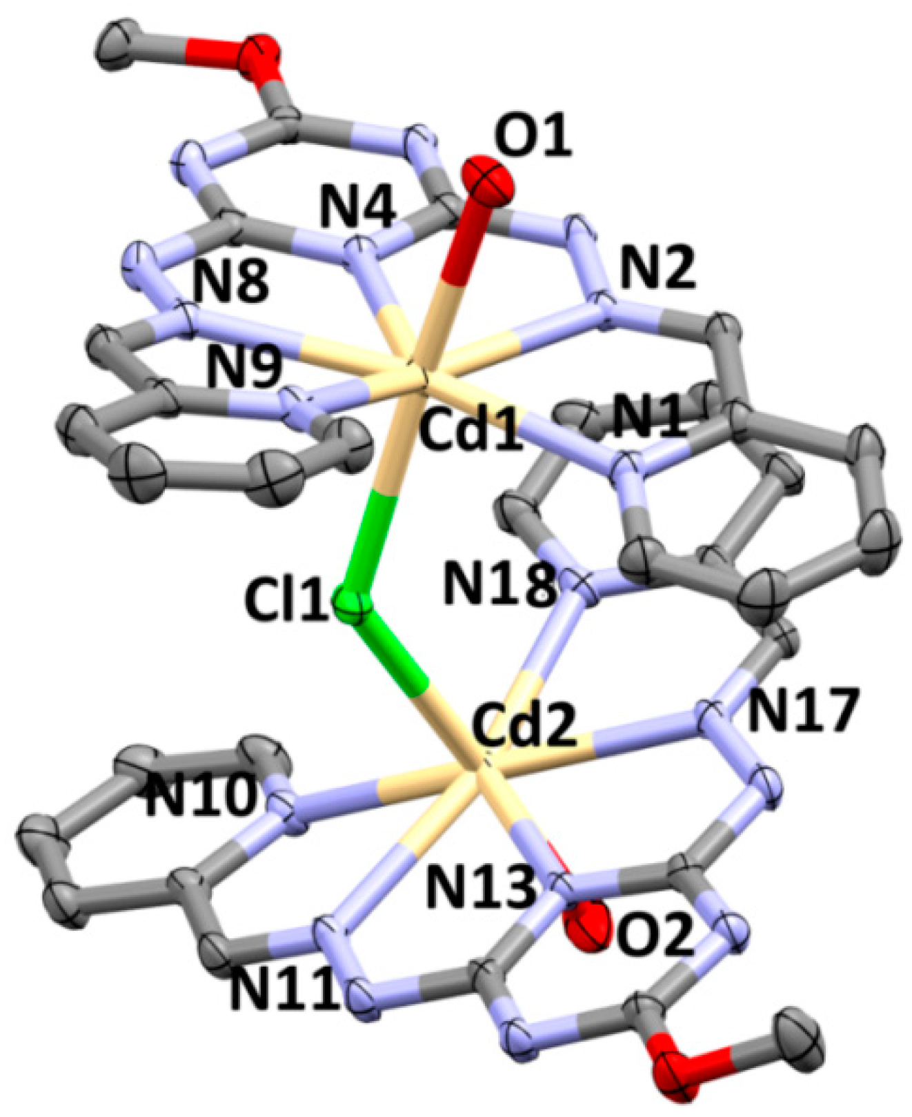
3.1. Crystal Structure Description

The molecular structure and atom numbering of the homoleptic [Cd(BDMPT)2](ClO4)2 complex (1) are illustrated in Figure 2. The structure crystallized in the orthorhombic crystal system and Cmca space group with Z = 8. It should be noted that only half of the complex is crystallographically independent; thus, the asymmetric unit of this complex comprised half of its molecular formula. The structure showed a pincer-type complex with two tridentate ligand (BDMPT) units coordinating the central metal ion (Cd(II)) in the inner sphere and two ionic perchlorate anions in the outer sphere. Each BDMPT ligand coordinating the Cd(II) ion via one N-atom from the s-triazine core (Cd1–N3; 2.331(2) Å) and one N-atom from each pyrazole moieties with Cd1–N1 and Cd1–N7 distances of 2.403(6) and 2.373(6) Å, respectively (Table 2). The former was found generally shorter than the two latter bonds indicating the stronger Cd–N(triazine) interaction than Cd–N(pyrazole). The two ligand molecules coordinating Cd(II) in a meridional fashion with trans N–Cd–N angles varied significantly (114.4(3)–175.9(2)°). As a result, the Cd(II) showed a hexa-coordinated environment with a highly distorted configuration compared to any of the well-known ideal geometries, trigonal prism and octahedral (Figure 2). With the aid of the continuous shape measure (CShM) tool [53,54,55,56], the values of the CShM are 12.6 and 12.5 against the perfect octahedron and trigonal prism, respectively. The almost equal and large values of the CShM in both cases confirmed that the coordination geometry around the Cd(II) ion is an intermediate case between the two extremes.
The structure of the coordinated ligand molecule showed slight deviations in the two pyrazole moieties from co-planarity with the s-triazine core. The two pyrazole moieties deviated only by 3.48–3.58° from the mean plane of the s-triazine core. The maximum distance between the s-triazine mean plane and any atom in the pyrazole moieties did not exceed 0.249 Å. Moreover, the two ligand molecules coordinating the Cd(II) ion are not fully perpendicular with each other, which explained the strong distortion of the coordination environment around Cd(II). The angle between the two mean planes passing through each ligand molecule was found to be 62.2°. Such a situation left large spaces between the complex cation units (Figure 3A) which were found occupied by the perchlorate anions. The latter connected the complex units by weak C–H⋯O hydrogen bonds, as shown in Figure 3B and listed in Table 3. The anion–π-stacking interaction is another feature of molecular packing in the crystal structure of 1. The s-triazine ring has two perchlorate anions found below and above it, with C⋯O contact distances of 3.179, 3.171, and 3.139 Å for the C6⋯O1, C7⋯O1, and C8⋯O4 interactions, respectively. Presentation of these interactions is shown in Figure 4.
The crystal structure of the dinuclear [Cd2(MBPT)2(H2O)2Cl](ClO4)3·4H2O complex 2 comprised one formula unit per asymmetric unit and four molecules per unit cell. It crystallized in the monoclinic crystal system and the centrosymetric P21/n space group. The experimental geometric parameters (bond lengths and angles) are listed in Table 4. This complex showed two hepta-coordinated Cd(II) centers with a distorted pentagonal bipyramidal coordination geometry. While the outer sphere contained three perchlorate anions and four crystallization water molecules, the inner sphere comprised two ligands (MBPT) acting as pentadentate N-chelate via one N from the s-triazine core, two N-atoms from the hydrazone and two N-atoms from the pyridyl moieties which coordinated the Cd(II) center in a pincer-like fashion (Figure 5). The axial positions are occupied by one terminal water molecule and one bridged chloride connecting the two Cd-centers. The Cd1–Cl1–Cd2 angle is 132.21° and the two Cd1–O1 (2.325(4) Å) and Cd2–O2 (2.322(4) Å) bond distances are identical. Due to such a bent bridged structure, the two MBPT molecules are not parallel to one another and the ligand (MBPT) molecules favored an anti-configuration to each other to minimize the steric repulsion between the two bulky organic ligands. Moreover, the Cd–N(triazine) bonds are shorter than any of the Cd–N(hydrazone) and Cd–N(pyridine) bonds. It is found that the two pyridyl moieties are significantly twisted from one another due to the short distance between the hydrogen atoms at the 6-position. The H1⋯H15 and H17⋯H31 intramolecular distances are 2.332 and 2.255 Å, respectively. In the Cd1–MBPT unit, the angles between the mean plane of the s-triaizne core and the plane passing through the pyridine moieties are 11.7° {N1C1C2C3C4C5} and 12.8° {C15C14C13C12C11N9}, while the corresponding values for the Cd2–MBPT unit are 6.1° {C17N10C21C20C19C18} and 12.1° {C29C28C27N18C31C30}, indicating the twist of the two ligand strands from one another.
The complex molecules are packed in the crystal via sets of N–H⋯N, N–H⋯O, and O–H⋯O hydrogen-bonding interactions, as listed in Table 5. The N–H⋯N hydrogen bonds connected the coordinated organic ligand molecules in two neighboring complex cations. Moreover, the perchlorate anions and water molecules connected the complex cations via the N–H⋯O and O–H⋯O hydrogen bonds (Figure 6).

3.2. Analysis of Molecular Packing

Hirshfeld surfaces mapped over dnorm, shape index (SI), and curvedness for the [Cd(BDMPT)2]2+ and [Cd2(MBPT)2(H2O)2Cl]3+ units of complexes 1 and 2, respectively, are shown in Figure S4 (Supplementary Materials). A summary of the most important contacts and their percentages are shown in Figure 7.
It is clear that the packing of complex units in the crystal is dominated mainly by H⋯H and O⋯H contacts for complex 1. All these interactions appeared as red spots in the dnorm Hirshfeld surface (Figure 8). The contributions of these contacts are 57.2% and 24.0% of the whole fingerprint area, respectively. The minimum H⋯H and O⋯H contact distances are 2.290 Å (H9A⋯H9C) and 2.549 Å (O4⋯H10A), respectively. In addition, the presence of C⋯O contacts (5.3%) confirmed the anion–π-stacking interactions between the free perchlorate anion and the s-triazine moiety with shortest contact distance of 3.139 Å (C8⋯O4).
On the other hand, the packing of the complex units of 2 in its crystal is dominated mainly by strong O⋯H and N⋯H hydrogen bonds where all appeared as red regions in the dnorm map, as well as sharp spikes in the decomposed fingerprint plots. Presentation of the dnorm surfaces and fingerprint plots of these interactions is shown in Figure 9. The percentages of the O⋯H and N⋯H contacts are 37.4% and 10.8% of the whole fingerprint area, respectively. It is obvious that, the common H⋯H contacts contributed less in the molecular packing of this complex (27.2%) compared to 1. In complex 2, the N–H⋯O hydrogen bridges are the shortest O⋯H contacts, where the O17⋯H7 and O19⋯H16 contact distances were found to be 1.764 Å and 1.820 Å, respectively, using Hirshfeld analysis. The N⋯H hydrogen bridges occurred among each two neighbouring complex units via the NH group as the H-bond donor and one of the s-triazine N-atoms as the H-bond acceptor. The N⋯H hydrogen-bond distances are 1.959 Å (N14⋯H3) and 1.979 Å (N5⋯H12). Interestingly, the bridged chloride ion not only connected the two Cd(II) centres but also connected the complex units via two Cl⋯H hydrogen bridges (2.9%): one with the protons from the crystal water and the other from the hydrazone N=CH group of a neighbouring organic ligand. The Cl⋯H contact distances were found to be 2.407 Å and 2.607 Å for the Cl1⋯H17A and Cl1⋯H10 contacts, respectively. It is worth noting that both complexes showed some weak C⋯H interactions, where all appeared as blue regions in the dnorm surface, indicating the lower importance of this type of contact in the molecular packing of the studied complexes. Curvedness and shape index maps did not show any signs of π–π stacking interactions in both complexes (Figure S4, Supplementary Materials).

3.3. Vibrational Spectra and TGA Analysis

The FTIR spectra of complexes 1 and 2 are shown in Figure S1 (Supplementary Materials). It is clear that the ν(C=N) mode appeared at 1619 cm−1 in both complexes, while it appeared at 1593 cm−1 and 1625 cm−1 for the free BDMPT and MBPT, respectively. On other hand, the ν(C=C) modes showed little variations due to complexation of Cd(II) with the organic ligand. The ν(C=C) modes appeared at a lower wavenumber of 1543 cm−1 for complex 1 compared to 1555 cm−1 in the free ligand. The corresponding vibrational wavenumbers for complex 2 are 1586 and 1537 cm−1 compared to 1586 and 1544 cm−1 for the free MBPT. The perchlorate vibrations appeared as a triple split band in the regions of 1136–1056 cm−1 and 1160–1077 cm−1 for complexes 1 and 2, respectively.
TGA analyses of the free ligands (BDMPT and MBPT) compared to the corresponding Cd(II) complexes (1 and 2, respectively) are shown in Figure 10. Both ligands started the mass loss in the temperature range of 60–67 °C, which is probably attributed to the evaporation of solvent or moisture contained in the sample, before starting to decompose in several steps at 185 and 235 °C for BDMPT and MBPT, respectively. On other hand, complex 1 started to decompose thermally at 273 °C. The first step in the temperature range of 273–369 °C showed a large mass loss of 34.7% (calculated 32.9%), probably due to the decomposition of one of the coordinated ligands, followed by a slow decomposition of the complex residue up to 800 °C. In case of complex 2, the first small step in the temperature range of 81–121°C corresponded to the loss of six water molecules with an experimental mass loss of 8.3% (calculated 7.9%). The remaining complex residue showed good thermal stability as indicated by the long flat plateau until 273°C. After that, a sudden large mass loss of 53.5% (calculated 51.2%) occurred due to the decomposition of the organic ligand molecules up to 361°C, followed by a slow decomposition of the complex residue up to 800 °C.

3.4. Density Functional Theory (DFT) Studies

In metal organic complexes, the interaction of metal ion (Cd(II)) with ligand groups leads to significant variations in their charge densities due to the electron transferences from the ligand as a Lewis base to the metal ion as a Lewis acid. A summary of the natural charges at cadmium and ligand groups in complexes 1 and 2 are collected in Table 6. The average net natural charges at the perchlorate counter anions are almost −1.0 e, indicating insignificant interactions with the metal ion. The slight changes in the net charges of the perchlorate anions could be attributed to their interactions with the organic ligand via hydrogen bonds or anion–π-stacking interactions. On the other hand, the natural charges at the Cd ions decreased significantly by 0.535 e for Cd1 in complex 1. The corresponding values for Cd1 and Cd2 in complex 2 are 0.641 and 0.618 e, respectively. In the former, the net electron density transferred from the two organic ligands is 0.484 e, while, in complex 2, the electron density transferred to the two Cd-centers is almost the same (0.371 and 0.331 e, respectively). In addition, the coordinated chloride (~0.290 e) ligand transferred a significant amount of electron density to the two Cd-ions, while each of the coordinated water molecules transferred only 0.093 e (average value) to the central metal ion.
In order to quantify the strength of interactions between the Cd(II) and the ligand donor atoms, the interaction energies at the Cd–N, Cd–O, and Cd–Cl bond critical points (BCPs) were calculated in the framework of atoms in molecules topology analysis [57,58,59,60,61,62,63,64]. The results of the topological parameters shown in Table 7 were also used to describe the nature of the Cd–N, Cd–O, and Cd–Cl metal–ligand interactions. The interaction energies calculated using the Espinosa relationship [65] showed very good correlation with the Cd–N distances (Figure 11). The interaction energies dramatically decreased with the increase in Cd–N distances, which agrees with previous studies [17,18,19]. On other hand, the total electron density ρ(r) values are less than 0.1 a.u., indicating predominant closed-shell interactions for the Cd–N, Cd–O, and Cd–Cl coordinate bonds, where shorter bonds have higher ρ(r) values than longer ones. On other hand, the results showed that shorter Cd–N interactions have negative total energy density H(r) and V(r)/G(r) ratios slightly more than one, indicating some covalent characteristics for these interactions, while the opposite is true for longer Cd–N bonds which mainly belong to closed-shell interactions with negligible covalent characteristics (Figure 12). It is clear that a cut-off of about 2.4 Å for the Cd–N distances could be considered as a border between the closed-shell interactions and those having significant covalent characteristics. The results also showed that Cd–Cl and Cd–O coordinate bonds have the main characteristics of closed-shell interactions with positive H(r) and V(r)/G(r) ratios ˂1.

3.5. Comparative Study

In our previous studies [17,18,19,66,67,68], we reported detailed structural studies on the Mn(II), Cd(II), and Ni(II) complexes of the neutral ligands BDMPT and MBPT. Here, we present a comparative discussion on the affinity of these ligands toward some divalent metal ions based on the reported X-ray structures of their well-known complexes and those presented here in this publication. For this task, we calculated the interaction energies of the [M–L]2+ complex cation units of these systems. The results are listed in Table 8. There is no doubt that such sterically hindered cavity-containing ligands have higher affinity to large metal ions such as Cd(II). It is clear that the Cd(II)–L interaction energies of [Cd(BDMPT)]2+ and [Cd(MBPT)]2+ are at least four times higher than the corresponding Mn(II) and Ni(II) complexes. It is so obvious that the large size M(II) could fit better in the ligand cavity and strongly bond to the N-atoms of BDMPT or MBPT ligands without suffering from high steric hinderance between the two ligand arms. In contrast, the presence of such bulky groups around the s-triazine core prevents these moieties from approaching each other to a certain limit, which explains the weaker interactions with the smaller M(II) ions such as Mn(II) and Ni(II). Another factor which slightly affects the interaction energies; it is the presence of a coordinating anion inside the coordination sphere. The presence of an anion directly coordinating to the M(II) ion weakens the interaction with the s-triazine ligand as a result of the compensation of the divalent metal ion positive charge by the negative charge from the anionic ligand.

4. Conclusions

The homoleptic [Cd(BDMPT)2](ClO4)2 (1) and heteroleptic [Cd2(MBPT)2(H2O)2Cl](ClO4)3·4H2O (2) s-triazine pincer-type complexes were synthesized and characterized using FTIR, NMR, and single-crystal X-ray diffraction techniques. In 1, the Cd(II) is hexa-coordinated with two tridentate N-chelates and the coordination geometry is significantly distorted compared to the octahedral and trigonal prism configurations. In 2, the two Cd(II) are hepta-coordinated with distorted pentagonal bipyramidal coordination geometry. The molecular packing in both complexes was analyzed using Hirshfeld analysis. Complex 1 thermally decomposed at higher temperature (273 °C) compared to the free ligand BDMPT (185 °C). On other hand, the coordinated organic ligand of 2 decomposed at 273 °C after losing the crystal and coordinated water molecules, while the free ligand MBPT decomposed at 235 °C. Using atoms in molecules, the shorter Cd–N coordinate bonds have higher covalent characteristics than the longer ones. The interaction energies of the BDMPT or MBPT ligands with metal ions having larger size are higher than those for smaller-size metal ions, in agreement with the steric preference of these ligands.

Supplementary Materials

The following are available online at https://www.mdpi.com/2073-4352/9/5/226/s1. Figure S1: FTIR spectra of complexes 1 and 2. Figure S2: 1H and 13C NMR spectra of complex 1. Figure S3: 1H and 13C NMR spectra of complex 2. Figure S4: Hirshfeld surfaces of the studied complexes.

Author Contributions

The preparations of the organic ligands were carried out by Z.A.M. and A.E.F. Also, they performed the elemental analysis, TGA, and NMR analyses; FTIR and X-ray analyses, together with the computational calculations, as well as the synthesis of complexes 1 and 2, were carried out by S.M.S. All authors contributed in the preparation of the first and final versions of the manuscript.

Funding

The Deanship of Scientific Research at King Saud University, Saudi Arabia funded this research.

Acknowledgments

The authors extend their appreciation to the Deanship of Scientific Research at King Saud University for funding this work through research group No. RGP-234 (Saudi Arabia).

Conflicts of Interest

The authors declare no conflicts of interest.

References

  1. Stang, P.J.; Olenyuk, B. Self-Assembly, Symmetry, and Molecular Architecture:  Coordination as the Motif in the Rational Design of Supramolecular Metallacyclic Polygons and Polyhedra. Acc. Chem. Res. 1997, 30, 502–518. [Google Scholar] [CrossRef]
  2. Braga, D.; Grepioni, F.; Desiraju, G.R. Crystal Engineering and Organometallic Architecture. Chem. Rev. 1998, 98, 1375–1406. [Google Scholar] [CrossRef] [PubMed]
  3. Yaghi, O.M.; Li, H.; Davis, C.; Richardson, D.; Groy, T.L. Synthetic Strategies, Structure Patterns, and Emerging Properties in the Chemistry of Modular Porous Solids. Acc. Chem. Res. 1998, 31, 474–484. [Google Scholar] [CrossRef]
  4. Purdy, A.P.; Gilardi, R.; Luther, J.; Butcher, R.J. Synthesis, crystal structure, and reactivity of alkali and silver salts of sulfonated imidazoles. Polyhedron 2007, 26, 3930–3938. [Google Scholar] [CrossRef]
  5. Zhang, Z.-T.; Shi, J.; He, Y.; Guo, Y.-N. Self-assembly and crystal structure of a barium sulfonatechrysin coordination polymer. Inorg. Chem. Commun. 2006, 9, 579–581. [Google Scholar] [CrossRef]
  6. Yang, X.-L.; Ren, S.-B.; Zhang, J.; Li, Y.-Z.; Du, H.-B.; You, X.-Z. Syntheses and structures of three coordination polymers based on 4-methylbenzenethiolates of Zn(II) and Cd(II) and bipyridine. J. Coord. Chem. 2009, 62, 3782–3794. [Google Scholar] [CrossRef]
  7. Ghoshal, D.; Maji, T.K.; Mostafa, G.; Lu, T.H.; Chaudhuri, N.R. A Three-dimensional honeycomb-Like network constructed with novel “Sinusoidal” One-Dimensional Chains via Hydrogen Bonding and π−π Interactions. Cryst. Growth Des. 2003, 3, 9–11. [Google Scholar] [CrossRef]
  8. Rashidi-Ranjbar, Z.; Hamidi, S.; Heshmatpour, F.; Morsali, A. Thermal, spectroscopic, X-ray powder diffraction, and structural studies on a new Cd(II) mixed-ligand coordination polymer. J. Coord. Chem. 2009, 62, 2022–2027. [Google Scholar] [CrossRef]
  9. Smith, G.; Wermuth, U.D.; Young, D.J.; White, J.M. Polymeric structures in the metal complexes of 5-sulfosalicylic acid: The rubidium(I), caesium(I) and lead(II) analogues. Polyhedron 2007, 26, 3645–3652. [Google Scholar] [CrossRef]
  10. Wang, C.-J.; Ren, P.-D.; Zhang, Z.-B.; Fang, Y.; Wang, Y.-Y. Synthesis and characterization of a nickel-organic framework encapsulating hetero-chiral helical water chains in the 1-D channels. J. Coord. Chem. 2009, 62, 2814–2823. [Google Scholar] [CrossRef]
  11. De Silva, C.-R.; Maeyer, J.-R.; Dawson, A.; Zheng, Z. Adducts of lanthanide β-diketonates with 2,4,6-tri(2-pyridyl)-1,3,5-triazine: Synthesis, structural characterization, and photoluminescence studies. Polyhedron 2007, 26, 1229–1238. [Google Scholar] [CrossRef]
  12. Ionova, G.; Raber, C.; Guillaumont, R.; Ionov, S.; Madic, C.; Krupa, D.; Guillaneux, J.-C. A donor–acceptor model of Ln(III) complexation with terdentate nitrogen planar ligands. New J. Chem. 2002, 26, 234–242. [Google Scholar] [CrossRef]
  13. Yan, C.; Chen, Q.; Chen, L.; Feng, R.; Shan, X.; Jiang, F.; Hong, M. crystal structures and luminescence behaviour of d10 Metal–Organic Complexes with multipyridine ligands. Aust. J. Chem. 2011, 64, 104–118. [Google Scholar] [CrossRef]
  14. Wu, G.; Wang, X.-F.; Guo, L.; Li, H.-H. Zn(II) and Cd(II) Complexes extended structures sustained by hydrogen bonding, π–π and C–H···π interactions. J. Chem. Crystallogr. 2011, 41, 1071–1076. [Google Scholar] [CrossRef]
  15. Glaser, T.; Lügger, T.; Fröhlich, R. Synthesis, crystal structures, and magnetic properties of a mono- and a dinuclearcopper(II) complex of the 2,4,6-tris(2-pyridyl)-1,3,5-triazine ligand. Eur. J. Inorg. Chem. 2004, 2004, 394–400. [Google Scholar] [CrossRef]
  16. Schwalbe, M.; Karnahl, M.; Görls, H.; Chartrand, D.; Laverdiere, F.; Hanan, G.-S.; Tschierlei, S.; Dietzek, B.; Schmitt, M.; Popp, J.; et al. Ruthenium polypyridine complexes of tris-(2-pyridyl)-1,3,5-triazine—Unusual building blocks for the synthesis of photochemical molecular devices. Dalton Trans. 2009, 4012–4022. [Google Scholar] [CrossRef] [PubMed]
  17. Soliman, S.M.; El-Faham, A. Synthesis, characterization, and structural studies of two heteroleptic Mn(II) complexes with tridentate N,N,N-pincer type ligand. J. Coord. Chem. 2018, 71, 2373–2388. [Google Scholar] [CrossRef]
  18. Soliman, S.M.; El-Faham, A. One pot synthesis of two Mn(II) perchlorate complexes with s-triazine NNN-pincer ligand; molecular structure, Hirshfeld analysis and DFT studies. J. Mol. Struct. 2018, 1164, 344–353. [Google Scholar] [CrossRef]
  19. Soliman, S.M.; El-Faham, A.; Elsilk, S.E.; Farooq, M. Two heptacoordinated manganese(II) complexes of giant pentadentate s-triazinebis-Schiff base ligand: Synthesis, crystal structure, biological and DFT studies. Inorg. Chim. Acta 2018, 479, 275–285. [Google Scholar] [CrossRef]
  20. Nawrot, I.; Machura, B.; Kruszynski, R. Thiocyanate cadmium(II) complexes of 2,4,6-tri(2-pyridyl)-1,3,5-triazine—Synthesis, structure and luminescence properties. J. Luminescence 2014, 156, 240–254. [Google Scholar] [CrossRef]
  21. Zhao, X.X.; Qin, Z.B.; Li, Y.H.; Cui, G.H. New Cd(II) and Zn(II) coordination polymers showing luminescent sensing for Fe(III) and photocatalytic degrading methylene blue. Polyhedron 2018, 153, 197–204. [Google Scholar] [CrossRef]
  22. Cheng, H.J.; Lu, Y.F.; Li, C.; Shu, Y.; Ma, J.; Li, W.H.; Yuan, R.X. Two Cd(II) coordination polymers based on 3,6-bis(imidazol-1-yl)carbazole: Syntheses, structures and photocatalytic properties. Inorg. Chem. Commun. 2016, 73, 12–15. [Google Scholar] [CrossRef]
  23. Gong, W.J.; Yao, R.; Li, H.X.; Ren, Z.G.; Zhang, J.G.; Lang, J.P. Luminescent cadmium(II) coordination polymers of 1,2,4,5-tetrakis(4-pyridylvinyl)benzene used as efficient multi-responsive sensors for toxic metal ions in water. Dalton Trans. 2017, 46, 16861–16871. [Google Scholar] [CrossRef] [PubMed]
  24. Wang, Q.; Wang, S.; Feng, X.; Wu, L.; Zhang, G.; Zhou, M.; Wang, B.; Yang, L. A Heat-Resistant and Energetic Metal–Organic Framework Assembled by Chelating Ligand. ACS Appl. Mater. Interfaces 2017, 9, 37542–37547. [Google Scholar] [CrossRef] [PubMed]
  25. Liu, X.; Gao, W.; Sun, P.; Su, Z.; Chen, S.; Wei, Q.; Xie, G.; Gao, S. Environmentally friendly high-energy MOFs: crystal structures, thermos stability, insensitivity and remarkable detonation performances. Green Chem. 2015, 17, 831–836. [Google Scholar] [CrossRef]
  26. Feng, Y.; Liu, X.; Duan, L.; Yang, Q.; Wei, Q.; Xie, G.; Chen, S.; Yang, X.; Gao, S. In situ synthesized 3D heterometallic metal–organic framework (MOF) as a high-energy-density material shows high heat of detonation, good thermo stability and insensitivity. Dalton Trans. 2015, 4, 2333–2339. [Google Scholar] [CrossRef]
  27. Wang, S.; Wang, Q.; Feng, X.; Wang, B.; Yang, L. Explosives in the Cage: Metal–Organic Frameworks for high-energy materials sensing and desensitization. Adv. Mater. 2017, 29, 1701898. [Google Scholar] [CrossRef] [PubMed]
  28. Zhang, Q.; Shreeve, J.M. Metal-organic frameworks as high explosives: a new concept for energetic materials. Angew. Chem. Int. Ed. 2014, 53, 2540–2542. [Google Scholar] [CrossRef]
  29. Bushuyev, O.S.; Brown, P.; Maiti, A.; Gee, R.H.; Peterson, G.R.; Weeks, B.L.; Hope-Weeks, L.J. Ionic Polymers as a New Structural Motif for High-Energy-Density Materials. J. Am. Chem. Soc. 2012, 134, 1422–1425. [Google Scholar] [CrossRef]
  30. Bushuyev, O.S.; Peterson, G.R.; Brown, P.; Maiti, A.; Gee, R.H.; Weeks, B.L.; Hope-Weeks, L.J. Metal-organic frameworks (MOFs) as safer, structurally reinforced energetic. Chem. Eur. J. 2013, 19, 1706–1711. [Google Scholar] [CrossRef]
  31. Li, S.; Wang, Y.; Qi, C.; Zhao, X.; Zhang, J.; Zhang, S.; Pang, S. 3D Energetic Metal–Organic Frameworks: Synthesis and Properties of High Energy Materials. Angew. Chem. Int. Ed. 2013, 52, 14031–14035. [Google Scholar] [CrossRef]
  32. Zhang, S.; Yang, Q.; Liu, X.; Qu, X.; Wei, Q.; Xie, G.; Chen, S.; Gao, S. High-energy metal–organic frameworks (HE-MOFs): Synthesis, structure and energetic performance. Coord. Chem. Rev. 2016, 307, 292–312. [Google Scholar] [CrossRef]
  33. McDonald, K.A.; Seth, S.; Matzger, A.J. Coordination Polymers with High Energy Density: An Emerging Class of Explosives. Cryst. Growth Des. 2015, 1, 5963–5972. [Google Scholar] [CrossRef]
  34. Blair, L.H.; Colakel, A.; Vrcelj, R.M.; Sinclair, I.; Coles, S.J. Metal–organic fireworks: MOFs as integrated structural scaffolds for pyrotechnic materials. Chem. Commun. 2015, 5, 12185–12188. [Google Scholar] [CrossRef]
  35. Zhang, J.; Du, Y.; Dong, K.; Su, H.; Zhang, S.; Li, S.; Pang, S. Taming Dinitramide Anions within an Energetic Metal–Organic Framework: A New Strategy for Synthesis and Tunable Properties of High Energy Materials. Chem. Mater. 2016, 28, 1472–1480. [Google Scholar] [CrossRef]
  36. Yang, L.L.; Tan, X.X.; Wang, Z.Q.; Zhang, X. Supramolecular Polymers: Historical Development, Preparation, Characterization, and Functions. Chem. Rev. 2015, 115, 7196–7239. [Google Scholar] [CrossRef]
  37. Li, Z.X.; Zhang, X.; Liu, Y.C.; Zou, K.Y.; Yue, M.L. Controlling the BET Surface Area of Porous Carbon by Using the Cd/C Ratio of a Cd–MOF Precursor and Enhancing the Capacitance by Activation with KOH. Chem. Eur. J. 2016, 22, 17734–17747. [Google Scholar] [CrossRef]
  38. Sheldrick, G.M.; SADABS. Program for Empirical Absorption Correction of Area Detector Data; University of Göttingen: Göttingen, Germany, 1996. [Google Scholar]
  39. Dolomanov, O.V.; Bourhis, L.J.; Gildea, R.J.; Howard, J.A.K.; Puschmann, H. OLEX2: A complete structure solution, refinement and analysis program. J. Appl. Cryst. 2009, 42, 339–341. [Google Scholar] [CrossRef]
  40. Sheldrick, G.M. A Short History of SHELX. Acta Cryst. A 2008, 64, 112–122. [Google Scholar] [CrossRef]
  41. Sheldrick, G.M. SHELXT—Integrated space-group and crystal-structure determination. Acta Cryst. A 2015, 71, 3–8. [Google Scholar] [CrossRef]
  42. Spek, A.L. Structure validation in chemical crystallography. Acta Cryst. 2009, D65, 148–155. [Google Scholar] [CrossRef] [PubMed]
  43. Hirshfeld, F.L. Bonded-atom fragments for describing molecular charge densities. Theor. Chim. Acta 1977, 44, 129–138. [Google Scholar] [CrossRef]
  44. Spackman, M.A.; Jayatilaka, D. Hirshfeld surface analysis. CrystEngComm 2009, 11, 19–32. [Google Scholar] [CrossRef]
  45. Spackman, M.A.; McKinnon, J.J. Fingerprinting intermolecular interactions in molecular crystals. CrystEngCommun 2002, 4, 378–392. [Google Scholar] [CrossRef]
  46. Bernstein, J.; Davis, R.E.; Shimoni, L.; Chang, N.-L. Patterns in hydrogen bonding: Functionality and graph set analysis in crystals. Angew. Chem. Int. Ed. 1995, 34, 1555–1573. [Google Scholar] [CrossRef]
  47. McKinnon, J.J.; Jayatilaka, D.; Spackman, M.A. Towards quantitative analysis of intermolecular interactions with Hirshfeldsurfaces. Chem. Commun. 2007, 3814–3816. [Google Scholar] [CrossRef]
  48. Crystal Explorer 17. 2017. Available online: http://hirshfeldsurface.net (accessed on 27 April 2019).
  49. Frisch, M.J.; Trucks, G.W.; Schlegel, H.B.; Scuseria, G.E.; Robb, M.A.; Cheeseman, J.R.; Scalmani, G.; Barone, V.; Mennucci, B.; Petersson, G.A.; et al. GAUSSIAN 09. Revision A02; Gaussian Inc.: Wallingford, CT, USA, 2009. [Google Scholar]
  50. Chai, J.D.; Head-Gordon, M. Long-range corrected hybrid density functionals with damped atom-atom dispersion corrections. Phys. Chem. Chem. Phys. 2008, 10, 6615–6620. [Google Scholar] [CrossRef] [PubMed]
  51. Glendening, E.D.; Reed, A.E.; Carpenter, J.E.; Weinhold, F. NBO Version 3.1, CI; University of Wisconsin: Madison, WI, USA, 1998. [Google Scholar]
  52. Lu, T.; Chen, F. Multiwfn: A multifunctional wave function analyzer. J. Comput. Chem. 2012, 33, 580–592. [Google Scholar] [CrossRef]
  53. Ok, K.M.; Halasyamani, P.S.; Casanova, D.; Llunell, M.; Alvarez, S. Distortions in octahedrally coordinated d0 transition metal oxides: A continuous symmetry measures approach. Chem. Mater. 2006, 18, 3176–3183. [Google Scholar] [CrossRef]
  54. Santiaqo, A.; David, A.; Llunell, M.; Pinsky, M. Continuous symmetry maps and shape classification. The case of six-coordinated metal compounds. New J. Chem. 2002, 26, 996–1009. [Google Scholar]
  55. Hagit, Z.; Shmuel, P.; David, A. Continuous symmetry measures. J. Am. Chem. Soc. 1992, 114, 7843–7851. [Google Scholar]
  56. Keinan, S.; Avnir, D. Quantitative Symmetry in Structure−Activity Correlations: The Near C2Symmetry of Inhibitor/HIV Protease Complexes. J. Am. Chem. Soc. 2000, 122, 4378–4384. [Google Scholar] [CrossRef]
  57. Bader, R.F.W. Atoms in Molecules: A Quantum Theory; Oxford University Press: Oxford, UK, 1990. [Google Scholar]
  58. Matta, C.F.; Hernandez-Trujillo, J.; Tang, T.-H.; Bader, R.F.W. Hydrogen-hydrogen bonding: A stabilizing interaction in molecules and crystals. Chem. Eur. J. 2003, 9, 1940–1951. [Google Scholar] [CrossRef]
  59. Grabowski, S.J.; Pfitzner, A.; Zabel, M.; Dubis, A.T.; Palusiak, M. Intramolecular H…H interactions for the Crystal Structures of [4-((E)-But-1-enyl)-2,6-dimethoxyphenyl]pyridine-3-carboxylate and [4-((E)-Pent-1-enyl)-2,6-dimethoxyphenyl]pyridine-3-carboxylate; DFT calculations on modeled styrene derivatives. J. Phys. Chem. B 2004, 108, 1831–1837. [Google Scholar] [CrossRef]
  60. Matta, C.F.; Castillo, N.; Boyd, R.J. Characterization of a closed-shell fluorine-fluorine bonding interaction in aromatic compounds on the basis of the electron density. J. Phys. Chem. A 2005, 109, 3669–3681. [Google Scholar] [CrossRef]
  61. Pendás, A.M.; Francisco, E.; Blanco, M.A.; Gatti, C. Bond paths as privileged exchange channels. Chem. Eur. J. 2007, 13, 9362–9371. [Google Scholar]
  62. Bobrov, M.F.; Popova, G.V.; Tsirelson, V.G. A topological analysis of electron density and chemical bonding in cyclophosphazenes PnNnX2n (X = H, F, Cl; n = 2, 3, 4). Russ. J. Phys. Chem. 2006, 80, 584–590. [Google Scholar] [CrossRef]
  63. Gatti, C. Chemical bonding in crystals: New directions. Z. Kristallogr. 2005, 220, 399–457. [Google Scholar] [CrossRef]
  64. Gibbs, G.V.; Downs, R.T.; Cox, D.F.; Ross, N.L.; Boisen, M.B., Jr.; Rosso, K.M. Shared and closed-shell O-O interactions in silicates. J. Phys. Chem. A 2008, 112, 3693–3699. [Google Scholar] [CrossRef]
  65. Espinosa, E.; Molins, E.; Lecomte, C. Hydrogen bond strengths revealed by topological analyses of experimentally observed electron densities. Chem. Phys. Lett. 1998, 285, 170–173. [Google Scholar] [CrossRef]
  66. Soliman, S.M.; El-Faham, A.; Elsilk, S.E.; Farooq, M. Synthesis and structure diversity of high coordination number Cd(II) complexes of large s-triazinebis-Schiff base pincer chelate. Inorg. Chim. Acta 2019, 488, 131–140. [Google Scholar] [CrossRef]
  67. Soliman, S.M.; El-Faham, A. Synthesis, molecular structure and DFT studies of two heteroleptic nickel(II) s-triazine pincer type complexes. J. Mol. Struct. 2019, 1185, 461–468. [Google Scholar] [CrossRef]
  68. Soliman, S.M.; El-Faham, A. Synthesis, X-ray structure and DFT studies of penta- and octa-coordinated Cd(II) complexes with s-triazineN-pincer chelate. J. Coord. Chem. 2019, in press. [Google Scholar] [CrossRef]
Figure 1. Structure of the s-triazine-based ligands, 2,4-bis(3,5-dimethyl-1H-pyrazol-1-yl)-6-methoxy-1,3,5-triazine (BDMPT) and 2-methoxy-4,6-bis(2-(pyridin-2-ylmsethylene)hydrazinyl)-1,3,5-triazine (MBPT).
Crystals 09 00226 g001
Figure 2. Structure showing the most relevant atom numbering (left) and the distorted octahedron (right) of complex 1.
Crystals 09 00226 g002
Figure 3. Packing of the complex cations and perchlorate anions along the ab plane (A) and the weak C–H⋯O hydrogen-bonding interactions connecting the complex units (B).
Crystals 09 00226 g003
Figure 4. The possible perchlorate–π-stacking interactions in 1.
Crystals 09 00226 g004
Figure 5. The structure and atom numbering of the asymmetric unit of 2. All hydrogen atoms, crystal water molecules, and perchlorate anions were omitted for more clarity.
Crystals 09 00226 g005
Figure 6. Part of the hydrogen-bond network connecting the complex units via N–H⋯N, N–H⋯O, and O–H⋯O hydrogen bonds.
Crystals 09 00226 g006
Figure 7. Summary of the intermolecular interactions and their percentages in the crystal structure of the studied complexes.
Crystals 09 00226 g007
Figure 8. The most important intermolecular contacts in the crystal structure of complex 1. The O⋯H and C⋯O contacts are indicated by yellow and black dotted lines, respectively.
Crystals 09 00226 g008
Figure 9. The most important intermolecular contacts in the crystal structure of complex 2. The O⋯H, N⋯H, and Cl⋯H are indicated by purple, turquoise, and green colors, respectively. The hydrophobic C…H interactions appeared weak and were omitted from this illustration for better clarity.
Crystals 09 00226 g009
Figure 10. Thermogravimetric analyses of complexes 1 and 2 compared to the free ligands.
Crystals 09 00226 g010
Figure 11. Inverse correlation between the interaction energy (Eint) and Cd–N distance of complexes 1 and 2.
Crystals 09 00226 g011
Figure 12. Relationship between the H(r) and V(r)/G(r) ratio and the Cd–N distances (Å) in complexes 1 and 2.
Crystals 09 00226 g012
Table 1. Crystal data and structure refinement for the studied complexes.
12
Empirical formulaC28H34CdCl2N14O10C32H42Cd2Cl4N18O20
Formula weight910.00 g/mol1365.43 g/mol
Temperature (K)293(2)293(2)
Crystal systemOrthorhombic Monoclinic
Space groupCmcaP21/n
a (Å)32.032(19)11.183(2)
b (Å)15.181(9)28.562(6)
c (Å)16.441(10)16.683(3)
α (°)9090
β (°)90104.019
γ (°)9090
Volume (Å3)7995(8)5170(2)
Z84
ρcalcg (cm3)1.5121.754
μ (mm−1)0.7481.119
F(000)36962736
Crystal size (mm3)0.31 × 0.24 × 0.130.30 × 0.06 × 0.02
RadiationMoKα (λ = 0.71073 Å)
2θ range for data collection/°4.96 to 50.004.72 to 52.74
Index ranges−37 ≤ h ≤ 38, −18 ≤ k ≤ 18, −19 ≤ l ≤ 19−13 ≤ h ≤ 13, −35 ≤ k ≤ 35, −20 ≤ l ≤ 20
Reflections collected91,955168,280
Independent reflections3588 (Rint = 0.0950)10515 (R(int) = 0.0881)
Data/restraints/parameters3588/0/25910515/18/731
Goodness-of-fit on F21.1151.064
Final R indexes (I>=2σ (I))R1 = 0.0803, wR2 = 0.1636R1 = 0.0406, wR2 = 0.0936
Final R indexes (all data)R1 = 0.1211, wR2 = 0.1883R1 = 0.0571, wR2 = 0.1007
Largest diff. peak/hole (e Å−3)0.87/−0.420.696/−0.530
CCDC1,906,7641,906,765
Table 2. Bond lengths (Å) and angles (°) for complex 1.
Bond Length
Cd1–N12.403(6)Cd1–N72.373(6)
Cd1–N32.331(5)
Bond Angle
N1–Cd1–N11114.4(3)N31–Cd1–N7114.8(2)
N31–Cd1–N1110.97(18)N3–Cd1–N767.3(2)
N3–Cd1–N166.66(18)N7–Cd1–N1180.3(2)
N3–Cd1–N11110.97(18)N71–Cd1–N11133.93(19)
N31–Cd1–N1166.66(18)N71–Cd1–N180.3(2)
N31–Cd1–N3175.9(2)N71–Cd1–N7121.9(3)
N31–Cd1–N7167.3(2)
(1)1+X,1 − Y,1 − Z
Table 3. Hydrogen bonds (Å and °) for complex 1.
D–H···AD···A (Å)D–H···A (°)
C14–H14C⋯O4(i)3.492(12)145.9(6)
C10–H10A⋯O4(ii)3.551(13)154.7(6)
(i) 1 − x,1 − y,1 − z; (ii) 1 − x,1/2 + y,1.5 − z.
Table 4. Bond lengths (Å) and angles (°) for complex 2.
Bond Length
Cd1–O12.325(4)Cd1–N92.436(3)Cd2–N172.450(3)
Cd1–N42.374(3)Cd1–Cl12.6195(11)Cd2–N102.464(3)
Cd1–N22.441(3)Cd2–O22.322(4)Cd2–N182.402(3)
Cd1–N82.435(3)Cd2–N132.389(3)Cd2–Cl12.6153(11)
Cd1–N12.381(3)Cd2–N112.429(3)
Bond Angle
O1–Cd1–N491.34(14)O1–Cd1–Cl1176.91(12)N13–Cd2–N1164.38(10)
O1–Cd1–N187.26(14)N4–Cd1–Cl191.49(8)O2–Cd2–N1783.63(13)
N4–Cd1–N1131.12(11)N1–Cd1–Cl189.95(9)N18–Cd2–N1767.08(11)
O1–Cd1–N896.97(13)N8–Cd1–Cl185.36(8)O2–Cd2–N1083.53(14)
N4–Cd1–N864.52(10)N9–Cd1–Cl196.61(8)N18–Cd2–N1099.21(11)
N1–Cd1–N8163.90(11)N2–Cd1–Cl1100.15(8)N17–Cd2–N10161.03(11)
O1–Cd1–N982.52(13)Cd2–Cl1–Cd1132.21(4)N13–Cd2–Cl191.45(9)
N4–Cd1–N9129.62(11)O2–Cd2–N1890.79(15)N11–Cd2–Cl184.54(8)
N1–Cd1–N998.63(12)N18–Cd2–N11164.58(11)N10–Cd2–Cl193.45(9)
N8–Cd1–N966.73(11)N13–Cd2–N1763.85(10)O2–Cd2–Cl1176.86(10)
O1–Cd1–N279.99(12)N11–Cd2–N17128.14(11)N18–Cd2–Cl190.56(9)
N4–Cd1–N264.18(10)N13–Cd2–N10129.96(11)N17–Cd2–Cl199.51(8)
N1–Cd1–N267.50(11)N11–Cd2–N1066.61(11)O2–Cd2–N1389.83(16)
N8–Cd1–N2128.49(11)O2–Cd2–N1193.44(14)
N9–Cd1–N2158.07(10)N13–Cd2–N18130.52(11)
Table 5. The hydrogen-bond parameters (Å, °) in the crystal structure of 2.
D–H···AD⋯AD–H⋯A
N3–H3⋯N14(i)2.941(5)164.7(2)
N12–H12A⋯N5(i)2.891(5)151.0(2)
N16–H16⋯O192.812(6)167.7(3)
N7–H7⋯O172.749(5)165.8(2)
O1–H1B⋯O5(ii)3.107(8)150.5(4)
O1–H1A⋯O182.716(7)159.7(1)
O2–H2B⋯O102.798(7)168.7(3)
O2–H2A⋯O132.730(2)134.3(1)
O18–H18A⋯O14(iii)3.025(9)123.1(2)
O18–H18B⋯O20(iv)2.900(1)149.2(7)
O17–H17B⋯O7(v)3.422(8)145.5(3)
O17–H17B⋯O6(v)3.024(8)154.3(4)
O19–H19B⋯O202.799(8)154.6(6)
(i) 1 + x,y,z; (ii) 1 − x,1 − y,1 − z; (iii) 1/2 − x, ½ + y,1.5−z; (iv) 1 − x,1 − y,1 − z and (v) −1 + x,y,−1 + z
Table 6. Natural charges at Cd and ligand groups calculated using WB97XD method and 6-311G(d,p) basis sets for nonmetal atoms and LANL2DZ for Cd.
1 2
Cd11.4646Cd11.3590
Cd21.3819
ClO4 a−0.9745ClO4 a−0.9726
BDMPT a0.2422MBPT b0.3707
MBPT c0.3315
H2O(1)0.0935
H2O(2)0.0918
Cl(1)−0.7103
a Average value; b lower and c higher atom numbering
Table 7. The atoms in molecules (AIM) topology parameters (a.u.) of the Cd–N, Cd–Cl, and Cd–O interactions of complexes 1 and 2.
ρ(r)G(r) aV(r) aH(r)V(r)/G(r)Eint kcal/mol
Complex 1
Cd1–N10.03610.0549−0.0553−0.00051.008517.36
Cd1–N20.04460.0691−0.0729−0.00381.055122.88
Cd1–N30.02940.0600−0.0616−0.00161.026219.32
Complex 2
Cd1–Cl10.02700.0427−0.04050.00210.949912.71
Cd2–Cl10.02850.0430−0.04140.00150.964813.00
Cd1–O10.03510.0615−0.06060.00090.984719.00
Cd2–O20.03680.0618−0.06160.00010.997819.34
Cd1–N10.03860.0599−0.0612−0.00131.021919.19
Cd1–N20.03160.0496−0.04830.00130.972915.15
Cd1–N40.03860.0609−0.0623−0.00151.024419.56
Cd1–N80.03190.0503−0.04900.00130.973715.37
Cd1–N90.03360.0513−0.05090.00040.992915.98
Cd2–N100.03180.0471−0.04630.00080.984014.53
Cd2–N110.03310.0512−0.05030.00090.982615.79
Cd2–N130.03800.0583−0.0597−0.00141.024618.73
Cd2–N170.03140.0482−0.04680.00140.971314.68
Cd2–N180.03710.0561−0.0568−0.00071.013317.83
a V(r) and G(r) are the potential and kinetic energy density, respectively.
Table 8. The calculated interaction energies of the [M(II)–L]2+ units.
Complex[M(II)–L]2+L aM(II) bEint d
[Mn(BDMPT)(H2O)2Cl]Cl−2152.5313−1001.9710−1150.1470−259.3372
[Mn(BDMPT) (H2O)3](ClO4)2·H2O−2152.5388−1001.9684−1150.1470−265.6799
[Mn(BDMPT)2](ClO4)2−2152.5342−1001.9643−1150.1470−266.5690 c
[Mn(BDMPT) (H2O)3](NO3)2·H2O−2152.5323−1001.9701−1150.1470−260.5259
[Ni(BDMPT) (H2O)2Cl]Cl−2509.8376−1001.9610−1507.3050−358.6813
[Cd(BDMPT)Cl2]−2761.6162−1001.9707−1757.4389−1384.6861
[Cd(BDMPT)(NO3)2(H2O)]−2761.6101−1001.9714−1757.4389−1380.3829
1−2761.7279−1001.9656−1757.4389−1457.9655 c
[Mn(MBPT)(MeOH)NO3]NO3·MeOH−2337.0686−1186.4005−1150.1470−326.9324
[Mn(MBPT)(H2O)2](NO3)2−2337.0039−1186.3295−1150.1470−329.0932 c
[Cd(MBPT)Cl2]H2O*1/2MeOH−2946.9519−1186.2843−1757.4389−2026.0395 c
[Cd(MBPT) (NO3)(MeOH)]NO3 MeOH−2947.1183−1186.3260−1757.4389−2104.2816
2−2947.2042−1186.3329−1757.4389−2161.3672 c
a L = BDMPT or MBPT; b M(II) = Cd(II), Mn(II), Ni(II); c average value; d Eint = Ecomplex − ( Emetal+ Eligand).

