Article

Endowing Ferroelectric Properties of Tetragonal Lysozyme Crystals through C60 Doping

Key Laboratory for Space Bioscience and Biotechnology, School of Life Sciences, Northwestern Polytechnical University, 127 Youyixi Road, Xi’an 710072, China
*
Author to whom correspondence should be addressed.
Crystals 2024, 14(4), 339; https://doi.org/10.3390/cryst14040339
Submission received: 30 December 2023 / Revised: 19 January 2024 / Accepted: 27 March 2024 / Published: 1 April 2024
(This article belongs to the Special Issue Feature Papers in Biomolecular Crystals in 2022-2023)

Abstract

:
The inherent nonpolarity of tetragonal lysozyme crystals excludes a ferroelectricity response. Herein, we present a demonstration of achieving measurable ferroelectricity in tetragonal lysozyme crystals through C60 doping. Ferroelectric characterizations revealed that C60-doped tetragonal lysozyme crystals exhibited typical characteristic ferroelectric hysteresis loops. Crystallographic structural analysis suggested that C60 doping may induce a reduction in the overall symmetry of tetragonal Lys@C60, leading to the observed ferroelectricity response. Moreover, the introduction of C60 facilitates efficient electron transport inside the crystal and influences the polarization of Lys@C60, further contributing to the observed ferroelectricity response. This work verifies that C60 doping can serve as a simple strategy to bestow novel ferroelectric properties to non-ferroelectric lysozyme crystals, potentially rendering them suitable for biocompatible and biodegradable application in implantable and wearable bioelectronics.

1. Introduction

Crystalline materials exhibit a variety of unique physical properties, such as piezoelectricity, pyroelectricity, and ferroelectricity, which can convert external stimuli (such as mechanical stress, heat, electricity, and light) into electrical and optical signals, making them ideal for biomedical applications [1,2]. Piezoelectricity, observed only in crystals with a non-centrosymmetric structure, involves the generation of electricity under stress [3]. Pyroelectricity, a subset of piezoelectricity, exhibits spontaneous polarization to generate electrical energy under temperature fluctuations [4]. Ferroelectricity is characterized by switchable spontaneous polarization, where the direction of electric dipoles can be re-orientated under a sufficiently high electric field [5,6]. The piezoelectricity, pyroelectricity, and ferroelectricity of crystalline materials are governed by their symmetry. Among the 32 point groups of the crystal, 21 lack non-centrosymmetry, and 20 of them exhibit piezoelectric effects. Within these crystal forms, 10 piezoelectric point groups are polarizable (1, 2, m, mm2, 3, 3m, 4, 4mm, 6 and 6mm), potentially indicating ferroelectric characteristics [7,8]. However, most crystalline materials are non-ferroelectric, restricting the development and application of ferroelectric materials [9]. Strategies that can feasibly transform non-ferroelectric materials into ferroelectric ones will extend their applications in energy storage and conversion.
We hypothesize that doping may endow non-ferroelectric materials with ferroelectric properties. Fullerene (C60) exhibits unique electric properties, and previous research has widely documented that C60 doping can induce a new emergent high charge conductance in electrically insulating protein assemblies [10]. Therefore, C60 doping may achieve the non-ferroelectric to ferroelectric transformation. Currently, research on the ferroelectricity of proteins is limited to fibril protein types [11,12,13], with elastin extensively studied in this context [14,15]. However, there is a relative lack of research on the ferroelectric properties of nonfibrillar proteins [16,17]. Lysozyme, a widely studied globular protein, has a non-centrosymmetric structure, satisfying the basic premise of piezoelectricity [8,18], while the most common tetragonal form of lysozyme crystals (P422) are nonpolar and lack ferroelectricity, making them an ideal model to check whether C60 doping can transform non-ferroelectric P422 tetragonal lysozyme crystals into ferroelectric ones.
In this study, we demonstrated that C60 doping can endow ferroelectric properties to tetragonal lysozyme crystals (P422), presenting a feasible strategy to prepare ferroelectric materials for bio-applications. Results from a conductive atomic force microscope (c-AFM) and switching-spectroscopy piezoresponse force microscopy (SS-PFM) revealed a significant increase in electrical conductance and ferroelectricity in P422 tetragonal lysozyme crystals after C60 doping, indicating that C60 doping can indeed serve as a feasible strategy to impart novel ferroelectric properties to non-ferroelectric protein crystals, extending the applications of protein crystals in electrical fields.

2. Methods

2.1. Preparation of Lys@C60 Tetragonal Crystals

Initially, a lysozyme–C60 complex solution (hereafter abbreviated as Lys@C60) was prepared using an ultrasonic method [19,20]. Specifically, 10 mg of lysozyme was mixed with 10 μg of C60 powder in a sodium acetate buffer (100 mM, pH 4.8). Subsequently, the mixed solution underwent ultrasonication on ice with a tip (50 mW, 2 s work, 6 s pause). The resulting solution was then centrifuged at 4 °C with 8000 rpm, and the supernatant was collected for further use.
To generate tetragonal crystals, a protein crystallization method was employed [21,22]. Briefly, an 80 mg/mL Lys@C60 solution was blended with crystallization precipitants (40 mg/mL NaCl). For the assessment of ferroelectric properties, Lys@C60 tetragonal crystals were grown on ITO-coated glass slides. Once the crystals had formed, the crystallization solution was discarded, and the crystals were washed three times with crystallization precipitants to eliminate any uncrystallized protein. Finally, the washed crystals were cross-linked using 1% glutaraldehyde to enhance the stability. Simultaneously, tetragonal lysozyme crystals were prepared using the same crystallization methods mentioned above for comparative analysis.

2.2. Characterization of Lys@C60 Tetragonal Crystals

Thermogravimetric analysis: The thermal stability of Lys@C60 crystals and lysozyme crystals was assessed using the thermogravimetric analyzer. A 5 mg freeze-dried sample underwent heating to 700 °C at a rate of 5 °C/min, and the corresponding data were recorded.
Structural analysis: The Lys@C60 crystal, suitable for X-ray single crystal diffraction, was obtained using the hanging-drop method at 293 K. A 2 μL drop, consisting of a 1:1 v/v mixture of 80 mg/mL Lys@C60 solution in 100 mM sodium acetate buffer (pH 4.8) and a reservoir solution of 40 mg/mL NaCl in 100 mM sodium acetate buffer (pH 4.8), was employed. After two days of incubations, the obtained crystal was flash-frozen by transferring it to an antifreeze solution containing 20% glycerol in the reservoir solution. Subsequently, the diffraction data were collected using a household diffractometer at 100 K with diffraction parameters set as follows: X-ray wavelength of 1.54179 Å, diffraction distance of 150 mm, exposure time of 5 min/photo, and a rotation angle of 1°. The acquired diffraction data were processed with HKL 2000, and then scaled with CCP4. The structure was solved by rigid-body refinement with phenix.refine in Phenix software (version 1.11) [23]. The protein structure refinement was finished with phenix.refine and REFMAC5 in the CCP4 suite. The structure rebuilding was performed by COOT with sigma-A weighted 2|Fo| − |Fc| and |Fo| − |Fc| electron density maps [24,25].

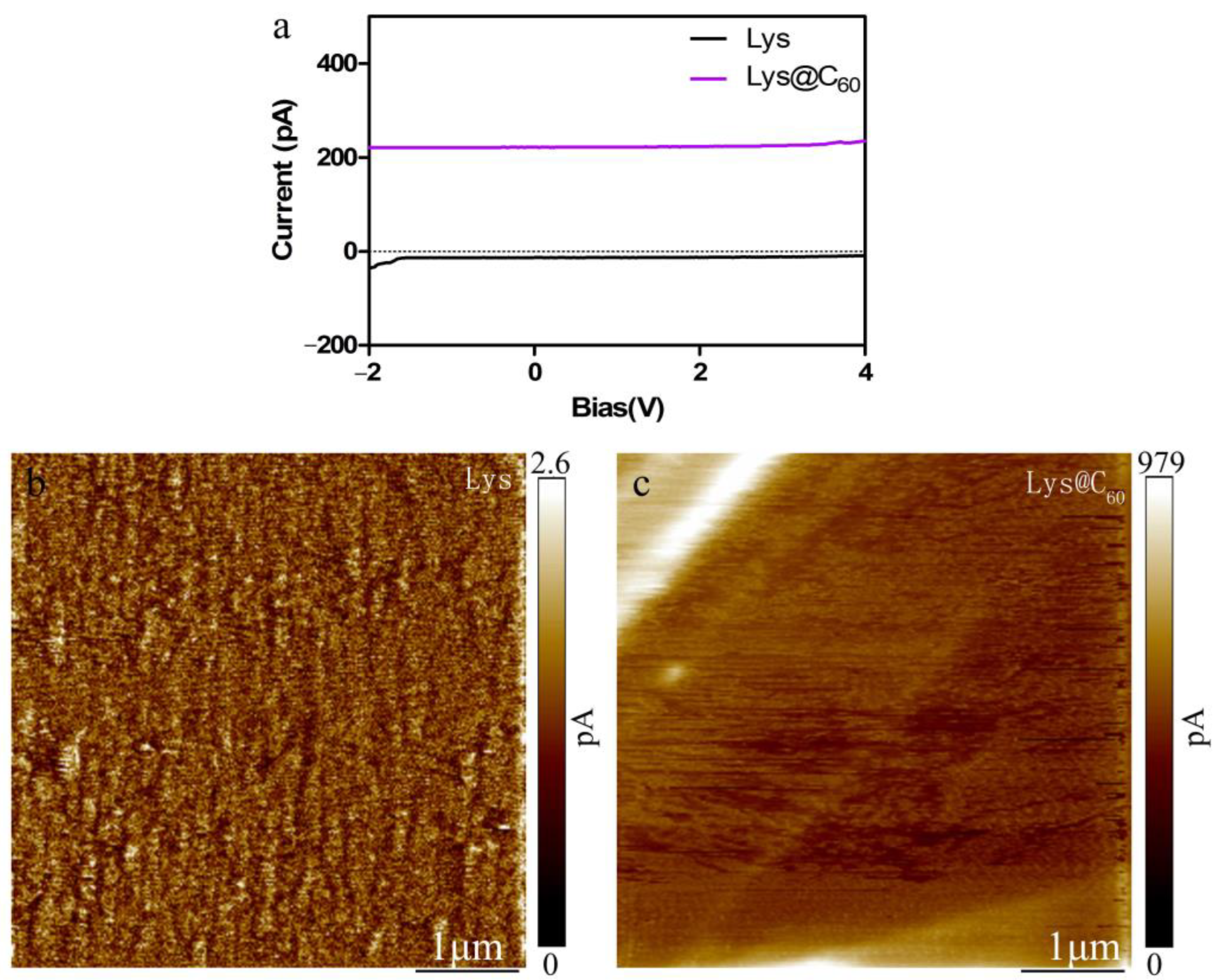
2.3. Conductive Performance of Lys@C60 Tetragonal Crystal

To directly measure the conductive properties of Lys@C60, a conductive atomic force microscope (c-AFM) was employed [26]. The ITO-Lys@C60 electrode was securely affixed to the sample stage using silver tape. The sample’s conductivity was assessed using a CONTSPT probe, and the resulting current was directly recorded through a preamplifier with a gain ranging from 109 to 1011 V/A. The c-AFM spectrum was obtained by applying a voltage of 5 V to the sample and subsequently scanning the sample surface.
The resistance of Lys@C60 was quantified through electrochemical impedance spectroscopy (EIS) using an electrochemical workstation. The experimental setup included a nickel foam–Lys@C60 crystal as the working electrode, a saturated calomel electrode as the reference electrode, a platinum electrode as the counter electrode, and a 1× PBS electrolyte. The bias voltage was maintained at 0 V throughout the experiment. The voltage of the current–time (I-T) curve was set to 0.5 V, and the scan time extended over a period of 30 min.

2.4. Ferroelectric Performance of Lys@C60 Tetragonal Crystals

To investigate the ferroelectric performance of Lys@C60 crystals and lysozyme crystals, switching spectroscopy piezoresponse force microscopy (SS-PFM) was employed [27]. The experimental setup involved fixing the ITO-Lys@C60 electrode or ITO-Lys electrode on the sample stage using silver tape. The tip’s oscillation frequency and amplitude were configured at 1 KHz and 50 nm, respectively. The alternating voltage (VAC) frequency was set at 10 KHz, and the direct voltage (VDC) range spanned from −5 V to 5 V. The scanning protocol involved an initial forward scan, followed by a reverse scan, and the results were recorded for subsequent analysis.

3. Result

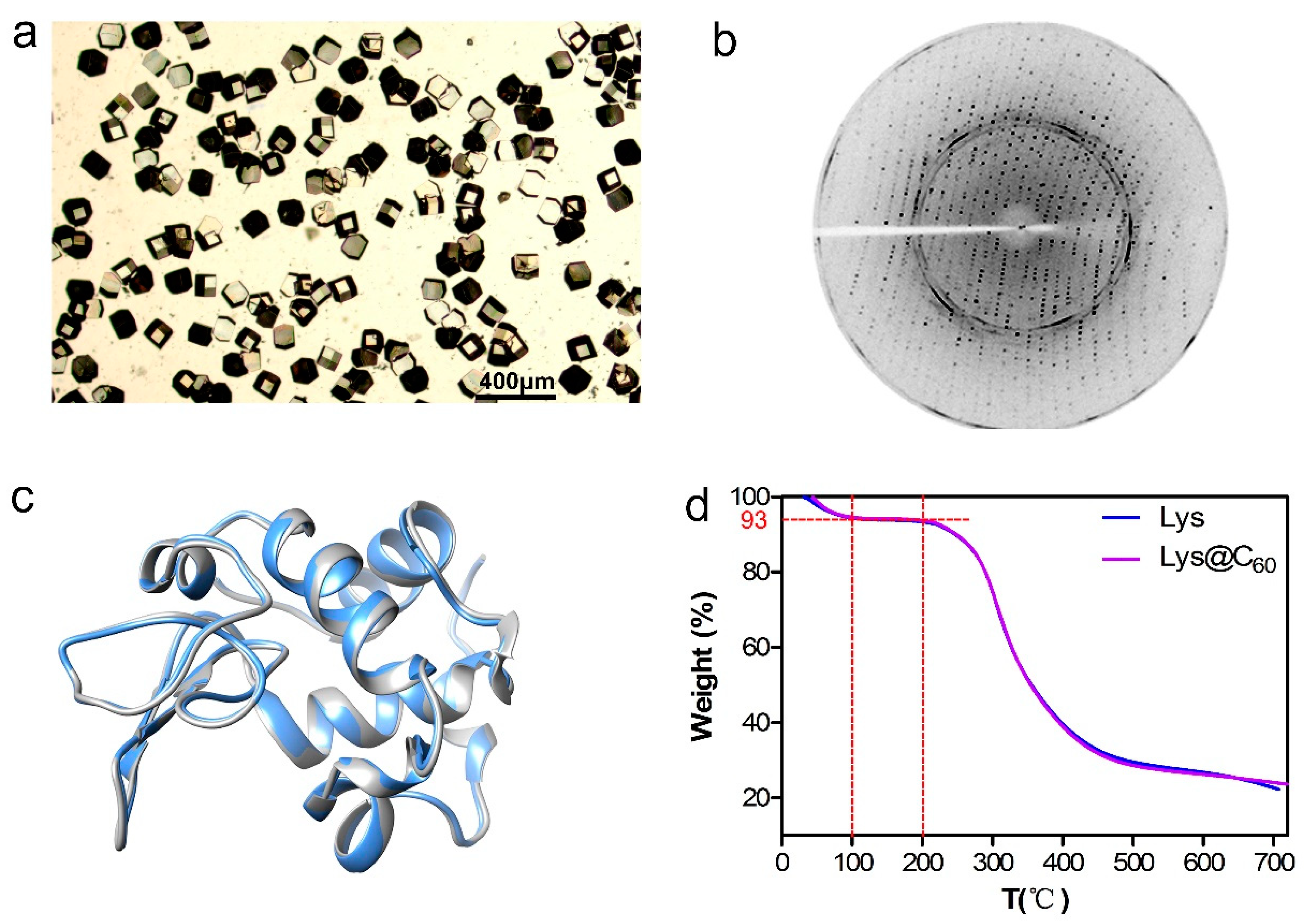
3.1. Characterization of Lys@C60 Crystal

The physical properties of protein crystals, such as ferroelectricity, are governed by their crystallographic symmetry [28]. Various lysozyme crystal forms can be obtained using different crystallization conditions [29]. In this work, tetragonal Lys@C60 crystals were obtained as expected. As shown in Figure 1a, the optical microscopy image illustrates the tetragonal form of Lys@C60 crystals on the ITO electrode. It is noteworthy that within the tetragonal crystal, two available crystals forms (Point Group 422 and Point Group 4) exist. The symmetry constraints play a pivotal role in determining the ferroelectric properties of these crystals [30]. Theoretically, Point Group 4 crystals are expected to be polar and exhibit ferroelectric properties, whereas chiral Point Group 422 crystals are not (Figure S1). The investigation into whether the C60 doping alters the symmetry is crucial before study the ferroelectric properties. Therefore, to accurately determine the point group symmetry of Lys@C60 crystals, X-ray single-crystal diffraction was employed for crystal structure determination. As depicted in Figure 1b, the highest diffraction resolution achieved for the Lys@C60 crystal is 2.1 Å. Subsequent structural analysis revealed that the obtained Lys@C60 crystal belongs to the same P43212 point group as the original lysozyme crystal. Detailed structural data are listed in Table S1. The diffraction spectra of lysozyme crystal are presented in Figure S2. After structural alignment (Figure 1c), it is evident that the structure of the Lys@C60 crystal remains consistent with that of the original lysozyme crystal. These results demonstrate that the doping of C60 does not affect the conformation and point group symmetry of lysozyme crystals.

3.2. Enhancing Stability of Lys@C60 Crystals for Ferroelectric Property Measurement

Protein crystals exhibit soft and brittle characteristics due to their higher water content, varying from 17.4% to 47.2% in different crystal forms [31]. To preserve their inherent structure and prevent cracking and degradation during the measurement of ferroelectric properties, the mechanical properties of proteins can be improved through chemical cross-linking [32]. In this study, Lys@C60 crystals underwent cross-linking with glutaraldehyde to enhance their stability. The cross-linked Lys@C60 crystals displayed significantly improved stability, maintaining their morphology at room temperature without alterations in space group symmetry and structure (Figure 1a–c). To further assess the stability of cross-linked Lys@C60 crystals, thermogravimetric analysis (TGA) was conducted. Figure 1d illustrates that the weight loss of Lys@C60 crystals occurs in three distinct stages. The first stage (0 °C–100 °C) involves the evaporation of 7% bound water in the Lys@C60 crystal. During the second stage (100 °C–200 °C), the mass of Lys@C60 crystal remains essentially constant, indicating that glutaraldehyde cross-linking significantly enhances the thermal stability of the crystal, enabling it to withstand temperatures up to 200 °C. The third stage (200 °C–700 °C) involves pyrolysis, resulting in an 80% mass loss for Lys@C60 crystals. The TGA results for lysozyme crystals are essentially consistent with those of Lys@C60 crystals, suggesting that the doping of C60 does not compromise the thermal stability of lysozyme crystals. Consequently, the stability of Lys@C60 crystals is substantially improved after cross-linking treatment, meeting the characterization requirements for evaluating ferroelectric performance.

3.3. Ferroelectric Evaluation of Lys@C60 Crystals

As previously stated, lysozyme crystals with the P43212 point group exhibit non-ferroelectric properties. In order to investigate whether C60 doping will endow ferroelectricity to P43212 point group lysozyme crystals, the ferroelectric performance of Lys@C60 crystals is evaluated with piezoresponse force microscopy (PFM). PFM has evolved as a powerful tool for measuring the conductivity and ferroelectric properties of materials at the nanoscale. Based on the inverse piezoelectric effect, a specified voltage is applied to the piezoelectric sample through the cantilever beam of the atomic force conductive microscope, inducing sample deformation. During scanning of the sample surface by the atomic force microscope probe, the deformation induced by the sample’s electric field can be detected. Additionally, utilizing the polar scanning method, the piezoelectric force microscope can measure the ferroelectric properties of the sample [33]. If the sample possesses ferroelectric properties, the spontaneous polarization of the sample will change with variations in the applied voltage, allowing the detection of the sample’s ferroelectric properties.
In our study, a piezoelectric force microscope was employed to apply a voltage of 5 V to the Lys@C60 crystal through a conductive probe, scanning the sample surface with an area of (5 × 5 μm2). When a crystal is polarized, the polarity can be detected through scanning. Figure 2a,b illustrate the absence of polarity (ferroelectric domain) in the lysozyme crystal, consistent with the theory that the P43212 lysozyme crystal cannot spontaneously polarize. In contrast, the Lys@C60 crystal, as anticipated, exhibited clear ferroelectric domains and domain walls in different directions in its amplitude diagram (Figure 2c). Moreover, the phase diagram (Figure 2d) revealed domain inversion, indicating that through C60 doping, Lys@C60 crystals can indeed exhibit spontaneous polarization. Therefore, Lys@C60 crystals may possess ferroelectric properties, offering promising prospects for further applications.
To provide further confirmation of ferroelectricity, the characteristic hysteresis loop of Lys@C60 crystals was measured using switching-spectroscopy piezoresponse force microscopy (SS-PFM). In the presence of ferroelectricity, the spontaneous polarization undergoes reversal under the applied field, resulting in a distinctive butterfly loop pattern. As illustrated in Figure 3a,b, the control group exhibits negligible butterfly curves and hysteresis loops for lysozyme crystals, aligning with the theoretical expectation that the P43212 crystal form of lysozyme lacks a ferroelectric effect. Contrastingly, as anticipated, the switching phase loop (Figure 3c) and butterfly-shaped amplitude (Figure 3d) for Lys@C60 clearly indicate its ferroelectric nature. Notably, the characteristic hysteresis loops display asymmetry, suggesting the presence of internal bias within the Lys@C60 crystal. This internal bias is hypothesized to arise from the bound water within Lys@C60 (hydration layer). Due to its strongly bound nature, this water is resistant to the switching direction. As corroborated by the thermogravimetric results (Figure 1d), approximately 7% bound water exists within Lys@C60 crystals. Therefore, these findings demonstrate that C60 doping can indeed bestow ferroelectric properties of the originally non-ferroelectric P43212 lysozyme crystal.
Encouraged by its ferroelectric properties, Lys@C60 may be used as potential bio-compatibility implantable devices in biomedical applications. The cytotoxicity of Lys@C60 in HEK 293 cells was assessed using the CCK-8 method. As depicted in Figure S3, the excellent biocompatibility of Lys@C60 was observed in HEK 293 cells treated with varying concentrations (0.1 mM, 0.2 mM, 0.3 mM, and 0.4 mM), indicating the biosafety of Lys@C60 for biomedical applications.

3.4. Explanation of Observed Ferroelectricity in Lys@C60 Crystals

3.4.1. Lowering Symmetry by C60 Doping

The emergent ferroelectricity of Lys@C60 crystals can be explained through two possible mechanisms. One is that doping C60 may lower the entire symmetry of the Lys@C60 crystal hybrid. Although the X-ray crystallography data determine the P43212 point group of Lys@C60 crystals, the absence of a C60 electron map suggests a disordered distribution of C60 within the lysozyme crystals (Figure 1c). This disordered C60 arrangement may lower the overall symmetry of the Lys@C60 crystal hybrid allowing for switchable spontaneous polarization and resulting in ferroelectricity.

3.4.2. Influence on Polarization by Emerging Conductivity

Another explanation involves C60 doping influencing the polarization of lysozyme crystals by its emerging conductivity. Assembling C60 into electrically insulating protein crystals has been documented to alter the charge distribution and achieve high conductance protein–C60 hybrid [10]. Thus, the influence of C60 on the polarization of Lys@C60 crystals can be detected by the conductivity. To assess the conductive performance of Lys@C60 crystals, conductive force microscopy (c-AFM) was employed. As shown in Figure 4a, lysozyme crystals alone exhibit electrical insulation with zero current, whereas, after C60 doping, Lys@C60 crystals display a weak current of 200 pA, indicating electrical conductivity. This observation is further supported by the current diagram from the conductive atomic force microscope. Again, the lysozyme crystal surface is electrical insulting (Figure 4b), while most of the Lys@C60 crystal surface is not conductive, linear areas with higher conductivity are dispersed across the surface, and the measured current represents an average. In the most conductive areas, currents can reach approximately 1000 pA (Figure 4c). The disorder distribution of C60 within the lysozyme crystal, as indicated by the absence of an electron map in the Lys@C60 crystal during structural analysis (Figure 1c), explains this phenomenon. Therefore, the introduction of C60 facilitates efficient electron transport inside the crystal, contributing to the observed ferroelectricity response.
The assessment of C60 doping enhancing the conductivity of Lys@C60 crystals is also indirectly supported by measuring the impedance of Lys@C60 crystals grown on nickel foam (Figure S4). As depicted in the provided Figure S5, Lys@C60 crystals exhibit a significant reduction in resistance compared to lysozyme crystals alone. It is crucial to emphasize that the measured impedance primarily reflects the resistance of the nickel foam. In reality, both Lys@C60 crystals and lysozyme crystals contribute to an overall increase in the resistance of the nickel foam. However, Lys@C60 crystals demonstrate less resistance compared to lysozyme crystals, indirectly suggesting the improved conductivity of Lys@C60. This additional evidence strengthens the conclusion of the enhanced conductive performance of Lys@C60 crystals resulting from C60 doping. In summary, the enhanced conductivity induced by C60 doping is a key factor in the newfound ferroelectric properties of Lys@C60 crystals.

4. Discussion

Ferroelectric materials, with their reorientable polarization, show great promise for energy storage and generation, particularly in implantable or wearable biomedical applications [34]. Protein crystals, owing to their unique structure symmetry and biodegradability, are considered ideal bio-ferroelectric materials. However, their inherent electrical insulation and fragile nature impede their application in electrical fields. This study aimed to develop methods to improve the stability and electrical properties of protein crystals for electro-mechanical applications.
The primary challenge in studying the electrical properties of protein crystals lies in their poor stability. Unlike other inorganic materials, the direct preparation of protein crystals into electrodes for electrical measurement is challenging. To overcome this issue, cross-linking with glutaraldehyde was employed, significantly enhancing the stability and mechanical properties of lysozyme crystals. Remarkably, even when exposed to temperatures as high as 200 °C, the lysozyme crystal structure remained intact (Figure 1d). Another significant challenge is the direct electroding of lysozyme crystals for electric-mechanical measurements. In this study, we successfully carried out the electroding of lysozyme crystals onto conductive substrates (ITO film and nickel foam) to enable electrochemical performance characterization (see Figure S2). And the conductive substrate did not affect Lys@C60 crystal preparation, allowing their use as electrodes for testing electrical properties.
Additionally, considering the poor electrical conductivity of protein crystals, efforts were made to enhance lysozyme crystal conductivity through C60 doping. The combined results from direct measurements of conductivity through atomic force microscopy and indirect measurements of resistance and current on nickel foam demonstrate that C60 doping does improve the conductivity of lysozyme crystals. The observed I-V curve of Lys@C60 crystals does not exhibit ohmic characteristics (Figure 4a), indicating that the doping content of C60 is low. Under low C60 doping conditions, a tunnel barrier is present in the crystal channel. The mechanism underlying this conductivity improvement is attributed to the disorderly arrangement of C60 within the lysozyme crystal, effectively acting as p-type doping and forming pairs of holes and electrons. Under the influence of an electric field, the movement of carriers within the crystal results in the generation of an electric current. C60 doping enhancing the conductivity of lysozyme crystals pave the way for new applications of protein crystals in the field of bioelectronics.
Surprisingly, C60 doping introduced intriguing ferroelectricity to P422 tetragonal lysozyme crystals. The characteristic ferroelectric hysteresis loops of Lys@C60 crystals were detected with SS-PFM (Figure 3), despite the theoretical expectation that P43212 tetragonal lysozyme crystals lack the ability for spontaneous polarization. Two potential explanations for the ferroelectricity of Lys@C60 crystals are proposed. First, C60 doping may lower the overall symmetry of the Lys@C60 crystal hybrid due to disordered C60 distribution, enabling switchable spontaneous polarization. Second, C60 doping might enhance the conductivity of Lys@C60 crystals, influencing the electric charge distribution and changing spontaneous polarization, resulting in ferroelectricity. The detailed mechanism warrants further exploration. In summary, with their natural biocompatibility and newfound ferroelectric effect, Lys@C60 crystals present a competitive alternative to traditional piezoelectric energy harvesting systems, especially in biomedical applications where they can be utilized as biodegradable implantable devices like pacemakers and artificial synapses.

5. Conclusions

In summary, we propose a simple C60 doping strategy to endow non-conductive and non-ferroelectric lysozyme crystals with conductive and ferroelectric properties. Our methodology involves enhancing the stability and electroding of Lys@C60 crystals for electric-mechanical measurements. Results from c-AFM and SS-PMF analyses demonstrate that C60 doping endows ferroelectricity and conductivity to tetragonal lysozyme crystals. The increased conductivity of Lys@C60 crystals is attributed to the disorderly arrangement of C60 within the lysozyme crystal, forming specific hole-electron pairs capable of carrying current under an electric field. Additionally, the enhanced conductivity and reduced symmetry contribute to the ferroelectricity of Lys@C60 crystals. Therefore, this work verifies that C60 doping can serve as a simple yet effective strategy to impart novel ferroelectric properties to lysozyme crystals, potentially rendering them suitable for energy storage and conversion to achieve biocompatible and biodegradable implantable and wearable bioelectronics applications.

Supplementary Materials

The following supporting information can be downloaded at: https://www.mdpi.com/article/10.3390/cryst14040339/s1, Figure S1: The physical properties of 32 point groups categorized depending on their symmetry; Figure S2: The diffraction spectra of lysozyme crystal; Figure S3: Cell viability of HEK 293 cells treated with various concentration of Lys@C60; Figure S4: Electroding of Lys@C60 crystals. (a) the ITO electrode with Lys@C60 crystals; (b) the Ni foam electrode with Lys@C60 crystals; Figure S5: The electrical properties of Lys@C60 crystals growth on Ni foam. (a) the EIS of Lys@C60 crystals; (b) the I-T curve of Lys@C60 crystals; Table S1: The diffraction data of Lys@C60 crystal and lysozyme crystals.

Author Contributions

Methodology, R.Z., X.L., W.G. and D.Y.; Software, W.G.; Validation, R.Z. and D.Y.; Formal analysis, X.L.; Investigation, R.Z.; Resources, D.Y.; Data curation, X.L.; Writing—original draft, R.Z.; Writing—review & editing, D.Y.; Visualization, R.Z.; Supervision, D.Y.; Project administration, D.Y.; Funding acquisition, D.Y. All authors have read and agreed to the published version of the manuscript.

Funding

This work was supported by the National Natural Science Foundation of China (NSFC) (Grant No. 82172063), the Science and Technology Program of Ali Region, Tibet (Program No. QYXTZX-AL2022-07), Key R&D Project in Shaanxi Province (Grant No. 2023-YBNY-068), and the Fundamental Research Funds for the Central Universities (Grant No. 23GH02021).

Institutional Review Board Statement

Not applicable.

Informed Consent Statement

Not applicable.

Data Availability Statement

Data are contained within the article.

Conflicts of Interest

The author declares no conflicts of interest.

References

  1. Liao, W.-Q.; Zeng, Y.-L.; Tang, Y.-Y.; Peng, H.; Liu, J.-C.; Xiong, R.-G. Multichannel control of multiferroicity in single-component homochiral organic crystals. J. Am. Chem. Soc. 2021, 143, 21685–21693. [Google Scholar] [CrossRef] [PubMed]
  2. Yuan, X.; Shi, J.; Kang, Y.; Dong, J.; Pei, Z.; Ji, X. Piezoelectricity, Pyroelectricity and Ferroelectricity in Biomaterials and Biomedical Applications. Adv. Mater. 2023, 36, 2308726. [Google Scholar] [CrossRef] [PubMed]
  3. Guerin, S.; Stapleton, A.; Chovan, D.; Mouras, R.; Gleeson, M.; McKeown, C.; Noor, M.R.; Silien, C.; Rhen, F.M.F.; Kholkin, A.L.; et al. Control of piezoelectricity in amino acids by supramolecular packing. Nat. Mater. 2018, 17, 180–186. [Google Scholar] [CrossRef] [PubMed]
  4. Panda, S.; Hajra, S.; Song, H.; Jo, J.; Kim, N.; Hwang, S.; Choi, Y.; Kim, H.G.; Kim, H.J.; Mishra, Y.K. Pyroelectric based energy harvesting devices: Hybrid structures and applications. Sustain. Energy Fuels 2023, 7, 5319–5335. [Google Scholar] [CrossRef]
  5. Hu, P.; Hu, S.; Huang, Y.; Reimers, J.R.; Rappe, A.M.; Li, Y.; Stroppa, A.; Ren, W. Bioferroelectric properties of glycine crystals. J. Phys. Chem. Lett. 2019, 10, 1319–1324. [Google Scholar] [CrossRef] [PubMed]
  6. Yun, Y.; Buragohain, P.; Li, M.; Ahmadi, Z.; Zhang, Y.; Li, X.; Wang, H.; Li, J.; Lu, P.; Tao, L.; et al. Intrinsic ferroelectricity in Y-doped HfO2 thin films. Nat. Mater. 2022, 21, 903–909. [Google Scholar] [CrossRef] [PubMed]
  7. Sirotin, Y.I.; Shaskol’skaia, M.P. Fundamentals of Crystal Physics; MIR Publishers: Moscow, Russia, 1982. [Google Scholar]
  8. Stapleton, A.; Noor, M.R.; Sweeney, J.; Casey, V.; Kholkin, A.L.; Silien, C.; Gandhi, A.A.; Soulimane, T.; Tofail, S.A.M. The direct piezoelectric effect in the globular protein lysozyme. Appl. Phys. Lett. 2017, 111, 142902. [Google Scholar] [CrossRef]
  9. Lee, D.H.; Lee, Y.; Cho, Y.H.; Choi, H.; Kim, S.H.; Park, M.H. Unveiled Ferroelectricity in Well-Known Non-Ferroelectric Materials and Their Semiconductor Applications. Adv. Funct. Mater. 2023, 33, 2303956. [Google Scholar] [CrossRef]
  10. Kim, K.-H.; Ko, D.-K.; Kim, N.H.; Paul, J.; Zhang, S.-Q.; Murray, C.B.; Acharya, R.; DeGrado, W.F.; Kim, Y.H.; Grigoryan, G. Protein-directed self-assembly of a fullerene crystal. Nat. Commun. 2016, 7, 11429. [Google Scholar] [CrossRef]
  11. Minary-Jolandan, M.; Yu, M.-F. Uncovering nanoscale electromechanical heterogeneity in the subfibrillar structure of collagen fibrils responsible for the piezoelectricity of bone. ACS Nano 2009, 3, 1859–1863. [Google Scholar] [CrossRef]
  12. Harnagea, C.; Vallières, M.; Pfeffer, C.P.; Wu, D.; Olsen, B.R.; Pignolet, A.; Légaré, F.; Gruverman, A. Two-dimensional nanoscale structural and functional imaging in individual collagen type I fibrils. Biophys. J. 2010, 98, 3070–3077. [Google Scholar] [CrossRef] [PubMed]
  13. Minary-Jolandan, M.; Yu, M.-F. Nanoscale characterization of isolated individual type I collagen fibrils: Polarization and piezoelectricity. Nanotechnology 2009, 20, 085706. [Google Scholar] [CrossRef] [PubMed]
  14. Liu, Y.; Cai, H.-L.; Zelisko, M.; Wang, Y.; Sun, J.; Yan, F.; Ma, F.; Wang, P.; Chen, Q.C.; Zheng, H.; et al. Ferroelectric switching of elastin. Proc. Natl. Acad. Sci. USA 2014, 111, E2780–E2786. [Google Scholar] [CrossRef] [PubMed]
  15. Liu, Y.; Wang, Y.; Chow, M.-J.; Chen, N.Q.; Ma, F.; Zhang, Y.; Li, J. Glucose suppresses biological ferroelectricity in aortic elastin. Phys. Rev. Lett. 2013, 110, 168101. [Google Scholar] [CrossRef] [PubMed]
  16. Stapleton, A.; Ivanov, M.S.; Noor, M.R.; Silien, C.; Gandhi, A.A.; Soulimane, T. Converse piezoelectricity and ferroelectricity in crystals of lysozyme protein revealed by piezoresponse force microscopy. Ferroelectrics 2018, 525, 135–145. [Google Scholar] [CrossRef]
  17. Denning, D.; Kilpatrick, J.I.; Fukada, E.; Zhang, N.; Habelitz, S.; Fertala, A.; Gilchrist, M.D.; Zhang, Y.; Tofail, S.A.M.; Rodriguez, B.J. Piezoelectric tensor of collagen fibrils determined at the nanoscale. ACS Biomater. Sci. Eng. 2017, 3, 929–935. [Google Scholar] [CrossRef] [PubMed]
  18. Miao, L.-P.; Ding, N.; Wang, N.; Shi, C.; Ye, H.-Y.; Li, L.; Yao, Y.-F.; Dong, S.; Zhang, Y. Direct observation of geometric and sliding ferroelectricity in an amphidynamic crystal. Nat. Mater. 2022, 21, 1158–1164. [Google Scholar] [CrossRef] [PubMed]
  19. Soldà, A.; Cantelli, A.; Di Giosia, M.; Montalti, M.; Zerbetto, F.; Rapino, S.; Calvaresi, M. C 60@ lysozyme: A new photosensitizing agent for photodynamic therapy. J. Mater. Chem. B 2017, 5, 6608–6615. [Google Scholar] [CrossRef] [PubMed]
  20. Brahmkhatri, V.; Atreya, H.S. Dynamics of Protein–Nanoparticle Interactions Using NMR. In NMR Spectroscopy for Probing Functional Dynamics at Biological Interfaces; Royal Society of Chemistry: London, UK, 2022; pp. 236–253. [Google Scholar]
  21. Zhou, R.; Ohulchanskyy, T.Y.; Xu, Y.; Ziniuk, R.; Xu, H.; Liu, L.; Qu, J. Tumor-microenvironment-activated NIR-II nanotheranostic platform for precise diagnosis and treatment of colon cancer. ACS Appl. Mater. Interfaces 2022, 14, 23206–23218. [Google Scholar] [CrossRef]
  22. Zhou, R.; Xu, H.; Qu, J.; Ohulchanskyy, T.Y. Hemoglobin nanocrystals for drugs free, synergistic theranostics of colon tumor. Small 2023, 19, 2205165. [Google Scholar] [CrossRef]
  23. Afonine, P.V.; Grosse-Kunstleve, R.W.; Echols, N.; Headd, J.J.; Moriarty, N.W.; Mustyakimov, M.; Terwilliger, T.C.; Urzhumtsev, A.; Zwart, P.H.; Adams, P.D. Towards automated crystallographic structure refinement with phenix.refine. Acta Crystallogr. Sect. D Biol. Crystallogr. 2012, 68, 352–367. [Google Scholar] [CrossRef]
  24. Emsley, P.; Cowtan, K. Coot: Model-building tools for molecular graphics. Acta Crystallogr. Sect. D Biol. Crystallogr. 2004, 60, 2126–2132. [Google Scholar] [CrossRef]
  25. Murshudov, G.N.; Vagin, A.A.; Dodson, E.J. Refinement of macromolecular structures by the maximum-likelihood method. Acta Crystallogr. Sect. D Biol. Crystallogr. 1997, 53, 240–255. [Google Scholar] [CrossRef] [PubMed]
  26. Lanza, M. Conductive Atomic Force Microscopy: Applications in Nanomaterials; John Wiley & Sons: Hoboken, NJ, USA, 2017. [Google Scholar]
  27. Jesse, S.; Baddorf, A.P.; Kalinin, S.V. Switching spectroscopy piezoresponse force microscopy of ferroelectric materials. Appl. Phys. Lett. 2006, 88, 062908. [Google Scholar] [CrossRef]
  28. Liu, H.-Y.; Zhang, H.-Y.; Chen, X.-G.; Xiong, R.-G. Molecular design principles for ferroelectrics: Ferroelectrochemistry. J. Am. Chem. Soc. 2020, 142, 15205–15218. [Google Scholar] [CrossRef] [PubMed]
  29. Tang, X.H.; Liu, J.J.; Zhang, Y.; Wang, X.Z. Study on the influence of lysozyme crystallization conditions on crystal properties in crystallizers of varied sizes when temperature is the manipulated variable. J. Cryst. Growth 2018, 498, 186–196. [Google Scholar] [CrossRef]
  30. Shi, P.-P.; Tang, Y.-Y.; Li, P.-F.; Liao, W.-Q.; Wang, Z.-X.; Ye, Q.; Xiong, R.-G. Symmetry breaking in molecular ferroelectrics. Chem. Soc. Rev. 2016, 45, 3811–3827. [Google Scholar] [CrossRef]
  31. Tait, S.L.; White, E.T.; Litster, J.D. Mechanical characterization of protein crystals. Part. Part. Syst. Charact. 2008, 25, 266–276. [Google Scholar] [CrossRef]
  32. Zhou, R.; Ohulchanskyy, T.Y.; Xu, H.; Ziniuk, R.; Qu, J. Catalase Nanocrystals Loaded with Methylene Blue as Oxygen Self-Supplied, Imaging-Guided Platform for Photodynamic Therapy of Hypoxic Tumors. Small 2021, 17, 2103569. [Google Scholar] [CrossRef] [PubMed]
  33. Bonnell, D.; Kalinin, S.; Kholkin, A.; Gruverman, A. Piezoresponse force microscopy: A window into electromechanical behavior at the nanoscale. MRS Bull. 2009, 34, 648–657. [Google Scholar] [CrossRef]
  34. Wang, W.; Li, J.; Liu, H.; Ge, S. Advancing versatile ferroelectric materials toward biomedical applications. Adv. Sci. 2021, 8, 2003074. [Google Scholar] [CrossRef] [PubMed]
Figure 1. Characterization of Lys@C60 crystals. (a) optical image of Lys@C60 tetragonal crystals; (b) the diffraction pattern of Lys@C60 crystal; (c) the structure alignment of Lys@C60 (blue color) and lysozyme (gray color); (d) the TGA analysis of Lys@C60 and lysozyme crystals.
Crystals 14 00339 g001
Figure 2. The ferroelectricity of lysozyme and Lys@C60 crystals. (a) The amplitude of lysozyme crystals; (b) the phase of lysozyme crystals; (c) the amplitude of Lys@C60 crystals; (d) the phase of Lys@C60 crystals.
Crystals 14 00339 g002
Figure 3. The ferroelectricity of lysozyme and Lys@C60 crystals. (a) The switching phase loop of lysozyme crystals; (b) the butterfly shape amplitude of lysozyme crystals; (c) the switching phase loop of Lys@C60 crystals; (d) the butterfly shape amplitude of Lys@C60 crystals.
Crystals 14 00339 g003
Figure 4. The electrical properties of Lys@C60 crystals. (a) The I-V curve of Lys@C60 crystals; (b) the c-AFM map of lysozyme crystals; (c) the c-AFM map of Lys@C60 crystals.
Crystals 14 00339 g004
Disclaimer/Publisher’s Note: The statements, opinions and data contained in all publications are solely those of the individual author(s) and contributor(s) and not of MDPI and/or the editor(s). MDPI and/or the editor(s) disclaim responsibility for any injury to people or property resulting from any ideas, methods, instructions or products referred to in the content.

