Article

Photocatalytic and Antimicrobial Activity of Titanium(IV)-Oxo Clusters of Different Core Structure

1
Department of Inorganic and Coordination Chemistry, Faculty of Chemistry, Nicolaus Copernicus University in Toruń, Gagarina 7, 87-100 Toruń, Poland
2
Department of Microbiology, Faculty of Biological and Veterinary Sciences, Nicolaus Copernicus University in Toruń, Lwowska 1, 87-100 Toruń, Poland
*
Authors to whom correspondence should be addressed.
Crystals 2023, 13(7), 998; https://doi.org/10.3390/cryst13070998
Submission received: 29 May 2023 / Revised: 19 June 2023 / Accepted: 20 June 2023 / Published: 22 June 2023
(This article belongs to the Special Issue Metal Oxides: Crystal Structure, Synthesis and Characterization)

Abstract

:
The purpose of this paper is to assess the relationship between the core architecture of titanium(IV)-oxo complexes (TOCs) known as {TiaOb} and their photocatalytic and antimicrobial activity. The following TOCs: [Ti6O4(OiBu)8(O2C13H9)8] · 2(CH3)2CO (1), [Ti6O6(OiBu)6(O2C13H9)6] (2), [Ti6O6(OiBu)6(O2C13H9)6] (3), [Ti3O(OiPr)8(O2C13H9)2] (4), and [Ti4O2(OiBu)10(O2C13H9)2] (5), where -O2C13H9represents 9-fluorene-carboxylate ligands, werestudied to investigate thiseffect. The structures of (1)–(5) were confirmed using single-crystal X-ray diffraction and spectroscopic methods. Since TOCs can be sensitive to hydrolysis processes, their photocatalytic and antimicrobial activity was evaluated after dispersing them in a polymer matrix, which acted as a protective agent against the aquatic environment. The results revealed that the photocatalytic activity of the studied TOCs follows the trend (2) > (5) > (4) > (1) in both the UV and visible ranges. All studied oxo complexes exhibited strong antibacterial activity against Gram-positive strains and weaker activity against Gram-negative strains. The proposed mechanism of the antimicrobial activity of TOCs assumes that this effect is associated with the generation of reactive oxygen species (ROS) on the surface of composite samples. Samples of PMMA + (1) 10 wt.% and PMMA + (5) 20 wt.%, in which both O and O2 paramagnetic species were observed in the electron paramagnetic spectroscopy (EPR) spectra, demonstrated the highest antimicrobial activity.

1. Introduction

A significant part of the research on new materials that improve the quality of our life involves materials based on titanium dioxide. These materials have diverse applications, including water treatment [1], photocatalysis [2,3], sun protection [4], self-cleaning surfaces [5], pharmaceutical and food industries [6,7], and the production of biocompatible and antibacterial implants [8,9]. An important direction for the application of TiO2-based materials, which exhibit photocatalytic properties, is their use in antibiotic elimination technologies [10]. The main requirement is extensive use of antibiotics, primarily in agriculture but also in medicine, which poses a huge threat to our environment. Therefore, it is important to develop new photocatalysts that are environmentally friendly, cost-effective to produce, and characterized by high activity in the photodecomposition of antibiotics [11,12]. However, TiO2-based materials face limitations in their practical use as photocatalysts due to their wide bandgap, which primarily requires ultraviolet excitation. Moreover, the recombination of electrons and light-induced holes easily occurs, reducing the photocatalytic activity of titania particles. Therefore, the main aim of these investigations is the development and production of a photocatalyst whose activity is shifted toward visible light.
To address this challenge, attention has turned to multinuclear oxo-titanium(IV) complexes (TOCs) with a core composed of {TiaOb} skeletons [13,14,15]. The advantage of these compounds is the ability to control their physicochemical, photocatalytic, and biological properties via changes in the core size and architecture of {TiaOb}, as well as the functionalization of carboxylate groups as stabilizing ligands [16,17,18,19,20]. From the perspective of TOC synthesis, the type of alkoxide groups, solvent, and temperature are also important factors [19,20,21]. Investigations on clusters with {Ti4O} and {Ti4O2} cores, stabilized by 4-tert-butylcatechol and N, N-diethylformamide ligands, used in the photo-induced direct degradation of methylene blue (MB) in visible light, are good examples of this [21]. The study shows that after 2.5 h, 98% degradation of MB concentration is observed, whereas, in the case of TiO2, only 15% degradation is observed at the same time. The better photocatalytic activity of TOCs is due to the narrowed band gap compared to TiO2 and the wide absorption band [21]. Functionalization of poly-oxo-titanate clusters with 1,1-ferrocene dicarboxylic acid ligands resulted in obtaining TOCs with {Ti8O4} and {Ti6O2} cores. The ligands cause an increase in the absorption maximum into the visible range and increase the electron transfer in 1,1-ferrocene dicarboxylic acid and Ti-oxo skeletons [22]. Moreover, obtaining sub-clusters with {Ti4O} and {Ti6O4} cores exhibit a better ability to degrade MB than each of the clusters used separately [23,24]. It should be noted that another way to increase the photocatalytic activity of titanium-based materials is to introduce an extra atom into the {TiaOb} core. The research of Wang et al. on clusters with the core {Ti4Co2O4}, {Ti5Cu4O6}, {Ti12Cd5O18} is an example [25].
The limited knowledge related to the bactericidal activity of TOCs is one of the factors that justify research on their use as antimicrobial agents. Swenson et al. pointed out the possibility of using TOCs as an antibacterial agent against Staphylococcus aureus for {Ti4O2} clusters stabilized by triclosan ligands [26]. However, these works focused on studying the antibacterial properties of the hydrolyzed complex, which may be used for drug delivery applications. When studying the use of transition metal oxides (e.g., TiO2, CuO, ZnO, Fe2O3) as antimicrobial agents, it was noted that after activation with light at a specific wavelength, reactive oxygen species (ROS) are generated on their surface [27,28,29,30]. According to these investigations, the formed ROS are responsible for microbial cell death (e.g., Escherichia coli (E. coli), Staphylococcus aureus (S. aureus), Rhizopus stolonifer, Rhizopus oryzae) [29]. Therefore, in our work, we decided to test this antimicrobial effect in the case of TOCs. The main issue that needed to be addressed was the protection of the oxo complex grains against potential hydrolysis processes [31,32]. For this reason, we chose to disperse TOCs grains in a polymer matrix (e.g., poly(methyl methacrylate) (PMMA)) [31,32]. The results of our earlier investigations of composites enriched with TOCs, containing the {Ti4O2} cores and stabilized by 4-aminobenzoic, 4-hydroxybenzoic, and 9-fluorene-carboxylate acid ligands, demonstrated their strong antimicrobial activity at levels of 85–99% against Gram-positive and Gram-negative bacteria [31,32]. Analysis of this data revealed that the highest activity was observed for TOCs stabilized by 9-fluorene-carboxylate ligands (O2C13H9) Początekformularza [32].
The presented paper focuses on the comparative analysis of the photocatalytic and antimicrobial activities of Ti(IV)-oxo complexes. These complexes contain {Ti6O6}, {Ti6O4}, {Ti4O2}, and {Ti3O} skeletons, and their structures were confirmed using X-ray single crystal diffraction. The stability of these oxo clusters was achieved through the use of a 9-fluorene-carboxylate ligand, along with -OiBu and -OiPr (only for {Ti3O}) ones. This systematic approach allows us to examine how the structure of the {TiaOb} core influences the properties of these complexes. Notably, our research contributes novelty by assessing the impact of the core structure on the antimicrobial activity exhibited by the synthesized oxo complexes.

2. Materials and Methods

2.1. General ProcedureandReagents

Titanium(IV) isobutoxide (Aldrich, St. Louis, MO, USA), titanium(IV) isopropoxide (Aldrich, St. Louis, MO, USA), 9-fluorene-carboxylic acid (TCI, Tokyo, Japan) were purchased commercially and were used without further purification. All solvents used in our experiments were dried, distilled before use, and stored in an argon atmosphere. The processes of Ti(IV) oxo-complexes syntheses were carried out using the standard Schlenk technique in the inert gas atmosphere (Ar) and at room temperature (RT).

2.2. Analytical Procedures

The structures of the isolated solid reaction products (crystals) were confirmed using vibrational spectroscopy methods, i.e., IR spectrophotometry (Perkin Elmer Spectrum 2000 FTIR spectrophotometer (400–4000 cm−1 range, KBr pellets)) and Raman microscopy (RamanMicro 200 spectrometer (PerkinElmer, Waltham, MA, USA)). Elemental analyses were performed on Elemental AnalyserVario Macro CHN (Elementar Analysensysteme GmbH, Langenselbold, Germany). Titanium content was gravimetrically determined as TiO2, according to a method described by Meth-Chon et al. [33]. Raman spectra were registered using a laser with the wavelength 785 nm, with a maximum power of 350 mW, in the range 200–3200 cm−1, using a 20 × 0.40/FN22 objective lens and an exposure time of 15 s each time. The diffuse reflectance UV-Vis (UV-VIS-DRS) spectra registered between 200 and 800 nm were used for the maximum absorption determination. The Jasco V-750 spectrophotometer was used in above mentioned works (JASCO Deutschland GmbH, Pfungstadt, Germany). The produced PMMA + TOCs film surfaces morphology was studied using a scanning electron microscope with field emission (SEM, Quanta 3D FEG, Houston, TX, USA).

2.3. Synthesis of [Ti6O4(OiBu)8(O2C13H9)8] · 2(CH3)2CO (1), [Ti6O6(OiBu)6(O2C13H9)6] (2), and [Ti6O6(OiBu)6(O2C13H9)6] (3)

A total of 0.184 g of 9-fluorene-carboxylic acid (0.875 mmol) was added to the solution of 0.3 g of titanium(IV) isobutoxide (0.875 mmol) in 2 mL of acetone, leading to a clear violet solution. The solution was left for crystallization. After 6–7 days, the mixture of crystals was formed, and the general yield basing on acid: (1) 15% (0.05 g), (2) 21% (0.07 g), (3) 5% (0.01 g). Using the optical microscope, the crystals of different shapes were separated and isolated from the reaction mixture. Elemental composition was determined for samples, and structures were solved by single-crystal X-ray diffraction.
Anal. Calc. for C116H138O26Ti6 (1): C, 62.31; H, 6.18; Ti, 12.89. Found: C, 62.14; H, 6.13; Ti, 12.56.13C NMR (solid state, 295 K, δ[ppm]): 19.7 (CH3), 31.3 (CH), 52.2, 53.4, 55.4, 56.8, 59.6, 73.6, 74.6 (CH2), 119.6, 120.7, 127.5, 141.5 (C(Ph)),175.4, 177.6, 178.7 (COO).
Anal. Calc. for C102H108O24Ti6 (2): C, 62.32; H, 5.49; Ti,14.60. Found: C, 62.17; H, 5.86;Ti, 14.64. 13C NMR (solid state, 295 K, δ[ppm]): 19.6 (CH3), 30.6 (CH), 53.7, 54.6, 73.7 (CH2), 121.3, 128.5, 140.7 (C(Ph)), 176.4, 177.9, 179.7 (COO).
Anal. Calc. for C102H108O24Ti6 (3): C, 62.32; H, 5.49; Ti,14.60. Found: C, 62.23; H, 5.76;Ti, 14.75. 13C NMR (solid state, 295 K, δ[ppm]): 19.6 (CH3), 30.6 (CH), 73.7 (CH2), 54.6, 121.3, 128.5, 140.7 (C(Ph)), 176.4, 177.9, 179.7 (COO).

2.4. Synthesis of [Ti3O(OiPr)8(O2C13H9)2] (4)

The complex was synthesized, as reported [34]. 0.184 g of 9-fluorene-carboxylic acid (0.875 mmol) was added to the solution of 1 mL of titanium(IV) isopropoxide (3.5 mmol) in 2 mL of acetone, leading to a clear yellow solution. The solution was left for crystallization (3–4 days). The yield basing on acid: 62% (0.36 g).
Anal. Calc. for C50H74O13Ti3 (4): C, 58.49; H, 7.21; Ti,14.04. Found: C, 58.54; H, 7.13; Ti, 13.95. 13C NMR (solid state, 295 K, δ[ppm]): 14.0, 19.5 (CH3), 30.9, 35.1 (CH), 55.4, 77.8 (CH2), 119.2, 127.6, 141.6 (C(Ph)), 179.6 (COO).

2.5. Synthesis of [Ti4O2(OiBu)10(O2C13H9)2] (5)

The complex was synthesized, as reported [35]. 0.184 g of 9-fluorene-carboxylic acid (0.875 mmol) was added to the solution of 1.19 g of titanium(IV) isobutoxide (3.5 mmol) in 2 mL of acetone, leading to a clear yellow solution. The solution was left for crystallization. The yield basing on acid: 78% (0.39 g).
Anal. Calc. for C68H108O16Ti4 (5): C, 59.28; H, 7.93; Ti, 13.94. Found: C, 58.17; H, 7.86; Ti, 13.64. 13C NMR (solid state, 295 K, δ[ppm]): 14.2, 19.3 (CH3), 34.8 (CH), 55.1, 77.3 (CH2), 119.3, 127.4, 141.9 (C(Ph)), 178.8 (COO).

2.6. Single Crystal X-ray Diffraction Measurements

The diffraction data of (1)–(3) were collected at 100 K on Rigaku XtaLAB Synergy (Dualflex) diffractometer with HyPix detector and monochromatedCuKα X-ray source (λ = 1.54184 Å). The data were processed, and the numerical absorption correction was applied using CrysAlis Pro [36]. The structures were solved by direct methods and refined with full-matrix least-squares procedure on F2 (SHELX-97 [37]. Heavy atoms were refined with anisotropic displacement parameters, whereas hydrogen atoms were assigned at calculated positions with thermal displacement parameters fixed to a value of 20% or 50% higher than those of the corresponding carbon atoms. We also observed positional disorder for some isobutanolate anions (O66 and O86 in (1), O71 and O81 in (2) and (3)). For the stability of the refinement process, several geometrical (DFIX) and thermal parameters (ISOR) restraints were applied for the disordered isobutanolates and acetone molecules. Additionally, EADP constraints were used for disordered BuiO anions. All figures were prepared in DIAMOND [38] and ORTEP-3 [39].

2.7. Preparation of PMMA + TOCs Composite Films

The composite films containing 10 or 20 wt.% of synthesized Ti(IV)-oxo clusters (TOCs) were prepared by a dispersion of the TOCs (ca. 0.12 or 0.25 g of (1), (2), (4), and (5) TOCs dispersed in 1 cm3 of THF) in the poly(methyl methacrylate) (PMMA) solution (1.0 g of PMMA dissolved in 5 cm3 of THF). The mixtures were stirred in an ultrasonic bath for 120 min; in the next step, they were poured into a glass Petri dish and left for the evaporation of the solvent at RT (glove box). The composite film thickness ca. 50 µm was characterized by Raman and IR spectroscopy and scanning electron microscopy. Due to the low synthesis efficiency of (3), the crystals of this oxo cluster were not used in our subsequent experiments. The received foils, after thorough drying in a vacuum, were cut into pieces of 1 × 1 cm for photocatalytic experiments and 3 × 3 cm for microbiological tests.

2.8. The Photocatalytic Activity Evaluation of PMMA + TOCs Composite Films

The photocatalytic activity of PMMA + TOCs foils (TOCs = (1), (2), (4), (5)) was estimated by monitoring the concentration changes of MB aqueous solution. Foil samples (10 × 10 mm) were preconditioned by exposure to UVA or Vis light (the range of radiation used to irradiate the samples were selected based on the analysis of their UV-VIS-DRS spectra) and after 28 h placed in quartz cuvettes with dye solution (V = 3.5 cm3 and C = 2.0 × 10⁻⁵ M). After 12 h in the dark, the solution was replaced by MB solution (C = 1.0 × 10⁻⁵ M). The prepared samples were exposed to UVA irradiation (18-W lamp, 340–395-nm range) and Vis light (range of 395–420 nm). MB absorbance at 664 nm was registered (Metertech SP-830 PLUS, Metertech, Inc., Taipei, Taiwan) every 2 h during 18 h (UV range) and 24 h (Vis range) of irradiation. The percentage of MB decolorization was calculated using the equation:
%   MB   decolorization = ( C 0 C t ) C 0 × 100
where C0 is an initial concentration of dye, and Ct is a dye concentration at a given time t [40].

2.9. EPR Studies

Electron paramagnetic resonance (EPR) spectroscopy was employed to confirm the formation of reactive oxygen species on the surface of the investigated composite samples. The measurements were conducted using an X-band EPR SE/X-2541M spectrometer (Radi-open, Poznań, Poland) with a 100 kHz modulation. The microwave frequency was monitored using a frequency meter, while the magnetic field was measured using an automatic NMR-type JTM-147 magnetometer (Radiopan, Poznań, Poland). The measurement conditions were as follows: microwave frequency of approximately 9.33 GHz, microwave power ranging from 6 to 58 mW, modulation amplitude between 0.2 and 1 mT, sweep ranging from 10 to 50 mT, sweep time from 1 to 8 min, time constant from 0.03 to 0.3 s, and receiver gain from 3.2 to 5 × 10⁵. Low-intensity signals were recorded in the accumulation mode. The measurements were performed at room temperature using foil samples that were simultaneously prepared for all studies. The portion utilized in the EPR measurements was exposed to sunlight. The catalytic and EPR measurements were conducted independently, approximately at the same time.

2.10. Antimicrobial Activity of PMMA + TOCs Composite Films

The antimicrobial activity of the samples was assessed against Gram-negative bacteria (Escherichia coli ATCC 25922, Escherichia coli ATCC 8739), Gram-positive bacteria (Staphylococcus aureus ATCC 25923, Staphylococcus aureus ATCC 6538), and Candida albicans ATCC 10231. Prior to the antimicrobial study, the tested PMMA + TOCs foils (20 × 20 mm) were sterilized using UVC for 15 min on both sides and then exposed to visible indoor light. The foils were placed in 12-well plates with 1 mL of microbial inoculum (1.0–3.3 × 10⁶ c.f.u. mL−1) in sterile deionized water. The plates were incubated for 24 h at 37 °C under humid conditions with gentle shaking at 80 rpm. The microbial density was adjusted to 0.5 on the McFarland scale using a densitometer (Biosan, Latvia). The inoculum was diluted 100 times with sterile deionized water to achieve the final concentration, which was determined by colony counts after spreading 100 µL of the inoculum on the surface of Tryptic Soy Agar (TSA; Becton Dickinson, Franklin Lakes, NJ, USA for bacteria) or Sabouraud Dextrose Agar (SDA; Becton-Dickinson for C. albicans) in Petri plates. The control consisted of the suspension of test microorganisms in a well without the test sample. After incubation, the inoculum was collected from the wells, and serial ten-fold dilutions were prepared. Then, 100 µL of the dilutions were spread on the appropriate medium in Petri dishes. The inoculated plates were further incubated for 24 h at 37 °C, and the colony-forming units (c.f.u) were counted. To express the antimicrobial activity of the tested samples, the reduction (R) index was calculated. The R index was determined using the formula: R = Ut − At, where Ut represents the common logarithm of the number of microbes in the microbial inoculum and At represents the common logarithm of the number of microbes in the treated microbial inoculum. An R-value of ≥2 indicates the biocidal activity of the sample.

3. Results

3.1. Structures of (1)–(5) Oxo Complexes

The reaction of the titanium(IV) isobutoxide with the 9-fluorene-carboxylic acid in a 1:1 molar ratio (Ti(iOBu)4/organic acid) in acetone as a solvent at room temperature in the argon atmosphere leads to the co-crystallization of three types of crystals (which differ in size and shape), after 6–7 days. The use of the optical microscope allowed for the separation of crystals and their isolation from the mother liquor (microscopy images are presented in Figure 1c and Figure 2b,c. The molecular structure of isolated crystals (Figure 1a,b and Figure 2a,d,e) was solved using the single-crystal X-ray diffraction method. The results of this investigation have been summarized in Table 1. The selected bond lengths and angles are presented in Table S1. CCDC 2224562, 2224563, and 2224564 contain the supplementary crystallographic data for (1), (2), and (3), respectively.These data can be obtained free of charge from The Cambridge Crystallographic Data Centre via www.ccdc.cam.ac.uk.data_request/cif (accessed on 1 May 2023).
Upon analyzing the collected data, it was found that the structure of the (1) cluster can be described by the following summary formula: [Ti6O4(OiBu)8(O2CC13H9)8] · 2(CH3)2CO. This cluster features a titanium-oxo core composed of flat {Ti3-(μ3-O)} motifs, with each motif being stabilized by two carboxylates and one alkoxide bridge (Figure 1a). The units mentioned earlier are interconnected through two {Ti2-(μ-O)} bridges and four carboxylate bridges. The coordination number 6 of the Ti(IV) central atoms is supplemented by six terminal alkoxide ligands. Figure 1b illustrates the densely packed arrangement of oxo clusters within the crystal lattice, which adopts a triclinic crystal system.
Analysis of crystal data of larger crystals isolated from the mother liquor (Figure 2b,c) proved the formation of hexanuclearTi(IV)-oxo clusters (2) and (3) with the general formula [Ti6O6(OiBu)6(O2CC13H9)6]. The differences in cluster packing in the crystal lattice of (2) and (3) are presented in Figure 2d,e. Selected interatomic distances and angles are shown in Table S1.
Crystals (4) and (5) were isolated from reaction mixtures, which composition was earlier described [34,35]. The structures of these complexes were confirmed using single-crystal X-ray diffraction.

3.2. Preparation of the PMMA + TOCs Composite Films and the Estimation of Their Properties

Composite materials were produced by dispersing 10 or 20 wt.% of micro grains of (1), (2), (4), and (5) in the PMMA matrix. The structure and distribution of TOCs micro grains within the polymeric matrix were assessed through the analysis of SEM images (Figure 3). The average grain size of composite materials containing 10% and 20% of TOCs (1) ranged from 30 to 70 µm. The blurry shape of (1) grains dispersed in the PMMA matrix sets them apart from the microcrystalline grains (2), (4), and (5) found in other composite samples. SEM image analysis indicated that the grain size of PMMA + (2), (4), and (5) composites (at both 10 and 20 wt.%) ranged from 3 to 15 µm.
To confirm the presence of (1) and (2) compounds in composite materials, the SEM/EDX method was used (Table 2, Figure S1). Elements, such as Ti, O, C, were found in composites compared to pure PMMA, where only O and C were found. An additional peak of Al comes from microscope equipment.
Another method used to confirm the presence of TOCs grains in the produced composites was the registration of Raman microscope maps (Figure 4). The use of an ultrasonic bath to achieve a uniform dispersion of TOCs grains in the PMMA solution can lead to a slight temperature increase, up to 50 °C. Under these conditions, the formation of a new system through the interaction of TOCs and PMMA is possible. The analysis of Raman maps confirmed that the grains of {Ti6O4} or {Ti6O6} dispersed in the PMMA matrix do not undergo structural changes upon introduction into the polymer. The analysis of Raman spectra recorded at selected sample points revealed the presence of regions with varying degrees of TOCs dominance. This phenomenon can be attributed to differences in the dispersion of TOCs grains within the PMMA matrix and/or aggregation processes. In the case of composites containing 10 wt.% addition of TOCs with {Ti6O4} and {Ti6O6} cores, the bands derived from the oxo-complexes are barely visible. This suggests that the oxo complex is highly fragmented and dispersed in the polymer matrix. However, when the TOCs concentration increases to 20 wt.%, areas dominated by TOCs grains can be observed, indicating a possible aggregation effect.

3.3. The Analysis of Thermal Properties of PMMA + TOCs Composites

Thermal properties of TOCs/PMMA composites were characterized by thermogravimetric analysis and differential scanning calorimetry. Measurements were carried out in the temperature range of 20–550 °C in an inert atmosphere. The results of both analyses are presented in Table 3 and Figure S2. As mentioned in our previous research [31], samples consisting of PMMA are characterized by a two-stage decomposition. Stage I results from the solvent evaporation needed to obtain the foil or from the decomposition of the PMMA monomer. This stage is characterized by a slight weight loss (5–15%) and occurs below 270 °C. In contrast, stage II is directly related to the thermal decomposition of the sample. This stage occurs in the temperature range of 270–450 °C and is characterized by a large weight loss (75–85%).
Differential scanning calorimetry (DSC) analysis of (1) and (2) revealed differences in the glass transition temperature (Tg) of these oxo complexes. In the case of (1) (Ti6O4 core), the glass transition temperature decreased from 99.6 °C for PMMA up to c.a. 71 °C, while for (2) (Ti6O6 core), this decrease was only slight, i.e., 90–91 °C. As mentioned earlier [31], in the case of pure PMMA, there is an exothermic peak (150 °C) due to an unreacted monomer. The composite’s DSC data analysis revealed that this transition was not found. Comparing temperatures associated with the decomposition transition onset of PMMA and composites exhibited that independently to TOCs type and its concentration in composite samples, significant changes in the decomposition temperature were not noticed.

3.4. UV–Vis Diffuse Reflectance Spectra (UV–Vis-DRS) of the Studied Samples

Solid-state UV-Vis DRS spectra of isolated compounds ((1), and (2)) were registered using magnesium oxide as a standard reference (Figure 5). The Kubelka-Munk (K-M) function versus light energy (i.e., K = f(hν), where K = (1−R)2/2R and R is the reflectance), was used for HOMO-LUMO gap determination. Synthesized TOCs exhibit absorption at λmax = 326 nm, which extends further into the high energy visible range with an absorption edge at 395–400 nm (Figure 5a). The HOMO-LUMO gaps (Ea) are almost the same in the case of (1) {Ti6O4} and (2) {Ti6O6} core and have values of 2.98 and 2.99 eV, respectively (Figure 5b and Figure S3). For comparison, the UV-Vis-DRS spectra were also registered for previously studied oxo-clusters (4) {Ti3O} and (5) {Ti4O2} (Figure S3). Analysis of these data exhibited that λmax amounted to 380 and 395 nm while Ea= 2.17 and 2.55 eV for (4) and (5), respectively.

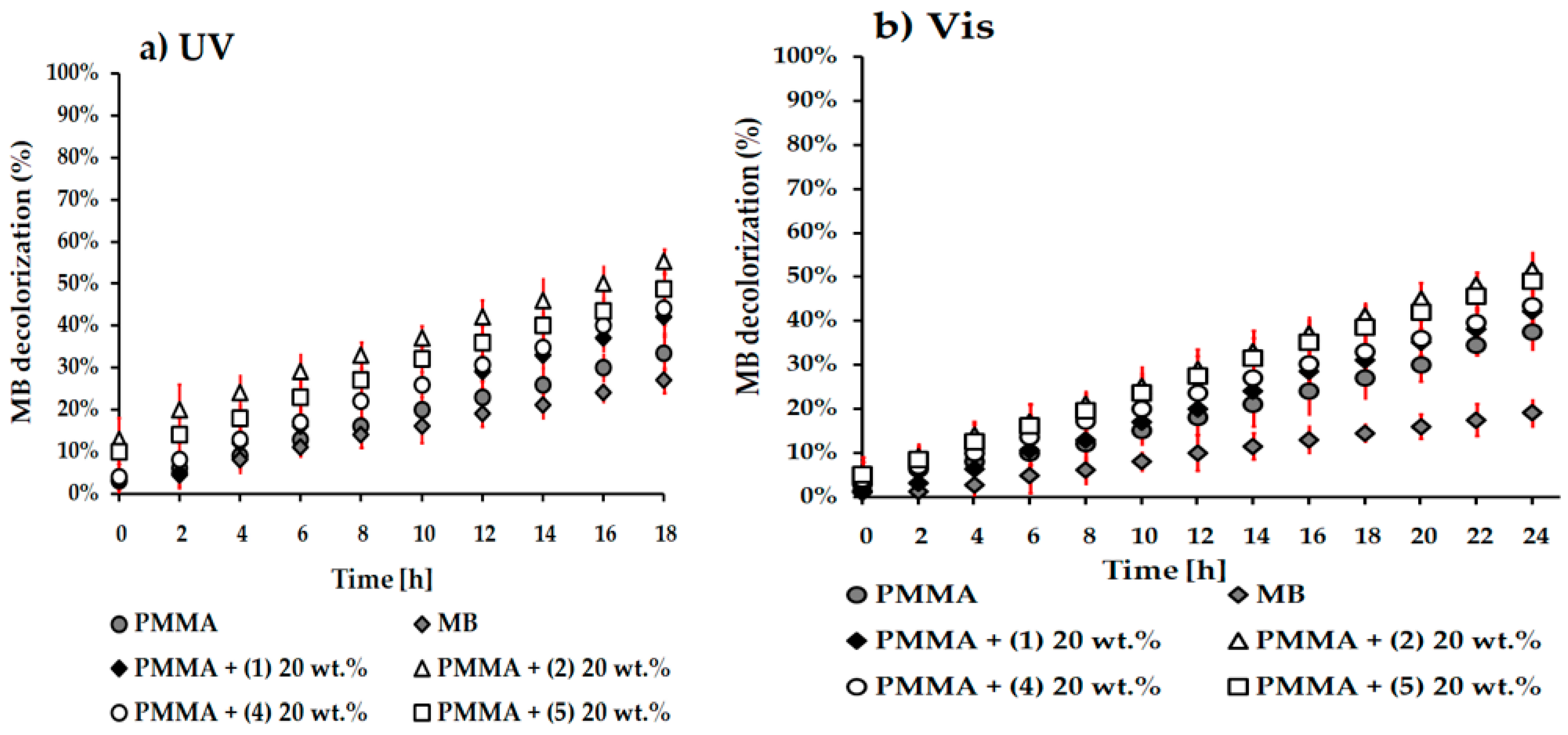
3.5. Photocatalytic Activity Studies of PMMA + TOCs Composite Films

The photocatalytic activity of PMMA + TOC 20 wt.% composite samples were assessed based on a decolorizing process of the methylene blue (MB) solution in which they were immersed and irradiated with UV or Vis radiation. The results for samples enriched with (1) ({Ti6O4}) and (2) ({Ti6O6}) cores have been compared with the previously characterized oxo complexes, i.e., {Ti3O} TOCs (4) and {Ti4O2} TOCs (5) [34,35]. The changes in MB decolorization versus the time of irradiation are presented in Figure 6. Analysis of this data revealed that the decolorization for studied composite samples proceeds faster in the UV range than in Vis one. The photocatalytic activity of PMMA + TOCs foils was higher than for pure PMMA. According to the results presented in Figure 6, Table 4, the PMMA + 20 wt.% of (2) ({Ti6O6} core) is the most active both in UV and Vis radiation.
The stability of photocatalytic activity in PMMA + TOCs composite samples (TOCs = (1), (2), (4), and (5)) was assessed over three measurement cycles under visible light irradiation (Figure 7). The results indicated that the photocatalytic activity of the tested systems remained consistent, as evidenced by a similar degree of MB discoloration observed in each cycle. By analyzing the data presented in Figure 6 and Figure 7, as well as Table 4, the influence of the {TiaOb} core structure on the photocatalytic activity of TOCs-enriched composites can be observed. The trend in the change of photocatalytic activity follows the sequence: {Ti6O6} (2) > {Ti4O2} (5) > {Ti3O} (4) > {Ti6O4} (1).

3.6. Electron Paramagnetic Resonance (EPR) Studies of PMMA + TOCs Composite Films

EPR spectroscopy was used to detect paramagnetic species on the surface of the synthesized materials. Pure PMMA polymer shows no EPR signal. However, paramagnetic centers are found in all samples of PMMA + TOCs (Figure 8).
Generally, the signals in the EPR spectra are weak, especially for composites with a 10 wt.% admixture of oxo-titanium(IV) complexes. However, the lines in the spectra of the samples with 20 wt.% TOCs addition are more difficult for analysis due to greater line overlap. Therefore, only the EPR spectra of the samples with 10 wt.% admixture of oxo-titanium(IV) complexes were analyzed in detail. Spectra registered for composites of PMMA + (1) 10 wt.% and PMMA + (2) 10 wt.% are presented in Figure 8. The EPR parameters and postulated types of observed paramagnetic species, especially ROS, are summarized in Table 5. Some EPR signals are marked with a question mark (Table 5. EPR) due to two different reasons (see Discussion). The first one concerns the g2-factor for O species in PMMA + (1) 10 wt.% sample, as the signals from both oxygen species may overlap. Further, concerns the g2-factor for Ti(III) in both PMMA + TOCs 10 wt.% samples, because the signals are almost on the noise level.

3.7. The Estimation of the Antimicrobial Activity of PMMA + TOCs Composite Folis

The studied PMMA + TOC composite foils showed superior biocidal activity (R index 5.3–5.4) against Gram-positive strains while their activity against Gram-negative strains was variable (R index 1.3–3.2). Although PMMA foils showed at least static activity (reduction index > 1–1.99) against E. coli reference strains, the PMMA + (5) 20 wt.% and PMMA + (1) 20 wt.% displayed biocidal activity (reduction index ≥ 2–3.2) against both these strains. None of the tested PMMA + TOC foils showed activity against yeasts of C. albicans (Table 6).

4. Discussion

The reaction between titanium isobutoxide and fluorene-9-carboxylic acid in a 1:1 molar ratio at room temperature, with acetone as the solvent, resulted in the co-crystallization of three complexes with the following summary formulas: [Ti6O4(OiBu)8(O2C13H9)8] · 2(CH3)2CO (1), [Ti6O6(OiBu)6(O2C13H9)6] (2), and [Ti6O6(OiBu)6(O2C13H9)6] (3). Single crystals, varying in shape and size (Figure 1c and Figure 2b,c), were manually isolated from the reaction mixture (optical microscope). Their structures were solved using the single-crystal X-ray diffraction method.
Analysis of the structural data revealed that [Ti6O4(OiBu)8(O2C13H9)8] · 2(CH3)2CO (1) crystallizes in the triclinic P-1 space group. Six titanium(IV) cations are almost coplanar, with the biggest shift reaching 0.106 Å. The core can be described as two coplanar titanium triangles (Figure 1a). In each triangle, the titanium(IV) cations are bridged by a µ3-O1 atom shifted by 0.389 Å from the Ti plane, while both triangles are joined by two µ-O2 bridges. In the former case, the Ti-O1 distances are 1.902 (3), 1.906 (3), and 2.066 (2) Å, with the longest one for the lateral Ti3 cation clearly indicating that this bridge is asymmetric. The Ti1-O2-Ti2 bridge is also asymmetric, with very short Ti-(µ-O2) bonds (1.755–1.869 (2) Å). In the reported compound, all titanium(IV) cations are found in a distorted octahedral environment, but the coordination sphere content is different. Ti1 and Ti2 are coordinated by two (O1 and O2) oxo anions, three oxygen atoms from carboxylate anions, and one bridging O26 (for Ti1) or terminal O86 (for Ti2) BuiO anion, whereas lateral Ti3 is found in an environment of O1 oxo anion, two carboxylates, and three BuiO anions (one bridging and two terminal). Carboxylate anions also form asymmetric bridges with distances differing by 0.03, 0.06, 0.13, and 0.04 for O11, O31, O51, and O71 carboxylate anions, respectively. Ti-ORCOO bonds introduce differences between Ti1 (three short bonds) and Ti2 (three long). Based on previous studies [14,17,18,31,32,41,42,43,44], it can be assumed that this structure is formed by the connection of two {Ti3O} subunits [14,43,44,45,46] through two μ-O bridges. Previous research has shown that a Ti(IV) complex (4) containing {Ti3O} cores can be isolated from a 4:1 mixture of isopropoxide and fluorene-9-carboxylic acid under similar reaction conditions [35].
It should be noted that the reaction conditions described for the 1:1 mixture also favor the formation of dense structures ((2), (3)) with a {Ti6O6} core stabilized by iso butoxide and fluorene-9-carboxylate ligands (Figure 2a) and are similar to previously studied hexanuclearTi(IV)-oxo complexes [14,43,44,45,46,47]. Analysis of the crystallographic data revealed that [Ti6O6(OiBu)6(O2C13H9)6] (2) (Figure 2d) and [Ti6O6(OiBu)6(O2C13H9)6] (3) (Figure 2e) are polymorphs crystallizing in monoclinic and tetragonal system, respectively (Table 1). Hence, only (2) will be described in detail. This oxo complex crystallizes in the P21/n space group with all atoms found in the general positions and half of the molecule given by the formula in the asymmetric unit. The common {Ti63-O)6} core is found in this structure, with Ti-Ti distances ranging from 3.1009 (9) Å to 3.1156 (9) Å for titanium located in different Ti3O3 hexagonal bases, and 3.4859 (9) Å to 3.5087 (9) Å for titanium from the same Ti3O3 hexagonal base. A Ti6O4 core is formed by two Ti3O3 hexagonal bases cross-linked by six carboxylates (Table S1). The coordination spheres of all titanium(IV) cations are composed of six oxygen atoms—three coming from oxo bridges arranged in a fac mode, two from two carboxylate anions, and one from terminal isobutanolate. O11 and O51 are coordinated in an asymmetric mode with two Ti-O distances differing by approximately 0.04 Å, whereas for the O31 anion, both Ti-O bonds were found to be identical within the experimental error. The terminal iso butoxide anions form the shortest bonds in the titanium coordination sphere. In this core, oxo anions connect three titanium atoms with distances falling into two ranges—1.880 (2) Å to 1.916 (3) Å and 2.142 (3) Å to 2.172 (3) Å. In the former case, they correspond to {Ti-(µ3-O)} distances for titanium and oxygen atoms from the same {Ti3O3} base, whereas the longest bond in Ti(IV) coordination occurs for Ti and O atoms from different bases. In the packing of (2), we observe densely packed [Ti6O6(OiBu)6(O2C13H9)6] clusters without any significant cavities, whereas in (3), significant voids were found, accounting for 1924.6 Å3 (9.2% of the cell volume). However, the model is complete as the highest peaks on the difference electron map do not exceed 1e/A3. In (2), the clusters interact via bulk ring moieties, forming both π-π interactions between strongly inclined C13H9 units and C-H…π interactions. Hence, intermolecular hydrogen bonds were not found, and isobutanolate was not involved in the crystal network formation. According to our previous results, when titanium isobutoxide and fluorene-9-carboxylic acid were reacted in a 4:1 molar ratio at room temperature, using acetone as the solvent, oxo complexes with the summary formula [Ti4O2(OiBu)10(O2C13H9)2] (5) were isolated [35].
One of the research directions exploring oxo-titanium(IV) complexes (TOCs) involves the evaluation of their physicochemical properties, particularly their photoinduced activity [31,34,35,48]. Liu et al. conducted a study where they introduced various carboxylate ligands (e.g.,propionic acid, pivalic acid, bromoacetic acid, trifluoroacetic acid, 2-naphthalenecarboxylic acid, 4-bromo-benzene carboxylic acid, pentafluorobenzoic acid) to the reactive coordination sites of the complex [Ti6O4(OiPr)10(O3P-Phen)2(OAc)2], resulting in successful modifications of the bandgap size (Ea) within the range of 3.6–3.0 eV [49]. In our earlier investigations, we also demonstrated the ability to modify the energy gap size within the range of Ea = 2.98–2.55 eV for complexes [Ti4O2(OiBu)10(O2CR″)2] through the functionalization of carboxylic ligands (R″ = PhNO2, PhCl, PhNH2, C13H9) [35,41]. Moreover, the results of these studies revealed that the complex stabilized by fluorene-9-carboxylate ligands (-O2CC13H9) exhibited photocatalytic activity both in the UV and visible range [35].
In this paper, we have focused on investigating the impact of the {TiaOb} core structure (while maintaining the same type of stabilized ligands) on the physicochemical properties and photocatalytic activity of oxo complexes. Previous research by Cui et al. explored these dependencies using a group of titanium-oxo complexes with different core architectures ((Ti4ClSal4), (Ti11ClSal8), and (Ti14ClSal8)), which were stabilized by isopropoxide and 4-chlorosalicylate (ClSal) ligands [50]. Analysis of the solid-state diffuse reflectance spectra (UV-Vis-DRS) of these oxo clusters revealed that the absorption maximum was observed in the UV range, with absorption edges at 450, 480, and 500 nm, respectively. The resulting optical band gaps exhibited a small range of variation, specifically between 2.8 and 3.0 eV. The photocatalytic activity of these compounds, evaluated through the photodecolorization of a methylene blue solution (MB) under sunlight irradiation, showed changes of approximately 78%, 73%, and 97% (after 32 min) for the (Ti4ClSal4), (Ti11ClSal8), and (Ti14ClSal8) clusters, respectively [50]. It is important to note that in all of these photocatalytic experiments, 30 mg of the oxo-cluster grains were added to 50 mL of MB solution. Our experiments focused on investigating the properties of oxo complexes containing {Ti3O} (4) [34], {Ti4O2} (5) [35], {Ti6O4} (1), and {Ti6O6} (2) cores, stabilized by fluorene-9-carboxylate ligands. Due to the potential hydrolysis of these compounds in water solutions, their photocatalytic and antimicrobial activities were evaluated for the composite systems, where 10 or 20 wt.% of TOCs were dispersed in the PMMA matrix. Examination of the SEM images of the composite films presented in Figure 3 revealed the presence of blurry-shaped (1) grains. This effect could be attributed to the interactions between polymer chains and molecules of this oxo complex. The evidence supporting this hypothesis is the noticeable difference in the glass transition temperature between the composite containing (1) and PMMA or PMMA + (2) (Table 3). SEM images of PMMA composites enriched with the (2), (4), and (5) complexes showed no blurry effect on the dispersed grains within the PMMA matrix (Figure 3). Analysis of the UV-Vis-DRS spectra for compounds (1)–(5) demonstrated a significant shift in the maximum absorption from the ultraviolet (UV) to the visible range, following this order: λmax = 365 nm for {Ti6O4} (1) and {Ti6O6} (2), λmax = 380 nm for {Ti3O} (4), and λmax = 395 nm for {Ti4O2} (5), with corresponding absorption edges at 400 nm (1) and (2), 420 nm (4), and 500 nm (5). These results indicate that the absorption of hexanuclear core structures ({Ti6O4} and {Ti6O6}) tends to shift towards the UV range, while in the case of oxo complexes consisting of simple units ({Ti3O} and {Ti4O2}), the shift occurs towards the visible range. The band gap values for the studied clusters exhibited the following changes: Ea= 2.17 eV for (4), 2.55 eV for (5), 2.98 eV for (1), and 2.99 eV for (2) (Figure S3). It is worth noting that these changes differ from the findings reported by Cui et al. [50], where the band gap values remained within a narrow range (Ea = 2.8–3.0 eV) and were highest for the smallest cluster (Ti4ClSal4).
The photocatalytic activity of the investigated titanium oxo clusters (TOCs) (1, 2, 4, and 5) incorporated into the PMMA matrix (PMMA + TOCs 20 wt.% systems) was evaluated by measuring the decolorization of a methylene blue (MB) solution. The aforementioned samples were immersed in the MB solution and exposed to UV (340–395 nm) and visible (395–420 nm) light. The analysis of the data presented in Figure 6 and Figure 4 revealed that the photocatalytic activity of the composite samples was more pronounced in the UV range. Specifically, composites containing TOCs with structurally dense cores, such as {Ti6O6} (2) and {Ti4O2} (5), exhibited higher activity compared to systems enriched with flat cores, namely {Ti3O} (4) and {Ti6O4} (1). For composite (1), it is important to consider potential interactions between the polymer chains and TOC molecules, which could potentially diminish the photocatalytic activity of the PMMA + composite (1). These photoinduced activities observed across all the studied composite samples were found to be reproducible processes (Figure 7). Notably, our samples demonstrated longer MB photodegradation times in comparison to previous studies on the photocatalytic activity of Ti(IV)-oxo clusters [21,23,24,50]. Analyzing the presented experimental data should consider that the photocatalytic activity is assessed for PMMA + TOCs systems in which only 20 wt.% of TOCs are dispersed in the polymer matrix [32,34,35].
Based on previous findings, we made the assumption that the photocatalytic activity of PMMA + TOCs samples is a result of their photoexcitation, leading to the generation of reactive oxygen species (ROS) [51,52,53,54,55]. Additionally, this effect is combined with the photoactivated reduction of Ti(IV) to Ti(III) [52,53,54]. To confirm the aforementioned phenomenon, we recorded EPR spectra of PMMA + TOCs (TOCs = (1) and (2)) samples (Figure 8). In the EPR spectrum of PMMA + (1), we observed anisotropic signals corresponding to O2 and O radicals, as well as Ti(III) (Figure 8a and Table 5). It is worth noting that some lines, particularly g2 for both oxygen species, may overlap. The formation of O2 and O paramagnetic centers involves electron removal and stabilization of the resulting radical through the reduction of Ti(IV) to Ti(III) [51,52]. According to the literature, the maximum g-factor for O2 is higher compared to O species [53]. For the PMMA + (2) composite, we obtained an EPR spectrum with improved resolution (Figure 8b), revealing the characteristic signal attributed to O paramagnetic species. However, the presence of O2 radicals cannot be entirely ruled out; their content on the sample surface is likely much lower. The electron transfer between Ti(IV) and Ti(III) was also confirmed in this sample. Similar spectral patterns were observed for the PMMA + (5) 20 wt.% composite in previous studies (Table 5) [35]. In the case of the PMMA + (4) 20 wt.% sample, the EPR spectrum indicated the presence of O2, O, and possibly Ti(III) forms, consistent with the results obtained for a composite containing a {Ti6O4} core built by Ti3O subunits. The signals observed in the PMMA + (4) 20 wt.% sample was relatively weak and exhibited a rapid decrease in intensity over time, indicating lower stability of the {Ti3O} core compared to other cores. It is important to note that other oxygen-based radicals, such as peroxy-type radicals (RCOO•) and organic-type adducts of O2, could potentially be indistinguishable from the superoxide radical anion (O2) using CW-EPR spectroscopy. However, this scenario is unlikely as the first two forms are unstable at room temperature and can only be detected at lower temperatures [55].
The antimicrobial activity of TiO2-based materials, specifically TiO2 coatings, is an important but relatively poorly understood issue. Research on TiO2-based materials with photocatalytic activity has shown their remarkable effectiveness in killing microorganisms on various surfaces [56]. Our previous investigations on the modification of titanium or Ti6Al4V alloy implants [57,58,59] revealed that the antimicrobial efficiency of TiO2 coatings is mainly influenced by factors such as surface structure and morphology, hydrophilicity, exposure to light (especially UV), and the presence of oxygen. Kumaravel et al. [56] suggest that antimicrobial activity depends on the interaction between microorganisms and the surface characteristics of the coating material, such as electrostatic interaction, pH, and morphology. They observed that after exposure to light, TiO2 coatings prevented the reactivation or regrowth of microbes, which may be associated with the generation of reactive oxygen species (ROS). Therefore, while researching the photocatalytic properties of Ti(IV)-oxo clusters, we also drew attention to their antimicrobial activity [31,32]. The significant advantage of using oxo complexes is their ability to reduce the energy gap from 2.9–3.2 eV for TiO2 to 2.2–3.0 eV for the studied group of TOCs, allowing them to absorb visible light. The core structure and type of carboxylate ligand significantly influences the band gap value in this context. Analysis of electron paramagnetic resonance (EPR) spectra indicated that stable ROS species formed on the surface of the composite film were oxygen radicals (O) for PMMA + TOCs (TOCs = (1), (2), and (5)) samples and superoxide radicals (O2) for PMMA + TOCs (TOCs = (1), (4), and (5)) samples. Previous studies on TiO2 materials have reported that the generation of ROS species can damage cells, hinder microbial growth and migration, and directly interact with the cell membrane [56,60]. This mechanism is also applicable to our PMMA + TOC samples. Based on the data presented in Table 6, we can conclude that all the PMMA + TOCs samples exhibited biocidal activity against S. aureus reference strains, and most of them also showed activity against E. coli strains. However, the antibacterial activity varied among different strains of Escherichia coli. The composite samples enriched with TOCs grains containing {Ti4O2} cores (5) and {Ti6O4} (1) demonstrated the highest antibacterial activity. It is worth noting that, similar to PMMA + (1) 10 wt%, the composite PMMA + (5) 20 wt.% also released both oxygen anion species (O2 and O), confirming the crucial role of reactive oxygen species (ROS) as antibacterial agents. The effects of O on microorganism cells have been poorly investigated. It is known that O reacts with the hydrocarbon bonds of microorganisms [60]. Singlet oxygen or other ROS can damage bacterial cells or other microbes through various mechanisms, including lipid peroxidation, oxidation of amino acids in proteins, protein cross-linking, and oxidative damage to nucleic acids. This damage disrupts the normal functioning of microbial cells [60,61]. ROS can severely impair cell walls and intrinsic structures, leading to rapid and irreversible inactivation. However, the effectiveness of microorganism inactivation depends on the intensity of ROS, the initial population of microorganisms, the exposed area, the environment, and the type of microorganism. In our case, the PMMA + (2) 20 wt.% system, which only generated O species on its surfaces, exhibited weaker activity against E. coli strains compared to PMMA + (1) 20 wt.%. [60].

5. Conclusions

The aim of this research was to evaluate the influence of the {TiaOb} core structure on the photocatalytic and antimicrobial activity of titanium(IV)-oxo clusters (TOCs). The clusters, denoted by the general formulas [Ti6O4(OiBu)8(O2C13H9)8] · 2(CH3)2CO (1), [Ti6O6(OiBu)6(O2C13H9)6] (2), [TiO3(OiPr)8(O2C13H9)2] (4), and [Ti4O2(OiBu)10(O2C13H9)2] (5), were used to investigate these effects.
Analysis of UV-Vis-DRS spectra revealed that the core structure of {TiaOb} influences the band gap energy of the isolated crystalline powders. The energy values varied as follows: 2.17 eV for {Ti3O} (4), 2.55 eV for {Ti4O2} (5), 2.98 eV for {Ti6O4} (1), and 2.99 eV for {Ti6O6} (2). The absorption maximum for the studied TOCs was detected in the UV range (360–395 nm), with an absorption edge at 400 nm ({Ti6O4} (1) and {Ti6O6} (2)), 420 nm ({Ti3O} (4)), and 500 nm ({Ti4O2} (5)). According to these data, the investigated oxo complexes can exhibit photocatalytic activity in both UV and visible light.
The photocatalytic and antimicrobial activity of the studied group of Ti(IV)-oxo complexes (TOCs), has been estimated for PMMA + TOCs composite systems. The assumption was the protection of TOCs before possible hydrolysis processes. Analysis of EPR spectra of the samples mentioned above proved the presence of natural photoexcitation of all PMMA + TOCs surface and the generation of ROS species. In the presence of composite samples, the photoinduced de-colorization of methylene blue solution processes revealed that oxo clusters stabilized by 9-fluorene carboxylate ligands are photocatalytic active both in the UV and Vis range. However, this activity was higher in the UV range. The results of conducted experiments revealed that photocatalytic activity of investigated TOCs decreases in the row: {Ti6O6} (2) > {Ti4O2} (5) > {Ti3O} (4) > {Ti6O4} (1). It should be noted that the low photocatalytic activity of PMMA + (1) could also be attributed to potential interactions between TOCs grains and polymer chains, which is supported by SEM images and DSC data.
In general, PMMA + TOCs composites exhibited greater antibacterial activity against Gram-positive Staphylococcus aureus strains compared to Gram-negative Escherichia coli strains. However, it is important to mention that the antibacterial activity against E. coli strains of PMMA samples enriched with TOCs increased in the following order: {Ti3O} (4) <{Ti6O6} (2) <{Ti4O2} (5) <{Ti6O4} (1). It is worth noting that on the surface samples demonstrating the highest antimicrobial activity, namely PMMA + (1) and PMMA + (5), were generated by both O2 and O oxygen anion species.
One of the key advantages of PMMA + TOCs composite materials enriched with oxo-Ti(IV) clusters featuring a defined {TiaOb} core structure is their antimicrobial activity in the visible range. This characteristic renders this type of composite suitable for preventing the spread of bacteria in high-risk public areas (e.g., hospitals, supermarkets, or malls) without relying on harmful UV light, which is currently widely utilized.

Supplementary Materials

The following supporting information can be downloaded at: https://www.mdpi.com/article/10.3390/cryst13070998/s1, Figure S1: SEM images and EDX patterns of pure PMMA and PMMA + 20(1)) and PMMA + 20(2)) composites; Figure S2: Thermogravimetric curves (TGA) (a,c) and the differential scanning calorimetry curves of (DSC) (b,d) of the TOCs (1),(2) and produced composite materials; Figure S3: Structures of {TaOb} cores in molecules (1), (2), (4), and (5) oxo complexes, and values of HOMO-LUMO gap; Figure S3: The results of the photocatalytic process repeatability studies of PMMA + TOCs 20 wt.% composite samples (TOCs = (1), (2), (4) and (5)) in the visible light; Table S1: Selected distances and angles in (1), (2), and (3) oxo complexes.

Author Contributions

Conceptualization, P.P. and A.R.; methodology, B.K., P.P. and A.R.; formal analysis, B.K., P.P., A.R., T.M., P.G. and G.W.; investigation, B.K., T.M., P.G. and G.W.; data curation, B.K.; writing—original draft preparation, B.K., P.P., T.M. and P.G.; writing—review and editing, A.R. and P.P.; supervision, P.P. and A.R. All authors have read and agreed to the published version of the manuscript.

Funding

This research received no external funding.

Data Availability Statement

Crystallographic data has been deposited at the Cambridge Crystallographic Data Centre with Deposit Nos. 2224562–2224564 as detailed in Table 1 and Table S1.

Conflicts of Interest

The authors declare no conflict of interest.

References

  1. Zaky, A.M.; Chaplin, B.P. Porous Substoichiometric TiO2 Anodes as Reactive Electrochemical Membranes for Water Treatment. Environ. Sci. Technol. 2013, 47, 6554–6563. [Google Scholar] [CrossRef]
  2. Jandam, N.; Serivalsatit, K.; Hunsom, M.; Pruksathorn, K. Ultrasound-Assisted Synthesis of Nonmetal-Doped Titanium Dioxide Photocatalysts for Simultaneous H2 Production and Chemical Oxygen Demand Removal from Industrial Wastewater. ACS Omega 2021, 6, 24709–24719. [Google Scholar] [CrossRef]
  3. Li, A.; Chen, S.; Yang, F.; Gao, H.; Dong, C.; Wang, G. Metalloporphyrin-Decorated Titanium Dioxide Nanosheets for Efficient Photocatalytic Carbon Dioxide Reduction. Inorg. Chem. 2021, 60, 18337–18346. [Google Scholar] [CrossRef]
  4. Trivedi, M.; Murase, J. Titanium Dioxide in Sunscreen. In Application of Titanium Dioxide; Janus, M., Ed.; InTech: London, UK, 2017; ISBN 978-953-51-3429-9. [Google Scholar]
  5. He, Z.; Wu, H.; Shi, Z.; Gao, X.; Sun, Y.; Liu, X. Mussel-Inspired Durable TiO2/PDA-Based Superhydrophobic Paper with Excellent Self-Cleaning, High Chemical Stability, and Efficient Oil/Water Separation Properties. Langmuir 2022, 38, 6086–6098. [Google Scholar] [CrossRef]
  6. Radtke, J.; Wiedey, R.; Kleinebudde, P. Alternatives to Titanium Dioxide in Tablet Coating. Pharm. Dev. Technol. 2021, 26, 989–999. [Google Scholar] [CrossRef]
  7. Younes, M.; Aquilina, G.; Castle, L.; Engel, K.; Fowler, P.; Frutos Fernandez, M.J.; Fürst, P.; Gundert-Remy, U.; Gürtler, R. Safety Assessment of Titanium Dioxide(E171) as Food Additive. EFS2 2021, 19, e06585. [Google Scholar]
  8. Kitagawa, I.L.; Miyazaki, C.M.; Pitol-Palin, L.; Okamoto, R.; de Vasconcellos, L.M.R.; Constantino, C.J.L.; Lisboa-Filho, P.N. Titanium-Based Alloy Surface Modification with TiO2 and Poly(Sodium4-Styrenesulfonate) Multilayers for Dental Implants. ACS Appl. Bio Mater. 2021, 4, 3055–3066. [Google Scholar] [CrossRef] [PubMed]
  9. Kaur, R.; Thakur, N.S.; Chandna, S.; Bhaumik, J. Sustainable Lignin-Based Coatings Doped with Titanium Dioxide Nanocomposites Exhibit Synergistic Microbicidal and UV-Blocking Performance toward Personal Protective Equipment. ACS Sustain. Chem. Eng. 2021, 9, 11223–11237. [Google Scholar] [CrossRef]
  10. Ganguly, P.; Mathew, S.; Clarizia, L.; Kumar, R.S.; Akande, A.; Hinder, S.; Breen, A.; Pillai, S.C. Theoretical and Experimental Investigation of Visible Light Responsive AgBiS2-TiO2 Heterojunctions for Enhanced Photocatalytic Applications. Appl. Catal. B Environ. 2019, 253, 401–418. [Google Scholar] [CrossRef]
  11. Cao, L.; Li, Y.-F.; Tong, Y.; Yang, R.; Sun, L.; Cao, Q.; Chen, R. A Novel Bi12TiO20/g-C3N4 Hybrid Catalyst with a Bionic Granum Configuration for Enhanced Photocatalytic Degradation of Organic Pollutants. J. Hazard. Mater. 2019, 379, 120808. [Google Scholar] [CrossRef] [PubMed]
  12. Li, S.; Wang, C.; Liu, Y.; Cai, M.; Wang, Y.; Zhang, H.; Guo, Y.; Zhao, W.; Wang, Z.; Chen, X. Photocatalytic Degradation of Tetracycline Antibiotic by a Novel Bi2Sn2O7/Bi2MoO6S-Scheme Heterojunction: Performance, Mechanism Insightand Toxicity Assessment. Chem. Eng. J. 2022, 429, 132519. [Google Scholar] [CrossRef]
  13. Cheng, K.; Chhor, K.; Kanaev, A. Solvent Effect on Nucleation-Growth of Titanium-Oxo-Alkoxy Nanoparticles. Chem. Phys. Lett. 2017, 672, 119–123. [Google Scholar] [CrossRef]
  14. Schubert, U. Chemical Modification of Titanium Alkoxides for Sol–Gel Processing. J. Mater. Chem. 2005, 15, 3701. [Google Scholar] [CrossRef]
  15. Liu, X.-X.; Chen, S.; Fang, W.-H.; Zhang, L.; Zhang, J. Amino-Polyalcohol-Solvothermal Synthesis of Titanium-Oxo Clusters: From Ti6 to Ti19 with Structural Diversity. Inorg. Chem. 2019, 58, 7267–7273. [Google Scholar] [CrossRef]
  16. Sanchez, C.; Rozes, L.; Ribot, F.; Laberty-Robert, C.; Grosso, D.; Sassoye, C.; Boissiere, C.; Nicole, L. “Chimie Douce”: A Land of Opportunities for the Designed Construction of Functional Inorganic and Hybrid Organic-Inorganic Nanomaterials. Comptes Rendus Chim. 2010, 13, 3–39. [Google Scholar] [CrossRef] [Green Version]
  17. Piszczek, P.; Radtke, A.; Muzioł, T.; Richert, M.; Chojnacki, J. The Conversion of Multinuclear μ-OxoTitanium (IV) Species in the Reaction of Ti(OiBu)4 with Branched Organic Acids; Results of Structural and Spectroscopic Studies. Dalton Trans. 2012, 41, 8261–8269. [Google Scholar] [CrossRef] [PubMed]
  18. Radtke, A.; Piszczek, P.; Muzioł, T.; Wojtczak, A. The Structural Conversion of Multinuclear Titanium(IV)μ-Oxo-Complexes. Inorg. Chem. 2014, 53, 10803–10810. [Google Scholar] [CrossRef]
  19. Papiernik, R.; Hubert-Pfalzgraf, L.G.; Vaissermann, J.; Goncalves, M.C.H.B. Synthesis and Characterization of New Titanium Hexanuclear Oxo Carboxylato Alkoxides. Molecular Structure of [Ti63-O)6(μ-O2CC6H4OPh)6(OEt)6]. J. Chem. Soc. Dalton Trans. 1998, 14, 2285–2288. [Google Scholar] [CrossRef]
  20. Lv, H.-T.; Li, H.-M.; Zou, G.-D.; Cui, Y.; Huang, Y.; Fan, Y. Titanium-Oxo Clusters Functionalized with Catecholate-Type Ligands: Modulating the Optical Properties through Charge-Transfer Transitions. Dalton Trans. 2018, 47, 8158–8163. [Google Scholar] [CrossRef]
  21. Wang, C.; Wang, S.-J.; Kong, F.-G. Calixarene-Protected Titanium-Oxo Clusters and Their Photocurrent Responses and Photocatalytic Performances. Inorg. Chem. 2021, 60, 5034–5041. [Google Scholar] [CrossRef]
  22. Liu, J.-J.; Li, N.; Sun, J.-W.; Liu, J.; Dong, L.-Z.; Yao, S.-J.; Zhang, L.; Xin, Z.-F.; Shi, J.-W.; Wang, J.-X. Ferrocene-Functionalized Polyoxo-Titanium Cluster for CO2 Photoreduction. ACS Catal. 2021, 11, 4510–4519. [Google Scholar] [CrossRef]
  23. Wang, J.-F.; Fang, W.-H.; Li, D.-S.; Zhang, L.; Zhang, J. Cocrystal of {Ti4} and {Ti6} Clusters with Enhanced Photochemical Properties. Inorg. Chem. 2017, 56, 2367–2370. [Google Scholar] [CrossRef] [PubMed]
  24. Gao, M.-Y.; Fang, W.-H.; Wen, T.; Zhang, L.; Zhang, J. Connecting Titanium-Oxo Clusters by Nitrogen Heterocyclic Ligands to Produce Multiple Cluster Series with Photocatalytic H2 Evolution Activities. Cryst. Growth Des. 2017, 17, 3592–3595. [Google Scholar] [CrossRef]
  25. Wang, C.; Liu, C.; Li, L.-J.; Sun, Z.-M. Synthesis, Crystal Structures, and Photochemical Properties of a Family of Heterometallic Titanium Oxo Clusters. Inorg. Chem. 2019, 58, 6312–6319. [Google Scholar] [CrossRef] [PubMed]
  26. Svenson, F.G.; Sensenbaeva, G.A.; Keesler, V.G. Mixed-Ligand Titanium “Oxo-Clusters”: Structural Insights into the Formation and Binding of Organic Molecules and Transformation into Oxide Nanostructures on Hydrolysis and Thermolysis. Eur. J. Inorg. Chem. 2017, 35, 4117–4122. [Google Scholar] [CrossRef]
  27. Khalil, A.T.; Ovais, M.; Ullah, I.; Ali, M.; Shinwari, Z.K.; Khamlich, S.; Maaza, M. Sageretia Thea (Osbeck.) Mediated Synthesis of Zinc Oxide Nanoparticles and Its Biological Applications. Nanomedicine 2017, 12, 1767–1789. [Google Scholar] [CrossRef] [PubMed]
  28. Wang, G.; Feng, H.; Hu, L.; Jin, W.; Hao, Q.; Gao, A.; Peng, X.; Li, W.; Wong, K.-Y.; Wang, H.; et al. An Antibacterial Platform Based on Capacitive Carbon-Doped TiO2 Nanotubes after Direct or Alternating Current Charging. Nat. Commun. 2018, 9, 2055. [Google Scholar] [CrossRef] [Green Version]
  29. Mutalik, C.; Lin, I.-H.; Krisnawati, D.I.; Khaerunnisa, S.; Khafid, M.; Widodo; Hsiao, Y.C.; Kuo, Y.-R. Antibacterial Pathways in Transition Metal-Based Nanocomposites: A Mechanistic Overview. Int. J. Nanomed. 2022, 7, 6821–6842. [Google Scholar] [CrossRef]
  30. Gupta, A.; Mumtaz, S.; Li, C.-H.; Hussain, I.; Rotello, V.M. Combatting Antibiotic-Resistant Bacteria Using Nanomaterials. Chem. Soc. Rev. 2019, 48, 415–427. [Google Scholar] [CrossRef]
  31. Piszczek, P.; Kubiak, B.; Golińska, P.; Radtke, A. Oxo-Titanium(IV) Complex/Polymer Composites—Synthesis, Spectroscopic Characterization and Antimicrobial Activity Test. IJMS 2020, 21, 9663. [Google Scholar] [CrossRef]
  32. Kubiak, B.; Radtke, A.; Topolski, A.; Wrzeszcz, G.; Golińska, P.; Kaszkowiak, E.; Sobota, M.; Włodarczyk, J.; Stojko, M.; Piszczek, P. The Composites of PCL and Tetranuclear Titanium(IV)-Oxo Complexes as Materials Exhibiting the Photocatalytic and the Antimicrobial Activity. IJMS 2021, 22, 7021. [Google Scholar] [CrossRef] [PubMed]
  33. Meth-Cohn, O.; Thorpe, D.; Twitchett, H.J. Insertion Reactions of Titanium Alkoxides with Isocyanates and Carbodiimides. J. Chem. Soc. C 1970, 132–135. [Google Scholar] [CrossRef]
  34. Janek, M.; Muzioł, T.M.; Piszczek, P. Trinuclear Oxo-Titanium Clusters: Synthesis, Structure, and Photocatalytic Activity. Materials 2019, 12, 3195. [Google Scholar] [CrossRef] [Green Version]
  35. Janek, M.; Radtke, A.; Muzioł, T.; Jerzykiewicz, M.; Piszczek, P. Tetranuclear Oxo-Titanium Clusters with Different Carboxylate Aromatic Ligands: Optical Properties, DFT Calculations, and Photoactivity. Materials 2018, 11, 1661. [Google Scholar] [CrossRef] [PubMed] [Green Version]
  36. E. CrysAlis Red and CrysAlis CCD; Oxford Diffraction Ltd.: Abingdon, UK, 2000.
  37. Sheldrick, G.M. Crystal Structure Refinement with SHELXL. Acta Crystallogr. C Struct. Chem. 2015, 71, 3–8. [Google Scholar] [CrossRef] [Green Version]
  38. Brandenburg, K.; Berndt, M. Diamond, Release 2.1e, Crystal Impact GbR; Scientific Research: Bonn, Germany, 2001. [Google Scholar]
  39. Farrugia, L.J. WinGX and ORTEP for Windows: An Update. J. Appl. Crystallogr. 2012, 45, 849–854. [Google Scholar] [CrossRef]
  40. Xu, H.; Ouyang, S.; Liu, L.; Reunchan, P.; Umezawa, N.; Ye, J. Recent Advances in TiO2-Based Photocatalysis. J. Mater. Chem. A 2014, 2, 12642–12661. [Google Scholar] [CrossRef]
  41. Janek, M.; Muzioł, T.M.; Piszczek, P. The Structure and Photocatalytic Activity of the Tetranuclear Titanium(IV) Oxo-Complex with 4-Aminobenzoate Ligands. Polyhedron 2018, 141, 110–117. [Google Scholar] [CrossRef]
  42. Piszczek, P.; Grodzicki, A.; Richert, M.; Wojtczak, A. Structural and Thermal Stability Studies of Ti(IV) Hexanuclear Oxo Trimethylacetato Isopropoxide Complex. Inorganica Chim. Acta 2004, 357, 2769–2775. [Google Scholar] [CrossRef]
  43. Schubert, U. Titanium-Oxo Clusters with Bi- and Tridentate Organic Ligands: Gradual Evolution of the Structures from Small to Big. Chem. Eur. J. 2021, 27, 11239–11256. [Google Scholar] [CrossRef]
  44. Schubert, U. En Route from Metal Alkoxides to Metal Oxides: Metal Oxo/Alkoxo Clusters. J. Sol.-Gel. Sci. Technol. 2023, 105, 587–595. [Google Scholar] [CrossRef]
  45. Fang, W.-H.; Zhang, L.; Zhang, J. Synthetic Strategies, Diverse Structures and Tuneable Properties of Polyoxo-Titanium Clusters. Chem. Soc. Rev. 2018, 47, 404–421. [Google Scholar] [CrossRef] [PubMed]
  46. Piszczek, P. Thermal Behaviour of Hexanuclear Titanium(IV) Oxo Isopropoxide Carboxylates, and Their Usability as a Single-Source TiO2 CVD Precursors. Polyhedron 2007, 26, 93–100. [Google Scholar] [CrossRef]
  47. Piszczek, P.; Richert, M.; Wojtczak, A. Crystal Structure and Spectral Characterization of Hexanuclear Oxo Titanium(IV) Clusters: [Ti6O6(OSi(CH3)3)6(OOCR)6] (R = But, CH2But, C(CH3)2Et). Polyhedron 2008, 27, 602–608. [Google Scholar] [CrossRef]
  48. Nguyen, H.L.; Vu, T.T.; Le, D.; Doan, T.L.H.; Nguyen, V.Q.; Phan, N.T.S. A Titanium Organic Framework: Engineering of the Band-Gap Energy for Photocatalytic Property Enhancement. ACS Catal. 2017, 7, 338–342. [Google Scholar] [CrossRef]
  49. Liu, J.-X.; Gao, M.-Y.; Fang, W.-H.; Zhang, L.; Zhang, J. Bandgap Engineering of Titanium-Oxo Clusters: Labile Surface Sites Used for Ligand Substitution and Metal Incorporation. Angew. Chem. Int. Ed. Engl. 2016, 55, 5160–5165. [Google Scholar] [CrossRef]
  50. Cui, Y.; Zou, G.-D.; Li, H.-M.; Huang, Y.; Fan, Y. 4-Chlorosalicylate-Stabilized Titanium-Oxo Clusters with Structures Mediated by Tetrazole and Their Photophysical Properties. Polyhedron 2019, 157, 177–182. [Google Scholar] [CrossRef]
  51. He, W.; Liu, Y.; Wamer, W.G.; Yin, J.-J. Electron Spin Resonance Spectroscopy for the Study of Nanomaterial-Mediated Generation of Reactive Oxygen Species. J. Food Drug Anal. 2014, 22, 49–63. [Google Scholar] [CrossRef] [Green Version]
  52. Xiong, L.-B.; Li, J.-L.; Yang, B.; Yu, Y. Ti3+ in the Surface of Titanium Dioxide: Generation, Properties and Photocatalytic Application. J. Nanomater. 2012, 2012, 831524. [Google Scholar] [CrossRef] [Green Version]
  53. Suriye, K.; Lobo-Lapidus, R.J.; Yeagle, G.J.; Praserthdam, P.; Britt, R.D.; Gates, B.C. Probing Defect Sites on TiO2 with [Re3(CO)12H3]: Spectroscopic Characterization of the Surface Species. Chem. Eur. J. 2008, 14, 1402–1414. [Google Scholar] [CrossRef]
  54. Dan-Hardi, M.; Serre, C.; Frot, T.; Rozes, L.; Maurin, G.; Sanchez, C.; Férey, G. A New Photoactive Crystalline Highly Porous Titanium(IV) Dicarboxylate. J. Am. Chem. Soc. 2009, 131, 10857–10859. [Google Scholar] [CrossRef] [PubMed]
  55. Richards, E.; Murphy, D.M.; Che, M. An EPR Characterization of Stable and Transient Reactive Oxygen Species Formed under Radiative and Non-Radiative Conditions. Res. Chem. Intermed. 2019, 45, 5763–5779. [Google Scholar] [CrossRef] [Green Version]
  56. Kumaravel, V.; Nair, K.M.; Mathew, S.; Bartlett, J.; Kennedy, J.E.; Manning, H.G.; Whelan, B.J.; Leyland, N.S.; Pillai, S.C. Antimicrobial TiO2 nanocomposite coatings for surfaces, dental and orthopedic implants. Chem. Eng. J. 2021, 416, 129071. [Google Scholar] [CrossRef]
  57. Radtke, A.; Topolski, A.; Jedrzejewski, T.; Kozak, W.; Sadowska, B.; Wieckowska-Szakiel, M.; Piszczek, P. Bioactivity Studies on Titania Coatings and the Estimation of Their Usefulness in the Modification of Implant Surfaces. Nanomaterials 2017, 4, 90. [Google Scholar] [CrossRef] [PubMed] [Green Version]
  58. Piszczek, P.; Radtke, A.; Ehlert, M.; Jędrzejewski, M.; Sznarkowska, A.; Sadowska, B.; Bartmański, M.; Kemal Erdoğan, Y.K.; Ercan, B.; Jedrzejczyk, W. Comprehensive Evaluation of the Biological Properties of Surface-Modified Titanium Alloy Implants. J. Clin. Med. 2020, 9, 342. [Google Scholar] [CrossRef] [Green Version]
  59. Ehlert, M.; Radtke, A.; Forbot, N.; Jędrzejewski, T.; Roszek, K.; Golińska, P.; Trykowski, G.; Piszczek, P. TiO2/HA and Titanate/HA Double Layer Coatings on Ti6Al4V Surface and Their Influence on In Vitro Cell Growth and Osteogenic Potential. J. Funct. Biomater. 2022, 13, 271. [Google Scholar] [CrossRef]
  60. Li, L.; Wu, F.; Chen, Y.; Xu, L.; Hao, X.; Chen, Y.; Sun, Y.; Xiong, G. Reactions of Microorganisms with Atomic Oxygen Radical Anions: Damage of Cells and Irreversible Inactivation. J. Nanomater. 2019, 2019, 2483060. [Google Scholar] [CrossRef]
  61. Luksiene, Z. Photodynamic therapy: Mechanism of action and ways to improve the efficiency of treatment. Medicina 2003, 9, 1137–1150. [Google Scholar]
Figure 1. The structure of [Ti6O4(OiBu)8(O2C13H9)8] · 2 (CH3)2CO (1) complex (crystallographic ball-stick scheme). (a) The {Ti6O4} core with labeled titanium and oxygen atoms. For clarity of the picture, all hydrogen atoms and most of carbon atoms are omitted. (b) The packing of TOCs in the crystal network of (1) along the b axis. (c) The optical microscope image of (1) crystals.
Crystals 13 00998 g001
Figure 2. Two polymorphous forms of [Ti6O6(OiBu)6(O2C13H9)6]—(2) and (3) structures. The {Ti6O6} core with labeled titanium and oxygen atoms—(a). For clarity of the picture, all hydrogen atoms and most carbon atoms are omitted. The optical microscope images of (2) and (3) crystals—(b) and (c), respectively. The differences in TOCs packing in the crystal network of (2) and (3) along b axis—(d) and (e), respectively.
Crystals 13 00998 g002
Figure 3. SEM images of PMMA + TOCs (TOCs = (1), (2), (4) and (5) composites.
Crystals 13 00998 g003
Figure 4. Raman maps and spectra of composites consist of PMMA + TOCs (TOCs = (1) 20 wt.% and (2) 20 wt.%.
Crystals 13 00998 g004
Figure 5. Solid-state UV-Vis-diffuse reflectance spectra (DRS) of the (1), (2) micro-grains (a), Kubelka-Munk function versus light energy plot for the HOMO-LUMO gap determination (b).
Crystals 13 00998 g005
Figure 6. Changes in the concentrations of the methylene blue (MB) solution as a function of time for composite materials PMMA + (1), (2), (4), (5) 20 wt.% irradiated with UV and Vis light.
Crystals 13 00998 g006
Figure 7. Recycling MB decolorization tests of composite materials PMMA + TOCs 20 wt.% (TOCs = (1), (2), (4), and (5)) irradiated with visible light.
Crystals 13 00998 g007
Figure 8. EPR spectra of PMMA + (1) 10 wt.% (a) and PMMA + (2) 10 wt.% (b). The experimental conditions: cut foil, room temperature, microwave frequency: 9.32851 (a), 9.32908 (b) GHz; microwave power: 40 (a), 58 (b) mW; modulation amplitude: 1 mT; sweep: 20 mT; sweep time: 4 min.; time constant: 0.1 s; receiver gain: 4 (a), 5 (b) ×105.
Crystals 13 00998 g008
Table 1. Crystal data and structure refinement for (1)–(3).
Empirical FormulaC116H138O26Ti6 (1)C108H108O24Ti6 (2)C108H108O24Ti6 (3)
Formula weight 2726.142077.342077.34
Temperature (K)100 (2)100 (2)100 (2)
Wavelength (Å)1.541841.541841.54184
Crystal system TriclinicMonoclinicTetragonal
Space group P-1P21/nI41/1
Unit cell dimensions [Å] and [°]a = 13.7398 (5) α = 103.877 (4)
b = 15.6157 (6) β =106.420 (4)
c =17.1768 (7) γ = 91.043 (3)
a = 14.2526 (9) α = 90
b = 16.1562 (9) β = 106.427 (6)
c = 22.7794 (11) γ = 90
a = 36.8090 (4) α = 90
b = 36.8090 (4) β = 90
c = 15.4415 (4) γ = 90
Volume [Å3]3417.2 (2)5031.3 (5)20,921.7 (7)
Z, calculated density [Mg/m3]1, 1.3252, 1.3718, 1.319
Absorption coefficient [mm−1]3.4564.3711.301
F(000)142821608640
Crystal size [mm3]0.060 × 0.050 × 0.0400.410 × 0.270 × 0.1100.120 × 0.080 × 0.030
Theta range for data collection2.774 to 74.497°3.293 to 74.496°3.104 to 74.499°
Index ranges−17 ≤ h ≤ 16
−19 ≤ k ≤ 19
−20 ≤ l ≤ 21
−17 ≤ h ≤ 17
−20 ≤ k ≤ 19
−22 ≤ l ≤ 28
−43 ≤ h ≤ 44
−41 ≤ k ≤ 46
−17 ≤ l ≤ 18
Reflections collected/unique49,80433,49871,517
Completeness to
theta = 30.866°
99.8%98.9%99.7%
Absorption correctionGaussianGaussianAnalytical
Max. and min. transmission0.973 and 0.8611.000 and 0.1490.888 and 0.729
Refinement methodFull-matrix least-square on F2Full-matrix least-square on F2Full-matrix least-square on F2
Data/restraints/parameters13,516/76/8879803/10/66910,156/24/643
Goodness-of-fit on F21.0301.0511.035
Final R indices [I > 2sigma(I)]R1a = 0.0654, wR2b = 0.1678R1a = 0.0710, wR2b = 0.1987R1a = 0.0678, wR2b = 0.1097
R indices (all data)R1a = 0.1055, wR2b = 0.1926R1aa = 0.0816, wR2b = 0.2103R1a = 0.1113, wR2b = 0.2214
Largest diff. peak and hole0.554 and −0.507 e.Å−30.759 and −0.7530 e.Å−30.961 and −0.483 e.Å−3
a R1 = Σ||F0| − |Fc||/Σ|F0|. b wR2 = [Σw(F02Fc2)2/Σ(w(F02)2)]1/2.
Table 2. SEM EDX quantities data. All values are given in mass percent (%).
CompositeCOAlTi
PMMA26.1072.231.67-
PMMA + (1) 10 wt.%53.0240.811.036.12
PMMA + (1) 20 wt.%53.1537.901.007.95
PMMA + (2) 10 wt.%51.7742.261.494.48
PMMA + (2) 20 wt.%52.4541.120.815.62
Table 3. Thermal parameters received from thermogravimetric analysis (TGA) and differential scanning calorimetry (DSC) of the composites (Tg = glass transition temperature, T = evaporation temperature of an unreacted monomer, Td/max = decomposition temperature (transition onset), Tmax = temperature in the thermal transition maximum, ∆m = thermal transition weight loss).
DSCTGA
SampleTg/°CT/°CTd/°CStage IStage IISolid Residue
Tmax/°C/Δm/%Tmax/°C/Δm/%%
PMMA99.6150.8315.8199.9/12365.1/853
(PMMA + (1) 10 wt.%)71.3-311.3200.2/15372.3/805
(PMMA + (1) 20 wt.%)71.5 315.3200.2/15375.2/7510
(PMMA + (2) 10 wt.%)90.3 316.2190.4/7375.3/849
(PMMA + (2) 20 wt.%)90.8 316.1219.3/5376.1/8510
Table 4. MB solution decolorization percentages and ΔA18 or ΔA24 parameters for the studied reactions in relation to the composites irradiated by UV or VIS light.
CompositeMB Decolorization (%)ΔA 18ΔA 18 in Reference to PMMA102 Rate Constant, h-1102 Rate Constant in Reference to PMMA, h-1
MB irradiated27.1 ± 3.10.270−0.0631.35 ± 0.03−0.34
PMMA33.3 ± 3.80.333-1.69 ± 0.03-
PMMA + (1) 20 wt.%42.0 ± 3.40.4210.0882.14 ± 0.040.45
PMMA+ (2) 20 wt.%55.2 ± 4.40.5510.2182.25 ± 0.060.56
PMMA+ (4) 20 wt.%44.2 ± 4.10.4420.1092.19 ± 0.050.50
PMMA+ (5) 20 wt.%48.6 ± 4.20.4850.1522.23 ± 0.060.54
CompositeMB Decolorization (%)ΔA 24ΔA 24 in Reference to PMMA102 Rate Constant, h-1102 Rate Constant in Reference to PMMA, h-1
MB irradiated19.1 ± 2.90.191−0.1820.79 ± 0.02−0.63
PMMA37.4 ± 3.50.373-1.42 ± 0.03-
PMMA + (1) 20 wt.%42.1 ± 3.10.4230.051.63 ± 0.030.21
PMMA+ (2) 20 wt.%51.6 ± 4.20.5190.1461.96 ± 0.050.54
PMMA+ (4) 20 wt.%43.4 ± 3.20.4350.0621.74 ± 0.040.32
PMMA+ (5) 20 wt.%49.1 ± 3.90.4900.1171.85 ± 0.050.43
Table 5. EPR data for PMMA + 10% TOCs (TOCs = (1) and (2)) composite samples. The samples were exposed to visible light prior to measurement.
Sampleg-FactorsSpeciesRef.
PMMA + (1) 10 wt.%2.025, 2.009, 2.003
2.018, ca. 2.008(?), 2.000
1.993, 1.986(?)
O2
O
Ti(III)
-
PMMA + (2) 10 wt.%2.017, 2.008, 1.999
1.993, 1.984(?)
O
Ti(III)
-
PMMA + (4) 20 wt.%2.025, 2.017, 2.007, 2.000,
1.993, 1.989(?)
O2
Ti(III)?
-
PMMA + (5) 20 wt.%2.024, 2.0095, 2.0034
2.003, −1.997
1.967, 1.957, 1.938
O2
O
Ti(III)
[32]
Table 6. Antimicrobial activity of PMMA + TOCs (TOCs = (1), (2), (4), and (5)) irradiated by visible light.
Microorganisms
No.Composite SampleEscherichia coli
ATCC 8739
Escherichia coli
ATCC 5922
Staphylococcus
aureus
ATCC 6538
Staphylococcus
aureus
ATCC 25923
Candida albicans
ATCC 10231
1PMMAnonenonenonenonenone
2PMMA + (1)
20 wt.%
2.62.05.35.40.45
3PMMA + (2)
20 wt.%
1.61.35.35.40.5
4PMMA + (4)
20 wt.%
1.33.25.35.40.6
5PMMA + (5)
20 wt.%
2.03.25.35.40.1
Disclaimer/Publisher’s Note: The statements, opinions and data contained in all publications are solely those of the individual author(s) and contributor(s) and not of MDPI and/or the editor(s). MDPI and/or the editor(s) disclaim responsibility for any injury to people or property resulting from any ideas, methods, instructions or products referred to in the content.

